R E V I E W
Neurodegeneration as a consequence of failed mitochondrial
maintenance
Mariusz Karbowski• Albert Neutzner
Received: 21 September 2011 / Revised: 18 November 2011 / Accepted: 22 November 2011 / Published online: 7 December 2011 Ó Springer-Verlag 2011
Abstract Maintaining the functional integrity of mito-chondria is pivotal for cellular survival. It appears that neuronal homeostasis depends on high-fidelity mitochon-dria, in particular. Consequently, mitochondrial dysfunction is a fundamental problem associated with a significant number of neurological diseases, including Parkinson’s disease (PD), Huntington’s disease (HD), Alzheimer’s disease (AD), amyotrophic lateral sclerosis (ALS) and various peripheral neuropathies, as well as the normal aging process. To ensure optimal mitochondrial function, diverse, evolutionarily conserved mitochondrial quality control mechanisms are in place, including the scavenging of toxic reactive oxygen species (ROS) and degradation of damaged mitochondrial proteins, but also turnover of whole organ-elles. In this review we will discuss various mitochondria-associated conditions, focusing on the role of protein
turnover in mitochondrial maintenance with special emphasis on neurodegenerative disorders.
Introduction
The involvement of mitochondria and their dysfunction in the pathogenic context of neurological disorders has been extensively debated and is now generally accepted [23, 110]. While mitochondria have long been known for their role in ATP generation through oxidative phosphorylation (OxPhos), many more diverse (patho)physiological roles for these organelles have been described during the last two decades. The traditional view of mitochondria as power-houses quietly lingering around/resting in the cytosol of cells is now replaced by the perspective of a dynamic mitochondrial network that not only physically connects remote cellular compartments (such as neuronal synapses) to the soma but which is also intrinsically involved in major cellular life and death decisions [99,129]. Beyond ATP generation, mitochondria are also involved in a number of critical pathways, including the buffering of calcium ions [89], lipid metabolism [35], the synthesis of iron-sulfur clusters [105], and the regulation of pro-grammed cell death [137].
This deep integration of mitochondria into cellular physiology is reflected by the sometimes dramatic conse-quences linked to mitochondrial dysfunction. Physiological aging as well as age-related diseases are frequently asso-ciated with decreased mitochondrial function. Thus, diverse maintenance mechanisms, operating to keep mito-chondria in a peak functional state, are of uttermost importance to prevent mitochondrial dysfunction-linked diseases, premature aging and associated cell death.
M. Karbowski (&)
Center for Biomedical Engineering and Technology and Department of Biochemistry and Molecular Biology, University of Maryland School of Medicine,
725 W. Lombard St., Baltimore, MD 21201, USA e-mail: [email protected]
A. Neutzner (&)
Department of Biomedicine, University of Basel, Hebelstrasse 20, 4031 Basel, Switzerland e-mail: [email protected] A. Neutzner
Department of Ophthalmology, University of Basel, Hebelstrasse 20, 4031 Basel, Switzerland
Mitochondrial damage and neurodegeneration: targets and triggers
Overview
Mitochondria are thought to be remnants of an ancient endosymbiotic event between an anaerobic, glycolytic eubacterium and an a-proteo bacterium around 1.5–2 bil-lion years ago that formed modern aerobic eukaryotic cells [52,57,107]. The presence of bacterial chromosome-like, circular mitochondrial DNA (mtDNA), a bacterial-type protein translation apparatus, and the double membrane similar to that found in a-proteo bacteria still bear witness of this union. This symbiotic relationship, while obviously beneficial, comes with its own set of challenges. Whereas the mitochondrial proteome consists of at least 1,000 pro-teins [100], mtDNA only codes for 13 of these proteins [6]. Through mitochondrial gene-transfer to the host cell gen-ome, the vast majority of originally a-proteo bacterial proteins is translated in the cytosol and has to be imported into mitochondria. Since the mitochondrial electron trans-port chain (ETC) consists of large multi-protein complexes with their subunits encoded by both the nuclear and mitochondrial genomes, nuclear as well as mitochondrial protein expression has to be tightly coordinated to avoid the production of superfluous and potentially harmful ETC subunits. Mitochondrial DNA is organized in a large, cir-cular, plasmid-like chromosome devoid of introns and histons, with each mitochondrial subunit harboring several hundred individual copies organized in so-called nucleoids located in the mitochondrial matrix compartment. Each nucleoid contains on average 1.4 copies of mtDNA, and although histones are not present, other proteins coat the mtDNA and seem to confer some protection [14,78]. Since the matrix-enclosing inner mitochondrial membrane (IMM) is the site of OxPhos, mtDNA is in close vicinity to the toxic by-products of OxPhos in the form of reactive oxygen species (ROS) [18]. Although a wide variety of DNA repair mechanisms that act on mtDNA are in place [13], clonal analyses of aged cells show the expansion of large mtDNA deletion mutants in single cells causing ultimately failure of the ETC as evidenced by isolated cytochrome c oxidase negative muscle fibers [48]. But DNA is not the only target for oxidative damage. Proteins are also susceptible to modification, for example carbon-ylation [37], rendering them inactive, thus potentially impacting proper mitochondrial function [38].
Mitochondrial energy production
The components of the ETC is responsible for the transfer of electrons from nutrients to oxygen are localized in the IMM [118]. Unlike most other cell types, neurons are
unable to meet their energy needs through glycolysis alone, and therefore are particularly dependent on a highly effi-cient ETC [113]. Consequently, insufficient mitochondrial energy production, caused by mutations impairing OxPhos components, is linked to mitochondrial disorders such as LHON (Leber’s hereditary optic neuropathy), MELAS (mitochondrial myopathy, encephalopathy, lactic acidosis, and stroke-like episodes), MERRF (myoclonic epilepsy with ragged red fibers), Kearns-Sayre syndrome or CPEO (chronic progressive external ophthalmoplegia). These diseases manifest themselves with neurological symptoms such as loss of vision, deafness, ataxia, seizures, external ophthalmoplegia, and cognitive impairment, but also muscle weakness [20, 21, 29, 87]. However, mutations directly affecting mitochondrial energy production (mostly mtDNA mutations) comprise a relatively small number of neurodegenerative diseases. This is in contrast to the large number of patients affected with sporadic, late-onset neu-rodegenerative disorders such as Parkinson’s (PD), Alzheimer’s (AD) or Huntington’s disease (HD). Although failure of mitochondrial energy production is associated with late-onset neurodegenerative diseases, it is still under debate whether diminished mitochondrial function is the primary cause of the disease. It is possible that subtle disturbances in mitochondrial maintenance and/or (protein) quality control, perhaps induced by dysfunctions in other cellular pathways, may over extended periods of time result in the accumulation of low-fidelity mitochondria unable to maintain sufficient ATP levels, thus leading to neuronal cell degeneration.
Oxidative stress
Closely linked to mitochondrial energy production, oxi-dative stress caused by mitochondrial reactive oxygen production (ROS) is often considered an underlying cause of neurodegeneration [81]. Mitochondria consume large amounts of oxygen during oxidative phosphorylation. Leakage of electrons from the ETC results in the genera-tion of superoxide anions (O2-) and, consequently, in ROS such as hydrogen peroxide or hydroxyl radicals [18]. These highly reactive species are capable of damaging proteins, membrane lipids, and DNA. Since the generation of O2 -during OxPhos appears to be unavoidable and is quasi an occupational hazard for mitochondria, several defense mechanisms, including ROS scavengers and ROS con-verting enzymes, are in place to deal with free radicals [117] and damaged proteins or DNA, respectively [13, 122]. It is estimated that up to 2% of all electrons trafficked through the ETC are transferred prematurely to oxygen in one-electron reduction reactions causing the formation of superoxide (O2-) in a process called electron leakage [18], although this value might be much lower in properly
functioning mitochondria in vivo. Oxidative stress occurs after the antioxidant defense is overwhelmed and damage to proteins and DNA occurs. A sub-par working ETC now is a major source of electrons available for transfer to molecular oxygen [125] and an already damaged ETC might incur even more oxidative damage causing a vicious cycle that results in even more ROS production [7]. In addition, under this scenario, oxidatively damaged mtDNA would increase generation of mutated ETC components, thus further increasing ROS production. This hypothesis was first formulated by Harman [61] and ultimately led to the free radical theory of aging in the mid-fifties. However, this ‘‘vicious cycle’’ hypothesis has recently been chal-lenged [124], and it is unclear to what degree mitochondrial ROS can cause sufficient mtDNA damage to impact the ETC in such a way that electron leakage is increased [53]. Interestingly, single cell analyses of mtDNA mutations revealed that individual cells contain one particular mtDNA mutation (instead of various random mutations) independently of the mutations found in neighboring cells in aged tissue [17, 48]. In addition, although the so called ‘‘mutator mouse’’ with an increased mtDNA mutation rate did show signs of premature aging, no elevated levels of ROS or oxidative damage were observed [124]. While these observations would argue against the ‘‘vicious cycle’’ hypothesis, it would support aging through accumulated mtDNA damage. Nonetheless, under certain conditions, increased ROS production is linked to neurodegeneration as exemplified below.
Friedreich’s ataxia (FA), caused by mutations in the gene coding for the mitochondrial protein frataxin [19], is an example of a neurodegenerative disorder linked to increased oxidative stress and ROS production [75]. Acting as a chaperone in mitochondrial iron transport, frataxin serves important functions in the biogenesis of iron–sulfur clusters essential for ETC and tricarboxylic cycle function [102]. In patients afflicted with Friedreich’s ataxia, silencing mutations result in decreased frataxin activity, causing the accumulation of Fe(II) in mitochondria, which in turn leads to increased ROS production. Interestingly, treatment of early stage FA patients with idebenone, an analog of the ETC component coenzyme Q10 results in an alleviation of neurological symptoms [56]. Another prominent link between increased ROS production and neurodegeneration is exemplified by toxins such as 1-methyl-4-phenyl-1,2,3,6-tetrahydropyridine (MPTP), the herbicide paraquat, or the pesticide rotenone, all known inhibitors of ETC complex I. While there is still insuffi-cient epidemiological data linking, e.g., chronic paraquat exposure to neurodegeneration associated with PD, acute exposure to MPTP causes PD-like symptoms in humans. In addition, these complex I inhibitors are widely used in inducible PD models in rodents [11].
In addition to the above-discussed conditions of PD and FA, Leber’s hereditary optic neuropathy (LHON) impres-sively highlights the link between energy production, oxidative stress and neurodegeneration. LHON, occurring at a prevalence of up to 1:20,000 in certain areas of Europe, is usually caused by one of three mutations in subunits within the NADH dehydrogenase (ETC complex I) [123]. The disease exhibits a maternal inheritance as expected from mtDNA-based disease mutations. In addition, LHON exhibits a strong male bias at a low penetration (up to 50% in males; around 10% in females) whose basis is still unclear [123]. Interestingly, the mitochondrial DNA-haplogroup background seems to influence the clinical expression of LHON mutations [65]. In most cases, LHON occurs isolated to the optic nerve, with only a small sub-group of patients displaying a plus-phenotype with additional neurological symptoms such as cerebellar ataxia or encephalomyopathy [58,88, 95]. Intriguingly, depend-ing on the underlydepend-ing disease causdepend-ing mtDNA mutation, some patients experience partial vision recovery even several years after disease onset [2]. Why LHON affects both eyes simultaneously (or over a short period of time) in mostly young, male adults is not clear. Although analyses of cells harboring LHON mutations did reveal a reduction of the respiratory capacity in the case of phenotypically more severe mutations, ROS production seemed elevated in LHON cells, independently of the underlying mutation [138]. In addition, the pronounced male bias and the sud-den, mostly bilateral onset suggest additional genetic but also environmental factors. Indeed, a role for smoking in triggering LHON onset was found [74], supporting the notion that excess ROS production may be an important factor in LHON pathogenesis. This notion gains further support by the fact that antioxidant treatment may shorten the interval between disease onset and potential vision recovery [85], although final proof for such treatment of LHON is still not available [9].
Misfolded proteins
It is well established that in various neurodegenerative diseases, misfolded proteins [e.g., mutated superoxide dis-mutase in amyotrophic lateral sclerosis (ALS), mutant huntingtin in HD or b-amyloid peptide in AD] accumulate on mitochondria and cause the functional decline of these organelles [47,50,80,109,128]. Recent evidence indicates that accumulation of misfolded proteins on mitochondria can lead to various functional defects of these organelles, including inhibition of mitochondrial fusion, bioenergetic defects as well as abnormal apoptosis [47,49,50,109,130]. Interestingly, the evidence indicates that mitochondrial accumulation of ALS-linked mutant superoxide dismutase 1 (mSOD1) leads to dramatic changes in the proteome of
spinal cord mitochondria [80]. ALS exhibits fast progress-ing muscle weakness caused by the degeneration of upper and lower motor neurons. It has been found that upon exposure to mSOD1, levels of *50 mitochondrial proteins, including several ETC components, as well as mitochon-drial fusion factor Mfn2 were altered, indicating a possible widespread effect of mSOD1 on mitochondrial function. Furthermore, although observed decreases in some of the mitochondrial proteins were attributed to mSOD1-induced inhibition of mitochondrial protein import, increases in the levels of several other proteins were also detected. It is tempting to speculate that accumulation of ‘‘foreign’’ toxic proteins on the surface of mitochondria might also affect mitochondrial protein turnover, leading to further distur-bances in mitochondrial proteostasis.
PD-causing misfolded a-synuclein mutants—that like mSOD1 have also been shown to associate with the cyto-plasmic site of the OMM and to induce mitochondrial dysfunctions [90]—did not affect spinal cord mitochondria in a similar to mSOD1 fashion. Thus, the question remains of how disease-linked mutant proteins effect mitochondrial function. However, alternative mechanisms underlying mSOD1-induced mitochondrial toxicity, including inhibi-tion of the voltage-dependent anion channel (VDAC), activation of apoptosis, as well as ETC impairments, have also been described for other mitochondria-toxic proteins, including mutant huntingtin and a-synuclein mutant [47, 49, 50, 69, 109, 130] suggesting a common pathogenic theme involving mitochondria. Further extending the complexity of misfolded protein-induced mitochondrial defects, a number of reports indicate that mitochondrial fusion and fission are also affected by the exposure to these toxic factors [90, 112, 115, 128, 130, 131]. Importantly, either physical interaction with proteins implicated in mitochondrial fusion or fission or direct association with the OMM appears to be important for mitochondrial dynamics impairment by mutant huntingtin [112, 115, 130], a-synuclein [90], b-amyloid [131] and mSOD1 [128], indicating a close relationship between misfolded protein-induced mitochondrial dysfunction and mitochondrial fusion and fission machineries [36]. To sum up, although the data strongly suggest that mitochondrial activity is directly affected by abnormal accumulation of neurode-generation-linked mutant proteins, the precise scenario of the events, including primary triggers and the extent to which mitochondrial defects contribute to disease patho-genesis remain to be established.
Degradation of mitochondrial proteins
Maintenance of mitochondrial proteostasis is best per-ceived as a three-tiered mechanism [122] (Fig.1). Through
the first mechanism, damaged mitochondrial networks and cells hosting them are removed by the induction of apop-totic cell death pathways [79]. The second tier is represented by the intracellular recycling/elimination of entire dysfunctional mitochondria through mitophagy, a mitochondria-specific form of autophagy [1,73], with the underlying molecular pathways being discussed in the context of neurodegenerative disorders in the accompany-ing article by Winklhofer (this issue). Another mechanism of mitochondrial protein quality control—targeted protein degradation or proteolytic processing of mitochondrial proteins and its connection to neurological disorders—will be covered in this review. It should be pointed out, though, that these three levels of mitochondrial quality control do not act autonomously, but are instead tightly linked to basic physiological processes including mitochondrial morphol-ogy regulation.
To maintain cellular and mitochondrial homeostasis, adjusting the protein inventory to cellular needs and removing superfluous and damaged proteins is essential. Several protein processing and degradation mechanisms are in place to perform protein quality control and ensure proper mitochondrial function (Fig.2).
Protein degradation in the mitochondrial matrix
The mitochondrial matrix is the site of the citric acid (Krebs) cycle responsible for generating reduction equiv-alents for use in the ETC across the IMM. Thus, protein quality control in the matrix is crucial for maintaining mitochondrial ATP production. Due to the close proximity of matrix components to the ROS producing complexes of the ETC, major oxidative damage to matrix resident pro-teins can occur. The enzyme aconitase, responsible for the conversion of citrate via cis-aconitate to iso-citrate in the
apoptosis/cell death
mitophagy
protein degradation respir
a tor y c ompetenc e mitochondrial damage
Fig. 1 Mitochondrial quality control. Several mechanisms are in place to prevent runaway mitochondrial damage in order to maintain respiratory competence. Continuous removal of damaged proteins paired with the autophagic digestion of non-functional mitochondrial subunits uphold overall mitochondrial respiration. Extensive mito-chondrial damage leads to the removal of the entire mitomito-chondrial network through programmed cell death, thus conserving overall fidelity of the host organism
Krebs cycle, is especially prone to oxidative damage. This is due to the presence of an iron–sulfur cluster in the active center of aconitase making this enzyme sensitive to inac-tivation by reactive oxygen.
Stemming from the endosymbiotic origin of mito-chondria, quality control in the matrix is performed by specialized, bacterial-type proteases, namely Lon and ClpX [122]. Lon belongs to the family of AAA? ATPases and forms homomeric complexes enclosing a proteolytic chamber analogous to cytosolic proteasomes. Lon is impli-cated in the clearance of oxidized, carbonylated proteins from the mitochondrial matrix with inactivated aconitase being a major substrate [94]. In several aging models, an age-dependent decrease in Lon activity was found [15]. Loss of Lon activity over time causes the accumulation of dam-aged aconitase, thus severely impacting mitochondrial energy production. Interestingly, overexpression of Lon in a fungal aging model caused considerable healthspan exten-sion accompanied by an increased resistance to oxidative stress and a decrease in carbonylated aconitase [83]. To date, no distinct neurodegenerative disorder is connected to Lon. However, it is tempting to speculate that a loss of Lon activity, and thus a decline in mitochondrial health during aging, might be a contributing or modifying factor triggering
neurodegenerative processes. Mitochondria already under stress due to mutational alteration of key factors might depend on Lon protease activity to buffer against mito-chondrial dysfunction during preclinical stages of neurodegenerative disorders.
Ubiquitin-dependent degradation of mitochondrial proteins
While mitochondrial protein turnover was long thought to be the domain of intramitochondrial specialized proteases, a role for the ubiquitin (Ub)-proteasome system (UPS) in maintaining mitochondrial function is now generally accepted [82]. The attachment of the small protein Ub to proteins, ubiquitination, is a versatile regulatory signal. Ubiquitination is a three-tiered process involving the action of Ub-activating (E1) and Ub-conjugating enzyme (E2s), and finally, Ub ligases (E3s), which confer substrate specificity to the final Ub transfer [32]. In most cases, ubiquitination enables, as well as causes, the recognition and subsequent proteolytic degradation of substrate pro-teins by the proteasome, a large cytosolic protease complex. The mitochondrial matrix, intermembrane space and IMM are devoid of ubiquitination machinery; however, several recently identified E3 Ub ligases residing in the OMM target mitochondrial proteins for Ub and protea-some-dependent degradation [71, 93]. In addition to the OMM-anchored E3s, soluble cytosolic Ub ligases such as Parkin (in-depth discussion by Winklhofer in the same issue) also act on mitochondrial proteins [72].
Analogous to the endoplasmic reticulum (ER), where damaged, misfolded or superfluous proteins are cleared through the ER-associated degradation (ERAD) pathway, mitochondrial proteins are under the surveillance of the OMM-associated degradation (OMMAD) pathway [93]. Ub ligases currently known to play a role in mitochondrial maintenance are the RING finger proteins MARCH5/ MITOL, MULAN, RTM9 (our unpublished observation) as well as in-between-RING-proteins IBRDC2 and Parkin [10,16,71,121,140]. There are striking parallels between the ER and mitochondria in terms of their molecular machineries for Ub-dependent protein degradation. Both the mitochondria and the ER harbor membrane-anchored RING finger Ub ligases facing the cytosol in a position to engage the UPS localized in the cytoplasm. The interiors of both organelles are devoid of ubiquitination machinery as well as proteasomes. In addition, the ERAD and the OMMAD pathways depend on the action of p97/valosin-containing protein (VCP). It was recently shown that p97 is involved in the retrotranslocation of ubiquitinated proteins from mitochondria [135]. The AAA-ATPase p97 forms hexameric rings which provide the necessary energy to extract ubiquitinated proteins from or transport them across
Fig. 2 Mitochondrial protein quality control. Each of the four mitochondrial compartments is under the surveillance of proteolytic machineries. Mitochondrial matrix proteins are degraded by bacterial-type Lon protease and by proteases anchored to the IMM. Proteases of the AAA-type, anchored inside the IMM and facing the matrix and the IMS compartment are responsible for degradation of membrane-anchored proteins of the IMM, the matrix as well as the IMS. OMM-anchored ubiquitin ligases cause the degradation of OMM and potentially IMS localized proteins through a process termed OMMAD
membranes for proteasomal degradation [26]. Interestingly, mutations in p97 were found to be causative for inclusion body myopathy with Paget disease of bone and fronto-temporal dementia (IBMPFD), pointing to an important role of p97 in neuronal survival [64,132]. While ERAD is a well-studied protein degradation mechanism, research into the OMMAD pathway and its role in mitochondrial protein quality control is still in its infancy. Interestingly, certain neurodegenerative disorders are linked to failed clearance of proteins from mitochondria. ALS might be influenced by the OMMAD Ub ligase MARCH5 [136] and mutated superoxide dismutase 1 (mSOD1) has been linked to some forms of familial ALS. Mislocalization of mSOD1 to mitochondria seems to be a key factor for some of the familial forms of ALS [69, 80,128]. While there is still controversy about the mechanism by which mSOD1 exerts its deleterious effects, MARCH5 was recently shown to cause the degradation of mSOD1, while MARCH5 knockdown by RNAi caused the stabilization of mSOD1 thus exacerbating cellular damage [136].
Besides its role in the degradation of mSOD1, MARCH5 is also involved in blunting polyQ toxicity. Expansion of a polyQ track in ataxin-3 causes Machado-Joseph (MJD), a neurological disease with late onset characterized by ataxia. It was recently shown that expression of MARCH5 reduces the accumulation of polyQ in mitochondria by inducing its ubiquitination and subsequent proteasomal degradation. Furthermore, knockdown of MARCH5 induced polyQ aggregate formation subsequently causing cytochrome c release and cell death [119]. However, MARCH5 is not only involved in the quality control of mutated proteins localized to mitochondria. Mitochondrial fission is also impacted by MARCH5 through increased recruitment of the fission protein Drp1 to mitochondrial scission sites fol-lowing MARCH5 mutant expression [71]. Thus, MARCH5 seems to integrate two distinct mitochondrial quality con-trol mechanisms: Ub-dependent protein degradation and maintenance of mitochondrial dynamics.
While MARCH5 is the first identified and best studied mitochondrial Ub ligase, other on mitochondria acting E3 Ub ligases were recently described. One of these mito-chondrial Ub ligases, IBRDC2 [10], belongs to the class of in-between-ring RING finger proteins characterized by a RING finger domain flanked on both sides by additional RING fingers. IBRDC2 was recently found to be involved in the regulation of Bax-dependent programmed cell death [10]. This puts IBRDC2 in the pathway of mitochondrial quality control on the cellular level. Although no direct connection between IBRDC2 and mitochondrial dysfunc-tion during neurodegeneradysfunc-tion has been established to date, the involvement of this Ub ligase in apoptosis regulation makes this protein an interesting candidate and target for further studies.
A third E3 enzyme found to be integral to the OMM is MAPL/GIDE/MULAN (will be referred to as MAPL thereafter). MAPL was reported to promote ubiquitination as well as sumoylation of substrate proteins. MAPL is involved in NF-jB signaling and regulation of mitochon-drial fission through the sumoylation of Drp1 [16], as well as programmed cell death [140]. In addition, MAPL was found to regulate a novel vesicle-based, cargo-selective transport route between mitochondria and peroxisomes [91].
The study of mitochondrial ubiquitin ligases and the degradation pathways governed by them is still in its infancy. Further research is needed to evaluate the molecular mechanisms and general importance of these proteins in maintaining mitochondrial function.
Protein processing and degradation on the IMM
The well-established connections between protein degrada-tion and/or processing on the inner mitochondrial membrane IMM and neurodegeneration highlight the importance of protein quality control on this protein-richest cellular membrane. Mitochondrial protein turnover and processing on the IMM rely on specialized membrane-anchored prote-ases part of a highly conserved family of AAA proteprote-ases and evolutionary akin to the bacterial FtsH protease [84]. These proteins are the IMS-facing i-AAA and the matrix-facing m-AAA proteases. In mammals, the i-AAA protease is composed of homo-oligomeric hexameric complexes formed by YME1L. In humans, m-AAA protease has a hexameric ring-like architecture made of either a hetero-meric complex consisting of the subunits paraplegin and AFG3L2 or a homomeric complex containing only AFG3L2 subunits [55]. Notably, dysfunctions of the m-AAA protease have been linked to neurodegenerative diseases such as hereditary spastic paraplegia (HSP) [22] and a form of dominant spinocerebellar ataxia [42]. Autosomal dominant spinocerebellar ataxias (SCA) are a heterogeneous group of neurological diseases associated with cerebellum degener-ation causing progressive gait, imbalance and limb ataxia. SCA28 associated with mutations in the AFG3L2 gene accounts for around 3% of SCA cases and is the only dom-inant ataxia associated with mitochondrial dysfunction so far [42]. Localization studies, where AFG3L2 was found to be highly expressed in Purkinje cells and the large neurons of the deep cerebellar nuclei, correlate the disease symptoms with an important function of AFG3L2 in these neurons. In addition, this tissue-specific expression of AFG3L2 might be due to a specific protein quality control need for highly complex neurons such as Purkinje cells, which can be pro-vided only by high levels of AFG3L2, which might also explain the highly selective neurological damage associated with SCA28.
The second neurological disorder involving the m-AAA protease is associated with autosomal recessive mutations in the SPG7 gene coding for paraplegin accounting for around 5% of cases of HSP [22]. Progressive and cell-specific axonal degeneration is a characteristic of HSP. In its pure form, HSP is characterized by slowly progressive spasticity and weakness of the limbs, with occasional mild peripheral neuropathies [12]. Additional symptoms of HSP may include cortical and cerebellar atrophy, amyotrophy, peripheral neuropathy, optic atrophy, deafness, as well as mental retardation [106]. The involvement of paraplegin connects defective mitochondrial proteolysis and/or pro-cessing with axonal degeneration. As for the pathogenesis of the paraplegin-associated HSP, whether the processing of a specific substrate or a general proteolytic defect is responsible for the observed axonal degeneration is not entirely clear. A defect in mitochondrial ribosome assem-bly and thus mitochondrial protein translation was suggested based on the observation of impaired processing of MRPL32 in paraplegin-deficient mice [96]. MRPL32 is a subunit of the 70S mitochondrial ribosome and as such essential for the translation of the mtDNA-encoded sub-units of the ETC. While this is an interesting potential disease mechanism, paraplegin-containing m-AAA prote-ase is also involved in the processing and degradation of other proteins such as OPA1 as discussed below in a dif-ferent context.
Disturbed mitochondrial morphology is involved in neurodegeneration
The mitochondrial fission and fusion machineries are dee-ply integrated into cellular physiology, and consequently, mutations in mitochondrial morphogens do not only result in aberrant morphology of the organelles, but are causally associated with a wide spectrum of neuropathologies. Regulation of mitochondrial dynamics
Unlike the classical textbook picture of a static bean-shaped organelle living a solitary life in the cytosol, mitochondria display a more dynamic behavior [99, 111, 133]. Most cells contain, at any given time, potentially hundreds of individual mitochondria. However, this organellar individuality is mostly short lived. Due to con-stantly ongoing, antagonistically acting fusion and fission events (Fig.3), mitochondria form dynamic networks that are constantly reshaped to meet diverse cellular demands. Two separate molecular machineries govern mitochon-drial morphology and dynamics. The coordinated fusion of the OMM and IMM is achieved by the concerted action of large GTPases [33]: whereas the OMM localized
mitofusins Mfn1 and Mfn2 are important for organelle tethering and fusion of the OMM [77, 108], OPA1 [40], localized to the IMM and the intermembrane space, regu-lates fusion of the IMM. Mitochondrial fission is also governed by a large GTPase, the dynamin-related protein Drp1 [103], which is capable of forming custom-tailored spirals around mitochondria that can constrict and mediate mitochondrial division [66]. While the mitochondrial fusion proteins are membrane-bound, Drp1 is cytosolic and its mitochondrial association and activity in mammalian cells depends on various accessory proteins [54, 70, 98, 101]. Drp1 is also regulated by posttranslational modifi-cation [16,24,25,30,63,120] as discussed in detail in the accompanying review by Oettinghaus and colleagues.
One can imagine the highly fused mitochondria as a sys-tem of power lines connecting the various cellular compartments, allowing for the rapid transport of energy in the form of the mitochondrial membrane potential required for a balanced production of ATP across the cell. On the other hand, fission of mitochondria produces small, mobile power units that can easily be transported to sites of high energy demand which the long, less mobile power lines cannot reach, for example neuronal synapses [68]. Through axonal transport small, highly efficient mitochondria are moved toward synapses, while inefficient or damaged mitochondria can be brought back to the soma either for repair or auto-phagic removal [86]. Taking this perspective, it also becomes apparent that disturbances in maintaining mitochondrial dynamics and/or transport mechanisms might severely impact cellular survival, especially that of neuronal cells [76]. The mitofusin Mfn2 and neurodegeneration
The connection between mitochondrial fission and neuro-degeneration is discussed in depth by Oettinghaus and
Fig. 3 Mitochondrial morphogens. Fission and fusion of mitochon-dria in mammalian cells are governed by large GTPases. Concerted fusion of the two mitochondrial membranes is performed by the action of the OMM localized mitofusins Mfn1 and Mfn2, and the IMM and IMS resident Opa1 protein. Mitochondria are divided by the action of the dynamin-related GTPase Drp1 after recruitment to the OMM by accessory factors. Several regulatory mechanisms are in place to shape the mitochondrial network according to cellular demand. Ubiquitin-dependent degradation of mitofusins and proteo-lytic processing of Opa1 modulate mitochondrial fusion rates
colleagues in the same issue. Thus, we restrict our dis-cussion to mitochondrial fusion processes and their connection to neurodegenerative disorders. Mutations in the mitofusin Mfn2 are causally linked to the development of Charcot-Marie-Tooth disease Type 2A (CMT2A) [141]. CMT is a common, inherited peripheral neuropathy affecting motor as well as sensory neurons. In CMT2A, an autosomal-dominant inherited, axonal CMT subtype, the severity of the disease varies, with some patients addi-tionally developing hearing loss and optic atrophy depending on the underlying Mfn2 mutation [31]. Inter-estingly, although Mfn1 and Mfn2 share significant sequence homology with each other, no connections between mutations in Mfn1 and CMT have been reported. While it has been suggested that mutated Mfn2 acts in a dominant-negative manner [8], it is still somewhat unclear how these mutations lead to neurodegeneration. Studies on patient-derived fibroblasts carrying certain Mfn2 muta-tions did not reveal obvious changes in mitochondrial morphology [4]. Mfn1 can compensate for such mutations through the formation of fusion-competent heteromeric wildtype Mfn1/mutated Mfn2 complexes. Thus, tissue-specific lack of Mfn1 expression might explain the observed effect on mitochondrial morphology and disease development. Alternatively, changes in Mfn2 activity might be subtle and might manifest themselves in certain, exquisitely sensitive tissues and only after extended peri-ods of time, as seen for some CMT2A patients with late onset and mild symptoms, where the typical manifestation age is between 10 and 50 years [31]. This notion is sup-ported by observations made in budding yeast where only one mitofusin ortholog, Fzo1, exists: expression of Fzo1 modified to harbor equivalent pathogenic Mfn2 mutations leads to severe impairment of fusion activity, accompanied by changes in protein stability associated with such Mfn2 mutations [5]. Interestingly, mitochondrial fusion is important for maintaining respiratory competence. Com-plete loss of mitochondrial fusion activity through Fzo1 deletion results in loss of mtDNA and the development of a petite phenotype in yeast [104]. In mice, severe impairment of fusion through the deletion of Mfn1 or Mfn2 results in the partial loss of mtDNA in fibroblasts. In addition, aberrant mitochondrial morphology in Purkinje cells from mfn2-/- mice is accompanied by impaired dendritic outgrowth and spine formation, ultimately resulting in the degeneration of these cells [27,28]. Lastly, it should be mentioned here that Mfn2 (but not Mfn1) is also expressed on endoplasmic reticulum (ER) mem-branes, forming a bridge between mitochondria and the ER, essential for Ca2? homeostasis [39, 114]. However, up to now, impaired calcium signaling due to Mfn2 mutations has not been implicated with CMT2A pathogenesis.
OPA1 processing and disease
Mitochondrial fusion is impacted on the posttranslational level by the ubiquitin-dependent turnover of mitofusin [92, 93, 121], as well as the proteolytic processing of OPA1 [45]. Furthermore, proteolytic processing of OPA1 also appears to be important for cellular and in particular neu-ronal homeostasis. OPA1 resides in the mitochondrial intermembrane space and is involved in IMM fusion, as well as in the regulation of mitochondrial cristae mor-phology [34, 79, 97]. Inhibition of OPA1 leads to spontaneous apoptosis, indicating that mitochondrial fusion and/or cristae regulation by this protein are critical for the control of mitochondrial steps in apoptosis [79,97]. Human mitochondria contain at least eight OPA1 isoforms that arise from alternative splicing, generating OPA1 isoforms that also contain sites for proteolytic processing generating even more shorter OPA1 isoforms [41, 67]. Present in approximately equal proportions, balanced expression of long and short OPA1 isoforms is required for mitochon-drial fusion [116]. The pattern of OPA1 isoforms, and, therefore, OPA1 processing, strongly depends on the functional competence of mitochondria. Consistent with this notion, the dissipation of the mitochondrial membrane potential (Dwm), or mitochondria-targeting apoptotic stimuli induce OPA1 cleavage, the loss of long isoforms, and thus, subsequent inhibition of mitochondrial fusion [43, 59]. The link between functional integrity of mito-chondria, OPA1 processing and mitochondrial fusion suggests that proteostasis of OPA1 might be central for coordinating mitochondrial deterioration, including the dissipation of Dwm together with the activation of mito-chondrial quality control mechanisms. Indeed, it has been suggested that fusion-deficient mitochondria are separated from the dynamic network of these organelles to facilitate their efficient mitophagic removal [126].
The generation of the various OPA1 isoforms through proteolytic processing is a field of intense research. The proteolytic processing of OPA1 (Mgm1 in yeast) is a conserved process from yeast to human; however, its role and especially the proteases involved differ [84]. Most likely due to the additional roles OPA1 has acquired, especially the anti-apoptotic function, several proteases are involved in OPA1 processing in mammals. OPA1 possess three protease cleavage sites, one of which is only present after alternative splicing. While all OPA1 variants contain a recognition site for metallopeptidase for the removal of the mitochondrial targeting sequence and the m-AAA protease S1 cleavage site, OPA1 splice variants that include alternative exon 5b contain an additional i-AAA protease S2 cleavage site. It was found that homo-oligo-meric m-AAA protease isoforms containing AFG3L2 appear to be more efficient in OPA1 processing than
paraplegin-containing isoforms of this protease [84]. However, paraplegin seems to have a role in OPA1 pro-cessing which might be crucial under certain conditions or certain tissues, e.g., neurons. Recently, OMA1, another mitochondrial protease, was linked to OPA1 processing, independently from the m-AAA protease during mito-chondrial dysfunction [45,62]. The role of the rhomboid-like protease PARL in OPA1 processing, based on studies in budding yeast suggested to be a processing protease for OPA1, however, is still debated [34,59].
OPA1 mutations are linked to about 60% of autosomal dominant optic atrophy (ADOA) cases [3, 40]; the most common inherited optic atrophy. ADOA is characterized by vision impairment resulting from loss of retinal gan-glion cells. Notably, ADOA shows marked variations in clinical phenotypes and varying degrees of vision loss, even among siblings carrying identical mutations in OPA1. Thus, one might speculate that mitochondrial defects, including mtDNA mutations or abnormal mitochondrial ROS generation could serve as an additional trigger leading to optic nerve loss in OPA1 mutation-linked ADOA. Since mitochondrial oxidative phosphorylation compensation may preserve vision in patients with OPA1-linked ADOA [127], disease-linked OPA1 mutations likely affect the activity of ETC. Whether this is due to inhibition of mitochondrial fusion changes in mitochondrial cristae morphology, or a more direct effect on the ETC remains to be established. However, in contrast to OPA1 protein depletion that results in almost complete inhibition of mitochondrial fusion, ADOA-linked mutations have no or only mild effects on this process [139]. Aberrant OPA1 processing, namely decreases in long isoforms of this protein, as well as abnormal mitochondrial network orga-nization also occur in a genetic model of premature aging (the ‘‘mutator mouse’’ that is expressing a proof-reading-deficient mutant of mtDNA polymerase-c [43]), suggesting a more common role for OPA1 in aging and disease.
The mechanisms by which lowered fusion of mito-chondria and/or abnormal cristae structure could affect organelle function are not yet understood within the complex as a whole. Yet, the high rates of mitochondrial network remodeling, including fusion of these organelles, have been thought to facilitate the complementation of mtDNA. Likely, mitochondrial fusion could serve as a mechanism of mitochondrial quality control by eliminat-ing locally concentrated defects in mitochondrial proteins by diluting them, in a similar manner as mtDNA mutations are complemented. Reciprocally, abnormally lowered fusion of mitochondria could lead to an increase in the local abundance of oxidatively modified macromolecules. Therefore, it is likely that minor defects in mitochondrial fusion might lead to partial degeneration of mitochondria in affected cells, and only cells particularly sensitive to
mitochondrial dysfunction would display more severe phenotypic changes.
Interestingly, mutations of paraplegin, an m-AAA pro-tease that is required for mitochondrial processing of OPA1 [67] have been linked to HSP [46, 134]. Around 5% of cases of HSP are caused by mutations in SPG7, encoding paraplegin. In its pure form, HSP is characterized by slowly progressive spasticity and weakness of leg and hip muscles [51], with occasional mild peripheral neuropa-thies. As in ADOA, severity of symptoms and age of onset can vary widely, even within the same family. The link between paraplegin mutations and OPA1 processing in the development of the disease needs to be further clarified. However, a number of proteases, including YME1 [60, 116], peptidase OMA1 [45, 62], and m-AAA protease subunits other than paraplegin [44], can also mediate OPA1 processing, suggesting that the mechanism of OPA1 pro-cessing may vary between different cells and be regulated by distinct stimuli. Furthermore, abnormal processing of other m-AAA protease substrates besides OPA1 might also be critical for spastic paraplegia pathogenesis.
The prevalence of optic nerve degeneration among mitochondrial disorders
When considering mitochondrial maintenance and function in connection to neurodegenerative disorders, the very common and often most prominent involvement of the
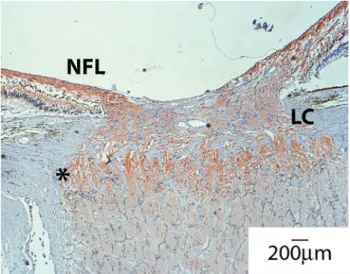
Fig. 4 Distribution of mitochondria around the optic nerve head. A section of human nerve was stained with anti-Cox4 antibodies to assess mitochondrial distribution. Shown is the optic nerve head with the retinal layers on the top. Note the strong Cox4 reactivity in the nerve fiber layer (NFL) before and especially after the axon left the globe (asterisk) and crossed the lamina cribrosa (LC). Also note the sharp decline in mitochondrial staining along the optic nerve. This coincides with the region where myelination of the optic nerve begins
optic nerve is striking. LHON and ADOA are mono-symptomatic and restricted to retinal ganglion cell (RGC) death. For other more complex syndromes, such as Leigh’s disease, MELAS, or MERRF, vision loss due to RGC degeneration is reported [21]. In addition, careful exami-nation of patients suffering from CMT2A and Friedreich’s ataxia, as well as Mohr-Tranebjerg syndrome, revealed optic nerve involvement. Why RGCs are especially vul-nerable to disturbances in mitochondrial dysfunction is still debated. Current models of optic neurodegeneration con-nected to mitochondrial dysfunction cite the special architecture of RGCs with mitochondria-enriched, non-myelinated parts in the retina and the optic nerve head and comparably mitochondria-poor, myelinated tracts along the optic nerve (Fig.4). It seems plausible to postulate that maintaining this mitochondrial gradient requires high fidelity on all levels of mitochondrial maintenance, namely fission/fusion, transport and biogenesis. In addition, com-pensatory mechanisms potentially active in other cell types might not be available under these circumstances. While increased mitochondrial mass may be sufficient to alleviate minor deficiencies in energy production due to mutations in
the ETC, the already high local concentration of mito-chondria in the unmyelinated part of RGCs might not allow for such a mechanism.
Another interesting aspect is the often less well-defined set of clinical manifestations of the above-mentioned dis-eases, which often feature a remarkable phenotypic overlap. For example, carriers of OPA1 mutations might present with the classical features of ADOA but may also present with peripheral neuropathy as seen in CMT or neurological symptoms as reported for HSP patients. The same is true of LHON and ADOA. While LHON normally follows an acute course and ADOA presents as a slowly progressive optic neuropathy, certain LHON cases follow the ADOA pattern of neuropathy while there are reports of ADOA patients with acute vision loss. These findings again highlight the shared common mitochondrial dysfunctions that might manifest themselves in different ways based on other genetic or environmental modifiers (Fig.5). In addition, these shared common features might allow gen-eralizing findings based on the study of these different disease entities and lead to a better understanding of neu-rodegenerative processes.
Fig. 5 Attack points of mitochondrial diseases. Protein degradation, balanced mitochondrial morphology and mitophagy promote the maintenance of a high fidelity, respiratory-competent mitochondrial network. Disturbances in mitochondrial morphology, impaired ETC through either acquired or inherited mtDNA mutations, as well as protein damage through oxidative stress cause damage to the mitochondrial network. The impact points of various neurodegener-ative diseases and their direct and also indirect effects on mitochondrial health are illustrated. Mitochondrial protein degrada-tion and processing is directly affected by disease mutadegrada-tions in SPG7/ paraplegin (mutated in hereditary spastic paraplegia) and AFG3L2
(mutated in spinocerebellar ataxia). Protein damage and oxidative stress are increased in patients suffering from Friedreich’s ataxia (FA) and amyotrophic lateral sclerosis (ALS). Inherited and also acquired mtDNA mutations impact the fidelity of the ETC in MELAS (mitochondrial encephalopathy, lactic acidosis, and stroke-like epi-sodes), MERRF (myoclonic epilepsy with ragged red fibers), LHON (Leber’s hereditary optic neuropathy) but also familial PD (Parkin-son’s disease). In addition, ETC function is indirectly affected via disturbed mitochondrial morphology in ADOA (autosomal dominant optic atrophy) as well as CMT2A (Charcott–Marie-Tooth Type 2A)
Conclusions
Mitochondrial dysfunction is at the heart of neurodegener-ative processes. The long-held belief that defects in mitochondrial respiration are responsible for the degener-ation and death of neurons during the course of most, if not all, neurodegenerative disorders is now being expanded by integrating more mitochondrial functions. While bioener-getics and associated oxidative stress are still crucial to the etiology of neurodegenerative diseases, mitochondrial protein quality control, mitochondrial dynamics and pro-grammed cell death pathways need to be integrated for a more comprehensive understanding of the link between mitochondrial dysfunction and neurodegeneration. With the understanding of the fundamental pathogenetic mechanisms leading to neurodegeneration, a clearer picture of differ-ences and similarities between different clinical disease entities emerges. In addition, the oftentimes striking degree of phenotypic expression of disease causing mutations and the symptomatic overlap between different diseases will likely be better understood with further acquisition of in-depth knowledge of the underlying mechanisms involving mitochondrial maintenance. In the future, an even more integrative view of mitochondrial dysfunction in the context of neurodegeneration is imperative for the successful development of effective treatment strategies aimed at combating common neurodegenerative diseases.
References
1. Abeliovich H (2007) Mitophagy: the life-or-death dichotomy includes yeast. Autophagy 3(3):275–277
2. Acaroglu G, Kansu T, Dogulu CF (2001) Visual recovery pat-terns in children with Leber’s hereditary optic neuropathy. Int Ophthalmol 24(6):349–355
3. Alexander C, Votruba M, Pesch UE, Thiselton DL, Mayer S, Moore A, Rodriguez M, Kellner U, Leo-Kottler B, Auburger G, Bhattacharya SS, Wissinger B (2000) OPA1, encoding a dyn-amin-related GTPase, is mutated in autosomal dominant optic atrophy linked to chromosome 3q28. Nat Genet 26(2):211–215 4. Amiott EA, Lott P, Soto J, Kang PB, McCaffery JM, DiMauro S, Abel ED, Flanigan KM, Lawson VH, Shaw JM (2008) Mitochondrial fusion and function in Charcot-Marie-Tooth type 2A patient fibroblasts with mitofusin 2 mutations. Exp Neurol 211(1):115–127
5. Amiott EA, Cohen MM, Saint-Georges Y, Weissman AM, Shaw JM (2009) A mutation associated with CMT2A neuropathy causes defects in Fzo1 GTP hydrolysis, ubiquitylation, and protein turnover. Mol Biol Cell 20(23):5026–5035
6. Anderson S, Bankier AT, Barrell BG, de Bruijn MH, Coulson AR, Drouin J, Eperon IC, Nierlich DP, Roe BA, Sanger F, Schreier PH, Smith AJ, Staden R, Young IG (1981) Sequence and organization of the human mitochondrial genome. Nature 290(5806):457–465
7. Balaban RS, Nemoto S, Finkel T (2005) Mitochondria, oxidants, and aging. Cell 120(4):483–495
8. Baloh RH, Schmidt RE, Pestronk A, Milbrandt J (2007) Altered axonal mitochondrial transport in the pathogenesis of Charcot-Marie-Tooth disease from mitofusin 2 mutations. J Neurosci 27(2):422–430
9. Barnils N, Mesa E, Munoz S, Ferrer-Artola A, Arruga J (2007) Response to idebenone and multivitamin therapy in Leber’s hereditary optic neuropathy. Arch Soc Esp Oftalmol 82(6): 377–380
10. Benard G, Neutzner A, Peng G, Wang C, Livak F, Youle RJ, Karbowski M (2010) IBRDC2, an IBR-type E3 ubiquitin ligase, is a regulatory factor for Bax and apoptosis activation. EMBO J 29(8):1458–1471
11. Berry C, La Vecchia C, Nicotera P (2010) Paraquat and Par-kinson’s disease. Cell Death Differ 17(7):1115–1125
12. Blackstone C, O’Kane CJ, Reid E (2011) Hereditary spastic paraplegias: membrane traffic and the motor pathway. Nat Rev Neurosci 12(1):31–42
13. Boesch P, Weber-Lotfi F, Ibrahim N, Tarasenko V, Cosset A, Paulus F, Lightowlers RN, Dietrich A (2011) DNA repair in organelles: Pathways, organization, regulation, relevance in disease and aging. Biochim Biophys Acta 1813(1):186–200 14. Bogenhagen DF, Rousseau D, Burke S (2008) The layered
structure of human mitochondrial DNA nucleoids. J Biol Chem 283(6):3665–3675
15. Bota DA, Van Remmen H, Davies KJ (2002) Modulation of Lon protease activity and aconitase turnover during aging and oxi-dative stress. FEBS Lett 532(1–2):103–106
16. Braschi E, Zunino R, McBride HM (2009) MAPL is a new mitochondrial SUMO E3 ligase that regulates mitochondrial fission. EMBO Rep 10(7):748–754
17. Bua E, Johnson J, Herbst A, Delong B, McKenzie D, Salamat S, Aiken JM (2006) Mitochondrial DNA-deletion mutations accumulate intracellularly to detrimental levels in aged human skeletal muscle fibers. Am J Hum Genet 79(3):469–480 18. Cadenas E, Davies KJ (2000) Mitochondrial free radical
gen-eration, oxidative stress, and aging. Free Radic Biol Med 29(3–4):222–230
19. Campuzano V, Montermini L, Molto MD, Pianese L, Cossee M, Cavalcanti F, Monros E, Rodius F, Duclos F, Monticelli A, Zara F, Canizares J, Koutnikova H, Bidichandani SI, Gellera C, Brice A, Trouillas P, De Michele G, Filla A, De Frutos R, Palau F, Patel PI, Di Donato S, Mandel JL, Cocozza S, Koenig M, Pandolfo M (1996) Friedreich’s ataxia: autosomal recessive disease caused by an intronic GAA triplet repeat expansion. Science 271(5254):1423–1427
20. Carelli V, Ross-Cisneros FN, Sadun AA (2004) Mitochondrial dysfunction as a cause of optic neuropathies. Prog Retin Eye Res 23(1):53–89
21. Carelli V, La Morgia C, Valentino ML, Barboni P, Ross-Cisneros FN, Sadun AA (2009) Retinal ganglion cell neurode-generation in mitochondrial inherited disorders. Biochim Biophys Acta 1787(5):518–528
22. Casari G, De Fusco M, Ciarmatori S, Zeviani M, Mora M, Fernandez P, De Michele G, Filla A, Cocozza S, Marconi R, Durr A, Fontaine B, Ballabio A (1998) Spastic paraplegia and OXPHOS impairment caused by mutations in paraplegin, a nuclear-encoded mitochondrial metalloprotease. Cell 93(6): 973–983
23. Cedarbaum JM, Blass JP (1986) Mitochondrial dysfunction and spinocerebellar degenerations. Neurochem Pathol 4(1):43–63 24. Cereghetti GM, Stangherlin A, Martins de Brito O, Chang CR,
Blackstone C, Bernardi P, Scorrano L (2008) Dephosphorylation by calcineurin regulates translocation of Drp1 to mitochondria. Proc Natl Acad Sci USA 105(41):15803–15808
25. Chang CR, Blackstone C (2007) Cyclic AMP-dependent protein kinase phosphorylation of Drp1 regulates its GTPase activity
and mitochondrial morphology. J Biol Chem 282(30):21583– 21587
26. Chapman E, Fry AN, Kang M (2011) The complexities of p97 function in health and disease. Mol Biosyst 7(3):700–710 27. Chen H, Detmer SA, Ewald AJ, Griffin EE, Fraser SE, Chan DC
(2003) Mitofusins Mfn1 and Mfn2 coordinately regulate mito-chondrial fusion and are essential for embryonic development. J Cell Biol 160(2):189–200
28. Chen H, McCaffery JM, Chan DC (2007) Mitochondrial fusion protects against neurodegeneration in the cerebellum. Cell 130(3):548–562
29. Chinnery PF, Howell N, Lightowlers RN, Turnbull DM (1997) Molecular pathology of MELAS and MERRF. The relationship between mutation load and clinical phenotypes. Brain 120(Pt 10):1713–1721
30. Cho DH, Nakamura T, Fang J, Cieplak P, Godzik A, Gu Z, Lipton SA (2009) S-nitrosylation of Drp1 mediates beta-amy-loid-related mitochondrial fission and neuronal injury. Science 324(5923):102–105
31. Chung KW, Kim SB, Park KD, Choi KG, Lee JH, Eun HW, Suh JS, Hwang JH, Kim WK, Seo BC, Kim SH, Son IH, Kim SM, Sunwoo IN, Choi BO (2006) Early onset severe and late-onset mild Charcot-Marie-Tooth disease with mitofusin 2 (MFN2) mutations. Brain 129(Pt 8):2103–2118
32. Ciechanover A, Iwai K (2004) The ubiquitin system: from basic mechanisms to the patient bed. IUBMB Life 56(4):193–201 33. Cipolat S, Martins de Brito O, Dal Zilio B, Scorrano L (2004)
OPA1 requires mitofusin 1 to promote mitochondrial fusion. Proc Natl Acad Sci USA 101(45):15927–15932
34. Cipolat S, Rudka T, Hartmann D, Costa V, Serneels L, Cra-essaerts K, Metzger K, Frezza C, Annaert W, D’Adamio L, Derks C, Dejaegere T, Pellegrini L, D’Hooge R, Scorrano L, De Strooper B (2006) Mitochondrial rhomboid PARL regulates cytochrome c release during apoptosis via OPA1-dependent cristae remodeling. Cell 126(1):163–175
35. Coleman RA, Lee DP (2004) Enzymes of triacylglycerol syn-thesis and their regulation. Prog Lipid Res 43(2):134–176 36. Costa V, Giacomello M, Hudec R, Lopreiato R, Ermak G, Lim
D, Malorni W, Davies KJ, Carafoli E, Scorrano L (2010) Mitochondrial fission and cristae disruption increase the response of cell models of Huntington’s disease to apoptotic stimuli. EMBO Mol Med 2(12):490–503
37. Dalle-Donne I, Rossi R, Giustarini D, Milzani A, Colombo R (2003) Protein carbonyl groups as biomarkers of oxidative stress. Clin Chim Acta 329(1–2):23–38
38. Dalle-Donne I, Aldini G, Carini M, Colombo R, Rossi R, Mil-zani A (2006) Protein carbonylation, cellular dysfunction, and disease progression. J Cell Mol Med 10(2):389–406
39. de Brito OM, Scorrano L (2008) Mitofusin 2 tethers endoplas-mic reticulum to mitochondria. Nature 456(7222):605–610 40. Delettre C, Lenaers G, Griffoin JM, Gigarel N, Lorenzo C,
Belenguer P, Pelloquin L, Grosgeorge J, Turc-Carel C, Perret E, Astarie-Dequeker C, Lasquellec L, Arnaud B, Ducommun B, Kaplan J, Hamel CP (2000) Nuclear gene OPA1, encoding a mitochondrial dynamin-related protein, is mutated in dominant optic atrophy. Nat Genet 26(2):207–210
41. Delettre C, Griffoin JM, Kaplan J, Dollfus H, Lorenz B, Faivre L, Lenaers G, Belenguer P, Hamel CP (2001) Mutation spec-trum and splicing variants in the OPA1 gene. Hum Genet 109(6):584–591
42. Di Bella D, Lazzaro F, Brusco A, Plumari M, Battaglia G, Pastore A, Finardi A, Cagnoli C, Tempia F, Frontali M, Veneziano L, Sacco T, Boda E, Brussino A, Bonn F, Castellotti B, Baratta S, Mariotti C, Gellera C, Fracasso V, Magri S, Langer T, Plevani P, Di Donato S, Muzi-Falconi M, Taroni F (2010)
Mutations in the mitochondrial protease gene AFG3L2 cause dominant hereditary ataxia SCA28. Nat Genet 42(4):313–321 43. Duvezin-Caubet S, Jagasia R, Wagener J, Hofmann S,
Trifu-novic A, Hansson A, Chomyn A, Bauer MF, Attardi G, Larsson NG, Neupert W, Reichert AS (2006) Proteolytic processing of OPA1 links mitochondrial dysfunction to alterations in mito-chondrial morphology. J Biol Chem 281(49):37972–37979 44. Duvezin-Caubet S, Koppen M, Wagener J, Zick M, Israel L,
Bernacchia A, Jagasia R, Rugarli EI, Imhof A, Neupert W, Langer T, Reichert AS (2007) OPA1 processing reconstituted in yeast depends on the subunit composition of the m-AAA pro-tease in mitochondria. Mol Biol Cell 18(9):3582–3590 45. Ehses S, Raschke I, Mancuso G, Bernacchia A, Geimer S,
Tondera D, Martinou JC, Westermann B, Rugarli EI, Langer T (2009) Regulation of OPA1 processing and mitochondrial fusion by m-AAA protease isoenzymes and OMA1. J Cell Biol 187(7):1023–1036
46. Elleuch N, Depienne C, Benomar A, Hernandez AM, Ferrer X, Fontaine B, Grid D, Tallaksen CM, Zemmouri R, Stevanin G, Durr A, Brice A (2006) Mutation analysis of the paraplegin gene (SPG7) in patients with hereditary spastic paraplegia. Neurology 66(5):654–659
47. Esteves AR, Arduino DM, Silva DF, Oliveira CR, Cardoso SM (2011) Mitochondrial Dysfunction: The Road to Alpha-Synuc-lein Oligomerization in PD. Parkinsons Dis 2011:693761 48. Fayet G, Jansson M, Sternberg D, Moslemi AR, Blondy P,
Lombes A, Fardeau M, Oldfors A (2002) Ageing muscle: clonal expansions of mitochondrial DNA point mutations and deletions cause focal impairment of mitochondrial function. Neuromuscul Disord 12(5):484–493
49. Ferreira IL, Cunha-Oliveira T, Nascimento MV, Ribeiro M, Proenca MT, Januario C, Oliveira CR, Rego AC (2011) Bio-energetic dysfunction in Huntington’s disease human cybrids. Exp Neurol 231(1):127–134
50. Filosto M, Scarpelli M, Cotelli MS, Vielmi V, Todeschini A, Gregorelli V, Tonin P, Tomelleri G, Padovani A (2011) The role of mitochondria in neurodegenerative diseases. J Neurol 258(10):1763–1774
51. Fink JK, Hedera P (1999) Hereditary spastic paraplegia: genetic heterogeneity and genotype-phenotype correlation. Semin Neu-rol 19(3):301–309
52. Frank S, Robert EG, Youle RJ (2003) Scission, spores, and apoptosis: a proposal for the evolutionary origin of mitochondria in cell death induction. Biochem Biophys Res Commun 304(3): 481–486
53. Fukui H, Moraes CT (2008) The mitochondrial impairment, oxidative stress and neurodegeneration connection: reality or just an attractive hypothesis? Trends Neurosci 31(5):251–256 54. Gandre-Babbe S, van der Bliek AM (2008) The novel
tail-anchored membrane protein Mff controls mitochondrial and peroxisomal fission in mammalian cells. Mol Biol Cell 19(6):2402–2412
55. Gerdes F, Tatsuta T, Langer T (2011) Mitochondrial AAA proteases—towards a molecular understanding of membrane-bound proteolytic machines. Biochim Biophys Acta. doi:
10.1016/j.bbamcr.2011.09.015
56. Gille G, Reichmann H (2011) Iron-dependent functions of mitochondria—relation to neurodegeneration. J Neural Transm 118(3):349–359
57. Gray MW, Burger G, Lang BF (1999) Mitochondrial evolution. Science 283(5407):1476–1481
58. Grazina MM, Diogo LM, Garcia PC, Silva ED, Garcia TD, Robalo CB, Oliveira CR (2007) Atypical presentation of Leber’s heredi-tary optic neuropathy associated to mtDNA 11778G [ A point mutation—a case report. Eur J Paediatr Neurol 11(2):115–118
59. Griparic L, Kanazawa T, van der Bliek AM (2007) Regulation of the mitochondrial dynamin-like protein Opa1 by proteolytic cleavage. J Cell Biol 178(5):757–764
60. Guillery O, Malka F, Landes T, Guillou E, Blackstone C, Lombes A, Belenguer P, Arnoult D, Rojo M (2008) Metallo-protease-mediated OPA1 processing is modulated by the mitochondrial membrane potential. Biol Cell 100(5):315–325 61. Harman D (1956) Aging: a theory based on free radical and
radiation chemistry. J Gerontol 11(3):298–300
62. Head B, Griparic L, Amiri M, Gandre-Babbe S, van der Bliek AM (2009) Inducible proteolytic inactivation of OPA1 mediated by the OMA1 protease in mammalian cells. J Cell Biol 187(7):959–966
63. Horn SR, Thomenius MJ, Johnson ES, Freel CD, Wu JQ, Coloff JL, Yang CS, Tang W, An J, Ilkayeva OR, Rathmell JC, Newgard CB, Kornbluth S (2011) Regulation of mitochondrial morphology by APC/CCdh1-mediated control of Drp1 stability. Mol Biol Cell 22(8):1207–1216
64. Hubbers CU, Clemen CS, Kesper K, Boddrich A, Hofmann A, Kamarainen O, Tolksdorf K, Stumpf M, Reichelt J, Roth U, Krause S, Watts G, Kimonis V, Wattjes MP, Reimann J, Thal DR, Biermann K, Evert BO, Lochmuller H, Wanker EE, Schoser BG, Noegel AA, Schroder R (2007) Pathological con-sequences of VCP mutations on human striated muscle. Brain 130(Pt 2):381–393
65. Hudson G, Carelli V, Spruijt L, Gerards M, Mowbray C, Achilli A, Pyle A, Elson J, Howell N, La Morgia C, Valentino ML, Huoponen K, Savontaus ML, Nikoskelainen E, Sadun AA, Salomao SR, Belfort R Jr, Griffiths P, Man PY, de Coo RF, Horvath R, Zeviani M, Smeets HJ, Torroni A, Chinnery PF (2007) Clinical expression of Leber hereditary optic neuropathy is affected by the mitochondrial DNA-haplogroup background. Am J Hum Genet 81(2):228–233
66. Ingerman E, Perkins EM, Marino M, Mears JA, McCaffery JM, Hinshaw JE, Nunnari J (2005) Dnm1 forms spirals that are structurally tailored to fit mitochondria. J Cell Biol 170(7): 1021–1027
67. Ishihara N, Fujita Y, Oka T, Mihara K (2006) Regulation of mitochondrial morphology through proteolytic cleavage of OPA1. EMBO J 25(13):2966–2977
68. Ishihara N, Nomura M, Jofuku A, Kato H, Suzuki SO, Masuda K, Otera H, Nakanishi Y, Nonaka I, Goto Y, Taguchi N, Mor-inaga H, Maeda M, Takayanagi R, Yokota S, Mihara K (2009) Mitochondrial fission factor Drp1 is essential for embryonic development and synapse formation in mice. Nat Cell Biol 11(8):958–966
69. Israelson A, Arbel N, Da Cruz S, Ilieva H, Yamanaka K, Sho-shan-Barmatz V, Cleveland DW (2010) Misfolded mutant SOD1 directly inhibits VDAC1 conductance in a mouse model of inherited ALS. Neuron 67(4):575–587
70. James DI, Parone PA, Mattenberger Y, Martinou JC (2003) hFis1, a novel component of the mammalian mitochondrial fission machinery. J Biol Chem 278(38):36373–36379 71. Karbowski M, Neutzner A, Youle RJ (2007) The mitochondrial
E3 ubiquitin ligase MARCH5 is required for Drp1 dependent mitochondrial division. J Cell Biol 178(1):71–84
72. Karbowski M, Youle RJ (2011) Regulating mitochondrial outer membrane proteins by ubiquitination and proteasomal degra-dation. Curr Opin Cell Biol 23(4):476–482
73. Kim I, Rodriguez-Enriquez S, Lemasters JJ (2007) Selective degradation of mitochondria by mitophagy. Arch Biochem Biophys 462(2):245–253
74. Kirkman MA, Yu-Wai-Man P, Korsten A, Leonhardt M, Dim-itriadis K, De Coo IF, Klopstock T, Chinnery PF (2009) Gene-environment interactions in Leber hereditary optic neuropathy. Brain 132(Pt 9):2317–2326
75. Klockgether T, Paulson H (2011) Milestones in ataxia. Mov Disord 26(6):1134–1141
76. Knott AB, Bossy-Wetzel E (2008) Impairing the mitochondrial fission and fusion balance: a new mechanism of neurodegener-ation. Ann N Y Acad Sci 1147:283–292
77. Koshiba T, Detmer SA, Kaiser JT, Chen H, McCaffery JM, Chan DC (2004) Structural basis of mitochondrial tethering by mitofusin complexes. Science 305(5685):858–862
78. Kukat C, Wurm CA, Spahr H, Falkenberg M, Larsson NG, Jakobs S (2011) Super-resolution microscopy reveals that mammalian mitochondrial nucleoids have a uniform size and frequently contain a single copy of mtDNA. Proc Natl Acad Sci USA 108(33):13534–13539
79. Lee YJ, Jeong SY, Karbowski M, Smith CL, Youle RJ (2004) Roles of the mammalian mitochondrial fission and fusion mediators Fis1, Drp1, and Opa1 in apoptosis. Mol Biol Cell 15(11):5001–5011
80. Li Q, Vande Velde C, Israelson A, Xie J, Bailey AO, Dong MQ, Chun SJ, Roy T, Winer L, Yates JR, Capaldi RA, Cleveland DW, Miller TM (2010) ALS-linked mutant superoxide dismutase 1 (SOD1) alters mitochondrial protein composition and decreases protein import. Proc Natl Acad Sci USA 107(49):21146–21151 81. Lin MT, Beal MF (2006) Mitochondrial dysfunction and oxi-dative stress in neurodegenerative diseases. Nature 443(7113): 787–795
82. Livnat-Levanon N, Glickman MH (2011) Ubiquitin–proteasome system and mitochondria—reciprocity. Biochim Biophys Acta 1809(2):80–87
83. Luce K, Osiewacz HD (2009) Increasing organismal healthspan by enhancing mitochondrial protein quality control. Nat Cell Biol 11(7):852–858
84. Martinelli P, Rugarli EI (2010) Emerging roles of mitochondrial proteases in neurodegeneration. Biochim Biophys Acta 1797(1): 1–10
85. Mashima Y, Kigasawa K, Wakakura M, Oguchi Y (2000) Do idebenone and vitamin therapy shorten the time to achieve visual recovery in Leber hereditary optic neuropathy? J Neuro-ophthalmol 20(3):166–170
86. Miller KE, Sheetz MP (2004) Axonal mitochondrial transport and potential are correlated. J Cell Sci 117(Pt 13):2791–2804 87. Moraes CT, DiMauro S, Zeviani M, Lombes A, Shanske S,
Miranda AF, Nakase H, Bonilla E, Werneck LC, Servidei S et al (1989) Mitochondrial DNA deletions in progressive external ophthalmoplegia and Kearns-Sayre syndrome. N Engl J Med 320(20):1293–1299
88. Murakami T, Mita S, Tokunaga M, Maeda H, Ueyama H, Kumamoto T, Uchino M, Ando M (1996) Hereditary cerebellar ataxia with Leber’s hereditary optic neuropathy mitochondrial DNA 11778 mutation. J Neurol Sci 142(1–2):111–113 89. Murgia M, Giorgi C, Pinton P, Rizzuto R (2009) Controlling
metabolism and cell death: at the heart of mitochondrial calcium signalling. J Mol Cell Cardiol 46(6):781–788
90. Nakamura K, Nemani VM, Azarbal F, Skibinski G, Levy JM, Egami K, Munishkina L, Zhang J, Gardner B, Wakabayashi J, Sesaki H, Cheng Y, Finkbeiner S, Nussbaum RL, Masliah E, Edwards RH (2011) Direct membrane association drives mito-chondrial fission by the Parkinson disease-associated protein alpha-synuclein. J Biol Chem 286(23):20710–20726
91. Neuspiel M, Schauss AC, Braschi E, Zunino R, Rippstein P, Rachubinski RA, Andrade-Navarro MA, McBride HM (2008) Cargo-selected transport from the mitochondria to peroxisomes is mediated by vesicular carriers. Curr Biol 18(2):102–108 92. Neutzner A, Youle RJ (2005) Instability of the mitofusin Fzo1
regulates mitochondrial morphology during the mating response of the yeast Saccharomyces cerevisiae. J Biol Chem 280(19): 18598–18603