501
Regression of
AIDS-Related Kaposi's
Sarcoma During Therapy with
Thalidomide
Rolf A. Soler, Mark Howard, Nicola S. Brink, Diana Gibb, Richard S. Tedder, and David Nadal
From the Infectious Diseases Unit, University Children's Hospital, Zurich, Switzerland; and the Division of Virology, Department of
Medical Microbiology, and the MRC HIV Clinical Trials Centre, UniversityCollege London Medical School, London, and University College London Hospitals NHS Trust, London, England
A 14-year-old girl with HIV infection and subcutaneous Kaposi's sarcoma (KS) received thalido-mide therapy for oral ulcers, resulting in regression of KS lesions, disappearance of KS-associated herpesvirus (KSHV) DNA from blood, and reduced viral load in tumor tissue. Administration of granulocyte colony-stimulating factor resulted in clinical exacerbation of KS and reappearance of KSHV DNA in blood.
Kaposi's sarcoma (KS), a proliferative disease of endothelial cell origin, is a common AIDS-defining illness in HIV-infected adults. Italso occurs infrequently in children with vertically acquired HTV infection [1]. As current therapies, including chemotherapy, systemic TFN-a, and local radiotherapy, have a limited effect [2], there is a need for alternative therapeutic approaches.
See the editorial by Levine on pages 504-5. A novel herpesvirus has been recently described as a possible cause of KS. In the United States, Chang and colleagues iso-lated two unique DNA fragments from a KS skin lesion and showed that they were from a previously unidentified y2 her-pesvirus, provisionally termed Kaposi's sarcoma-associated herpesvirus (KSHV) [3]. Further investigations showed an as-sociation between detectable KSHV DNA in peripheral blood and KS and, more importantly, that the detection of KSHV DNA in peripheral blood from HIV-infected individuals pre-dicted progression to KS [4].
We report the case of a 14-year-old girl with vertically ac-quired HTV infection and KS whose tumor clinically regressed while she was receiving treatment with thalidomide for oral ulcers [5]. Regression of the tumor was paralleled by the disap-pearance ofKSHV DNA from peripheral blood and a reduction of viral load in tumor tissue.
Case Report
A 14-year-old girl with vertically acquired HIV infection who immigrated from Zaire to Switzerland developed
subcuta-Received 15 February 1996; revised 19 April 1996.
Reprints or correspondence: Dr. David Nadal, Infectious Diseases Unit, University Children's Hospital, Steinwiesstrasse 75, CH-8032 Zurich, Switzer-land.
Clinical Infectious Diseases 1996;23:501-3
©1996 by The University of Chicago. All rights reserved.
1058--4838/96/2303-0013$02.00
neous nodular lesions on both arms, the left upper leg, and the right eyelid. Histologic examination of specimens from the lesions revealed proliferation of spindle-shaped cells and of vascular structures with large endothelial cells; on the basis of these findings, the diagnosis of KS was made. She had been well until the age of 9 years, when she experienced the first of several episodes of pneumonia. She subsequently developed various HTV-associated diseases, including recurrent oral can-didiasis, lymphocytic interstitial pneumonitis, and
Pneumo-cystis cariniipneumonia (one episode). Her CD4+ cell count had been 0/ILL for 14 months before the diagnosis of KS was made.
At the time that the diagnosis of KS was made, her therapy included co-trimoxazole, oral amphotericin B, didanosine, and monthly iv immunoglobulin infusions; this therapy remained unchanged except for the temporary use of antibiotics to treat acute infections. She did not receive cytotoxic or immuno-modulating chemotherapy or radiotherapy. The KS lesions in-creased slowly in number and size over a 4-month period.
At the age of 14.3 years the patient developed painful oral ulcers. Histologic examination of specimens from the ulcers as well as conventional cultures for bacteria, mycobacteria, fungi, and viruses revealed no infectious cause. After topical and systemic corticosteroid therapy failed, therapy with thalido-mide was started at a dose of 3 mg/(kg· d) [5]. During 3 weeks of treatment with thalidomide, a coincidental decrease in the size of the existing KS-associated lesions was noted, and no new lesions developed. Therapy with thalidomide was therefore continued after the oral ulcers resolved.
Two months later, no new lesions appeared and the number of lesions decreased from 29 to 13. Treatment with thalidomide was successfully augmented by local radiotherapy to two digital nodes, which caused restricted distal interphalangeal joint mo-bility, and to the right upper lid (for cosmetic reasons).
The patient's course of therapy was further complicated by Candida albicans septicemia that required iv therapy with am-photericin B together with granulocyte colony-stimulating fac-tor (G-CSF) because of severe neutropenia. This therapy was associated with an increase in the size of the remaining KS
502 Soler et al.
em
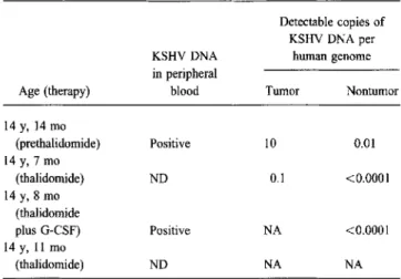
1996;23(September)Table 1. Detection of Kaposi's sarcoma-associated herpesvirus DNAinthe blood and tumor tissue of an HIV-infected girl before and during treatment with thalidomide.
Detectable copies of KSHV DNA per
KSHVDNA human genome
in peripheral
Age (therapy) blood Tumor Nontumor
14 y, 14 mo (prethalidomide) Positive 10 0.01 14 y, 7 mo (thalidomide) ND 0.1 <0.0001 14 y, 8 mo (thalidomide plus G-CSF) Positive NA <0.0001 14y, 11 mo (thalidomide) ND NA NA
NOTE. G-CSF granulocyte colony-stimulating factor; KSHV Kaposi's sarcoma-associated herpesvirus; NA =not analyzed; ND =not detected.
lesions. Therapy with G-CSF was stopped, and the size of the tumor decreased clinically. As of this writing, the patient has been receiving thalidomide therapy for 7 months. Her tumor bulk is stable and she has not experienced any adverse effects from thalidomide.
Methods
Detection ofKSHV. We examined blood, tumor tissue, and healthy skin for the presence of KSHV DNA with use of nested PCR amplification (previously described in [4]). DNA was extracted from the tissue samples by digestion with 10% SDS buffer containing 10 mMTris-HCI, 10 mMEDTA, and 10mM NaCl, together with proteinase K at 1ILg/mL, and tested for KSHV DNA and human DNA. This test involved amplification of the human single copy gene encoding pyruvate dehydroge-nase by PCR, which has been shown to have a sensitivity of a single input copy (data not shown). Quantification of human and KSHV DNA was performed by endpoint dilution. To con-trol for PCR inhibitors, samples that did not contain KSHV DNA were spiked with known amounts of virus genome.
HIV viral load. Quantitative measurement of HIV viral RNA was performed on stored serum samples with use of a commercial PCR test (Amplicor HIV Monitor, Roche Diagnos-tic Systems, Branchburg, NJ); the manufacturer's instructions were followed.
Results
KSHV DNA was detected in blood, tumor tissue, and healthy skin before treatment with thalidomide was started (Table 1). In contrast, blood obtained 3 months after thalidomide therapy was started contained no detectable KSHV DNA, and tumor
tissue showed an approximate 100-fold decrease in viral load. However, 6 days after starting G-CSF therapy, KSHV DNA again became detectable in blood and was associated with a clinical increase in tumor bulk. At this time, viral DNA was still undetectable in healthy tissue. Tumor tissue was not avail-able for analysis. Blood taken 3 months after G-CSF therapy was stopped contained no detectable KSHV DNA. No gross histological changes were noted in tumor tissues obtained be-fore thalidomide therapy was started vs. in those obtained 3 months later.
Two stored serum samples were available for retrospective quantitative measurement of HIV viral load. One sample, drawn when the patient was 14 years old (before therapy with didanosine was started), contained 106,441 copies of HIV RNA per mL. The other sample, obtained when the patient was 14.6 years old (3 months after thalidomide treatment was started), harbored 9,563 copies of HIV RNA per mL.
Discussion
The observation of clinical regression of KS lesions in a patient with vertically acquired HIV infection who was receiv-ing therapy with thalidomide raises interestreceiv-ing questions, espe-cially as this regression was paralleled by a virological re-sponse, with KSHV DNA becoming undetectable in blood and being reduced in tumor tissue. Although the clinical assessment of KS tumor bulk is imprecise and the observations were made in a single patient, the clinical and virological effects are bio-logically plausible.
Thalidomide inhibits monocyte TNF-a by degrading
TNF-a mRNA [6]. It also inhibits angiogenesis induced by basic fibroblast growth factor, which might explain its terato-genic potential [7]. Furthermore, thalidomide induces T helper cell type 2 (i.e., antiinflammatory) and inhibits T helper cell type 1 (i.e., proinflammatory) cytokine production [8]. TNF-a, basic fibroblast growth factor, and T helper cell type I cytokines such as IFN-'Ystimulate spindle cell transformation and mitosis of KS cells [9]. This finding could explain the clinical effect of thalidomide on our patient's KS lesions.
In contrast, G-CSF stimulates proliferation of human endo-thelial cells [10] and may, therefore, have counterbalanced the effect of thalidomide, resulting in a rapid increase in KS tumor bulk when the patient started receiving G-CSF therapy. This clinical deterioration was paralleled by the reappearance of KSHV DNA in peripheral blood (table 1).
There is no other plausible explanation for the apparent re-gression and enlargement ofKS lesions in our patient following administration of thalidomide and G-CSF since other therapy was unchanged throughout the period of observation, the pa-tient did not receive acyclovir, and the level of immunodefi-ciency remained unaltered. Itis not clear whether thalidomide has a direct effect on KSHV DNA, resulting in changes in the viral load. In vitro studies using KS cell lines [11] and KSHV-containing cell lines [12] may help to elucidate this question.
em 1996;23 (September) Thalidomide Therapy for Kaposi's Sarcoma 503
Our patient's HIV viral load while she was receiving treat-ment with thalidomide was less than 10% of the viral load before thalidomide therapy was started. However, determina-tion of the viral load was done retrospectively on stored serum and not on plasma because the latter was not available. Since the patient also started receiving didanosine therapy during the interval between collection of specimens, the reduction of HIV load cannot be attributed solely to thalidomide treatment. Nev-ertheless, thalidomide has been shown to inhibit replication of HIV in vitro and in vivo [13].
Although this case suggests that thalidomide may be effica-cious in the treatment of a patient with HIV-related KS, conven-tional chemotherapy, radiotherapy, and IFN-a should remain first-line therapeutic options. However, we suggest that thalido-mide should be further studied as a therapeutic agent for the treatment of HIV-induced KS and that its use may be justified in patients with disseminated KS who are considered unsuitable for or who are unable to tolerate conventional therapeutic inter-ventions.
References
1. Peterman TA, Jaffe HW, Beral V. Epidemiologic clues to the etiology of Kaposi's sarcoma. AIDS 1995; 7:605-11.
2. Cho J, ChachuaA.Kaposi's sarcoma. CUff Opin Oncol 1992;4:667-73. 3. Chang Y, Cesarman E, Pessin MS, et al. Identification of herpesvirus-like DNA sequences in AIDS-associated Kaposi's sarcoma. Science 1994;
266:1865-9.
4. Whitby D, Howard MR, Tenant-Flowers M, et al. Detection of Kaposi sarcoma associated with herpesvirus in peripheral blood of HIV-infected individuals and progression to Kaposi's sarcoma. Lancet 1995; 346: 799-802.
5. Soler RA, Migliorati C, Van Waes H, Nadal D. Thalidomide therapy of mucosal ulceration in AIDS. Arch Dis Child 1996; 74:64-5. 6. Moreira AL, Sampaio EP, Zmuidzinas A, Frindt P, Smith KA, Kaplan G.
Thalidomide exerts its inhibitory action on tumor necrosis factor alpha by enhancing mRNA degradation. JExp Med 1993; 177:1675-80. 7. D'Amato RJ, Loughnan MS, Flynn E, Folkman 1.Thalidomide is an
inhibitor of angiogenesis. Proc Nat! Acad Sci USA 1994;91:4082-5. 8. McHugh SM, Rifkin IR, Deighton J, Wilson AB, Lachman PJ, Lockwood
CM. The immunosuppressive drug thalidomide induces T helper cell type 2 (Th2) and concomitantly inhibits Thl cytokine production in mitogen- and antigen-stimulated human peripheral blood mononuclear cell cultures. Clin Exp ImmunoI1995;99:160-7.
9. Fiorelli V, Gendelman R, Samaniego F, Markham PD, Ensoli B. Cytokines from activated T cells induce normal endothelial cells to acquire the phenotypic and functional features of AIDS-Kaposi's sarcoma spindle cells.JClin Invest 1995;95:1723-34.
10. Bussolino F, WangJM,Defilippi P, et al. Granulocyte- and granulocyte-macrophage colony-stimulating factors induce human endothelial cells to migrate and proliferate. Nature 1989; 337:471-3.
11. Lunardi-Iskandar Y, Bryant IL, Zeman RA, et al. Tumorigenesis and metastasis of neoplastic Kaposi's sarcoma cell line in immunodeficient mice blocked by a human pregnancy hormone. Nature 1995; 375: 64-8.
12. Renne R, Zhong W, Hemdier B, et al. Lytic growth of Kaposi's sarcoma-associated herpesvirus (herpesvirus 8) in culture. Nature Medicine 1996; 2:342-6.
13. Makonkawkeyoon S, Limson-Probe RNR, Moreira AL, Schauf V, Kaplan G. Thalidomide inhibits the replication of human immunodeficiency virus type 1. Proc Natl Acad Sci USA 1993; 90:5974-8.