Selenium absorption and retention from a selenite- or selenate-fortified
milk-based formula in men measured by a stable-isotope technique
Peter Van Dael*, Lena Davidsson², Rafael MunÄoz-Box, Laurent B. Fay and Denis Barclay
Nestle Research Centre, PO Box 44, Vers-chez-les-Blanc, CH-1000 Lausanne, Switzerland (Received 11 January 2000 ± Revised 10 July 2000 ± Accepted 04 August 2000)
The present study was designed to determine the apparent absorption and retention of the inorganic Se compounds SeO322and SeO422, which are commonly used for Se fortification of
clinical nutrition products and infant formulas. Ten healthy men were fed a milk-based formula labelled with 40mg Se as74SeO
322or76SeO422on two consecutive days using a randomised
crossover design. Se stable-isotope analysis of 9 d complete collections of urine and faeces was used to calculate apparent Se absorption and retention. Se retention from74SeO
322(41´0 (SD
8´4) %) and from 76SeO
422 (46´0 (SD 7´9) %) was not significantly different P . 0´05:
However, Se absorption was significantly higher from SeO422than from SeO322(91´3 (SD1´4)
% v. 50´2 (SD7´8) %, P , 0´05: Urinary excretion of the administered dose was 9´2 (SD1´8) %
for 74SeO
322 and 45´3 (SD 8´2) % for 76SeO422 P , 0´05: Urinary Se excretion kinetics
differed significantly for the two Se compounds; 90 % of the total urinary Se was excreted after 121 h for74SeO
322and after 40 h for76SeO422 P , 0´05: These results suggest that although
Se absorption and urinary excretion differ for SeO322 and SeO422, both Se compounds are
equally well retained when administered at a relatively low dose (40 mg Se). The nutritional impact of Se fortification of foods would thus be expected to be similar when SeO422or SeO322
are used.
Selenium fortification: Selenium absorption: Selenium retention: Inorganic selenium compounds: Stable isotopes
Se is an essential trace element for human subjects (National Research Council, 1989). Se is an integral part of glutathione peroxidase (GSH-Px), an enzyme involved in cellular protection against oxidative damage, and of iodothyronine deiodinase which catalyses the conversion of thyroxine into triiodothyronine (Zachara, 1993). Several other selenoproteins, such as selenoprotein P, have been isolated, but their physiological role is still not fully elucidated (Zachara, 1993). In addition, Se has been reported to play a role in the maintenance of optimal immune response (Chandra, 1997), and the role of Se in the emergence of viral mutations has been documented (Beck, 1996). Recently, the impact of Se supplementation on the incidence of certain cancers (Clark et al. 1996) was reported.
Low dietary Se intake has been associated with a number of deficiency symptoms, of which Keshan disease, a fatal cardiomyopathy endemic to low-Se areas in China, is the
most extensively documented (Lockitch, 1989). Popula-tions at risk of impaired Se status are patients on long-term parenteral or enteral nutrition without Se supplementation (Levander, 1984) and infants consuming infant formulas with a low Se content (Litov & Combs, 1991). Although no overt clinical symptoms of Se deficiency have been diagnosed in these individuals, cases of cardiac and muscular dystrophies have been reported in patients on long-term total parenteral nutrition (Casey & Hambidge, 1985; Hambidge, 1985; Vinton et al. 1987; Kelly et al. 1988).
Food fortification with Se is not widely used, except for clinical products such as enteral and parenteral nutrition products and infant formulas. Inorganic Se compounds, SeO322or SeO422, are typically used for food fortification
and information about bioavailability, absorption and retention of these Se compounds in human subjects is needed to estimate the nutritional impact of Se fortification.
Abbreviations: GSH-Px, glutathione peroxidase; Hb, haemoglobin; HGAAS, hydride-generation atomic absorption spectrometry. * Corresponding author: Dr Peter Van Dael, fax +41 21 785 8563, email [email protected]
²Present address: Laboratory for Human Nutrition, Institute of Food Science, Swiss Federal Institute of Technology, PO Box 474, CH-8803 RuÈschlikon, Switzerland.
Previous studies of Se bioavailability have focused mainly on the comparison between inorganic Se, in particular SeO322, and organic Se compounds, selenomethionine or
Se-rich yeast (Alfthan et al. 1991; Xia et al. 1992; NeÁve, 1995). Only very limited data on Se bioavailability from inorganic compounds (SeO322and SeO422) are available.
Furthermore, until now, no information about Se absorption and retention from SeO322 and SeO422 after intake of
relatively small amounts of the Se compounds, relevant to food fortification, has been reported. The recent develop-ment of stable-isotope techniques offers the possibility of monitoring absorption and retention of trace elements without introducing radiation exposure (SandstroÈm et al. 1993).
The aim of the present study was to compare apparent Se absorption and retention from SeO322and SeO422in men
using a stable-isotope technique. The inorganic Se compounds were added to a milk-based formula in quantities relevant to food fortification. A milk-based formula was selected as the food vehicle since it represents the most common products fortified with Se (i.e. products used for enteral nutrition or infant formulas).
Materials and methods Subjects
Ten healthy men, 24±36 (mean 30) years old, were recruited from personnel at the Nestle Research Centre, Lausanne, Switzerland. The aims and procedures of the study were explained verbally and in writing before enrolment in the study. All subjects gave written informed consent. A medical examination was carried out before enrolment. Medication, dietary supplements and high-Se food items (i.e. offal meat, fish and other seafoods) were excluded during the study. In all other respects subjects maintained their normal lifestyle and dietary habits during the study. None of the subjects consumed dietary supple-ments containing Se.
The study protocol was reviewed and approved by the external ethical committee of the Nestle Research Centre and followed the guidelines of the Helsinki Declaration regarding human subjects.
Stable isotope labels
Highly-enriched elemental74Se (74Se 98´2 %, 76Se 1´8 %,
other Se isotopes ,0´1 %),76Se (74Se 1´2 %,76Se 98´5 %, 77Se 0´2 %, other Se isotopes ,0´03 %) and 82Se (74Se
0´06 %, 76Se 0´62 %, 77Se 0´56 %, 78Se 1´76 %, 80Se
4´8 %, 82Se 92´2 %) were purchased from Isotec (St.
Quentin, France). Elemental Se was converted into
74SeO
322,76SeO422 and82SeO322. SeO322was prepared
by dissolving a precise amount of elemental Se (approxi-mately 2´5±10 mg; weighed on a high-precision analytical balance) into 1 ml concentrated HNO3. The solution was
heated for 1 h at 608C under N2. The clear solution
(approximately 0´2 ml) was made up to 20 ml with deionised water, filtred through a 0´22 mm Teflon filter and stored at 48C. SeO422 labels were prepared from the
SeO322 solution after evaporation to dryness at 1008C
under N2. H2O2(5 ml) was added to the white precipitate to
oxidise SeO322. The solution was gently heated to 708C
and 0´2 ml KOH (2 mol/l) was added. The volume was reduced under N2to about 1 ml at 708C and the oxidation
step repeated three times. The solution was evaporated to dryness, the white precipitate redissolved in deionised water and 0´1 ml concentrated HNO3 was added. Finally,
the solution was made up to 20 ml, filtered through a 0´22 mm Teflon filter and stored at 48C.
Speciation of SeO322 and SeO422 stable-isotope
solu-tions was performed by continuous-flow hydride-genera-tion atomic absorphydride-genera-tion spectrometry (HGAAS) based on the principle that only SeO322, but not SeO422, can be
directly measured by HGAAS on reaction with a NaBH4
reductant solution (Van Dael et al. 1995). Determination of SeO422by HGAAS requires an HCl reduction step before
HGAAS analysis.
Se concentration of the stable-isotope solutions was verified by continuous-flow HGAAS (Van Dael et al. 1995). For each isotope, stock solutions of 100 mg Se/ml in HNO3(0´01 mol/l) were prepared.
Test meals
A commercial ready-to-feed milk-based formula was used as the test meal (Carnation Follow-Upw; Carnation,
Glen-dale, CA, USA). The product contained (g/l): protein 32´5, fat 26´6, carbohydrate 85´6. Each test meal was prepared by the addition of 40 mg Se as74SeO
322or76SeO422to 500 g
formula. Test meals were prepared 15 h before adminis-tration and stored at 48C until consumed.
Study design
A randomised crossover design was employed. Labelled test meals (74SeO
322- or76SeO422-fortified formula) were
administered after an overnight fast on two consecutive days. No food or drink was allowed for 3 h following intake of the labelled test meals. Complete collection of faeces and urine were made for 10 d, starting immediately after intake of the first test meal. Individual portions of faeces were collected in acid-washed plastic containers. Urine was collected in acid-washed plastic bottles in 4 h portions during the first 4 d followed by 24 h collections for the remaining period of the study. A baseline 24 h urine sample was collected the day preceding the study. All excreta were labelled with the subject's code, date and hour of collection and immediately frozen at 2408C. Faecal material was freeze-dried and homogenised using acid-washed mortars and pestles. A faecal marker, 100 mg Brilliant blue, was used to determine the start of the faecal monitoring. A gelatine capsule containing the dye was ingested on the evening before intake of the first test meal. All individual faecal and urine samples (4 h and 24 h collections) were analysed for total Se content and Se isotopic ratios.
A venous blood sample (20 ml) was drawn after an overnight fast into heparinised tubes for assessment of Se status. Plasma and erythrocytes were separated by cen-trifugation (2000 g at 48C for 15 min). Erythrocytes were
washed twice with saline (0´9 g NaCl/l). Plasma and erythrocyte samples were stored at 2808C until analysed.
Analytical methods
Total Se and stable-isotope ratios in samples of faeces and urine were determined after acid digestion and derivatisa-tion by GC±MS (Van Dael et al. 1998). Briefly, samples of urine and faeces were acid-digested using a mixture of HNO3±HClO4(3:1, v/v) followed by the addition of HCl to
convert all Se to SeO322, which was then derivatised with
4-nitro-phenylene-diamine to form the volatile piazselenole before the analysis by GC±MS. Total Se in the faecal and urine samples was determined by isotope-dilution GC±MS using82SeO
322as a spike. The Se analysis was validated
against the standard reference material NIST 2670 toxic metals in freeze-dried urine (National Institute of Standards and Technology, Gaithersburg, MD, USA).
Total Se in the milk-based formula was determined by continuous-flow HGAAS (Van Dael et al. 1995) and validated against standard reference material NIST 1549 non-fat milk powder (National Institute of Standards and Technology).
Plasma and erythrocyte Se levels were determined by continuous-flow HGAAS (Van Dael et al. 1995) and expressed as mg Se/l and mg Se/g haemoglobin (Hb) respectively. The analysis was validated against a com-mercial whole-blood and serum quality-control material (Seronorm trace elements whole-blood batch no. 030016 and serum batch no. 010017; Nycomed, Oslo, Norway). Hb was analysed by the cyanomethaemoglobin technique (Makarem, 1974).
Plasma and erythrocyte GSH-Px activities were deter-mined according to Belsten & Wright (1995) and expressed per mg protein and per g Hb respectively. Plasma protein concentrations were determined using a colorimetric method (Pierce BCA, Rockford, IL, USA).
Apparent selenium absorption and retention Apparent Se absorption and retention were calculated according to Turnlund et al. (1993). Apparent absorption of
74Se and76Se was calculated as the difference between the
administered dose and the total amount of stable isotopes excreted in faeces. All faecal material collected from the appearance of the faecal marker in the stools until day 9 after administration of the74Se and76Se isotope labels was
included in the calculation of apparent 74Se and 76Se
absorption respectively. Apparent Se retention was based on apparent absorption and adjusted for the amount of74Se
and76Se excreted in urine collected over 9 d following the
administration of the specific isotope.
In addition, cumulative apparent Se absorption was calculated based on the faecal excretion during consecutive 24 h faecal collections (i.e. 24 h, 48 h, 72 h etc.) after administration of the labelled test meals. Similarly, cumulative urinary Se excretion was calculated based on consecutive 24 h urinary collections. Se retention of the stable-isotope doses was calculated as the difference between the cumulative excretion of 74Se and 76Se in
faeces and urine.
Statistical evaluation
Differences in Se absorption, retention and urinary excretion between SeO322 and SeO422 were evaluated
using the t test procedure for crossover studies according to Hills & Armitage (1979). Results are expressed as means and standard deviations, and statistical significance is defined as P , 0´05:
Results
Plasma Se concentration was 81´5 (SD 14´4; range 60´5± 104´0) mg Se/l, erythrocyte Se was 0´304 (SD0´047; range 0´248±0´385) mg Se/g Hb, plasma GSH-Px was 6´4 (SD0´8; range 5´3±7´7) units/g protein and erythrocyte GSH-Px activity was 52´4 (SD 6´6; range 41´9±62´8) units/g Hb. Baseline urine Se concentration was 27´8 (SD 7´5; range 15´7±36´4) mg Se/l and 24 h urinary Se excretion was 32´1 (SD6´1; range 23´8±39´4) mg Se. The Se level of the milk-based formula was 12´6 (SD0´4) mg Se/l (6´0 mg Se/500 g formula). At least 97´5 % of the elemental stable-isotope labels were transformed into 74SeO
322, 76SeO422 and 82SeO
322.
Apparent Se retention from 74SeO
322 (41´0 (SD 8´4 %;
range 21´9±52´4) %) and 76SeO
422 (46´0 (SD 7´9; range
33´3±56´1) %) was not significantly different. However, apparent Se absorption and urinary excretion from the two Se compounds differed considerably. Se absorption was significantly P , 0´05 higher from SeO422(91´3 (SD1´4;
range 89´5±93´4) %) than from SeO322(50´2 (SD7´8; range
31´9±59´2) %). Urinary excretion of the administered dose represented 9´2 (SD1´8) % 74SeO322and 45´3 (SD8´2) % 76SeO
422, corresponding to 18 and 50 % Se absorbed from
SeO322and SeO422respectively.
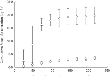
Cumulative faecal Se excretion (Fig. 1) differed significantly P , 0´05 between the two Se compounds; 90 % of the total faecal Se (74Se and76Se) was excreted
after 152 (SD 18) h and 65 (SD 21) h for 76SeO 422 and 74SeO22 respectively. The corresponding times for 95 %
excretion were 177 (SD14) h and 99 (SD21) h for76SeO422
and74SeO
322respectively P , 0´05: Cumulative urinary
Se excretion (Fig. 2) also differed significantly P , 0´05 between the two Se compounds; 90 % of the total urinary Se (74Se and76Se) was excreted after 40 (SD 30) and 121
(SD 51) h for 76SeO422 and 74SeO322 respectively. The
corresponding times for 95 % excretion were 84 (SD 52) and 144 (SD55) h for76SeO
422and74SeO322respectively
P , 0´05: Under the present experimental conditions, apparent Se retention based on cumulative faecal and urinary excretion of74Se and76Se showed that Se retention
was not significantly different between SeO322and SeO422
P . 0´05 based on 120 h (74SeO 322 44´7 (SD 8´0) %, 76SeO 422 50´0 (SD 7´2) %) or 168 h (74SeO322 42´3 (SD 8´2) %,76SeO 42247´6 (SD7´6) %). Apparent Se retention
based on 120 or 168 h cumulative faecal and urinary excretion did not differ significantly from data based on 9 d collections P . 0´05:
No effect of the order of administration of labelled test meals was detected for Se absorption, urinary excretion or retention. For 74SeO
322 Se retention was positively
76SeO
422 Se retention and urinary excretion were
nega-tively correlated (r 20´99, P , 0´05: Discussion
The present study demonstrated that in men, mean apparent Se retention was similar for SeO322and SeO422added to a
milk-based infant formula (41 % v. 46 %), although large differences in absorption and urinary excretion were found. The study design and the dose of 40 mg Se for each isotope were based on nutritional considerations. Preliminary
analytical data showed that accurate Se stable-isotope measurements in urine and faeces were possible after intake of 40 mg Se and demonstrated the analytical feasability of the study (Van Dael et al. 1998). The labelled test meals were administered in a crossover design on two consecutive days. A milk-based formula was selected as the test meal because milk is the most common raw material for dietetic foods such as enteral nutrition products or infant formulas. These milk-based formulas, often the only dietary source of energy and nutrients, are naturally low in Se and often fortified with Se in order to meet dietary Se requirements.
25.0 20.0 15.0 10.0 5.0 0.0 Cumulati ve faecal S e excretion ( µ g S e )
Period after administration of dose (h)
0 50 100 150 200 250
Fig. 1. Cumulative faecal selenium (74Se and76Se) excretion by healthy men after a single oral dose of 40 mg selenium as74SeO
322(K) or76SeO422(W) added to a milk-based formula. Values are means with their standard errors represented by vertical bars for ten subjects. For details of experimental procedures, see p. 159. 25 20 15 10 5 0 Cumulati ve urinary S e excretion ( µ g S e )
Period after administration of dose (h)
0 50 100 150 200 250
Fig. 2. Cumulative urinary selenium (74Se and76Se) excretion by healthy men after a single oral dose of 40 mg selenium as 74SeO
322 (K) or
76SeO
422(W) added to a milk-based formula. Values are means with their standard errors represented by vertical bars for ten subjects. For details of experimental procedures, see p. 159.
Thus, information about Se retention and absorption from these products at relevant fortification levels is needed. The daily Se intake in Switzerland is estimated to be 60±70 mg Se/d (Pfannhauser, 1994). The administration of 40 mg Se as SeO322 or SeO422 on two consecutive days increased
the total dietary Se intake of the subjects to levels comparable with the upper range of Se intakes in Europe (van Dokkum, 1995). Previous data on Se retention and absorption were based on Se doses ranging up to 200 mg Se (i.e. considerably higher than the daily dietary Se intake in most countries). Our data are therefore more relevant to Se retention and absorption from Se-fortified foods within the range of habitual dietary Se intakes in Europe. The subjects in the present study were healthy European men with adequate Se status, as judged from blood Se status indicators and 24 h urinary Se excretion (Robberecht & Deelstra, 1984; Van Dael & Deelstra, 1993). Se retention data for SeO322and SeO422found in the present study are
in agreement with those of Thomson & Robinson (1986), who reported Se retention from a 1000 mg dietary Se supplement in New Zealand adults using a chemical-balance technique (mean retention (%) SeO32240, SeO422
32). Percentage Se retention was comparable with that found in the present study despite the much higher doses of Se administered. Our data on Se retention from the test meal labelled with SeO322 are within the wide range of
reported results (30±75 %) based on stable-isotope techni-ques (Christensen et al. 1983; Sirichakwal et al. 1985; Martin et al. 1989; Mangels et al. 1990; Finley, 1999). The reasons for the wide range in SeO322retention values are
not known, although it can be assumed that factors such as differences in study design, stable-isotope dose, dietary regimen and normal Se intake may play a role.
Previous Se retention studies differed considerably in the number of days (5±21) used in the faecal and urinary collection period (Christensen et al. 1983; Sirichakwal et al. 1985; Martin et al. 1989; Mangels et al. 1990; Finley, 1999). Our data indicate that Se retention for SeO322and
SeO422based on 120 h collections of faeces and urine was
not significantly different from 9 d balances. However, although our data suggest that complete faecal and urine collection periods shorter than 9 d may be appropriate for studies of Se retention, it is important to consider carefully the collection time, in relation to factors such as gastrointestinal passage time and dietary habits.
Although retention of the two inorganic forms of Se was found to be similar, a large difference was observed in the percentage absorption of SeO322and SeO422; SeO422was
almost completely absorbed (91 %) compared with 50 % for SeO322. The between-subject variation in Se absorption
was larger for SeO322 (CV 15 %) than for SeO422 (CV
1´5 %), and confirmed previously reported data on inter-individual variations in SeO322 absorption (Martin et al.
1989). A wide range of apparent Se absorption values (35± 85 %) based on stable-isotope techniques have been reported for SeO322(Janghorbani et al. 1982; Christensen
et al. 1983; Kasper et al. 1984; Sirichakwal et al. 1985; Martin et al. 1989; Patterson et al. 1989; Mangels et al. 1990; Ducros et al. 1991; Finley, 1999). SeO322has been
reported to interact with the lumen content, and therefore it is likely that dietary habits influence Se absorption from
SeO322 (Vendeland et al. 1992). Martin et al. (1989)
reported that the percentage SeO322 absorption in adults
fed a Se-restricted diet (18 mg Se/d) was significantly higher than that of a group fed 118 mg Se/d (89 v. 58 respectively). Recently, Finley (1999) also reported sig-nificant differences in percentage SeO322 absorption
between subjects at low (32´6 mg Se/d) and high (226´5 mg Se/d) Se intake, but contrary to the data of Martin et al. (1989), higher mean Se absorption (38 % v. 15 %) was reported at the higher Se intake. As in the present study, Thomson & Robinson (1986) found high Se absorption from SeO422 (94 (SD 4) %) based on the
classical balance technique. Finley (1999) reported Se absorption from SeO422in the range of 68±76 % based on
a stable isotope technique. Our absorption data and those of Thomson & Robinson (1986) for both SeO422and SeO322
were substantially higher than those reported by Finley (1999). In agreement with the data in human subjects, higher Se absorption from SeO422 v. SeO322 was also
reported in animals (Wolffram et al. 1985; Vendeland et al. 1992). In those studies SeO422 was shown to be rapidly
transported across the brush border by a carrier-mediated mechanism, whereas SeO322 was absorbed by passive
diffusion and showed a strong tendency to bind to the brush border.
Renal excretion is the major pathway for elimination of absorbed Se (Oster & Prellwitz, 1990). In the present study Se excretion from SeO422was high; approximately half the
Se absorbed from SeO422 was re-excreted in urine
compared with only 20 % for Se absorbed from SeO322.
The urinary excretion of Se absorbed from SeO422was also
very rapid; 87 % of the total urinary Se excretion was recovered within 24 h of administration while 90 % of the total Se excretion of SeO322 was completed after 3 d.
Thomson & Robinson (1986) found similar differences for total urinary Se excretion and excretion kinetics between SeO422and SeO322after administration of a single dose of
Se (1 mg). Previous stable-isotope data in adults reported urinary excretion for SeO322 which was similar to our
findings (Martin et al. 1989). The rapid and high Se excretion from SeO422is probably related to its more rapid
absorption and metabolism compared with SeO322
(Thom-son & Robin(Thom-son, 1986).
As an alternative to inorganic Se, organic Se could be considered for food fortification. Se retention from organic Se compounds such as selenomethionine and Se-rich yeast has been demonstrated to be high at about 80±90 % (Mangels et al. 1990; Alfthan et al. 1991). Organic Se may not only be incorporated into Se-specific proteins such as GSH-Px and iodothyronine deiodinase but also non-specifically into blood proteins such as albumin and Hb due to homology between selenomethionine and methio-nine. It has been shown that organic and inorganic Se compounds are equally well utilised for incorporation into GSH-Px but that organic Se gives higher plasma and erythrocyte Se levels (Alfthan et al. 1991; Xia et al. 1992; Thomson et al. 1993; NeÁve, 1995). The metabolic similarity between selenomethionine and methionine has been suggested as the reason for Se toxicity at high Se intakes (Whanger, 1998).
at risk of low Se intake, such as patients with chronic or acute disease or infants, should ensure adequate absorption and retention of Se. Thus, fortification of nutritional products with SeO422 could be considered to be more
efficient than SeO322. Nevertheless, SeO422 may not be
the ideal fortificant for individuals with impaired renal function or preterm infants with immature renal function, since SeO422 might not be as efficiently excreted as in
healthy individuals. For patients with malabsorption syndromes SeO422may be the more appropriate
fortifica-tion compound, since Se absorpfortifica-tion from SeO322has been
shown to be markedly reduced in patients with short bowel syndrome (20 %) compared with healthy adults (82 %; Rannem et al. 1996). Finally, concern has been raised about the potential pro-oxidative properties of SeO322 and its
lower stability compared with SeO422when added to foods
(Smith et al. 1995; Tyrala et al. 1996). Although these issues need to be further investigated, they are additional factors potentially in favour of the use of SeO422 for Se
fortification of foods.
In conclusion, the results from the present study indicate that SeO322and SeO422, when administered within normal
dietary intake ranges, were equally well retained in healthy men, although large differences in absorption and urinary excretion were observed. Thus, the nutritional impact of foods fortified with either of these inorganic Se compounds can be assumed to be similar in healthy individuals, at least at the level of Se intake evaluated in the present study.
Acknowledgements
The authors are grateful for the expert technical assistance of Mrs K. Longet, Mrs S. Metairon, Mrs I. Bartholdi and Mrs J. Clough, and for the fruitful discussions with Dr R. Hurrell and Dr P. Kastenmayer.
References
Alfthan G, Aro A, Arvilommi H & Huttunen JK (1991) Selenium metabolism and platelet glutathione peroxidase activity in healthy Finnish men: effects of selenium yeast, selenite, and selenate. American Journal of Clinical Nutrition 53, 120±125. Beck MA (1996) The role of nutrition in viral disease. Journal of
Nutritional Biochemistry 7, 683±690.
Belsten JL & Wright AJA (1995) European Community ± FLAIR common assay for whole-blood glutathione peroxidase (GSH-Px). Results of an interlaboratory trial. European Journal of Clinical Nutrition 49, 921±927.
Casey CE & Hambidge KM (1985) Trace minerals. In Vitamin and Mineral Requirements in Preterm Infants, pp. 153±184 [RC Tsang, editors]. New York: Marcel Dekker.
Chandra RK (1997) Nutrition and the immune system: an introduction. American Journal of Clinical Nutrition 66, 460S±463S.
Christensen MJ, Janghorbani M, Steinke FH, Istfan N & Young VR (1983) Simultaneous determination of absorption of selenium from poultry meat and selenite in young men: application of a triple stable-isotope method. British Journal of Nutrition 50, 43±50.
Clark LC, Combs GF & Turnbull BW (1996) Effects of selenium supplementation for cancer prevention in patients with carcinoma of the skin. Journal of the American Medical Association 276, 1957±1963.
Ducros V, Favier A & Guigues M (1991) Selenium bioavailability as selenite (74Se) and as a selenium drug (76Se) by stable isotope methodology. Journal of Trace Elements and Electro-lytes in Health and Disease 5, 145±154.
Finley JW (1999) The retention and distribution by healthy young men of stable isotopes of selenium consumed as selenite, selenate or hydroponically-grown broccoli are dependent on the isotopic form. Journal of Nutrition 129, 865±871.
Hambidge KM (1985) Trace elements in human nutrition. In Nutrition in Pediatrics. Basic Science and Clinical Applica-tions, pp. 7±45 [WA Walker and JB Watkins, editors]. Boston, MA: Little Brown Co.
Hills M & Armitage P (1979) The two-period cross-over clinical trial. British Journal of Clinical Pharmacology 8, 7±20. Janghorbani M, Christensen MJ, Nahapetian A & Young VR
(1982) Selenium metabolism in healthy adults: quantitative aspects using the stable isotope74SeO
322. American Journal of
Clinical Nutrition 35, 647±654.
Kasper LJ, Young VR & Janghorbani M (1984) Short-term dietary selenium restriction in young adults: quantitative studies with the stable isotope 74SeO
322. British Journal of Nutrition 52,
443±455.
Kelly DA, Coe AW, Shenkin A, Lake BD & Walker-Smith JA (1988) Symptomatic selenium deficiency in a child on home parenteral nutrition. Journal of Gastroenterology and Nutrition 7, 783±786.
Levander OA (1984) The importance of selenium in total parenteral nutrition. Bulletin of the New York Academy of Medicine 60, 144±155.
Litov R & Combs G (1991) Selenium in pediatric nutrition. Pediatrics 87, 339±351.
Lockitch G (1989) Selenium: clinical significance and analytical concepts. CRC Critical Reviews in Clinical Laboratory Sciences 27, 483±539.
Makarem A (1974) Trace elements in human nutrition. In Principles and Techniques, 2nd ed., pp. 1125±1147 [RJ Henry, DC Cannon and JW Winkelman, editors]. Hagerstown, MD: Harper & Row.
Mangels AR, Moser-Veillon PB, Patterson KY & Veillon C (1990) Selenium utilization during human lactation by use of stable-isotope tracers. American Journal of Clinical Nutrition 52, 621±627.
Martin RF, Janghorbani M & Young VR (1989) Experimental selenium restriction in healthy adult humans: changes in selenium metabolism studied with stable-isotope methodology. American Journal of Clinical Nutrition 49, 854±861.
National Research Council (1989) Recommended Dietary Allow-ances, 10th ed. Washington, DC: National Academy Press. NeÁve J (1995) Human selenium supplementation as assessed by
changes in blood selenium concentration and glutathione peroxidase activity. Journal of Trace Elements in Medicine and Biology 9, 65±76.
Oster A & Prellwitz W (1990) The renal excretion of selenium. Biological Trace Element Research 24, 119±146.
Patterson BH, Levander OA, Helzlsouer K, McAdam PA, Lewis SA, Taylor PR, Veillon C & Zech LA (1989) Human selenite metabolism: a kinetic model. American Journal of Physiology 257, R556±R567.
Pfannhauser W (1994) Neue Forschungsergebnisse uÈber Selen (New research data on selenium). ErnaÈrhrung 18, 75±76. Rannem T, Hylander E, Ladefoged K, Staun M, Tjellesen L &
Jarnum S (1996) The metabolism of [75Se]selenite in patients
with short bowel syndrome. Journal of Parenteral and Enteral Nutrition 20, 412±416.
Robberecht HJ & Deelstra HA (1984) Selenium in human urine. Determination, speciation and concentration levels. Talanta 7, 497±508.
SandstroÈm B, Fairweather-Tait S, Hurrell R & van Dokkum W (1993) Methods for studying mineral and trace element absorption in humans using stable isotopes. Nutrition Research Reviews 6, 71±95.
Sirichakwal PP, Young VR & Janghorbani M (1985) Absorption and retention of selenium from intrinsically labeled egg and selenite as determined by stable isotope studies in humans. American Journal of Clinical Nutrition 41, 264±269.
Smith AM, Chen LW & Thomas MR (1995) Selenate fortification improves selenium status of term infants fed soy formula. American Journal of Clinical Nutrition 61, 44±47.
Thomson CD & Robinson MF (1986) Urinary and fecal excretions and absorption of a large supplement of selenium: superiority of selenate over selenite. American Journal of Clinical Nutrition 44, 659±663.
Thomson CD, Robinson MF, Butler JA & Whanger PD (1993) Long-term supplementation with selenate and selenomethio-nine: selenium and glutathione peroxidase (EC 1.11.1.9) in blood components of New Zealand women. British Journal of Nutrition 69, 577±588.
Turnlund JR, Keyes WR & Peiffer GL (1993) Isotope ratios of molybdenum determined by thermal ionisation mass spectro-metry for stable isotope studies of molybdenum metabolism in humans. Analytical Chemistry 65, 1717±1722.
Tyrala EE, Borschel MW & Jacobs JR (1996) Selenate fortification of infant formulas improves the selenium status of preterm infants. American Journal of Clinical Nutrition 64, 860±865.
Van Dael P, Barclay D, Longet K, Metairon S & Fay LB (1998) Determination of selenium stable isotopes by gas
chromatography-mass spectrometry with negative chemical ionisation. Journal of Chromatography 715B, 314±317. Van Dael P & Deelstra H (1993) Selenium. International Journal
of Vitamin and Nutrition Research 63, 312±316.
Van Dael P, Van Cauwenbergh R, Deelstra H & Calomme M (1995) Determination of selenium in human serum by long-itudinal Zeeman correction and flow injection hydride genera-tion atomic absorpgenera-tion spectrometry. Atomic Spectroscopy 16, 251±257.
van Dokkum W (1995) The intake of selected minerals and trace elements in European countries. Nutrition Research Reviews 8, 271±302.
Vendeland SC, Butler JA & Whanger PD (1992) Intestinal absorption of selenite, selenate, and selenomethionine in the rat. Journal of Nutritional Biochemistry 3, 359±365.
Vinton NE, Dahlstrom KA, Strobel CT & Ament ME (1987) Macrocytosis and pseudo-albinism: manifestations of selenium deficiency. Journal of Pediatrics 111, 711±777.
Whanger PD (1998) Metabolism of selenium in humans. Journal of Trace Elements in Experimental Medicine 11, 227±240. Wolffram S, ArduÈser F & Scharrer E (1985) In vivo intestinal
absorption of selenate and selenite by rats. Journal of Nutrition 115, 454±459.
Xia Y, Zhao X, Zhu L & Whanger PD (1992) Metabolism of selenate and selenomethionine by a selenium-deficient popula-tion of men in China. Journal of Nutripopula-tional Biochemistry 3, 202±210.
Zachara BA (1993) Mammalian selenoproteins. Journal of Trace Elements in Health and Disease 6, 137±151.