LABORATORY INVESTIGATION
Ocular penetration of caspofungin in a rabbit uveitis model
David Goldblum&Kathrin Fausch&Beatrice E. Frueh&
Regula Theurillat&Wolfgang Thormann&
Stefan Zimmerli
Received: 26 May 2006 / Revised: 11 September 2006 / Accepted: 16 September 2006 / Published online: 22 November 2006 # Springer-Verlag 2006
Abstract
Background Little is known about the ocular penetration of echinocandin antifungals. We studied the ocular distribution of systemically administered caspofungin in a rabbit uveitis model.
Methods Caspofungin (1 mg/kg per day) was given intravenously to rabbits as a single dose or as repeated daily doses on 7 days starting 24 h after induction of unilateral uveitis by intravitreal endotoxin injection. Cas-pofungin concentrations were determined by high-perfor-mance liquid chromatography in the cornea, aqueous humor, vitreous humor, and serum 4, 8, 16, and 24 h after
administration of a single dose and 24 h after the last of seven doses.
Results The mean caspofungin concentration in the aque-ous of the inflamed eye 4 and 8 h after single-dose
administration was 1.30 ± 0.39 μg/ml and 1.12±0.34
μg/ml, respectively. Drug concentrations decreased to
0.24±0.09 μg/ml at 16 h and 0.26±0.14 μg/ml at 24 h.
In the vitreous of inflamed eyes drug levels were undetectable at all time points. No drug was found in the aqueous of inflamed eyes 24 h after the last of seven repeated doses, and the vitreous only contained trace amounts. In the corneas of inflamed eyes concentrations
reached 1.64±0.48 μg/g at 4 h, peaked at 2.16±1.14 μg/g
at 8 h, and declined to 1.87±0.52μg/g and 1.49±0.48 μg/g
at 16 and 24 h, respectively. After repeated dosing, corneal
concentrations of caspofungin were 0.8 and 1.0 μg/g and
below the limit of detection in two of four animals. In non-inflamed eyes no drug was detectable in the aqueous and vitreous humor, and the corneas at any time point.
Conclusions In our model, caspofungin reached therapeu-tically relevant levels in the aqueous and cornea but not in the vitreous humor of inflamed eyes. Intraocular drug deposition was critically dependent on a disrupted blood-eye barrier. These findings suggest a limited role for caspofungin in the treatment of fungal endophthalmitis. Keywords Caspofungin . Pharmacokinetics .
Ocular penetration . Animal model
Introduction
Caspofungin is a first-in-class echinocandin with potent antifungal activity against Candida and Aspergillus spp., the dominant human fungal pathogens. In vitro and in vivo This study was supported in part by an independent research grant
from Merck & Co., Inc., Switzerland. S.Z. has received additional independent research grants and drugs from the same organisation which had no influence on the design of the present study, the data analysis and the interpretation of results. We the authors have full control of all primary data and we agree to allow Graefe’s Archive for Clinical and Experimental Ophthalmology to review our data upon request.
D. Goldblum
:
K. Fausch:
B. E. FruehClinic of Ophthalmology, University Hospital, Inselspital,
3010 Bern, Switzerland
K. Fausch
:
R. Theurillat:
W. ThormannDepartment of Clinical Pharmacology, University of Bern,
Murtenstr. 35, 3010 Bern, Switzerland
S. Zimmerli (*)
Institute for Infectious Diseases, University of Bern,
P.O. Box 61, Friedbuehlstr. 51, 3010 Bern, Switzerland
caspofungin is fungicidal against nearly all Candida spp. including fluconazole-resistant strains. Randomized clinical trials with caspofungin in patients with candidemia, invasive candidiasis, and Candida esophagitis demonstrate equivalent efficacy to amphotericin B (AmB), with
sub-stantially fewer toxic effects [1, 5, 16, 24]. In addition,
caspofungin is as effective as liposomal AmB in the empiric antifungal treatment of neutropenic patients with persistent fever and has been licensed for the treatment of severe pulmonary aspergillosis in patients who fail to
respond or are intolerant to therapy with AmB [26].
Fungal infections of the eyes are important causes of morbidity and blindness; certain ophthalmic mycoses may
even be life-threatening [14, 28]. Keratitis is the most
frequent manifestation [20], but intraocular structures as
well as orbit, lids, lacrimal apparatus, conjunctiva, and sclera may also be infected.
A recently published case series suggests that caspofun-gin in combination with voriconazole may be useful for the
treatment of endogenous Candida endophthalmitis [2]. The
same drug combination has been used to successfully treat a patient with endophthalmitis due to Aspergillus fumigatus
[6]. Clinical reports on the utility of caspofungin
mono-therapy for ocular infections are scarce and conflicting. Cure has been reported for a patient with endogenous Candida glabrata endophthalmitis presenting with a single
retinal lesion and mild retinitis [18]. In another reported
case of an ocular yeast infection, a patient with more advanced endogenous endophthalmitis due to C. albicans failed to respond to caspofungin. After 9 days of treatment with standard doses, his intravitreal caspofungin level was
undetectable by a sensitive assay [8]. Successful treatment
with caspofungin has also been reported in a patient with Acremonium endophthalmitis that failed to respond to AmB
[4]. In a rabbit model, topical caspofungin was as effective
as AmB for the treatment of Candida keratitis [9].
Caspofungin concentrations in homogenates of whole non-inflamed rat eyes 0.5 and 24 h after a 2.0-mg/kg i.v. bolus injection of radioactively labeled drug were 0.52 and
0.30μg Eq/g, respectively [23]. Due to its high molecular
weight (1,213 kD) caspofungin may not cross the unin-flamed blood-eye barrier that is thought to be impermeable for molecules exceeding 500 kD. Indeed, the degree of rupture of the blood-eye barrier in inflammation may be critical for the intraocular deposition of this and other echinocandins.
Only detailed knowledge of the pharmacokinetics of caspofungin in ocular inflammation will provide a base for rational treatment decisions. The disposition of caspofungin to various ocular compartments after systemic treatment is unknown. The aim of this study was to characterize the ocular distribution of intravenously administered caspofun-gin in a rabbit uveitis model. For that purpose,
high-performance liquid chromatography (HPLC) assays for the determination of caspofungin concentrations in the cornea, aqueous humor, vitreous humor, and serum had to be developed.
Materials and methods Animals
Twenty adult (nine male) Burgundy fawn rabbits (2.3– 4.9 kg) were used in this study. They were provided by an authorized breeding center and were kept in individual cages under well-defined and standardized conditions (in a humidity and temperature controlled room, with a cycle of 13 h of light and 11 h of dark). They received standard dry food and water ad libitum. All eyes were initially examined with a handheld slit lamp. Only animals without any signs of ocular inflammation were included. All experiments
were conducted in accordance with the “Principles of
laboratory animal care” (NIH publication No. 85-23, revised 1985), the OPRR Public Health Service Policy on the Humane Care and Use of Laboratory Animals (revised 1986), and the US Animal Welfare Act, as amended, and were approved by the Swiss Federal and local Ethics and Agricultural Committees.
Anesthesia
Animals were anesthetized with intramuscular ketamine (35 mg/kg of body weight; Ketalar, Parke-Davis, Ann Arbor, MI, USA) and xylazine (5 mg/kg; Xylapan, Chassot, Bern, Switzerland). Novocaine 0.2% (Inselspital Pharmacy, Bern, Switzerland) eye drops were used for topical anesthesia. All rabbits were anesthetized once for the induction of uveitis and placement of the central venous catheter. This vascular access was established by surgical placement of a subcutaneous silastic central venous catheter that permitted one-time or repeated drug administrations
without further anesthesia [25].
Induction of experimental uveitis
After washing the ocular surface with sterile novocaine 0.2%, 100 ng (for animals given a single drug dose) or
50 μg (for animals given multiple drug doses) of
lipopolysaccharide (LPS) from Escherichia coli (Sigma,
St. Louis, MO, USA) diluted in 10 μl of sterile saline
solution were injected [13]. The higher LPS dose was
administered to maintain intraocular inflammation over the
treatment period [7]. LPS was injected through the sclera
into the vitreous using a 30–1/2 G needle connected to a Hamilton syringe. The injection was performed in the right
eye of each animal, taking care to avoid damage to the lens. The left eyes served as controls. Inflammation was evaluated by clinical observation of iris and conjunctival hyperemia.
Single-dose studies
Single-dose kinetics of caspofungin acetate (Cancidas, Merck, Whitehouse Station, NJ, USA) were studied over 24 h. Immediately before use, caspofungin acetate was dissolved in sterile 0.9% sodium chloride to a concentration of 0.5 mg/ml. Twenty-four hours after induction of uveitis, 16 rabbits were given 1 mg/kg caspofungin over 6 min by steady bolus injection via a central venous catheter. The drug dosage used corresponds to the standard dose recommended for the treatment of human fungal infections and is well tolerated by rabbits. Four animals per time point were sacrificed by cervical dislocation and subsequent exsanguination at 4, 8, 16, and 24 h after drug administra-tion. Blood samples were collected during bleeding, and serum was separated by centrifugation. Aqueous humor was drawn from the freshly enucleated eyes with a tuberculin syringe using a 30-gauge needle. The cornea was excised at the limbus. After sectioning the eyes just behind the lens, vitreous humor was obtained by dissecting it carefully from the retina.
Multiple-dose study
Twenty-four hours after induction of uveitis, four rabbits were given doses of 1 mg/kg caspofungin by steady intravenous bolus via a central venous catheter that were repeated daily for 7 consecutive days (total seven doses). Twenty-four hours after the last dose, the rabbits were sacrificed and samples were prepared for examination as described above.
Analytical procedures
Samples were stored at −70°C until analysis. Blank
aqueous humor, blank vitreous humor, and blank corneas were obtained from porcine eyes collected at the abattoir. Blank plasma was prepared from bovine blood. Caspofun-gin acetate was a kind gift of D. Sanglard, Institute of Microbiology, University of Lausanne, Lausanne, Switzer-land, and 4-hexylresorcinol (internal standard) was from Fluka (Buchs, Switzerland). Chemicals used were of analytical or HPLC grade. Caspofungin in serum and plasma was extracted using Sep-Pak C18 extraction cartridges (Waters, Milford, MA, USA) conditioned with 1 ml of methanol and 2 ml of distilled water. Two hundred
and fifty μl of serum or plasma mixed with 25 μl of a
12.5μg/ml internal standard solution and 750 μl of water
was centrifuged at 14,000 g for 2 min and applied to the cartridge. After rinsing with 2 ml water, the sample was eluted with 1 ml of methanol containing 4% concentrated acetic acid, evaporated to dryness at 40°C under a gentle
stream of air, and reconstituted in 225μl of mobile phase.
For analysis of aqueous humor, 100μl of sample and 20 μl
of a 1.25μg/ml internal standard solution were mixed with
200 μl of methanol containing 4% acetic acid. After
centrifugation and evaporation, it was reconstituted in
100 μl of mobile phase prior to ultrasonic mixing
(Bransonic 92, Bender & Hobein, Zurich, Switzerland) for 5 min, and in case of turbid samples, centrifugation at 14,000 g for 1 min. Vitreous samples were homogenized with an Ultra-Turrax microhomogenizer
(IKA-Labortech-nik, Staufen, Switzerland) at high speed for 10 s; 125μl of
vitreous, 25 μl of a 1.25 μg/ml internal standard solution,
and 250 μl of methanol containing 4% acetic acid were
mixed, centrifuged at 14,000 g for 1 min, and the clear
supernatant was evaporated and redissolved in 125 μl of
mobile phase. Pieces of cornea weighing 20–50 mg were
added to 100 μl of distilled water, vortex mixed, and
centrifuged at 14,000 g. The sample was cut into small
pieces with scissors, mixed with 20 μl of a 1.25 μg/ml
internal standard solution, sonicated for 15 min, diluted
with 200 μl of methanol containing 4% acetic acid, and
centrifuged. The supernatant was evaporated and
reconsti-tuted with 100 μl of mobile phase. Recoveries for
caspofungin in serum, aqueous, vitreous, and cornea were determined to be approximately 65, 86, 91, and 87%, respectively.
Drug levels were determined as total caspofungin concentrations by HPLC with fluorescence detection using
a protocol adapted from [19]. Briefly, an LC-Module I plus
autosampler and pump (Waters, Milford, MA, USA) were connected to an LC 304 fluorescence detector (Linear Instruments, Reno, NV, USA). A 250/8/4.6 Nucleosil 300 wide bore C8 column (Macherey-Nagel, Düren, Germany) packed with 5 micron material was used. The mobile phase comprised two solutions (A and B) that were mixed in a volume/volume ratio (v/v) of 62:38 for vitreous and aqueous and a ratio of 60:40 (v/v) for serum and cornea. Solvent A was 0.1% trifluoroacetic acid (adjusted to pH 3 with diethylamine) and solvent B was acetonitrile. After a run time of 11 min, the column was washed for 4 min with a solution containing solvents A and B in a ratio (v/v) of 50:50 and reequilibrated for 5 min with mobile phase. Elution was performed at a flow rate of 1.4 ml/min for vitreous and aqueous, 1.3 ml/min for cornea, and 1.0 ml/min for serum. The column temperature was maintained at 30°C. Excitation and emission wavelengths were set at
224 and 300 nm, respectively. For all assays, 50 μl of
sample was injected. The concentrations of the lowest
aqueous and vitreous, and 0.025μg for cornea (for 40 mg
cornea: 0.625 μg/g cornea), were taken as quantification
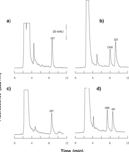
limits. Intraday variabilities (n=4) were <7% in all cases. All control samples were stable during the 3-month study period and interday variabilities were below 10% for plasma, aqueous, and vitreous, and below 20% for cornea. Typical electropherograms obtained with aqueous humor
are presented in Fig.1.
Results Aqueous
To study drug penetration into the aqueous of inflamed and uninflamed control eyes, samples harvested immediately postmortem from eyes with iatrogenic uveitis leading to disruption of blood-eye barriers were analyzed for
caspo-fungin concentration. Results were compared to those obtained from similarly harvested aqueous from contralat-eral, uninflamed eyes with intact blood-eye barriers.
Four and 8 h after intravenous injection of a single dose of 1 mg/kg caspofungin, the mean drug concentration in the aqueous of the inflamed eye was 1.30±0.39 and 1.12±
0.34μg/ml, respectively. These concentrations are in excess
of the minimum inhibitory concentration of most Candida and Aspergillus spp. Sixteen and 24 h after treatment, the
concentration decreased to 0.24±0.09 and 0.26±0.14μg/ml,
respectively (Fig.2a). Caspofungin was undetectable in the
aqueous of contralateral non-inflamed left eyes at all time points.
a)
b)
c)
d)
0 4 8 12 IST 20 mAU 0 4 8 12 0 4 8 12 IST CAS 0 4 8 12 CAS IST ISTTime (min)
Fluorescence
(300
nm)
Fig. 1 Typical chromatograms of caspofungin (CAS). a Blank porcine aqueous fortified with internal standard (IST). b Four-fold diluted aqueous of an in-flamed rabbit eye harvested 4 h after a single-dose injection
containing 1.30μg/ml
caspo-fungin. c Aqueous of a non-inflamed eye obtained 8 h after a single dose of caspofungin. d Porcine aqueous spiked with caspofungin at a concentration
of 0.5μg/ml
Fig. 2 a–c Mean (±SD) caspofungin concentration over time in a
aqueous, b corneas of LPS-treated eyes, and c serum of rabbits given a single intravenous dose of caspofungin at 1 mg/kg of body weight (n = 4 per time point)
0.0 0.5 1.0 1.5 2.0 2.5 3.0 3.5 4 8 16 24 time (h) A q ue ous c a s pof ungi n c o nc e n tr a ti o n (µ g /m l) 0 0.5 1 1.5 2 2.5 3 3.5 4 8 16 24 time (h)
Corneal caspofungin concentration
(µg/ g) 0 0.5 1 1.5 2 2.5 3 3.5 4 8 16 24 time (h)
Serum caspofungin concentration
(µg/ml)
a
b
In the multiple-dose study animals were treated with repeated daily doses of 1 mg/kg of body weight for 7 days. Caspofungin concentration was below the level of deter-mination in all the aqueous samples of inflamed and control eyes collected 24 h after the last of seven doses
(Fig.3).
Vitreous
After single-dose administration of 1 mg/kg caspofungin, drug concentrations in all the vitreous samples from both inflamed and non-inflamed eyes were below the level of
detection (0.05μg/ml). Twenty-four hours after the last of
seven intravenous doses, caspofungin levels were undetect-able in all vitreous samples with one exception: in one
inflamed eye a caspofungin concentration of 0.08 μg/ml
was found (Fig.3).
Cornea
Topical antifungals are the treatment of choice for fungal keratitis because high drug concentrations can be achieved in the cornea, particularly when the corneal epithelium is damaged, and systemic toxicity is minimal. In advanced infections with threatening penetration into the anterior chamber, however, systemic antifungals are often added. We were therefore interested in determining the corneal penetration of intravenous caspofungin. Four hours after single-dose treatment, the mean caspofungin concentration
in the cornea of inflamed eyes was 1.64 ±0.48 μg/g.
Corneal drug concentrations also exceeded aqueous levels after 8, 16, and 24 h at 2.16±1.14, 1.87± 0.52, and 1.49±
0.48μg/g, respectively (Fig.2b). Caspofungin levels in the
corneas of uninflamed eyes were undetectable at all time points, suggesting that disruption of the blood-eye barriers was critical for corneal drug deposition.
Corneal drug concentrations in inflamed eyes of rabbits
treated with multiple doses were 0.82μg/g, and 1.03 μg/g
in two animals, and undetectable in the other two (Fig.3).
Caspofungin was below the level of detection (0.625μg/g)
in uninflamed eyes after repeated daily dosing. Serum
Four hours after single-dose administration of caspofungin, the mean drug concentration in serum was 1.91±0.46
μg/ml (Fig. 2c). It fell to 0.47±0.11, 0.08±0.01, and 0.03
±0.01 μg/ml after 8, 16, and 24 h, respectively. Serum
level 24 h after the last of seven daily caspofungin doses
was 0.17±0.06μg/ml (Fig.3)
Discussion
Despite treatment, fungal endophthalmitis results in loss of useful vision in a majority of patients. New antifungal drugs with good ocular penetration would be a welcome addition to our therapeutic armamentarium. In spite of its established efficacy for the treatment of a broad range of infections due to Candida and Aspergillus spp., caspofun-gin has only rarely been used to treat fungal infections of
the eyes and no clinical studies have been done [1,16,24].
In this study we aimed to characterize the penetration into various ocular compartments of systemically administered caspofungin in a rabbit uveitis model. For that purpose, adequate assays had to be at hand. Several HPLC assays for the measurement of caspofungin concentrations in plasma, urine, and various tissues have been described in the literature
[3, 11, 12, 17, 19]. No assay for the determination of
caspofungin in ocular tissues has been found. The HPLC
method of Schwartz et al. [19] was adapted to quantify
caspofungin in samples of aqueous, vitreous, cornea, and
0 0.1 0.2 0.3 0.4 0.5 0.6 0.7 0.8 0.9 1
aqueous vitreous cornea serum
C a s pof ungi n c onc e n tr a ti on (µ g /m l, µ g /g r esp e c ti ve ly )
Fig. 3 Mean (+SD) caspofun-gin concentrations in aqueous, vitreous, and corneas of in-flamed eyes as well as serum after repeated-dose administra-tion. Samples were taken 24 h after the last of seven daily doses of 1 mg/kg caspofungin administered via a central ve-nous catheter (n = 4). Drug lev-els were undetectable in the aqueous and the vitreous
serum/plasma. The assays, which were found to be sensitive and robust, are all based upon the use of a C8 column and mobile phases composed of an aqueous, 0.1% trifluoroacetic acid solution at pH 3, and acetonitrile, but differ in sample preparation. Solid phase extraction with C18 cartridges is employed for serum analysis, whereas protein precipitation with acidified methanol is used for the determination of caspofungin in aqueous, vitreous, and cornea. The lower limit of quantification (LLQ) of our assay for serum and
plasma was 0.1μg/ml and was significantly lower for ocular
tissues. This compares favorably with the sensitivity of
published HPLC assays with LLQs for plasma of 0.15μg/ml
[11] and 0.125μg/ml [17]. In the paper by [19] a diol solid
phase extraction column was used to extract caspofungin from human plasma and urine. In our laboratory, the use of such a polar sorbent did not reveal any recovery for the investigated cyclic hexapeptide. With a C18 cartridge, however, a respectable recovery was obtained. Furthermore, there was no difference between plasma and serum.
In inflamed eyes caspofungin concentrations exceeding
1μg/ml were measured in the aqueous during the first 8 h
postdosing and in the cornea for 24 h after single-dose administration. These concentrations exceed the minimal inhibitory concentrations of most Candida and Aspergillus spp. and are associated with relevant antifungal activity in
animal models [15].
Corneal concentrations in inflamed eyes substantially exceeded both serum and aqueous levels: they peaked at
8 h and remained above 1.4μg/ml for 24 h. The absence of
detectable drug levels from corneas of uninflamed eyes suggests that disruption of the blood-eye barrier is as critical for corneal penetration as for other ocular compart-ments. The same observation also makes it improbable that significant amounts of drug might have penetrated through the tear film although drug concentrations in tears were not established.
In murine models, the kidneys, liver, and large intestine were found to have higher exposure levels to caspofungin than
plasma and serum [12,15]. Differences in the extent of protein
binding may explain the high corneal drug concentrations we found. Retention of the water-soluble caspofungin macromol-ecule in the proteoglycans of the corneal stroma may be another reason for corneal drug accumulation.
Caspofungin levels in the vitreous were undetectable in all animals after single-dose administration and in all but one after repeated dosing. Thus, even in the presence of a disrupted blood-eye barrier caspofungin appears to poorly penetrate into this compartment. A similar finding was recently reported in a patient whose Candida albicans endophthalmitis failed to respond to caspofungin. After 9 days of therapy, the level of caspofungin in the vitreous of his infected eye was below 50 ng/ml while his blood level
at the time of vitrectomy was 3.3μg/ml [8].
Using caspofungin at higher doses than currently recommended might result in higher intraocular drug deposition and possibly increased efficacy; reports on paradoxical effects of increased caspofungin doses in animal models, however, warrant caution with this
ap-proach [21,22,27].
The serum concentrations of caspofungin we measured at 4 and 8 h postdosing are in good agreement with reported
results for rabbit plasma [11]. This is also true for the slight
drug accumulation we observed in serum following administration over 7 days. Compared to serum drug levels we found ocular caspofungin concentrations to decline more slowly, an observation that is consistent with the reported plasma pharmacokinetics of caspofungin that are
largely determined by tissue distribution [12].
The unilateral uveitis model we used allows for the direct observation, in the same animal, of the influence on drug disposition of a disrupted blood-eye barrier. With a sensitive HPLC assay drug levels were undetectable in aqueous, vitreous, and cornea of non-inflamed eyes. Our findings suggest that caspofungin does not penetrate the intact blood-eye barrier. This may be explained in part by the drug’s high molecular weight and its level of protein
binding in serum of 96% [12].
Repeated dosing was not found to lead to higher intraocular drug concentrations. On the contrary, caspofun-gin levels were lower in both aqueous and cornea than at the same interval after single-dose administration. This may be due to a decreasing intensity of LPS-induced uveitis over time in spite of the fact that higher endotoxin doses
were used in the multiple-dose study [10]. There is no
evidence that in the absence of fungal infection caspofungin may directly influence the host’s immune response and thus modulate the intensity of, e.g., iatrogenic uveitis.
In established fungal endophthalmitis the treatment of choice is vitrectomy followed by local injection of an antifungal agent, usually amphotericin B. In endogenous endophthalmitis that results from hematogenous fungal dissemination, additional systemic treatment is indicated. In cases where vitrectomy must be postponed or where it may be contraindicated, control of endophthalmitis must rely on the local effects of systemically administered
antifungals [2]. The poor vitreal penetration of caspofungin
in our experimental system suggests it may be of limited usefulness in the treatment of endogenous endophthalmitis where hematogenously disseminated fungal elements reach the vitreous first, after establishing infection in the choroid. At present, the i.v. administration of an antifungal agent is not standard for the treatment of fungal keratitis. Nevertheless, many clinicians use adjuvant systemic treat-ment to prevent spread into the anterior chamber and progression to exogenous endophthalmitis. In this chal-lenging clinical situation there might only be minimal
intraocular inflammation. Because caspofungin only rea-ches potentially therapeutic concentrations in cornea and aqueous humor when the blood-eye barrier is disrupted, its role as preemptive treatment in exogenous fungal endoph-thalmitis may be equally limited.
Caution is warranted in extrapolating the results of this rabbit model of iatrogenic uveitis to human fungal endophthalmitis. While caspofungin has similar pharmaco-kinetics in humans and rabbits and the rabbit model is of established relevance for human eye diseases, the disrup-tion of the blood-eye barrier resulting from endotoxin-induced uveitis may differ from that found in fungal
endophthalmitis [17].
In summary, our model suggests that caspofungin ocular penetration is critically dependent on disruption of the blood-eye barrier. This may limit its usefulness in fungal endophthalmitis. Studies in animals with fungal endoph-thalmitis are needed to further evaluate the drug’s potential.
Acknowledgements The authors thank Gyula Vucic and Daniel
Mettler, DVM from the Department of Clinical Research, University of Bern, for catheter implantation; Monika Kilchenmann and Hans Liechti, members of the ophthalmic laboratory, for expert technical assistance and Dr. S. Shaw for his kind assistance in the SPE serum/ plasma extraction of caspofungin.
This study was supported in part by an independent research grant from Merck & Co., Inc., Switzerland.
References
1. Arathoon EG, Gotuzzo E, Noriega LM, Berman RS, DiNubile MJ, Sable CA (2002) Randomized, double-blind, multicenter study of caspofungin versus amphotericin B for treatment of oropharyngeal and esophageal candidiases. Antimicrob Agents
Chemother 46:451–457
2. Breit SM, Hariprasad SM, Mieler WF, Shah GK, Mills MD, Grand MG (2005) Management of endogenous fungal endoph-thalmitis with voriconazole and caspofungin. Am J Ophthalmol 139:135–140
3. Chavez-Eng CM, Schwartz M, Constanzer ML, Matuszewski BK (1999) Determination of a cyclic hexapeptide, a novel antifungal agent, in human plasma by high-performance liquid chromatog-raphy with ion spray and turbo ion spray tandem mass spectrometric detection. J Chromatogr B Biomed Sci Appl
721:229–238
4. Cornely OA, Schmitz K, Aisenbrey S (2002) The first
echino-candin: caspofungin. Mycoses 45(Suppl)3:56–60
5. Denning DW (2003) Echinocandin antifungal drugs. Lancet
362:1142–1151
6. Durand ML, Kim IK, D’Amico DJ, Loewenstein JI, Tobin EH,
Kieval SJ, Martin SS, Azar DT, Miller FS 3rd, Lujan BJ, Miller JW (2005) Successful treatment of Fusarium endophthalmitis with voriconazole and Aspergillus endophthalmitis with voriconazole
plus caspofungin. Am J Ophthalmol 140:552–554
7. Francis P, Lee JW, Hoffman A, Peter J, Francesconi A, Bacher J, Shelhamer J, Pizzo PA, Walsh TJ (1994) Efficacy of unilamellar liposomal amphotericin B in treatment of pulmonary aspergillosis in persistently granulocytopenic rabbits: the potential role of
bronchoalveolar d-mannitol and serum galactomannan as markers
of infection. J Infect Dis 169:356–368
8. Gauthier GM, Nork TM, Prince R, Andes D (2005) Subthera-peutic ocular penetration of caspofungin and associated treatment failure in Candida albicans endophthalmitis. Clin Infect Dis 41:
e27–e28
9. Goldblum D, Frueh BE, Sarra GM, Katsoulis K, Zimmerli S (2005) Topical caspofungin for treatment of keratitis caused by Candida albicans in a rabbit model. Antimicrob Agents Chemother 49:1359–1363
10. Goldblum D, Rohrer K, Frueh BE, Theurillat R, Thormann W, Zimmerli S (2002) Ocular distribution of intravenously adminis-tered lipid formulations of amphotericin B in a rabbit model.
Antimicrob Agents Chemother 46:3719–3723
11. Groll AH, Gullick BM, Petraitiene R, Petraitis V, Candelario M, Piscitelli SC, Walsh TJ (2001) Compartmental pharmacokinetics of the antifungal echinocandin caspofungin (MK-0991) in rabbits.
Antimicrob Agents Chemother 45:596–600
12. Hajdu R, Thompson R, Sundelof JG, Pelak BA, Bouffard FA, Dropinski JF, Kropp H (1997) Preliminary animal pharmacoki-netics of the parenteral antifungal agent mk-0991 (L-743,872).
Antimicrob Agents Chemother 41:2339–2344
13. Kulkarni PS, Mancino M (1993) Studies on intraocular inflam-mation produced by intravitreal human interleukins in rabbits. Exp
Eye Res 56:275–279
14. Levin LA, Avery R, Shore JW, Woog JJ, Baker AS (1996) The spectrum of orbital aspergillosis: a clinicopathological review. Surv Ophthalmol 41:142–154
15. Louie A, Deziel M, Liu W, Drusano MF, Gumbo T, Drusano GL (2005) Pharmacodynamics of caspofungin in a murine model of systemic candidiasis: importance of persistence of caspofungin in tissues to understanding drug activity. Antimicrob Agents
Chemother 49:5058–5068
16. Mora-Duarte J, Betts R, Rotstein C, Colombo AL, Thompson-Moya L, Smietana J, Lupinacci R, Sable C, Kartsonis N, Perfect J (2002) Comparison of caspofungin and amphotericin B for
invasive candidiasis. N Engl J Med 347:2020–2029
17. Sandhu P, Xu X, Bondiskey PJ, Balani SK, Morris ML, Tang YS, Miller AR, Pearson PG (2004) Disposition of caspofungin, a novel antifungal agent, in mice, rats, rabbits, and monkeys.
Antimicrob Agents Chemother 48:1272–1280
18. Sarria JC, Bradley JC, Habash R, Mitchell KT, Kimbrough RC, Vidal AM (2005) Candida glabrata endophthalmitis treated successfully with caspofungin. Clin Infect Dis 40:e46-e48 19. Schwartz M, Kline W, Matuszewski B (1997) Determination of a
cyclic hexapeptide (L-743 872), a novel pneumocandin antifungal agent in human plasma and urine by high-performance liquid chromatography with fluorescence detection. Anal Chim Acta
352:299–307
20. Srinivasan R, Kanungo R, Goyal JL (1991) Spectrum of oculomycosis in south India. Acta Ophthalmol (Copenh)
69:744–749
21. Stevens DA, Espiritu M, Parmar R (2004) Paradoxical effect of caspofungin: reduced activity against Candida albicans at high
drug concentrations. Antimicrob Agents Chemother 48:3407–
3411
22. Stevens DA, White TC, Perlin DS, Selitrennikoff CP (2005) Studies of the paradoxical effect of caspofungin at high drug
concentrations. Diagn Microbiol Infect Dis 51:173–178
23. Stone JA, Xu X, Winchell GA, Deutsch PJ, Pearson PG, Migoya EM, Mistry GC, Xi L, Miller A, Sandhu P, Singh R, deLuna F, Dilzer SC, Lasseter KC (2004) Disposition of caspofungin: role of distribution in determining pharmacokinetics in plasma. Antimicrob Agents Chemother 48:815–823
24. Villanueva A, Arathoon EG, Gotuzzo E, Berman RS, DiNubile MJ, Sable CA (2001) A randomized double-blind study of
caspofungin versus amphotericin for the treatment of candidal
esophagitis. Clin Infect Dis 33:1529–1535
25. Walsh TJ, Bacher J, Pizzo PA (1988) Chronic silastic central venous catheterization for induction, maintenance and support of persistent granulocytopenia in rabbits. Lab Anim Sci
38:467–471
26. Walsh TJ, Teppler H, Donowitz GR, Maertens JA, Baden LR, Dmoszynska A, Cornely OA, Bourque MR, Lupinacci RJ, Sable CA, dePauw BE (2004) Caspofungin versus liposomal
amphotericin B for empirical antifungal therapy in patients with
persistent fever and neutropenia. N Engl J Med 351:1391–1402
27. Wiederhold NP, Kontoyiannis DP, Chi J, Prince RA, Tam VH, Lewis RE (2004) Pharmacodynamics of caspofungin in a murine model of invasive pulmonary aspergillosis: evidence of
concen-tration-dependent activity. J Infect Dis 190:1464–1471
28. Yohai RA, Bullock JD, Aziz AA, Markert RJ (1994) Survival factors in rhino-orbital-cerebral mucormycosis. Surv Ophthalmol 39:3–22