Emerging roles of pathogens in
Alzheimer disease
Judith Miklossy*
Chronic spirochetal infection can cause slowly progressive dementia, cortical
atrophy and amyloid deposition in the atrophic form of general paresis. There
is a significant association between Alzheimer disease (AD) and various types
of spirochete (including the periodontal pathogen Treponemas and Borrelia
burgdorferi), and other pathogens such as Chlamydophyla pneumoniae and
herpes simplex virus type-1 (HSV-1). Exposure of mammalian neuronal and
glial cells and organotypic cultures to spirochetes reproduces the biological
and pathological hallmarks of AD. Senile-plaque-like beta amyloid (A
β)
deposits are also observed in mice following inhalation of C. pneumoniae in
vivo, and A
β accumulation and phosphorylation of tau is induced in neurons
by HSV-1 in vitro and in vivo. Specific bacterial ligands, and bacterial and
viral
DNA
and
RNA
all
increase
the
expression
of
proinflammatory
molecules, which activates the innate and adaptive immune systems.
Evasion of pathogens from destruction by the host immune reactions leads
to persistent infection, chronic inflammation, neuronal destruction and A
β
deposition. A
β has been shown to be a pore-forming antimicrobial peptide,
indicating that A
β accumulation might be a response to infection. Global
attention and action is needed to support this emerging field of research
because dementia might be prevented by combined antibiotic, antiviral and
anti-inflammatory therapy.
Alzheimer disease (AD) is characterised by a
slowly progressive decline of memory and
cognition. Alzheimer described the characteristic
cortical
senile
plaques
and
neurofibrillary
tangles in the brain of a 51-year-old woman
with presenile dementia (Refs 1, 2). Because the
presenile form, with onset before the age of 65,
is identical to the most common form of senile
dementia, today, the term AD is used for the
designation of both presenile and senile cases
(Refs 3, 4).
Senile plaques were first described by Blocq
and Marinesco (Ref. 5). Redlich (Ref. 6) first
observed senile plaques in the brains of two
patients
with
senile
dementia.
Recently,
particularly from the use of the Gallyas silver
technique (Ref. 7), neuropil threads or curly
fibres were recognised as further characteristic
International Alzheimer Research Centre, Prevention Alzheimer Foundation, Martigny-Combe,
Switzerland
*Corresponding author: Judith Miklossy, 1921 Martigny-Croix CP 16, 1921, Switzerland. E-mail:
judithmiklossy@bluewin.ch
1
Emerging
roles
of
pa
thogens
in
Alzheimer
disease
lesions of AD. Granulovacuolar degeneration is
another typical alteration of neurons (Ref. 8).
Other important features include neuronal and
synaptic loss (Ref. 9), Hirano bodies, reactive
astrocytosis and microgliosis.
AD is a form of amyloidosis. The amyloid
substance aggregated in the brain is a small,
∼4 kDa amyloid beta peptide (Aβ). It is released
by
β- and γ-secretase cleavage from a larger
120 kDa
transmembrane
amyloid
precursor
protein (APP) (Refs 10, 11) and was purified and
partially sequenced by Glenner and Wong
(Ref. 12). APP exhibits features of a glycosylated
cell-surface receptor and was shown to be a
proteoglycan core protein (Ref. 13). APP is
phylogenetically
highly
conserved
and
constitutively expressed by various cells other
than neurons, including immune cells (Ref. 14).
Human platelets, peripheral lymphocytes and
leukocytes produce the major isoforms of APP,
and after activation, they secrete amyloidogenic
Aβ (Refs 15, 16, 17, 18).
Aβ1–42
has a higher ability to aggregate than
the shorter Aβ1–40
(Refs 19, 20). Aβ exists in
soluble
nontoxic
monomers,
strongly
toxic
soluble oligomers and in the form of less toxic
insoluble fibrils. The soluble oligomers of
Aβ1–42
are the most toxic (Refs 21, 22, 23, 24,
25,
26).
They
form
annular
or
pore-like
structures that are indistinguishable from a
class of pore-forming bacterial toxins (Refs 24,
25, 27), which cause rapid calcium influx
through the targeted cell membranes (Refs 28,
29, 30). Recent in vitro and in vivo studies
showed that Aβ is an antimicrobial peptide
(AMP)
that
targets
bacterial
membranes
(Ref. 31). AMPs have proinflammatory activities
and have a role in innate immune responses
(Ref. 31).
Neurofibrillary tangles and neuropil threads
contain paired helical filaments (PHFs) (Refs 32,
33). The major component of PHFs is the
microtubule-associated protein tau, which is in a
pathological hyperphosphorylated state that
abolishes microtubule assembly (Refs 34, 35).
Sequestration
of
peptidyl-prolyl
cis/trans
isomerase NIMA interacting 1 (PIN1) is one
theory that explains the formation of these
pathological fibrillar lesions (Ref. 36).
Various hypotheses have been proposed to
explain the pathogenesis of AD (Refs 37, 38, 39,
40, 41, 42, 43). A significant proportion of
early-onset AD is inherited as an autosomal dominant
trait (Ref. 44). Missense mutations of the APP
gene located on chromosome 21 (Refs 45, 46) are
responsible for 5% of all early-onset familial AD
cases (Ref. 44). Presenilin-1 (PS1) gene mutations
are most frequent in early-onset familial AD
(Refs 37, 47). More than 80 different PS1
missense mutations or amino acid deletions
have been identified (Refs 37, 47, 48, 49, 50).
Presenilin-2 (PS2) mutations are responsible for
another subset of early-onset familial AD
(Refs 47, 51). As originally shown by Hardy
(Ref. 39), mutations in these pathogenic genes
alter the processing of APP and result in an
increase in amyloidogenic Aβ1–42
and Aβ1–43
(Refs 50, 52, 53, 54, 55, 56, 57, 58, 59). The
epsilon 4 allele of apolipoprotein E (APOE
ε4) is
an important risk factor for late-onset AD,
which also correlates with increased Aβ burden
(Ref. 60). Finally, there is an association between
AD and polymorphisms of various other genes,
which include a growing number of genes,
implicated in immune defence mechanisms
(Refs 61, 62).
The relation between the two major biological
markers
of
AD,
Aβ (Refs 38, 41) and
hyperphosphorylated tau (Refs 34, 35), is not
clear. Soluble Aβ and tau strongly interact
(Ref. 63), and APP is expressed in neurofibrillary
tangles
(Ref.
64),
suggesting
that
these
apparently different pathologies are linked.
Alterations
of
various
neurotransmitters,
neuropeptides and hormones are reported to
occur in AD (Refs 65, 66, 67, 68). The cholinergic
hypothesis
is
based
on
the
alteration
of
acetylcholine synthesis, transport and release
(Refs 69, 70). Oxidative damage to proteins,
lipids and nucleic acids (Refs 71, 72, 73, 74) and
mitochondrial dysfunction (Ref. 75) are also
significant contributors to the pathogenesis of
AD. The role of various metals, including
aluminium (Refs 76, 77) and iron (Ref. 78) was
proposed several decades ago. Direct modulation
of APP processing by metal ions, including Ca
2+,
Zn
2+,
Fe
2+/Fe
3+and
Al
3+,
suggests
that
disrupted metal homeostasis also leads to
increased APP levels (Ref. 79). The calcium
homeostasis hypothesis indicates that sustained
deregulation of cytosolic calcium represents the
common final pathway for neuronal death in AD
(Ref. 80). Dysregulation of ubiquitylation or
glycosylation processes (Refs 81, 82) has been
shown in AD. Vascular lesions, including
cerebral hypoperfusion and disturbed brain
2
Emerging
roles
of
pa
thogens
in
Alzheimer
disease
microcirculation, are also important factors
(Refs 83, 84, 85, 86, 87, 88, 89, 90, 91). Factors
representing a risk for atherosclerosis (Refs 92,
93),
are
also
risk
factors
for
AD.
Early
involvement of the olfactory system in AD
(Ref. 94) led to the
‘olfactory hypothesis’, which
suggests that putative pathogenic agents might
access the brain by the olfactory pathways
(Refs 95, 96, 97). Deregulation of various
signalling pathways, apoptosis, craniocerebral
trauma, exercise, environmental and nutritional
factors, among others, are also implicated in the
pathogenesis of AD.
The critical role of chronic inflammation and
the importance of interleukin (IL)-1 signalling
in AD is now widely recognised (Refs 98,
99, 100). A series of inflammatory mediators,
including
cytokines,
chemokines,
proteases,
adhesion molecules, free radicals, pentraxins,
prostaglandins, anaphylatoxins and activated
complement proteins, is present at the site of
cortical lesions in AD (Refs 101, 102, 103). The
membrane attack complex (MAC, C5b-9) is also
associated with plaques, tangles and neuropil
threads (Refs 100, 104). Use of nonsteroidal
anti-inflammatory drugs reduces the risk of AD
(Refs 105, 106, 107, 108).
Nearly a century ago, Fischer, Alzheimer and
their colleagues (Refs 2, 109) discussed the
possibility that microorganisms could have a
role in the formation of senile plaques. That a
slow-acting unconventional infectious agent,
acquired at an early age and requiring decades
to become active, might be involved in AD was
considered by several authors (Refs 110, 111). A
growing number of recent observations indicate
that infectious agents are involved in the
pathogenesis of AD. Here, I review historical
and recent observations on infectious agents
related to AD and analyse the significance of the
association and causal relationship.
Analogies between AD and the atrophic
form of general paresis
Historical observations show that the clinical and
pathological hallmarks defining AD are similar to
those occurring in the atrophic form of general
paresis, a chronic bacterial infection (Refs 112,
113, 114, 115, 116, 117, 118). In 1913, Noguchi
and Moore (Ref. 119) provided conclusive
evidence that spirochetes are responsible for
slowly progressive dementia, cortical atrophy
and local amyloidosis.
General paresis of the insane, paretic dementia
or
dementia
paralytica
is
a
chronic
meningoencephalitis
caused
by
the
direct
invasion of brain parenchyma by Treponema
pallidum. Two forms are distinguished: the
infiltrative and the atrophic form (Refs 114, 117).
In the infiltrative form, mood disorders and
psychosis predominate, and lymphoplasmocytic
meningoencephalitis
is
the
characteristic
pathology (Refs 117, 118). The atrophic form is
characterised by slowly progressive dementia
and cortical atrophy, which is accentuated in the
frontotemporal
regions
(Refs
114,
115).
Spirochetes form masses, plaques or colonies
(Fig. 1) and disseminate as individual filaments,
which are restricted to the cerebral cortex
(Fig. 1) (Refs 114, 115). These spirochetal masses
and individual spirochetes are morphologically
identical to senile plaques (Fig. 1) and neuropil
threads (Fig. 1). Pacheco e Silva (Refs 114, 115),
reported that the number of spirochetes and
spirochetal
‘plaques’, which are numerous in the
hippocampus and frontal cortex, increases in
parallel with the severity of cortical atrophy
(Refs 114, 115). Lymphoplasmocytic infiltrates
are absent. Severe neuron loss is accompanied
by reactive microgliosis and astrocytosis and by
accumulation of
‘paralytic iron’ (Ref. 120). The
occurrence of neurofibrillary tangles is also
documented in general paresis (Refs 113, 118,
121, 122), and the local amyloid (Ref. 123), as in
AD, consists of Aβ (Ref. 124).
Analogies between AD and other
age-related chronic inflammatory disorders
Pathogens
can
produce
slowly
progressive
chronic
diseases.
Following
the
pioneering
work of Warren and Marshall (Ref. 125), it is
today established that Helicobacter pylori causes
stomach ulcers. Infectious agents are also linked
to atherosclerosis, cardio- and cerebrovascular
disorders (Refs 126, 127, 128, 129, 130, 131,
132, 133, 134), chronic lung diseases (Refs 135,
136, 137), inflammatory bowel diseases and
neuropsychiatric disorders (Refs 138, 139, 140,
141, 142, 143).
Chlamydophila
(Chlamydia) pneumoniae (Refs 126,
127), H. pylori (Refs 128, 129) and several
periodontal pathogens, including invasive oral
spirochetes (Refs 130, 131) and herpes viruses,
have been found in human atherosclerotic
lesions.
Some
of
them
also
enhanced
atherosclerosis
in
experimental
animals
3
Emerging
roles
of
pa
thogens
in
Alzheimer
disease
(Refs 131, 132). These pathogens were also
reported to be associated with AD (Refs 144,
145, 146, 147, 148, 149, 150, 151).
Epidemiological studies have confirmed that
several of these chronic inflammatory disorders
are associated with AD (Refs 152, 153, 154, 155).
In addition, they are all linked to periodontal
polybacterial disorders, which are primarily
caused by Gram-negative bacteria (Refs 156, 157,
158, 159). Spirochetes and herpes viruses are
predominant periodontal pathogens (Refs 160,
161, 162, 163), and C. pneumoniae is a major upper
respiratory tract pathogen. An infectious origin
might give a comprehensive explanation of the
common cellular and molecular mechanisms,
inflammatory
processes
and
common
inflammatory gene polymorphisms involved in
these chronic inflammatory disorders and AD
(Refs 61, 164, 165).
Evidence for the association of pathogens
with AD
Spirochetes
Spirochetes are Gram-negative, helical bacteria,
which
possess
endoflagella,
taxonomically
distinguishing them from other bacteria. There
are over 200 different spirochetal species or
phylotypes (Ref. 166). Spirochetes are causative
agents of important human diseases such as
syphilis, pinta, yaws, bejel, Lyme disease,
Vincent angina, relapsing fever, leptospirosis,
ulcerative gingivitis and various periodontal
disorders (Ref. 167). The major Borrelia species
causing
Lyme
disease
are
B.
burgdorferi
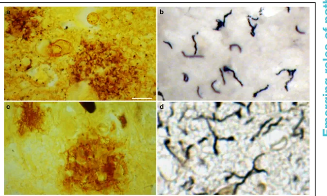
Distribution of spirochetes in the atrophic form of general paresis and in the
frontal cortex of an Alzheimer disease patient with Lyme neuroborreliosis
Expert Reviews in Molecular Medicine © 2011 Cambridge University Press
a b
c d
Figure 1. Distribution of spirochetes in the atrophic form of general paresis and in the frontal cortex of an Alzheimer disease (AD) patient with Lyme neuroborreliosis. Spirochetes form plaque-like masses (a,c) and disseminate as individual filaments (b,d) in general paresis (a,b) and AD (c,d), which are identical to senile plaques and curly fibres. (a,c) Bosma Steiner silver technique for the visualisation of spirochetes. (b) T. pallidum-specific polyclonal antibody. (d) Gallyas silver technique. Scale bars: 50μm (a), 40 μm (c), 10μm (b,d). All images were obtained from previously published studies: a (Ref. 124); b (Ref. 175); c, d (Ref. 149).
4
Emerging
roles
of
pa
thogens
in
Alzheimer
disease
(Ref. 170), B. afzelii, B. garinii and B. valaisiana.
Relapsing fever is caused by B. recurrentis. Oral
spirochetes
are
predominant
periodontal
pathogens that are highly prevalent in the
population and comprise diverse Treponema
species (Refs 166, 171). Several are invasive
(Refs 172, 173): they include T. denticola, T.
socranskii,
T.
pectinovorum,
T.
amylovorum,
T. lecithinolyticum, T. maltophilum, T. medium
and T. putidum (Refs 166, 171, 174). T. vincentii
causes
Vincent
angina,
a
necrotising
fusospirochetal disease (Ref. 167).
Because spirochetes are strongly neurotropic
(Ref. 167), it was expected that several types of
spirochetes, in an analogous way to T. pallidum,
might cause dementia, plaque- and tangle-like
lesions, Aβ deposition and consequently might
be involved in the pathogenesis of AD. To detect
all types of spirochete, neutral techniques
need to be used (Ref. 146). Using dark-field
microscopy, spirochetes were detected in the
cerebrospinal fluid (CSF), in the blood and in
the brain in 14 definite AD cases tested
(Table 1). Spirochetes were not found in 13
age-matched controls without any AD-type cortical
changes
(Ref.
146).
Silver-stained
helically
shaped spirochetes were also detected by
electron microscopy. Spirochetes were isolated
from the cerebral cortex in these 14 AD cases
(Table 1), and in three of them, they were
cultivated from the brain in a selective medium
for B. burgdorferi (Ref. 146). Spirochetes were
detected and isolated from the brains of eight
additional AD cases derived from another
laboratory and in the blood of five living
patients with clinically diagnosed AD-type
dementia (Ref. 186; Table 1). Four healthy
controls did not show spirochetes. Taxonomical
analyses showed that the helically shaped
microorganisms
belong
to
the
order
Spirochaetales
(Ref.
187).
To
ensure
the
consistency of these results, the detection of
spirochetes was also performed using various
other techniques, including histochemistry,
dark-field
microscopy,
atomic
force
microscopy,
electron
microscopy
and
immunoelectron
microscopy,
immunohistochemistry
using
spirochete and bacterial peptidoglycan
(PGN)-specific antibodies (Refs 146, 147, 149, 186, 187,
176, 175, 188, 189), and detection of specific and
nonspecific bacterial DNA (Refs 149, 176). PGN
is the building block of the cell wall of
Gram-negative and Gram-positive bacteria; however,
mycoplasma and chlamydiae lack detectable
PGN (Refs 190, 191). The morphology of
spirochetes detected by spirochete- or
PGN-specific antibodies is identical (Fig. 2; compare
also Fig. 7 G and H of Ref. 175).
PGN-immunoreactive spirochetes were detected in 32
definite AD cases and in 12 cases with mild or
moderate AD-type cortical changes.
Other authors found no evidence of spirochetes
in the brains of seven AD cases by dark-field or
electron
microscopy
(Ref.
192).
However,
spirochetes were observed in the blood of one of
22 living patients with AD-type dementia
(Table 1). The spirochete observed by these
authors
corresponded
to
the
vegetative,
regularly spiral form. They suggested that it
could correspond to an oral spirochete. Whether
the atypical, pleomorphic spirochetal forms,
which commonly occur in infected tissues, blood
and CSF (Refs 167, 175, 177, 193), were
considered by the authors is not clear.
Periodontal pathogen Treponemas
Using molecular and immunological techniques,
six of seven periodontal Treponema species,
namely T. socranskii, T. pectinovorum, T. denticola,
T. medium, T. amylovorum
and T. maltophilum,
were identified in the brains of AD patients
using species-specific polymerase chain reaction
(PCR; Table 1). At least one oral Treponema
species was present in 14 of 16 AD brains,
compared with 4 out of 18 controls (Ref. 147). T.
pectinovorum
and T. socranskii antigens were
observed in 15 of 16 AD brains and in 7 of 18
controls. In the hippocampus and in the frontal
cortex. Six different Treponema species were
detected in one AD patient, five species in four,
four or three species each in one AD case, and
one species in seven AD brains. Two Treponema
species were observed in one control and one
species in the other three positive controls.
These results reinforce previous observations
(Ref. 146) and indicate that these periodontal
pathogen spirochetes, in an identical way to T.
pallidum
and B. burgdorferi, have the ability to
invade the brain, persist in the brain, and cause
dementia,
cortical
atrophy
and
amyloid
deposition.
Borrelia burgdorferi
The causative agent of Lyme disease is transmitted
by the bite of infected ticks (Ref. 170). Neurological
complications occur in about 15% of affected
5
Emerging
roles
of
pa
thogens
in
Alzheimer
disease
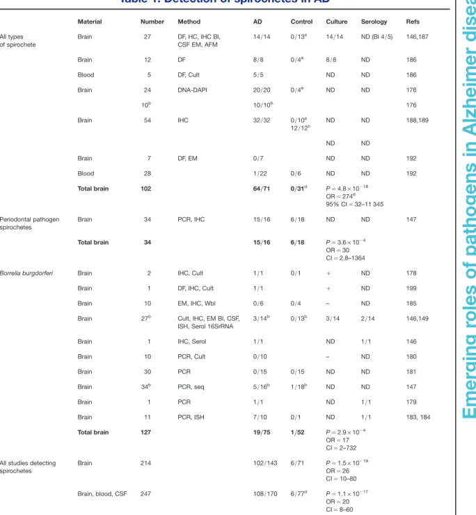
Table 1. Detection of spirochetes in AD
Material Number Method AD Control Culture Serology Refs
All types of spirochete Brain 27 DF, HC, IHC Bl, CSF EM, AFM 14/14 0/13a 14/14 ND (Bl 4/5) 146,187 Brain 12 DF 8/8 0/4a 8/8 ND 186 Blood 5 DF, Cult 5/5 ND ND 186 Brain 24 DNA-DAPI 20/20 0/4a ND ND 176 10b 10/10b 176 Brain 54 IHC 32/32 0/10a 12/12c ND ND 188,189 ND ND Brain 7 DF, EM 0/7 ND ND 192 Blood 28 1/22 0/6 ND ND 192 Total brain 102 64/71 0/31d P= 4.8 × 10⁻¹⁸ OR= 274d 95% CI= 32–11 345 Periodontal pathogen spirochetes Brain 34 PCR, IHC 15/16 6/18 ND ND 147 Total brain 34 15/16 6/18 P= 3.6 × 10⁻⁴ OR= 30 CI= 2.8–1364
Borrelia burgdorferi Brain 2 IHC, Cult 1/1 0/1 + ND 178
Brain 1 DF, IHC, Cult 1/1 + ND 199
Brain 10 EM, IHC, Wbl 0/6 0/4 – ND 185
Brain 27b Cult, IHC, EM Bl, CSF,
ISH, Serol 16SrRNA
3/14b 0/13b 3/14 2/14 146,149
Brain 1 IHC, Serol 1/1 ND 1/1 146
Brain 10 PCR, Cult 0/10 – ND 180 Brain 30 PCR 0/15 0/15 ND ND 181 Brain 34b PCR, seq 5/16b 1/18b ND ND 147 Brain 1 PCR 1/1 ND 1/1 179 Brain 11 PCR, ISH 7/10 0/1 ND 1/1 183, 184 Total brain 127 19/75 1/52 P= 2.9 × 10⁻⁴ OR= 17 CI= 2–732 All studies detecting
spirochetes Brain 214 102/143 6/71 P= 1.5 × 10⁻¹⁹ OR= 26 CI= 10–80 Brain, blood, CSF 247 108/170 6/77d P= 1.1 × 10⁻¹⁷ OR= 20 CI= 8–60
Data reviewed in the literature with respect to the detection of all types of spirochetes using neutral techniques and the specific detection of periodontal pathogen Treponemas and Bb. Results of the statistical analysis are given for each group and for all studies together. AD indicates number of AD cases positive for spirochetes/ number of AD cases analysed; Control indicates number of control cases positive for spirochetes/number of control cases analysed. AD, Alzheimer disease; Bl, blood; CSF, cerebrospinal fluid; EM, electron microscopy; AFM, atomic force microscopy; DF, dark-field microscopy; HC, histochemistry (Warthin and Starry, Bosma-Steiner silver stain for spirochetes); IHC, immunohistochemistry; ISH, in situ hybridisation; PCR, polymerase chain reaction; Bb, Borrelia burgdorferi; Wbl, Western blot; P is exact value of the significance calculated by Fischer test; OR, odds ratio; CI, 95% confidence interval;+, positive; –, negative; ND, not determined; seq, sequencing.
aControls without any AD-type changes.
bCases from previous studies, which were subtracted when the total number of cases of studies was considered. cCases with mild or moderate AD-type cortical changes.
dWhere the number of positive controls was zero, in order to calculate the OR and 95% CI, one positive case was added to the control group.
6
Emerging
roles
of
pa
thogens
in
Alzheimer
disease
individuals. Dementia and subacute presenile
dementia both occur in Lyme disease (Refs 146,
149, 194, 195, 196, 197, 198). B. burgdorferi was
first detected in the brains of two AD patients
(Table 1) by MacDonald and Miranda (Ref. 178)
and MacDonald (Ref. 199). This spirochete was
detected in the cerebral cortex by dark-field
microscopy and with a specific antibody against
B. burgdorferi. This species was also detected and
cultivated from the brains of three definite AD
cases in an initial series of 14 AD cases
(Ref. 146). Molecular characterisation, using 16S
rRNA gene sequence analysis, identified these
spirochetes as B. burgdorferi sensu stricto (s.s.)
(Ref.
149).
Electron
microscopy
analysis
confirmed that these spirochetes possess 10–15
endoflagella typical of Borrelia species. Two of
the three AD patients had a positive CSF
serology for B. burgdorferi, and the 31 kDa outer
surface protein A (OspA) band, which is highly
specific for B. burgdorferi, was detected by
western blot in these three AD cases (Ref. 149).
The pathological changes found in the brain
were identical to those occurring in the atrophic
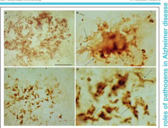
Spirochetes detected in the frontal cortex of neuropathologically confirmed
Alzheimer disease cases
Expert Reviews in Molecular Medicine © 2011 Cambridge University Press
a b
c d
Figure 2. Spirochetes detected in the frontal cortex of neuropathologically confirmed Alzheimer disease cases. Spirochetes in an immature senile plaque detected by a cocktail of antibodies against Borrelia burgdorferi (a), in a mature plaque detected by in situ hybridisation (b, arrows) using Borrelia-specific probes, and in an amorphous plaque detected by antibacterial peptidoglycan antibody (c). (d) The central part of panel c at higher magnification. Arrows indicate helical spirochetes with the same morphology as observed in b. Scale bars: 80μm (a), 30 μm (b), 80 μm (c), 20 μm (d). Images are from previously published studies: a,b (Ref. 149); c,d (Ref. 188).
7
Emerging
roles
of
pa
thogens
in
Alzheimer
disease
form of general paresis (Ref. 149). The cortical
distribution of spirochetes in masses or colonies
was identical to those of senile plaques, and the
morphology
of
individual
spirochetes
was
identical to those of curly fibres (Ref. 149). B.
burgdorferi
antigens colocalised with Aβ in
cortical plaques and in leptomeningeal and
cortical arteries containing amyloid deposits.
Neurofibrillary
tangles
were
also
immunoreactive for B. burgdorferi. OspA and
flagellin genes were detected in AD-type lesions
using in situ hybridisation (ISH) (Fig. 2b). In
additional AD patients with concurrent Lyme
neuroborreliosis, B. burgdorferi-specific antigens
(Ref. 146) and DNA (Ref. 179) were observed.
Using species-specific PCR, B. burgdorferi DNA
was detected in 5 of 16 AD patients tested and
in 1 of 18 controls, all of which also had oral
Treponema
spirochetes (Ref. 147). Finally, B.
burgdorferi-specific DNA was detected by both
ISH and PCR in the hippocampus in 7 of 10
pathologically confirmed AD cases (Refs 183,
184) (Table 1).
Pappolla and colleagues (Ref. 185) failed to detect
B. burgdorferi
in six AD patients. They stated that
they could not exclude other spirochetes not
detected by their methods. In all other studies
where B. burgdorferi was not detected, evidence is
lacking on whether the analysed AD patients had
Lyme neuroborreliosis or not (Refs 180, 181)
(Table 1). Similarly, the analysis of the serology of
B. burgdorferi
alone, as a result of the low
incidence of Lyme dementia compared with AD,
might give false-negative results (Refs 180, 182).
To demonstrate the role of B. burgdorferi, AD
patients with Lyme neuroborreliosis should be
analysed.
Taken together, these observations show that
various authors detected and cultivated various
types of spirochetes from the brains of AD
patients. Coinfection with several types of
spirochetes occurs.
Chlamydophyla pneumoniae
Several authors reported the existence of an
association between C. pneumoniae, an obligate
intracellular respiratory pathogen, and AD
(Refs 144, 200, 201, 202, 203) (Table 2). C.
pneumoniae-specific DNA was detected in the
brains in 90% of sporadic AD patients and in 5%
of controls (Ref. 144) (Table 2). Two mRNAs,
encoding KDO transferase and a 376 kDa
protein, specific to C. pneumoniae, were also
identified in frozen AD brain tissue by reverse
transcriptase-PCR (RT-PCR).
Immunohistochemical analyses of AD brains
showed C. pneumoniae in microglia, perivascular
macrophages and astrocytes, and in about 20%
of neurons (Refs 144, 200, 201, 203, 207, 211).
They were commonly found in brain regions
showing the characteristic neuropathology of
AD (Refs 144, 200, 201). The presence of
C. pneumoniae
in the brains of AD patients was
also confirmed by immunoelectron microscopy
using specific monoclonal antibody against
the outer membrane protein of C. pneumoniae.
Electron
and
immunoelectron
microscopy
identified both chlamydial elementary bodies
and reticulate bodies (RBs) (Refs 212, 144). That
the replicative RB form was also detected in
glial cells, neurons and pericytes indicates that a
viable and transcriptionally active form of
the microorganism is present in these cells
(Refs 201, 213, 214, 215). Pleomorphic forms of
C. pneumoniae
were also observed (Ref. 213).
C. pneumoniae-specific DNA was detected in the
CSF in a significantly higher number of cases
in
AD
patients
(43.9%)
than
in
controls
(10.6%) (Ref. 202). C. pneumoniae was cultured
from various brain samples of AD patients
originating from different geographic regions of
North America (Refs 144, 201, 215) and was also
isolated from the CSF (Ref. 202). Tor-1 and Phi-1
isolates were characterised by PCR assays
targeting C. pneumoniae-specific genes Cpn0695,
Cpn1046
and tyrP. Two groups, using
paraffin-embedded brain samples, failed to detect
C. pneumoniae
in AD or in controls by PCR
(Refs 216, 217). In another study, C. pneumoniae
was detected in 2 of 15 AD cases and in 1 of 5
controls (Ref. 204).
Other bacteria
Propionibacterium acnes, an atypical anaerobic
bacterium, was identified in biopsy specimens of
the frontal cortex in three of four AD patients
and in one of five controls with cerebral tumour.
The P. acnes positive control was an elderly
patient with cardiovascular risk factors and
glioblastoma (Ref. 205). P. acnes was identified
by
microbiological
methods
and
by
gas
chromatography. The bacterium was cultivated
from
frontal
cortical
biopsy specimens
in
Schaedler blood agar, at 35°C, under anaerobic
conditions (Refs 205, 206). P. acnes has long been
considered to be a commensal bacillus of the
8
Emerging
roles
of
pa
thogens
in
Alzheimer
disease
skin. Recent observations showed that P. acnes, the
causative agent of acne vulgaris, is implicated in
various infections, including brain abscesses,
endocarditis, endophthalmitis and osteomyelitis
(Refs 208, 209). P. acnes was also shown to be a
predominant periodontal pathogen (Ref. 218).
By haematogenous dissemination, it can reach
and infect various organs, including the brain.
Stabilisation of the clinical symptoms and
memory improvement were observed in two
AD
cases
treated
with
P.
acnes-sensitive
cephalosporine combined with enalapril and
oestrogen (Ref. 206). The author pointed to
microangiopathy as the underlying pathology
(Refs 219, 220). Because the number of AD cases
analysed is low, further studies should be
encouraged to determine any association of P.
acnes
with AD.
Actinomycetes
have also been suggested to be
involved in AD, with an incidence four times
higher than in other pathological conditions
(Ref. 221). Ultrastructural analysis revealed that
the fibronectin-immunopositive fibrillary lesions
in senile plaques, which were negative for
neuronal, glial and macrophage markers, are
compatible with filamentous microorganisms
and
might
correspond
to
Actinomycetes
(Ref. 221). It is noteworthy that Actinobacillus
actinomycetemcomitans
is a frequent periodontal
pathogen (Ref. 222) and that Nocardia asteroides
was reported to cause Parkinson-like symptoms
in experimental animals (Ref. 223).
Finally, the causative agent of stomach ulcers,
H. pylori
(Ref. 125), has also been suggested to
be associated with AD (Refs 224, 225, 226).
Serum IgG and IgA antibodies against H. pylori
occurred in a higher percentage in the group of
30 AD patients compared with 30 controls
(Ref.
224).
The
difference
is
statistically
significant
as
determined
by
post-hoc
Table 2. Detection of Chlamydophyla pneumoniae and other bacteria in AD
Material
Number
Method
AD
Control
Refs
C. pneumoniae Brain 38 PCR, EM, IHC, RT-PCR, Cult 17/19 1/19 144 Brain 25 PCR, IHC 0/25 216 Brain 20 PCR, IHC 0/20 217 Brain 20 PCR, Cult 2/15a 1/5a 204 Brain 21 PCR, ISH 21/21 0/1 200 Brain 52 PCR, Cult, RT-PCR 20/25 3/27 201 CSF 104 PCR, Cult 25/57 5/47 202 Total brain 177 P= 4.5 × 10⁻⁷ OR= 8.7 CI= 3.1–29.5 60/125 5/52 Brain and CSF 281 P= 9.8 × 10⁻¹¹ OR= 7.8 CI= 3.7–17.8 85/182 10/99 Propionibacterium acnes Brain 3 Cult 3/4 1/5 219, 220 H. pylori Stomach 80 HC 44/50 14/30 225
AD, number of AD cases with positive detection/number of AD cases analysed; Control, number of control cases with positive detection/number of control cases analysed; CSF, cerebrospinal fluid; PCR, polymerase chain reaction; RT-PCR, reverse transcriptase-PCR; EM, electron microscopy; IHC, immunohistochemistry; Cult, culture. P, exact value of significance following Fischer test; OR, odds ratio; CI, 95% confidence interval values.
a
Positive in at least one of several samples.
9
Emerging
roles
of
pa
thogens
in
Alzheimer
disease
comparison (Ref. 210). An almost twofold higher
prevalence of gastric H. pylori infection was
observed in clinically diagnosed AD patients
and in patients with mild cognitive decline
(Refs 225, 227) than in controls. H. pylori-specific
IgG antibody levels were also significantly
higher in the blood and CSF of AD patients than
in controls (Ref. 226). Additional studies would
be of interest in detecting whether H. pylori is
present in the brain and to analyse the
possibility of a causal relationship between H.
pylori
and AD.
Herpes simplex virus type-1 (HSV-1) and
other viruses
HSV-1 is a common neurotropic virus that infects
around 70% of the population after the age of 50
(Refs 150, 228, 229, 230). HSV-1 DNA was
detected (Table 2) in brain samples using ISH in
some elderly patients with dementia (Ref. 231).
Three other studies failed to detect HSV-1 by
ISH in AD or in control brains (Refs 232, 233,
234,
235).
Increasing
numbers
of
recent
observations have detected HSV-1 DNA in the
brain in AD (Refs 150, 151, 228, 229, 236, 237,
238). Using PCR, Jamieson et al. (Ref. 150)
observed HSV-1 DNA in the brains of a high
proportion of elderly subjects, with or without
AD, which was absent or less frequent in young
controls (Ref. 228). Several authors showed that
HSV-1 is a significant risk factor (Table 3) when
present in AD patients who are carriers of
APOE
ε4 (Refs 236, 237, 238), but this is not
supported by Beffert et al. (Refs 239, 240). In situ
PCR showed that HSV-1 DNA is localised to
senile plaques (Ref. 243). Ninety per cent of
senile plaques in AD and 80% in normal ageing
subjects contained viral DNA (Ref. 241).
Table 3. Detection of HSV-1 in AD
Material
Number
Method
AD
Control
P value
Refs
Brain 6 ISH 2/3 1/3 0.423 231 Brain 5 ISH 0/3 0/2 232 Brain 23 ISH 0/18 0/5 233 Brain 13 ISH 0/8 0/5 234 Brain 6 ISH 0/4 0/2 235 Brain 14 PCR 8/8 6/6 150 Brain 36+ 10 PCR 14/21 9/15 (0/5 middle aged; 0/5 young) 228 Brain 155 PCR 73/98 41/57 0.485 229 Brain 90 PCR 36/46 28/44 0.295 (P< 0.0001, APOEε4) 236 Brain 69 PCR 14/46 5/23 0.295 (P< 0.0047, APOEε4) 237 Brain 109 PCR 45/61 30/48 (P< 0.0001, APOE ε4) 238 Brain 110 PCR 54/74 26/36 0.82 239, 240 Total brain 556 210/ 344 118/212 P= 0.2152 OR = 1.25 CI= 0.9–1.8
Number of Alzheimer cases with positive detection/number of Alzheimer cases analysed; Control, number of control cases with positive detection/number of control cases analysed. P, value of significance following Fischer test; OR, odds ratio; CI, 95% confidence interval values;+, positive; –, negative; AD, Alzheimer disease; CSF, cerebrospinal fluid; HSV-1, herpes simplex virus type-1; IHC, immunohistochemistry; ISH, in situ hybridisation; PCR, polymerase chain reaction; APOEε4: epsilon 4 allele of apolipoprotein E.
10
Emerging
roles
of
pa
thogens
in
Alzheimer
disease
Anti-HSV-1 antibodies as detected in the CSF by
enzyme-linked immunosorbent assay (ELISA)
were also significantly higher in AD patients than
in younger controls, but without a significant
difference between the AD and age-matched
control groups (Ref. 242). Increased titres of
HSV-1 IgGs, which characterise past infection, were
observed in AD cases and age-matched controls,
without a significant difference between the two
groups (Refs 243, 244). In a large prospective
study, in addition to HSV-1 IgG, the presence of
IgM, which characterises active primary infection
or reactivation of the infection, was also assessed
in the sera of 512 elderly patients, initially free
of dementia. During 14 years of follow-up, 77
AD cases were diagnosed. In contrast to IgG,
IgM-positive subjects showed a significantly
higher risk of developing AD, indicating that
reactivation of HSV-1 seropositivity is correlated
with AD (Ref. 245).
Other herpes viruses were also detected in the
brain using PCR: human herpes virus 6 (HHV6)
types A and B, herpes simplex virus type-2
(HSV-2) and cytomegalovirus (CMV). HHV6,
HSV-2 and CMV were observed in 70%, 13%
and 36% of AD patients and in 40%, 20% and
35% of controls, respectively. The differences
between
the
groups were
not
statistically
significant (Ref. 246).
In addition to CMV, a possible association
between HLA-BW15 and AD has also been
reported (Ref. 247). A recent study revealed that
elderly subjects with high levels of antibody
against CMV develop more severe cognitive
decline over 4 years (Ref. 248) compared with
controls.
The adenovirus early region 1A (E1A) gene
and
its
expression
using
ISH
and
immunohistochemistry were analysed in five
AD
cases
and
in
two
controls.
Reactive
microglial cells in both AD (5/5) and control
brain tissue (2/2) showed positive hybridisation
and immunoreactive expression of adenovirus
E1A, indicating a monocyte- or
microglia-mediated entry of adenovirus into the central
nervous system (CNS) (Ref. 249).
Borna disease virus (BDV) was linked to
affective disorders and schizophrenia (Refs 139,
142). A few attempts have been made to analyse
the prevalence of BDV antibodies and BDV p40
gene coding sequences in AD (Refs 250, 251,
252), which did not show an association
(Table
3).
Following
Borna-virus-induced
infection in APP(Tg2576) transgenic mice, a
reduction of cortical and hippocampal Aβ
deposits
was
observed
(Ref.
253).
One
explanation
is
that
the
stimulation
and
activation of microglia might produce this effect.
The human immunodeficiency virus type-1
(HIV-1) (Ref. 254) is able to induce the biological
hallmarks of AD; however, the virus is present in
the brains of mostly young patients suffering
from acquired immune deficiency syndrome
(AIDS). The virus invades mostly macrophages
and glial cells and the virus itself does not
reproduce the pathological hallmarks of AD.
However, by affecting immune defences, HIV-1
facilitates
infection
by
various
pathogens,
including, among
others, HSV-1, CMV, C.
pneumoniae
and spirochetes (Refs 255, 256, 257, 258).
Correlates of infection risk and AD
Based on the data available on the association of
spirochetes, C. pneumoniae and HSV-1 with AD,
contingency tables were used to analyse the
strength of the association and the risk of
infection in AD. In those studies where all types
of spirochetes were detected using neutral
techniques, spirochetes were observed in the
brain in 90.1% (64
/71) of AD cases and were
absent
in
controls
without
any
AD-type
changes. This difference is significant (Table 1),
and the association remains significant when
cases where spirochetes were analysed in the
blood
are
also
included.
The
association
between periodontal pathogen spirochetes and
AD is also statistically significant. They were
detected in the brain in 93.7% of AD cases and
in 33.3% of controls. B. burgdorferi was about 13
times more frequent in AD cases than in
controls, a statistically significant difference. It is
noteworthy that B. burgdorferi was detected in
all AD cases with a positive serology or where
spirochetes were cultivated from the brain
(Table 1). Taken together, in all studies where
spirochetes or their specific species (periodontal
Treponema
spirochetes or B. burgdorferi) were
detected in the brain, it can be concluded that
the frequency of spirochetes is more than eight
times higher in AD cases (90
/131; 68.7%) than in
controls (6
/71; 8.41%). That spirochetes were
cultivated from the brains of AD patients
indicates that viable spirochetes are present in
advanced stages of dementia. They can sustain
persisting infection and inflammation and cause
neuronal destruction (Ref. 148).
11
Emerging
roles
of
pa
thogens
in
Alzheimer
disease
The frequency of C. pneumoniae was about five
times higher in AD cases (60/125; 48%) than in
controls (5/52; 9.6%). The difference remains
significant
when
those
cases
where
C.
pneumoniae
was detected in the CSF were also
included (Table 1). That C. pneumoniae was
cultivated from the brain (Refs 200, 213) and
CSF (Ref. 202) and that replicative RBs were
present in glial cells, neurons and pericytes in
AD indicate that this microorganism is also
present in a viable, active form in the brain in AD.
There is no significant difference between the
frequency of HSV-1 detected in the brain in AD
patients compared with age-matched control.
However, a significant difference was observed
between APOE
ε4-positive and -negative AD
carriers,
as
reported
by
several
authors
(Refs 236, 237, 238, 259, 260), except for Beffert
and colleagues (Refs 239, 240). A significant
association of AD with ongoing or reactivated
HSV-1 infection was reported (Ref. 245).
Evidence for a causal role of pathogens
in AD
Additional studies have brought further evidence
in favour of a probable causal relationship
between spirochetes, C. pneumoniae, HSV-1 and
AD. Exposure of primary mammalian neuronal
and glial cells to spirochetes, namely to B.
burgdorferi,
which
can
be
cultivated
and
propagated
in
pure
culture,
generated
thioflavin-S-positive and A
β-immunoreactive
amyloid
plaques
and
tangle-
and
granulovacuolar-like
formations
in
vitro
(Refs 175, 261). In situ, in the
spirochete-induced A
β plaques, synchrotron infrared
microspectroscopy
analysis
revealed
the
presence of a
β-pleated sheet conformation
(Ref. 261). Spirochete-induced increases in A
β,
APP and phosphorylated tau were all detected
by western blot in infected cell cultures
(Ref.
261).
This
additional
experimental
evidence indicates that spirochetes are able to
induce an AD-type host reaction and reproduce
the
defining
pathological
and
biological
hallmarks of AD (Refs 175, 261). Similar in
vitro studies performed on CNS organotypic
cultures,
which
aim
to
replace
in
vivo
experiments,
showed
identical
results
(Ref. 261). Reference B. burgdorferi spirochetes
(strain B31) and those cultivated from the AD
brain (strains ADB1 and ADB2) invaded
neurons and glial cells and induced nuclear
fragmentation in vitro, indicating that the
spirochetes cultivated from the brains of AD
patients are invasive and cause neuronal and
glial damage and apoptosis (Refs 175, 261).
Spirochetes
occur
in
both
extra-
and
intracellular locations. They invade neurons in
vitro, in primary neuronal cultures (Refs 175,
261), in
the cerebral cortex
and
in
the
trigeminal ganglia of AD patients (Refs 146,
147, 148, 149, 175, 183, 184, 261). Historical
observations and illustrations showing that
chronic spirochetal infection can reproduce the
clinical, pathological and biological hallmarks
of AD strongly support a causal relationship
between
spirochetal
infection
and
AD
(Refs 114, 115, 124). All these observations
indicate that various types of spirochetes,
including B. burgdorferi and several periodontal
pathogen spirochetes, in an analogous way to
T.
pallidum,
can
cause
dementia,
cortical
atrophy and the pathological and biological
hallmarks of AD.
Astrocytes and microglia infected in vitro with
C.
pneumoniae
display
inclusions
that
are
indistinguishable from those characteristic of
active infection of the standard HEp-2 host cell
line (Refs 214, 215). It was reported that chronic
or persistent infection of CNS cells with C.
pneumoniae
can affect apoptosis in AD, in both a
pro- and antiapoptotic manner (Refs 203, 214,
215). Furthermore, infection of BALB/c mice by
intranasal inhalation of C. pneumoniae initiated
Aβ1–42
deposits in the brain that resembled
senile plaques (Ref. 262). Antibiotic treatment
following C. pneumoniae infection limited the
number of induced amyloid plaques in vivo
(Ref. 263).
The glycoprotein B (gB) of HSV-1 has a highly
homologous sequence to a fragment of Aβ
(Ref. 264). Synthetic peptides derived from this
region accelerate fibrillar aggregation of Aβ in
vitro. They can self-assemble into fibrils, which
are ultrastructurally indistinguishable from Aβ
and are neurotoxic at a similar dose to Aβ. It
was proposed that HSV-1 might act as a
‘seed’
for senile plaque formation (Ref. 265). It was
also shown that HSV-1 is associated with APP
during its anterograde transport, which might
affect APP degradation and synaptic function
(Ref. 265).
Exposure of cultured cells to HSV-1 results in
increased intracellular Aβ levels in neurons and
glial cells as analysed by immunocytochemistry,
12
Emerging
roles
of
pa
thogens
in
Alzheimer
disease
ELISA and western blot. Tau phosphorylation was
also observed at a number of sites that are
phosphorylated in AD (Refs 266, 267, 268). It
was suggested that the association of viral DNA
and senile plaques is very likely to be causal,
because HSV-1 is able to increase the level of Aβ
in neurons of infected mice in vivo (Ref. 266).
Evidence for underlying mechanisms
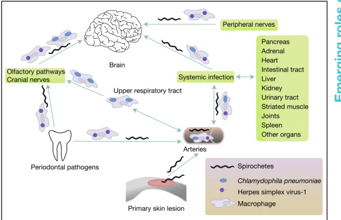
Sources and dissemination
Spirochetes, C. pneumoniae and HSV-1 are all able
to invade the brain (Fig. 3) and generate latent and
persistent chronic infection (Refs 144, 145, 146, 147,
148, 149, 228, 229, 269). The strong neurotropism of
spirochetes is well known (Ref. 167). In addition to
haematogenous dissemination, spirochetes can
spread through the lymphatic system and along
nerve fibre tracts (Ref. 167). Periodontal invasive
spirochetes were detected in the trigeminal
ganglia
and
along
the
trigeminal
nerve
(Ref. 147). They might also propagate along the
fila olfactoria and tractus olfactorius, which
would enable them to reach the CSF, the septal
and hippocampal regions in the earliest stages
of the disease. This would be in harmony with
the olfactory hypothesis (Refs 94, 95, 96) and the
early involvement of the olfactory tract and bulb
(Ref. 97).
Through infected circulating monocytes, C.
pneumoniae
can also spread by haematogenous
dissemination and, by crossing the blood–brain
barrier, infect the brain. C. pneumoniae is an
upper respiratory tract pathogen and can reach
the brain through the olfactory system (Ref. 95).
Intranasal inhalation of C. pneumoniae initiated
plaque-like Aβ1–42
deposits in the brain in
BALB/c mice, and C. pneumoniae-specific DNA
was detected by PCR in the olfactory bulb in
AD (Ref. 262).
HSV-1 infection might also reach the brain
through the olfactory system, by infection of
cranial and peripheral nerves and their ganglia
and
through
haematogenous
dissemination
(Refs 270, 271). The virus can reside in the brain
Pancreas
Arteries
Systemic infection
Other organs Upper respiratory tract
Brain Periodontal pathogens Adrenal Heart Intestinal tract Liver Kidney Urinary tract Striated muscle Joints Spleen
Primary skin lesion
Peripheral nerves
Olfactory pathways Cranial nerves
Spirochetes
Chlamydophila pneumoniae
Herpes simplex virus-1 Macrophage
Sources and dissemination of pathogens associated with Alzheimer disease
Expert Reviews in Molecular Medicine © 2011 Cambridge University Press
Figure 3. Sources and dissemination of pathogens associated with Alzheimer disease.
13
Emerging
roles
of
pa
thogens
in
Alzheimer
disease
in a latent form and be reactivated by peripheral
infection, stress or immunosuppression (Ref. 268).
Neuroinflammation and TLR signalling
Persisting, poorly degradable bacterial remnants
in
mammalian
tissues
act
as
chronic
inflammatory stimuli (Refs 272, 273, 274).
Lipopolysaccharides (LPSs), PGN and various
bacterial
lipoproteins
elicit
a
variety
of
proinflammatory responses and might represent
an important source of inflammation in AD.
Complex interactions between the innate and
adaptive immune systems have a major role in
infection (Ref. 275). The functions of innate
immunity allow host cells to recognise most
microorganisms,
execute
proinflammatory
defences and start adaptive immune responses.
Bacteria attach to host cells through a variety of
cell-surface
components,
including
surface
amyloid proteins, which interact with host
proteases (Refs 276, 277, 278, 279, 280, 281, 282,
283). Proteolysis of the extracellular matrix
allows bacteria to penetrate the basement
membrane and invade host cells.
It is the innate immune system that provides the
first line of defence against microorganisms.
Pattern recognition receptors (PRRs), such as
Toll-like receptors (TLRs), recognise unique
structures
of
invading
microorganisms
(Ref. 284). Patients with genetic defects related
to signalling pathways activated by TLRs
frequently suffer from severe recurrent infection
(Refs 285, 286, 287, 288, 289). In the absence of
TLRs,
death
from
experimental
sepsis
is
significantly
enhanced
(Ref.
287).
Various
bacteria, including spirochetes, activate TLR
signalling and interact with CD14 (Refs 290, 291,
292). T. pallidum and B. burgdorferi or their
synthetic membrane lipoproteins are major
inflammatory mediators (Refs 293, 294) and
activate
both
the classical and
alternative
complement pathways, which opsonise and kill
pathogens without the need for antibody.
Complex
interactions
between
pathogen-associated molecular patterns (PAMPs), PRRs
and TLR signalling pathways have a major role
in linking innate and adaptive immunity and
maintaining
pathogen-free
host
tissues
(Ref. 275). TLRs activate two major signalling
pathways (Fig. 4). The core pathway activated
by most TLRs leads to the activation of
transcription factor nuclear factor-kappa B
(NF-κB) and the mitogen-activated protein kinases
(MAPKs) p38 and Jun kinase (JNK). The second
pathway is activated by TLR3 and TLR4 and
results in the activation of both NF-κB and
interferon regulatory factor-3 (IRF3), allowing
the induction of another set of inflammatory
genes, including the antiviral interferon-β gene
(IFNB).
Activation of p38 MAPKs during bacterial
infection was also shown to be crucial in the
local production of cytokines such as IL-8
(Ref. 295) and in the development of effective
immune responses in vivo (Ref. 296).
Toxin-induced p38 MAPK activation requires pore
formation and is inhibited by the relief of
osmotic stress.
Pore-forming toxins are the most common class
of bacterial protein toxins and are often important
virulence factors (Ref. 297). Pore-forming bacterial
toxins
are
typically
oligomers
of
soluble,
monomeric proteins or peptides, which form
transmembrane channels. Channel formation in
the membrane of targeted cells, which triggers
cellular ion imbalance, is a widely used form of
bacterial attack (Refs 298, 299). These
pore-forming
bacterial
toxins
generate
calcium-dependent and lipid-mediated signalling on
host cell surfaces, leading to a variety of events
such as tyrosine phosphorylation (Ref. 300),
actin rearrangement (Ref. 301), NF-κB activation
(Ref. 302) and regulation of gene expression
through histone modification (Ref. 303).
Mammals also use bacterial porin-like proteins
such as perforins as part of their innate immune
defences (Ref. 304). Aβ1–42
was shown to be an
AMP that belongs to the innate immune system.
Toxic oligomers of Aβ1–42
bind to lipid bilayers
of bacterial membranes and enveloped viruses,
triggering Ca
2+influx and bacteriolysis or viral
destruction.
Deregulation
of
the
balance
between host cell destruction by pathogens
and
pathogen
destruction
by
the
host
immune systems will influence the outcome of
infections.
The innate and alternative pathways through
the common membrane attack complex (MAC,
C5b9) cause bacteriolysis (Refs 305, 306, 307).
Cellular
and
humoral
components
of
the
immune system reactions are both associated
with AD (Refs 98, 99, 100, 101, 102). The
adaptive
immune
system
kills
pathogens
through the formation of specific antibodies
directed
against
invading
microorganisms.
Monocytes,
macrophages
and
microglia
14
Emerging
roles
of
pa
thogens
in
Alzheimer
disease
Pore-forming bacterial toxins and bacterial amyloids
Ca2+
Ca2+
Pore formation on host cell membrane
Cell membrane Celullar ion imbalance
Cytolysis, neuronal and glial cell destruction
Bacterial proliferation Persistent infection Evasion Bacterium Antibodies Bacterium Bacterium Cell membrane
Gene expression cytokines
IL-1,IL-6, IL-8, IL-18 TNFα, COX2, IFNβ Gene expression B7.1- Naive T cells IFN-responsive genes Bacterium Bacterium Nucleus IRF8 CREB NF NF γ-secretase Increased Aβ AMP function of Aβ β-secretase AMPs PRRs PAMPs Aβ1-42 Aβ CpG DNA viral RNA FLA BLP, PGN LTA TLR 5 TLR1,6 TLR2 APP TLR10,2 LPS LBP c-Jun CD14 MALP2 κB c-Jun IRF3 TLR3,7-9 TLR4 p38 JNK eNOS Akt Apoptosis Soluble oligomers, pore formation on bacterial lipid membrane
Schematic representation of signalling pathways implicated in host–pathogen
interactions in Alzheimer disease
Expert Reviews in Molecular Medicine © 2011 Cambridge University Press
Complement, MAC
Activation of innate and adaptive immune responses
Bacterial killing Bacterial antigen Y Y Y Y Y Y Y Y Y Y Y Y Y Y κB MD2 Caspase1 NF-kB p38 MAPK TLR4 TRL9 TRL3
Figure 4. Schematic representation of signalling pathways implicated in host–pathogen interactions in Alzheimer disease. Pattern recognition receptors (PRRs) recognise conserved structural components of microorganisms, called pathogen-associated molecular patterns (PAMPs) or ligands, which include peptidoglycan (PGN), lipoteichoic acid (LTA), flagellin (FLA), bacterial lipoprotein (BLP) and nucleic acid structures, such as bacterial CpG DNA or viral RNA, unique to bacteria and viruses. Lipopolysaccharide (LPS) is recognised following its binding to lipoprotein binding protein (LBP). Only Toll-like receptors (TLRs) have a cytoplasmic domain for signal transduction. Plasma-membrane-localised TLRs include TLR1, TLR2, TLR4, TLR5, TLR6 and TLR10, whereas endosomal TLRs include TLR3, TLR7, TLR8 and TLR9. TLR9 is responsible for the recognition of CpG islands of bacterial DNA. Complex interactions between pathogen-associated molecular patterns (PAMPs), pattern recognition receptors (PRRs) and TLR signalling pathways have a major role in linking innate and adaptive immunity and maintaining pathogen-free host tissues. Mammals use antimicrobial peptides (AMPs) as part of their innate immune defences to destroy invading pathogens. In contrast, in a similar way, pore-forming bacterial surface proteins are also able to destroy host cells. Evasion of pathogens from destruction by the host immune defences will result in chronic persistent infection and host cell destruction. APP, amyloid precursor protein; Aβ, amyloid beta; CD14, cluster of differentiation 14; NF-κB, nuclear factor-kappa B; MALP2, mycoplasma diacylated lipopeptide 2; p38 MAPK, mitogen-activated protein kinase; JNK, c-Jun kinase; MAC, membrane attack complex; MD2, lymphocyte antigen 96; Akt, serine/threonine protein kinase Akt1; IRF, interferon regulatory factor; IL, interleukin; TNF, tumour necrosis factor; COX2, cyclooxygenase 2; IFN, interferon, CREB, cAMP response element binding.