Architecture of the Saccharomyces cerevisiae Origin Recognition Complex Bound to Origins of DNA Replication
by
Daniel Gyejun Lee
B. Sc. Genetics University of Alberta, 1993
Submitted to the Department of Biology
in Partial Fulfillment of the Requirements for the Degree of
Doctor of Philosophy in Biology at the
Massachusetts Institute of Technology
September, 1999
© 1999 Daniel Gyejun Lee. All rights Reserved.
The author hereby grants to MIT permission to reproduce and to distribute publicly paper and electronic copies of this thesis document in whole or in part.
Signature of Author:
Department of Biology August 31, 1999
Certified by:
Stephen P. Bell Associate Professolof Biology Tiesis Slipeqvisor
Accepted by:
Alan D. Grossman Professor of Biology Co-Chair, Committee for Graduate Students MASSACHUSETTS INSTITUTE
Architecture of the Saccharomyces cerevisiae Origin Recognition Complex Bound to Origins of DNA Replication
by
Daniel Gyejun Lee
B.Sc. Genetics University of Alberta, 1993
Submitted to the Department of Biology on August 31, 1999, in Partial Fulfillment of the Requirements for the Degree of Doctor of Philosophy in Biology
ABSTRACT
The Origin Recognition Complex (ORC) is thought to be required for the initiation of DNA replication in all eukaryotes. In Saccharomyces cerevisiae, ORC is bound to origins of DNA replication throughout the cell cycle and directs the assembly of higher-order protein-DNA complexes during G1. I have investigated the architecture of
yeast ORC bound to origin DNA. Determination of DNA residues important for ORC-origin association indicated that ORC interacts preferentially with one strand of the ARS] origin. DNA binding assays using ORC complexes lacking one of the six subunits
demonstrated that the DNA binding domain of ORC requires the coordinate action of five of the six ORC subunits. Protein-DNA crosslinking studies suggested that recognition of origin sequences is mediated by two groups of ORC subunits making sequence-specific contacts with two distinct regions of the DNA. Thus, the DNA-binding surface of ORC is formed by the coordinate action of multiple subunits.
Electron microscopy (EM) of ORC showed that the complex has a shape
consistent with the structure determined by protein-DNA crosslinking. These EM studies found that ORC is an elongated molecule with three lobes. Estimates of the molecular mass of ORC using EM were consistent with this elongated complex containing one copy of each of the six ORC subunits. ORC bound origin DNA along its long axis and
interacted with approximately 50 base pairs as predicted from DNase I protection assays of ORC bound to origin DNA.
To examine the fate of ORC when origin DNA is unwound during replication initiation, I determined the effect of single-stranded DNA (ssDNA) on ORC. I showed that ORC binds ssDNA and that the ssDNA-bound form of ORC is distinct from that bound to double-stranded origin DNA. EM studies demonstrated that ssDNA stabilizes a bent conformation of ORC whereas origin DNA stabilizes an extended form of ORC. In addition, ssDNA stimulates the ORC-ATPase activity, whereas origin-containing DNA inhibits it. I propose that the unwinding of origin DNA activates an ssDNA-controlled ORC conformational switch that contributes to the remodeling of the origin-associated protein complexes assembled during G1.
Thesis Supervisor: Stephen P. Bell Title: Associate Professor of Biology
Dedicated, with love
to my parents,
Jai-Hyung and Boon-Ok Lee
and
to my wife, Tallessyn
Acknowledgements
For day-to-day support during my graduate career, I wish to acknowledge all the members of the Bell lab. I am grateful for your technical help, for supplying important reagents, for countless discussions on all topics scientific and otherwise, and for tremendous emotional support and friendship. Special thanks go out to Oscar Aparicio who survived five years as my bay-mate. Thanks for putting up with my music and for loaning me your daughter so that she could be in my wedding.
For intellectual guidance and encouragement, I thank the members of my thesis committee, Tania Baker, Andindya Dutta, Carl Pabo and Phillip Sharp. You have provided important constructive criticism of my work and have helped me to be a more careful and rigorous scientist.
I am especially indebted to my thesis supervisor, Stephen Bell, for his support and mentorship. You have been the biggest influence on my development as a scientist and I am especially thankful for the opportunity to work in an environment in which the
science is thorough and always comes first. In the future, I hope to model my own lab after the way in which you have managed yours.
Finally, for continued love and support, before, during and after grad school, I thank my parents and my wife. I couldn't have done it without you.
Table of Contents
Abstract 2 Dedication 4 Acknowledgements 5 Table of Contents 6 Chapter I. Introduction 9The Replicon Model in Eukaryotes 11
A Two-Step Process for Regulating Initiation in Yeast 13 Properties of the Known pre-RC and RC Components 17
Properties of the Cell-Cycle Regulated Kinases 22
Overview 24
References 26
Chapter II. Architecture of the Origin Recognition Complex Bound
to Yeast Origins of Replication 32
SUMMARY 33
INTRODUCTION 34
RESULTS 39
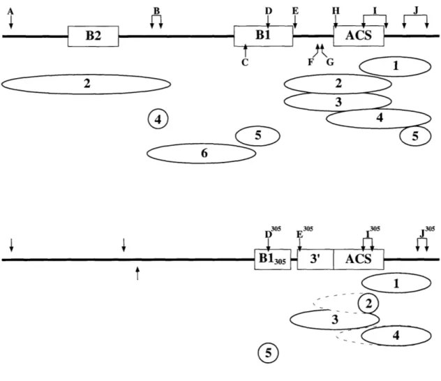
Residues of ARS1 Required for ORC-DNA Binding 39 ORC Bends DNA at Some but not All Origins 44
Orc6p is Not Required for DNA Binding 44
Organization of ORC Subunits at ARS1 47
ORC Subunit Organization at ARS305 52
ORC Subunits in the Major Groove of ARS1 54
DISCUSSION 58
Conservation of ORC-DNA Interactions at the Origin Core 58 Implications of ORC-Origin Architecture for ORC Function 61
ORC Subunit Interactions 62
EXPERIMENTAL PROCEDURES 65
REFERENCES 72
Chapter III. Electron Microscopy of ORC 75
SUMMARY 76
INTRODUCTION 77
RESULTS 79
ORC is an Elongated Complex with Three Lobes 80
Characterization of dsDNA Binding 80
DISCUSSION AND FUTURE PERSPECTIVES 90
EXPERIMENTAL PROCEDURES 95
REFERENCES 100
Chapter IV. Regulation of ORC Conformation and ATPase Activity: Evidence for a Single-Stranded DNA-Controlled Conformational
Switch 102
SUMMARY 103
INTRODUCTION 104
RESULTS 108
ORC Binds ssDNA in an ATP-Independent Manner 108 ORC-ssDNA Binding and ORC-dsDNA Binding are Mutually
Exclusive 110
ORC Prefers to Bind ssDNA Molecules 90 Nucleotides or Longer 112 ssDNA and dsDNA Have Opposite Effects on ORC-ATPase
Activity 115
ssDNA Alters the Conformation of ORC 118
DISCUSSION 122
Does ORC Interact with ssDNA in vivo? 123
The Transition Between the two States of ORC may be Coupled
to Origin Unwinding 124
EXPERIMENTAL PROCEDURES 129
REFERENCES 135
Chapter V. Conclusions 138
The Subunits of the Origin Recognition Complex Act in a
Coordinate Fashion 140
Regulation of Cdc6p via an ATPase Switch 141
ATPase Switches in Other Proteins Involved in DNA Metabolism 145
A High-Resolution Structure of ORC 151
Understanding ORC Function in the Context of the Nucleus:
Pre-RC Components and Cell-Cycle Regulated Kinases 153 Understanding ORC Function in the Context of the Nucleus:
Specialized Sub-Nuclear Structures 155
Concluding Remarks 156
References 159
Appendix A. Regions of ORC Required for ssDNA Binding 163
RESULTS AND DISCUSSION 164
Orc6p and the N-terminus of Orcip are not Required for
ssDNA Binding 164
The C-terminal Region of Orcip is Required for ssDNA Binding 166
Southwestern Blot Analysis of ORC 168
Attempts to Crosslink ORC Subunits to ssDNA 170 Subcomplexes of ORC Generated by V8 Protease Digestion
are Active for DNA Binding 170
EXPERIMENTAL PROCEDURES 173
REFERENCES 175
Appendix B. Attempts to Characterize the ssDNA-Induced
Conformational Change of ORC 176
INTRODUCTION 177
RESULTS AND DISCUSSION 177
Native Gel Mobilities of ORC-ssDNA and ORC-dsDNA
Complexes 177
Limited Proteolysis of ORC 179
Interactions with Other Proteins 181
EXPERIMENTAL PROCEDURES 184
REFERENCES 187
Chapter I
Introduction
The replication of a genome is a logistically complex task. The process itself is
an impressive feat, involving the synthesis of tens or thousands of megabases of DNA in
eukaryotes, with high fidelity, in a short period of time (-20 minutes for yeast and as
little as 3-4 minutes for fruit fly embryos). Moreover, the process must be precisely
regulated each cell cycle to ensure that the entire genome is duplicated but that no portion
is over-replicated. Both too little and too much replication can have deleterious
consequences for an organism.
In 1964, Jacob, Brenner and Cuzin proposed a simple, yet powerful model for the
initiation of DNA replication that continues to be influential today (Jacob et al., 1964).
The replicon model postulated that the initiation of DNA replication would require a
positive trans-acting factor called the initiator protein, that would activate replication via
a cis-acting sequence called the replicator. The replicator is therefore a genetically
defined element that is required for replication, whereas the more frequently used term, replication origin, refers to the actual initiation site of DNA synthesis as determined by
physical mapping techniques. Although the replicator and the origin need not necessarily
overlap (reviewed in Stillman, 1993), the two elements are generally coincident and I will
hereafter refer only to origins.
The replicon model has since been supported by the study of genomic replication
in bacteria, phage and eukaryotic viruses. These experiments have indicated that the
initiator generally performs three functions (first proposed by Bramhill and Kornberg, 1988; reviewed in Baker and Bell, 1998). First, initiator proteins bind their cognate origins of replication in a sequence specific manner and thereby select the site at which
DNA replication will begin. Second, binding of the initiator often induces distortions in the origin DNA, facilitating the generation of the single-stranded DNA template for
polymerase action. Finally, initiator proteins recruit other replication proteins required
for the assembly of replication forks at the origin.
The initiator protein and the early steps of replication are likely targets for the
regulatory mechanism that ensure the fidelity of genomic replication. From a practical
standpoint, it makes sense to regulate steps that occur before the stable complex of
replication proteins bound to origins is converted into a moving replication machine. If
replication elongation was a major target of cell cycle regulation and was routinely
interrupted, the resulting replication intermediates would generate undesirable regions of
genomic instability. Indeed, studies of related processes such as transcription and
translation have shown that the initiation phase is a key target for regulation (Sachs and
Buratowski, 1997). Furthermore, studies of the replication of eukaryotic viruses such as
the Simian Virus 40 (SV40) have demonstrated that the viral genome replicates more
than once per cell cycle. Because SV40 replication requires only one viral-encoded
protein (the SV40 initiator protein, T-Antigen) and all other activities are provided by the
mammalian host cell (reviewed in Stillman, 1994), the simplest explanation is that
proteins involved in initiation and not the DNA synthesis machinery are the major target
for cell-cycle regulation.
The Replicon Model in Eukaryotes
Studies of DNA replication in the budding yeast Saccharomyces cerevisiae
strongly support the hypothesis that eukaryotic cells adhere to the replicon model. S.
cerevisiae is the only eukaryote that has both well-defined origins of replication and a
putative initiator protein. Yeast origins were first identified as genomic DNA sequences
capable of supporting the autonomous replication of episomal DNA (Autonomous
Replicating Sequences or ARSs; Hsiao and Carbon, 1979; Stinchcomb et al., 1979).
Many of these elements were subsequently shown to act as origins of replication in their
normal chromosomal context (reviewed in Newlon and Theis, 1993). Yeast origins are
modular in nature and contain the conserved 11 base-pair ARS Consensus Sequence
(ACS) that is essential for ORC-DNA binding and origin function in vivo, as well as additional elements that enhance origin function (referred to as B elements; Bell, 1995).
The structure of yeast origins will be discussed in greater detail in Chapter II.
A candidate eukaryotic initiator protein was first identified in S. cerevisiae as a six protein complex called the Origin Recognition Complex (ORC). ORC was purified
as an activity that bound specifically to the ARSJ origin in vitro in the presence of ATP
(Bell and Stillman, 1992). ORC was subsequently shown to bind yeast origins of
replication in vivo (Aparicio et al., 1997; Diffley and Cocker, 1992; Santocanale and
Diffley, 1996; Tanaka et al., 1997). The six ORC subunits are referred to as Orc1p
through Orc6p in order of decreasing mass and all six proteins are essential for the
viability of yeast cells (Bell et al., 1993; Bell et al., 1995; Li and Herskowitz, 1993; Loo
et al., 1995). Conditional mutations in ORC subunits result in cell-cycle abnormalities
and high plasmid loss rates consistent with defects in DNA replication (Foss et al., 1993;
Loo et al., 1995; Micklem et al., 1993). These mutations also lead to decreased origin
usage in vivo (Fox et al., 1995; Liang et al., 1995) and decreased origin binding in vivo
(Aparicio et al., 1997).
Although origins of replication have been difficult to define in multi-cellular
eukaryotes (reviewed in Diffley, 1996), analogs of ORC subunits have been discovered in
numerous species (reviewed in (Dutta and Bell, 1997). In Xenopus laevis and Drosophila
melanogaster, these proteins form a six-protein complex similar to that seen in yeast.
Futhermore, studies of replication either in vivo or in vitro strongly suggest that ORC is
required for DNA replication in all eukaryotic species (Chesnokov et al., 1999; Landis et
al., 1997; Pasero et al., 1997; reviewed in Dutta and Bell, 1997). In addition to the
putative initiator protein, other replication proteins involved in the initiation process (see
below) are conserved throughout evolution as are the components of the DNA synthesis
machinery (reviewed in Baker and Bell, 1998). This high degree of conservation among
replication proteins argues that the mechanisms for both the initiation and the elongation
phases of DNA replication are similar in all eukaryotes.
A Two-Step Process for Regulating Initiation in Yeast
Multiple aspects of replication must be controlled to ensure faithful genomic
duplication. First, all eukaryotic DNA is restricted to the appropriate time during the cell
cycle. In normal cycling cells, DNA synthesis begins only after cell division has been
completed, and replication does not occur again until there has been an intervening
mitotic phase. Second, the large size of the eukaryotic genome combined with its
organization into multiple chromosomes requires that bidirectional replication be initiated
from multiple origins (reviewed in Diffley, 1996). Furthermore, origins of replication are
temporally regulated: rather than being activated simultaneously at the beginning of each
S-phase, origins initiate replication at characteristic times during S-phase (reviewed in
Diller and Raghuraman, 1994; Simon and Cedar, 1996). The problem of multiple origins
firing throughout S-phase requires mechanisms to ensure that each origin initiates only
once per cell cycle and that an origin that is passively replicated does not initiate. These
regulatory mechanisms are enforced by controlling the assembly of replication complexes
(see below).
ORC is known to possess at least two of the three general properties of initiator
proteins: it specifically binds origin DNA and it recruits other replication proteins to
origins. In yeast, ORC binds to origins of replication throughout most or all of the cell
cycle and directs the assembly of higher order complexes prior to the initiation of DNA
replication (Figure 1). In vivo DNase I protection assays and chromatin
immunoprecipitation (CHIP) experiments suggest that ORC alone is present at the origin
during the G2- and M-phases to form what has been termed the postreplicative complex
(post-RC; Aparicio et al., 1997; Diffley et al., 1994; Tanaka et al., 1997). In GI, ORC
recruits additional replication proteins to origins, including Cdc6p and the MCM proteins
(in that order), to form the prereplicative complex (pre-RC). Initially, the pre-RC was
defined as a structure that extended the ORC-specific DNase I footprint at origins during
GI (Diffley et al., 1994). This larger region of protection was dependent on the activity of the Cdc6p protein (Cocker et al., 1996). Subsequent in vivo chromatin
immunoprecipitation assays demonstrated that Cdc6p as well as the MCMs are recruited
to origins by ORC (Aparicio et al., 1997; Tanaka et al., 1997), and the term pre-RC has
since come to include MCMs.
After the formation of pre-RCs, Cdc45p and the replicative polymerases are recruited to origins in a manner correlated with the time of replication initiation (Aparicio
et al., 1997; Zou and Stillman, 1998). This higher-order complex containing Cdc45p and
the polymerases has been named the replicative complex (RC). During S-phase, Cdc6p
is degraded (Piatti et al., 1995), and MCM proteins and Cdc45p are released from origins
and appear to move with the DNA polymerases as part of the replication fork (Aparicio et
al., 1997). ORC remains at the origin to repeat this process in the following cell cycle.
The requirement for an ORC-dependent assembly of replication proteins on DNA is
likely conserved throughout evolution, as replication in Xenopus extracts requires
chromatin association of ORC, Cdc6p and MCM proteins, with the same dependence as
seen in yeast (reviewed in Diffley, 1996).
Cell Cycle Regulation of Replication Complexes
C
Cs
Cdc45p MCMs
Cell C
0
Cdc6p
ycle Kinases
*
DNA Polymerases
4-Pre-RC
RC
Post-RC
G1r
S
RC
M
G2
Cell Cycle Kinases
0)
Post-RC
Figure 1
Figure 1. Cell-Cycle Regulation of Replication Complexes. In G2, ORC (green oval) is bound to origin DNA to form the postreplicative complex (post-RC). During G1, ORC recruits Cdc6p and MCM proteins to the origin to form a prereplicative complex
(pre-RC). Close to the G1-S transition when B-type cell-cycle kinases are activated, Cdc45p
and the DNA polymerases are recruited to origins to form a replicative complex (RC).
The activity of kinases activate the RC, remodeling its components to release the
polymerases, as well as the MCMs and Cdc45p which move as part of the replication fork. High levels of kinase activity during S-, G2- and M-phases prevent the formation
of further pre-RCs. See text for more details.
During each cell cycle, the assembly and disassembly of origin-associated
complexes is controlled by the action of cell-cycle regulated kinases. In yeast, the
assembly of pre-RCs occurs during G1 when Cdc6p is abundant and when levels of
B-type cyclin-dependent kinase (CDK) activity are low (B-B-type CDKs are active during S-, G2- and M-phases; see below). Entry into S-phase requires the activation of the S-phase CDKs (Clb5p- and Clb6p-associated CDKs) and the activity of the Cdc7p/Dbf4p kinase. In a CDK-dependent event, each origin is activated, resulting in the dismantling of the RC. ORC binds origin DNA to form a post-RC and must be prevented from
re-forming new pre-RCs and re-initiating replication until the next Gi-phase. Interestingly,
the activities of the B-type CDKs prevent pre-RC formation during S-, G2-, and
M-phases (Dahmann et al., 1995; Piatti et al., 1996; Tanaka et al., 1997). Thus, the cell
cycle can be split into two phases with respect to kinase and pre-RC activity. During G1, when B-type kinase activity is low, pre-RCs can form but cannot be activated. When
Clb5p- and Clb6p-associated kinase activities peak during S-phase, pre-RCs can be converted into RCs and activated, but no further pre-RC formation is allowed until the
next Gi-phase when B-type CDKs return to low levels.
Properties of the Known pre-RC and RC Components
As described above, ORC remains bound to origin DNA throughout the cell
cycle, and the first component of the pre-RC recruited to DNA by ORC is Cdc6p
(Aparicio et al., 1997; Cocker et al., 1996; Tanaka et al., 1997). Cdc6p is required for an
early event in DNA replication (Hartwell, 1976) and interacts both genetically and
physically with ORC (Liang et al., 1995; Wang et al., 1999; reviewed in Dutta and Bell).
Both the mRNA and protein levels for Cdc6p fluctuate during the cell cycle, resulting in
a peak of Cdc6p at the M-G1 transition and a second at the G1-S transition (Piatti et al.,
1995). The instability of Cdc6p is likely due to a combination of kinases and the cell-cycle regulated degradation machinery. Cdc6p is a target for phosphorylation by the
Clb5-associated CDK in vivo and in vitro (Elsasser et al., 1996; Piatti et al., 1996). Furthermore, Cdc6p is degraded by the Cdc4/34/53 ubiquitin-mediated proteolysis
pathway involved the regulated destruction of proteins at the G1-S transition (Drury et
al., 1997; Piatti et al., 1996). No causal relationship has yet been determined for
phosphorylation and degradation of Cdc6p; however, the related Schizosaccharomyces
pombe protein, Cdc 18p, has been shown to be targeted for degradation by CDK
phosphorylation (Jallepalli et al., 1997).
Although Cdc6p may normally be destroyed prior to the initiation of replication,
the requirement for its removal remains unclear. A stable variant of Cdc6p that is no
longer a substrate for Cdc4/34/53 mediated degradation has no consequence for
cell-cycle progression is budding yeast (Drury et al., 1997). These data are in contrast to
work in S. pombe in which stabilized forms of Cdc I8p are potent activators of
re-replication (Jallepalli et al., 1997). This difference suggests that redundant mechanisms
operate in S. cerevisiae to ensure that replication occurs once and only once. It should be
noted that Xenopus and mammalian Cdc6p are stable proteins that appear to be controlled by regulated nuclear localization. These proteins are nuclear in G1 but are relocalized outside of the nucleus around the time of DNA replication (Coleman et al., 1996; Jiang et
al., 1999; Petersen et al., 1999; Saha et al., 1998).
Cdc6p and ORC are both required for the origin association of the next
component of the pre-RC, the mini-chromosome maintenance (MCM) proteins (Aparicio
et al., 1997; Donovan et al., 1997; Tanaka et al., 1997). The MCMs are a family of six related proteins (named Mcm2p through Mcm7p) first identified in yeast by screening for mutations defective in the maintenance of ARS-containing plasmids or cell-cycle
progression (Maine et al., 1984; Moir et al., 1982). These six proteins are similar in
sequence, particularly in a region that contains a putative DNA-dependent ATPase
domain (Koonin, 1993). Despite these sequence similarities, the MCM proteins cannot
functionally substitute for one another as they are each essential for cell viability
(reviewed in Dutta and Bell, 1997). MCM family members have been identified in
organisms from S. pombe to humans, and in each case, data suggests that the MCM
proteins function together in a complex. Various sub-complexes of MCMs have been
observed in yeast, Drosophila, Xenopus and human cells (reviewed in Dutta and Bell, 1997), although recent work has detected a hetero-hexameric complex containing all six MCM proteinsfrom S. pombe and human cells (Adachi et al., 1997; Fujita et al., 1998).
Recent data has suggested that MCM proteins are the replicative helicase. A
weak helicase activity has been detected for a complex containing the human Mcm4p,
Mcm6p and Mcm7p in the presence of hydrolyzable ATP or dATP (Ishimi, 1997).
Furthermore, in vivo chromatin immunoprecipitation assays in yeast have shown that, after being loaded at origins, MCMs appear to move away from origins during S-phase
with similar kinetics as the DNA polymerases, arguing that MCMs are a component of
the replication fork (Aparicio et al., 1997). Less compelling arguments derive from the
observation that many other replicative helicases (including those from Escherichia coli, SV40, and the bacteriophages T4 and T7) form hexamers that can encircle DNA
(reviewed in Baker and Bell, 1998). Electron microscopy of purified MCM proteins
from S. pombe has revealed a globular structure with a central cavity, suggesting that this
complex may also encircle DNA (Adachi et al., 1997). Furthermore, Cdc6p, which is
required for loading MCMs onto DNA, has recently been shown to be related to
prokaryotic and eukaryotic clamp loaders, protein complexes that assemble a ring-shaped
processivity factor around the DNA to improve the processivity of the DNA polymerase
(Perkins and Diffley, 1998). In isolated chromatin preparations following MCM loading, Cdc6p and ORC can be removed from the DNA by salt treatments that only partially
remove the MCMs (Donovan et al., 1997). These data are consistent with the hypothesis
that MCMs are loaded onto DNA by Cdc6p to form a hexameric ring that encircles the
DNA and is resistant to removal by salt due to its topological linkage.
In addition to Cdc6p, MCM proteins are also putative targets for regulation by
cell-cycle dependant kinases. Cdc7/Dbf4p is a kinase required for entry into S-phase (see
below). A point mutation in mcm5 was isolated as a bypass suppressor of cdc7 null
mutants (and dbf4 mutants; Hardy et al., 1997). These data argue either that
Cdc7p/Dbf4p activates Mcm5p and that the mcm5 mutation causes it to be constitutively activated, or that Mcm5p has both positive and negative roles in DNA replication and
that kinase activity overcomes the repressive function. Mcm2p also interacts genetically
with Cdc7p/Dbf4p (a screen for suppressors of an mcm2-1 mutation identified a mutation
in dbf4; Lei et al., 1997), and a negative role for Mcm2p has been suggested by the
finding that murine Mcm2p inhibits the helicase activity of the human Mcm4p, 6p, and
7p complex (Ishimi et al., 1998). If yeast Mcm2p also acts to repress DNA replication, then phosphorylation by Cdc7p/Dbf4p may relieve this repression. Finally, various
MCM proteins have been shown to interact physically with or serve as in vitro substrates
for Cdc7p kinase activity in yeast, Xenopus and human cells (Lei et al., 1997; Roberts et
al., 1999; Sato et al., 1997).
The activity of CKDs may also negatively regulate MCM function at the end of
S-phase. In mammalian cells, Mcm2p and 4p are typically hyperphosphorylated and
released from chromatin in G2 (Fujita et al., 1998). When a cell line carrying a
temperature-sensitive mutation in the Cdc2 kinase (the mammalian CDK required for
B-type kinase activity) was tested at the non-permissive temperature, both phosphorylation
of Mcm2p and 4p and the release of MCMs from chromatin were impaired. Other work
has shown that the use of the serine-threonine protein kinase inhibitor
6-dimethylaminopurine (DMAP) allows nuclei isolated from G2 HeLa cells to replicate in
Xenopus egg extracts, whereas untreated G2 nuclei do not normally replicate (Coverley et
al., 1996). DMAP treatment allows the MCMs that are normally released from chromatin
in G2 nuclei to reassociate with chromatin DNA and allows for replication even in
extracts that are immuno-depleted of Xenopus MCM proteins (Coverley et al., 1998).
Following MCM loading, Cdc45p is recruited to origins. CDC45 was first
isolated as a mutation defective for cell cycle progression and showed genetic
interactions with two MCM genes (Moir et al., 1982). Further characterization of this
gene product demonstrated that it has a role in the initiation of DNA replication (Zou et
al., 1997) and that this protein likely acts at the same time as Cdc7p/Dbf4p during
replication initiation (Owens et al., 1997). Cdc45p is recruited to origins at a time that
correlates with the loading of the replicative DNA polymerases and the firing of origins
(Aparicio et al., 1997; Zou and Stillman, 1998). Like the MCMs, Cdc45p also appears to
move from the origins in a manner that suggests it is part of the replication fork (Aparicio
et al., 1997). In Xenopus cells, Cdc45p is required for the loading of DNA Polymerase a
and it co-localizes with the polymerase, consistent with a role for Cdc45p in the moving
replication fork (Mimura and Takisawa, 1998). Unlike Cdc6p and MCMs, which are
found to load onto both early and late-firing origins at the same time, the association of
Cdc45p with chromatin is temporally regulated such that it binds to late origins after it
has been recruited to early origins (Aparicio et al., 1999). Furthermore, cell-cycle
checkpoints that block DNA replication in response to DNA damage or stalled replication
forks prevent the association of Cdc45p with late origins. Since Cdc45p is required for
loading DNA polymerases onto DNA in yeast (Aparicio et al., 1999) as well as Xenopus,
it is a likely target for the regulatory mechanisms that prevent late origins from firing
until the appropriate time during S-phase or in the presence of activated cell-cycle
checkpoints.
Properties of the Cell-Cycle Regulated Kinases
Cell cycle progression is controlled by the action of cyclin dependent kinases
(CDKs) whose activities rise and fall in a characteristic manner during the cell-cycle
(reviewed in Murray and Hunt, 1993). These enzymes are activated by associating with a
regulatory subunit called a cyclin, and the levels of cyclins are regulated in a cell-cycle
dependent manner. Thus, the oscillating levels of cyclins (in part) lead to the oscillating
levels of CDK activity. Cyclins fall into three general categories depending on the
cell-cycle stage in which they are most active: GI cyclins, S-phase cyclins, and mitotic
cyclins . In S. cerevisiae the S-phase and mitotic kinase activities depend on six B-type
(or CLB) cyclins, CLB 1-6 (reviewed in Mendenhall and Hodge, 1998; Roberts, 1999).
Normally, Clb5p and Clb6p stimulate S-phase kinase activity and Clb1p - Clb4p are associated with mitotic kinase activity. As described above, B-type cyclins both activate
pre-RCs at the G1-S transition (either directly or indirectly) and prevent pre-RC
formation during S-, G2- and M-phases. B-type cyclins may also perform a function
required during the elongation phase of DNA replication since ongoing DNA synthesis in
a soluble yeast system requires the activity of the Clb5-Cdc28 kinase (Duncker et al.,
1999).
Analyses of clb5 and clb6 mutant cells have supported their role in promoting
S-phase. In a clb5 mutant, DNA replication initiates at the appropriate time but lasts almost
twice as long as a conventional S-phase (Epstein and Cross, 1992; Kuhne and Linder,
1993). A clb6 mutant has a normal onset and duration of phase, but the start of
phase in a cib5 clb6 double mutant is substantially delayed (Schwob and Nasmyth, 1993).
Interestingly, the length of S-phase in these double mutant cells (driven by the mitotic
cyclins, Clblp-Clb4p) is similar to that in wild-type cells, implying that the prolonged
S-phase phenotype of the cib5 mutant is suppressed by the additional mutation in clb6.
Clb5p is also involved in temporal regulation of DNA replication (Donaldson et al., 1998). Clb6p normally activates early but not late origins; therefore, in a clb5 cell (in which only Clb6p is active during S-phase), late origins are not activated, resulting in a
longer S-phase. Clb5p can activate both early and late origins, resulting in an apparent
normal S-phase in clb6 cells (in which only Clb5p is active during S-phase). Finally, in
cib5 clb6 double mutants, the onset of DNA replication is delayed because S-phase
cannot be initiated until the mitotic kinases are active. However, since the mitotic
kinases activate both early and late origins in the correct sequence, the duration of
S-phase is normal. The mechanism by which early and late replication origins are
selectively activated remains unclear. It is not yet known whether Cdc45p loading onto
late origins is prevented in clb5 cells in which these origins do not initiate.
In addition to the CDKs, a new class of cell-cycle regulated kinases required for
cell-cycle progression have been identified (reviewed in Johnston et al., 1999). Cdc7p is
a kinase required in S. cerevisiae for DNA replication (Hartwell, 1973). This enzyme
interacts with Dbf4p, a protein whose levels oscillate during the cell-cycle in a manner
reminiscent of cyclins (Cheng et al., 1999). The levels of Dbf4p increase at the onset of
S-phase and decline rapidly as cells exit from mitosis and begin the next cell cycle.
Cdc7p and Dbf4p-related proteins have been identified in fission yeast and metazoans,
and this class of proteins has therefore been named the Dbf4p dependent kinases (DDKs).
Rather than being required simply to trigger DNA replication, Cdc7p/Dbf4p
appears to act directly on individual origins throughout S-phase (Bousset and Diffley,
1998; Donaldson et al., 1998). The requirement for Cdc7p activity at each origin is consistent with a previous finding that Dbf4p could be localized to the ARS1 origin
(Dowell et al., 1994). These authors used a "one-hybrid" assay in which a library was screened to identify genomic fragments fused to a transcriptional activator that could induce transcription of reporter genes downstream of the ARS1 sequence - Dbf4p was identified multiple times in this screen. The origin-interacting domain of Dbf4p can be
separated from its Cdc7p-interacting domain; however, both are essential for yeast cell
viability.
Various mutations in the ARS1 sequence interfered with the ability of Dbf4p to be recruited to the origin (Dowell, et al., 1994). A mutation in the essential ACS element or
the important B 1 element (see Chapter II, Figure 1) that abolishes or reduces ORC binding, respectively, also abolished or reduced Dbf4p-origin association. Interestingly,
a mutation in the B2 element also reduced the strength of the Dbf4p-origin association.
Unpublished chromatin immunoprecipitation data from our lab has demonstrated that the same mutation in B2 reduces MCM loading at the ARS] origin (0. Aparicio and S. P. B., unpublished observations). Given the numerous direct interactions observed between Cdc7p and MCM proteins (Lei et al., 1997; Roberts et al., 1999; Sato et al., 1997), it is likely that Cdc7p/Dbf4p is brought to origins via an interaction that requires MCM
proteins.
Overview
The overall goals of my thesis research were to understand the mechanisms
underlying the initiation of DNA replication and the regulation of this process in
eukaryotes. I chose to focus on elucidating the biochemical properties of the S.
cerevisiae Origin Recognition Complex, the central player in the initiation process and a
possible target of cell-cycle regulation. When I began this work, little was known about
the functions of ORC, except than that it bound origin DNA in the presence of ATP and
that it displayed genetic interactions with multiple other genes involved in DNA
replication. I therefore began by characterizing the known property of ORC, DNA
binding. In Chapter II, I describe the elucidation of the architecture of ORC bound to
origin DNA, with descriptions of the DNA residues important for ORC-origin
association, the subunits of ORC directly contacting DNA and the arrangement of these
subunits with respect to each other and the origin sequences. In Chapter III, I have taken
advantage of electron microscopy (EM) to examine individual molecules of ORC and to
address questions regarding the shape of ORC and stoichiometry of its subunits, and to
directly examine the structure of the associated origin DNA. Finally, in Chapter IV, I
have determined that ORC likely exists in two distinct states during the cell-cycle. In
addition to the form of ORC bound to double-stranded origin DNA, ORC adopts a
different conformation (as assayed by EM) when bound to single-stranded DNA. This
change in conformation is associated with a switch in (at least) one biochemical property
of ORC and may be a key mechanism in the regulation of DNA replication. Thus, the
experiments described in this thesis are relevant for understanding both the mechanism of
replication initiation and the manner in which this process is controlled.
REFERENCES
Adachi, Y., Usukura, J. and Yanagida, M. (1997). A globular complex formation by Ndal and the other five members of the MCM protein family in fission yeast.
Genes Cells 2, 467-79.
Aparicio, 0. M., Sout, A. M. and Bell, S. P. (1999). Differential assembly of Cdc45p and DNA polymerases at early and lat origins of DNA replication. Proc Natl Acad Sci USA 96, in press.
Aparicio, 0. M., Weinstein, D. M. and Bell, S. P. (1997). Components and Dynamics of DNA Replication Complexes in S. cerevisiae: Redistribution of MCM Proteins
and Cdc45p During S Phase. Cell 91, 59-69.
Baker, T. A. and Bell, S. P. (1998). Polymerases and the replisome: machines within machines. Cell 92, 295-305.
Bell, S. P. (1995). Eukaryotic replicators and associated protein complexes. Curr. Opin. Gen. Dev. 5, 162-167.
Bell, S. P., Kobayashi, R. and Stillman, B. (1993). Yeast origin recognition complex functions in transcription silencing and DNA replication. Science 262, 1844-1849. Bell, S. P., Mitchell, J., Leber, J., Kobayashi, R. and Stillman, B. (1995). The
multi-domain structure of Orc1p reveals similarity to regulators of DNA replication and transcriptional silencing. Cell 83, 563-568.
Bell, S. P. and Stillman, B. (1992). ATP-dependent recognition of eukaryotic origins of DNA replication by a multiprotein complex. Nature 357, 128-134.
Bousset, K. and Diffley, J. F. (1998). The Cdc7 protein kinase is required for origin firing during S phase [published erratum appears in Genes Dev 1998 Apr 1; 12(7):1072]. Genes Dev 12, 480-90.
Cheng, L., Collyer, T. and Hardy, C. F. (1999). Cell cycle regulation of DNA replication initiator factor Dbf4p. Mol Cell Biol 19, 4270-8.
Chesnokov, I., Gossen, M., Remus, D. and Botchan, M. (1999). Assembly of functionally active drosophila origin recognition complex from recombinant proteins [In Process Citation]. Genes Dev 13, 1289-1296.
Cocker, J. H., Piatti, S., Santocanale, C., Nasmyth, K. and Diffley, J. F. (1996). An essential role for the Cdc6 protein in forming the pre-replicative complexes of budding yeast. Nature 379, 180-182.
Coleman, T. R., Carpenter, P. B. and Dunphy, W. G. (1996). The Xenopus Cdc6 protein is essential for the initiation of a single round of DNA replication in cell-free extracts. Cell 87, 53-63.
Coverley, D., Wilkinson, H. R. and Downes, C. S. (1996). A protein kinase-dependent block to reinitiation of DNA replication in G2 phase in mammalian cells. Exp Cell Res 225, 294-300.
Coverley, D., Wilkinson, H. R., Madine, M. A., Mills, A. D. and Laskey, R. A. (1998). Protein kinase inhibition in G2 causes mammalian Mcm proteins to reassociate
with chromatin and restores ability to replicate. Exp Cell Res 238, 63-9. Dahmann, C., Diffley, J. F. and Nasmyth, K. A. (1995). S-phase-promoting
cyclin-dependent kinases prevent re-replication by inhibiting the transition of replication origins to a pre-replicative state. Curr Biol 5, 1257-69.
Diffley, J. F. (1996). Once and only once upon a time: specifying and regulating origins of DNA replication in eukaryotic cells. Genes Dev 10, 2819-30.
Diffley, J. F., Cocker, J. H., Dowell, S. J. and Rowley, A. (1994). Two steps in the assembly of complexes at yeast replication origins in vivo. Cell 78, 303-316. Diffley, J. F. X. and Cocker, J. H. (1992). Protein-DNA interactions at a yeast replication
origin. Nature 357, 169-172.
Diller, J. D. and Raghuraman, M. K. (1994). Eukaryotic replication origins: control in space and time. Trends Biochem Sci 19, 320-5.
Donaldson, A. D., Fangman, W. L. and Brewer, B. J. (1998). Cdc7 is required throughout the yeast S phase to activate replication origins. Genes Dev 12, 491-501.
Donaldson, A. D., Raghuraman, M. K., Friedman, K. L., Cross, F. R., Brewer, B. J. and Fangman, W. L. (1998). CLB5-dependent activation of late replication origins in
S. cerevisiae. Mol Cell 2, 173-82.
Donovan, S., Harwood, J., Drury, L. S. and Diffley, J. F. (1997). Cdc6p-dependent loading of Mcm proteins onto pre-replicative chromatin in budding yeast. Proc Natl Acad Sci U S A 94, 5611-6.
Dowell, S. J., Romanowski, P. and Diffley, J. F. (1994). Interaction of Dbf4, the Cdc7 protein kinase regulatory subunit, with yeast replication origins in vivo [see comments]. Science 265, 1243-6.
Drury, L. S., Perkins, G. and Diffley, J. F. (1997). The Cdc4/34/53 pathway targets Cdc6p for proteolysis in budding yeast. Embo J 16, 5966-5976.
Duncker, B. P., Pasero, P., Braguglia, D., Heun, P., Weinreich, M. and Gasser, S. M. (1999). Cyclin B-cdk1 kinase stimulates ORC- and Cdc6-independent steps of semiconservative plasmid replication in yeast nuclear extracts. Mol Cell Biol 19, 1226-41.
Dutta, A. and Bell, S. P. (1997). Initiation of DNA replication in eukaryotic cells. Ann. Rev. Cell Dev. Biol. 13, 293-332.
Elsasser, S., Lou, F., Wang, B., Campbell, J. L. and Jong, A. (1996). Interaction between yeast Cdc6 protein and B-type cyclin/Cdc28 kinases. Mol Biol Cell 7, 1723-35.
Epstein, C. B. and Cross, F. R. (1992). CLB5: a novel B cyclin from budding yeast with a role in S phase. Genes Dev 6, 1695-706.
Foss, M., McNally, F. J., Laurenson, P. and Rine, J. (1993). Origin recognition complex (ORC) in transcriptional silencing and DNA replication in S. cerevisiae [see comments]. Science 262, 1838-44.
Fox, C., Loo, S., Dillin, A. and Rine, J. (1995). The origin recognition complex has essential functions in transcriptional silencing and chromosomal replication.
Genes and Development 9, 911-924.
Fujita, M., Yamada, C., Tsurumi, T., Hanaoka, F., Matsuzawa, K. and Inagaki, M. (1998). Cell cycle- and chromatin binding state-dependent phosphorylation of human MCM heterohexameric complexes. A role for cdc2 kinase. J Biol Chem 273, 17095-101.
Hardy, C. F., Dryga, 0., Seematter, S., Pahl, P. M. and Sclafani, R. A. (1997).
mcm5/cdc46-bobl bypasses the requirement for the S phase activator Cdc7p. Proc Natl Acad Sci U S A 94, 3151-5.
Hartwell, L. H. (1973). Three additional genes required for deoxyribonucleic acid synthesis in Saccharomyces cerevisiae. J Bacteriol 115, 966-74.
Hartwell, L. H. (1976). Sequential function of gene products relative to DNA synthesis in the yeast cell cycle. J Mol Biol 104, 803-17.
Hsiao, C.-L. and Carbon, J. (1979). High-frequency transformation of yeast by plasmids containing the cloned ARG4 gene. Proc. Natl. Acad. Sci. USA 76, 3829-3833. Ishimi, Y. (1997). A DNA helicase activity is associated with an MCM4, -6, and -7
protein complex [published erratum appears in J Biol Chem 1998 Sep 4;273(36):23616]. J Biol Chem 272, 24508-13.
Ishimi, Y., Komamura, Y., You, Z. and Kimura, H. (1998). Biochemical function of mouse minichromosome maintenance 2 protein. J Biol Chem 273, 8369-75. Jacob, F., Brenner, S. and Cuzin, F. (1964). On the regulation of DNA replication in
bacteria. Cold Spring Harbor Symp Quant Biol 28, 329-348.
Jallepalli, P. V., Brown, G. W., Muzi-Falconi, M., Tien, D. and Kelly, T. J. (1997). Regulation of the replication initiator protein p65cdc18 by CDK phosphorylation. Genes Dev 11, 2767-79.
Jiang, W., Wells, N. J. and Hunter, T. (1999). Multistep regulation of DNA replication by Cdk phosphorylation of HsCdc6. Proc Natl Acad Sci U S A 96, 6193-8.
Johnston, L. H., Masai, H. and Sugino, A. (1999). First the CDKs, now the DDKs. Trends Cell Biol 9, 249-52.
Koonin, E. V. (1993). A common set of conserved motifs in a vast variety of putative nucleic acid-dependent ATPases including MCM proteins involved in the initiation of eukaryotic DNA replication. Nucleic Acids Res 21, 2541-7.
Kuhne, C. and Linder, P. (1993). A new pair of B-type cyclins from Saccharomyces cerevisiae that function early in the cell cycle. Embo J 12, 3437-47.
Landis, G., Kelley, R., Spradling, A. C. and Tower, J. (1997). The k43 gene, required for chorion gene amplification and diploid cell chromosome replication, encodes the
Drosophila homolog of yeast origin recognition complex subunit 2. Proc. Natl.
Acad. Sci. USA 94, 3888-3892.
Lei, M., Kawasaki, Y., Young, M. R., Kihara, M., Sugino, A. and Tye, B. K. (1997). Mcm2 is a target of regulation by Cdc7-Dbf4 during the initiation of DNA
synthesis. Genes Dev 11, 3365-74.
Li, J. J. and Herskowitz, I. (1993). Isolation of ORC6, a component of the yeast origin recognition complex by a one-hybrid system. Science 262, 1870-1874.
Liang, C., Weinreich, M. and Stillman, B. (1995). ORC and Cdc6p interact and
determine the frequency of initiation of DNA replication in the genome. Cell 81, 667-676.
Loo, S., Fox, C. A., Rine, J., Kobayashi, R., Stillman, B. and Bell, S. P. (1995). The origin recognition complex in silencing, cell cycle progression, and DNA replication. Mol. Biol. Cell. 6, 741-756.
Maine, G. T., Sinha, P. and Tye, B. K. (1984). Mutants of S. cerevisiae defective in the maintenance of minichromosomes. Genetics 106, 365-85.
Mendenhall, M. D. and Hodge, A. E. (1998). Regulation of Cdc28 cyclin-dependent protein kinase activity during the cell cycle of the yeast Saccharomyces cerevisiae. Microbiol Mol Biol Rev 62, 1191-243.
Micklem, G., Rowley, A., Harwood, J., Nasmyth, K. and Diffley, J. F. (1993). Yeast origin recognition complex is involved in DNA replication and transcriptional silencing. Nature 366, 87-9.
Mimura, S. and Takisawa, H. (1998). Xenopus Cdc45-dependent loading of DNA polymerase alpha onto chromatin under the control of S-phase Cdk. Embo J 17, 5699-707.
Moir, D., Stewart, S. E., Osmond, B. C. and Botstein, D. (1982). Cold-sensitive cell-division-cycle mutants of yeast: isolation, properties, and pseudoreversion studies. Genetics 100, 547-63.
Newlon, C. S. and Theis, J. F. (1993). The structure and function of yeast ARS elements. Current Opinions in Genetics and Development 3, 752-758.
Owens, J. C., Detweiler, C. S. and Li, J. J. (1997). CDC45 is required in conjunction with CDC7/DBF4 to trigger the initiation of DNA replication. Proc Natl Acad Sci U S A 94, 12521-6.
Pasero, P., Braguglia, D. and Gasser, S. M. (1997). ORC-dependent and origin-specific initiation of DNA replication at defined foci in isolated yeast nuclei. Genes Dev 11, 1504-1518.
Perkins, G. and Diffley, J. F. (1998). Nucleotide-dependent prereplicative complex assembly by Cdc6p, a homolog of eukaryotic and prokaryotic clamp-loaders. Mol Cell 2, 23-32.
Petersen, B. 0., Lukas, J., Sorensen, C. S., Bartek, J. and Helin, K. (1999).
Phosphorylation of mammalian CDC6 by cyclin A/CDK2 regulates its subcellular localization. Embo J 18, 396-410.
Piatti, S., Bohm, T., Cocker, J. H., Diffley, J. F. and Nasmyth, K. (1996). Activation of S-phase-promoting CDKs in late GI defines a "point of no return" after which Cdc6 synthesis cannot promote DNA replication in yeast. Genes Dev 10, 1516-31. Piatti, S., Lengauer, C. and Nasmyth, K. (1995). Cdc6 is an unstable protein whose de
novo synthesis in GI is important for the onset of S phase and for preventing a 'reductional' anaphase in the budding yeast Saccharomyces cerevisiae. Embo J 14, 3788-3799.
Roberts, B. T., Ying, C. Y., Gautier, J. and Maller, J. L. (1999). DNA replication in vertebrates requires a homolog of the Cdc7 protein kinase. Proc Natl Acad Sci U S A 96, 2800-4.
Roberts, J. M. (1999). Evolving ideas about cyclins. Cell 98, 129-32.
Sachs, A. B. and Buratowski, S. (1997). Common themes in translational and transcriptional regulation. Trends Biochem Sci 22, 189-92.
Saha, P., Chen, J., Thome, K. C., Lawlis, S. J., Hou, Z. H., Hendricks, M., Parvin, J. D. and Dutta, A. (1998). Human CDC6/Cdcl8 associates with Orci and cyclin-cdk and is selectively eliminated from the nucleus at the onset of S phase. Mol Cell Biol 18, 2758-67.
Santocanale, C. and Diffley, J. F. X. (1996). ORC- and Cdc6-dependent complexes at active and inactive chromosomal replication origins in Saccharomyces cerevisiae. The EMBO Journal 15, 6671-6679.
Sato, N., Arai, K. and Masai, H. (1997). Human and Xenopus cDNAs encoding budding yeast Cdc7-related kinases: in vitro phosphorylation of MCM subunits by a putative human homologue of Cdc7. Embo J 16, 4340-51.
Schwob, E. and Nasmyth, K. (1993). CLB5 and CLB6, a new pair of B cyclins involved in DNA replication in Saccharomyces cerevisiae. Genes Dev 7, 1160-75.
Stillman, B. (1993). Replicator renaissance. Nature 366, 506-507.
Stillman, B. (1994). Smart Machines at the DNA Replication Fork. Cell 78, 725-728. Stinchcomb, D. T., Struhl, K. and Davis, R. W. (1979). Isolation and characterisation of a
yeast chromosomal replicator. Nature 282, 39-43.
Tanaka, T., Knapp, D. and Nasmyth, K. (1997). Loading of an Mcm protein onto DNA replication origins is regulated by Cdc6p and CDKs. Cell 90, 649-660.
Wang, B., Feng, L., Hu, Y., Huang, S. H., Reynolds, C. P., Wu, L. and Jong, A. Y. (1999). The essential role of Saccharomyces cerevisiae CDC6 nucleotide-binding site in cell growth, DNA synthesis, and OrcI association. J Biol Chem 274, 829 1-8.
Zou, L., Mitchell, J. and Stillman, B. (1997). CDC45, a novel yeast gene that functions with the origin recognition complex and Mcm proteins in initiation of DNA replication. Mol Cell Biol 17, 553-63.
Zou, L. and Stillman, B. (1998). Formation of a preinitiation complex by S-phase cyclin CDK-dependent loading of Cdc45p onto chromatin. Science 280, 593-596.
Chapter II
Architecture of the Origin Recognition Complex Bound to Yeast Origins of Replication
An earlier version of this chapter was published as a 1997 manuscript entitled
"Architecture of the Yeast Origin Recognition Complex Bound to Origins of DNA
Replication" (Molecular and Cellular Biology 17:7159-7168). The authors were Daniel
G. Lee and Stephen P. Bell.
I thank Robert Batey, Tania Baker, Brian Dynlacht and Larry Stem for providing helpful technical advice and Tania Baker, Robert Sauer and Peter Sorger for critical
comments on the published manuscript.
SUMMARY
The replication of DNA in many organisms requires the binding of a protein
called the initiator to DNA sites referred to as origins of replication. Analyses of multiple
initiator proteins bound to their cognate origins have provided important insights into the
mechanism by which DNA replication is initiated. To extend this level of analysis to the
study of eukaryotic chromosomal replication, I have investigated the architecture of the
Saccharomyces cerevisiae Origin Recognition Complex (ORC) when bound to yeast
origins of replication. Determination of DNA residues important for ORC-origin
association indicated that ORC interacted preferentially with one strand of the ARS1
origin of replication. DNA binding assays using ORC complexes lacking one of the six
subunits demonstrated that the DNA binding domain of ORC required the coordinate
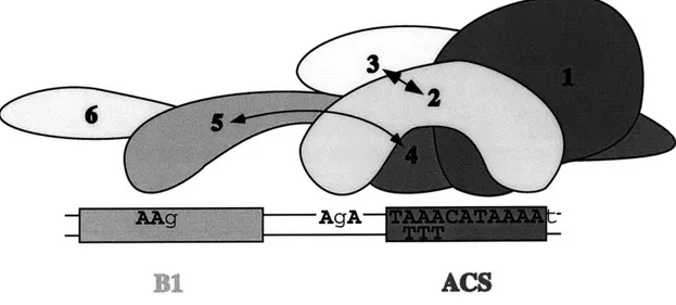
action of five of the six ORC subunits. Protein-DNA crosslinking studies suggested that
recognition of origin sequences is mediated primarily by two different groups of ORC
subunits that make sequence-specific contacts with two distinct regions of the DNA.
Implications of these findings for ORC function and the mechanism of initiation of
eukaryotic DNA replication are discussed.
INTRODUCTION
The initiation of DNA replication is a complex process involving multiple
regulated steps, including the selection of the initiation site on the DNA, unwinding of
the DNA helix, and the assembly of a multi-protein replication machine. Studies of the
replication of bacteria, phage, and eukaryotic viral genomes have established that a
protein called the initiator binds its cognate origin of DNA replication in a
sequence-specific manner. Once bound, initiator proteins often participate in other aspects of
replication initiation including facilitating origin unwinding and the recruiting other
replication proteins to the origin (reviewed in Baker and Bell, 1998; Kornberg and Baker,
1992). Detailed analyses of initiator proteins bound to their cognate origins have been important in determining how these proteins function during replication initiation. Our
aim is to extend this type of analysis to a putative eukaryotic chromosomal initiator
protein bound to an origin of DNA replication.
The Saccharomyces cerevisiae Origin Recognition Complex (ORC) clearly
performs at least two of the three general functions of initiator proteins. As described in
Chapter I, ORC binds to origins of DNA replication in vitro and in vivo and ORC recruits
other replication proteins to origins to assemble the pre-RC. In this chapter, I describe
work aimed at characterizing the first activity, sequence-specific DNA binding. The
ability of ORC to bind origins is essential for yeast cell viability. Mutations in origin
sequences that reduce or eliminate origin function also reduce or eliminate ORC binding
in vitro (Bell and Stillman, 1992; Rao and Stillman, 1995; Rowley et al., 1995) and in vivo (Aparicio et al., 1997; Tanaka et al., 1997). In addition, most conditional mutations
in ORC genes lead to decreased origin usage (Fox et al., 1995; Liang et al., 1995) and
decreased origin binding in vivo (Aparicio et al., 1997; 0. Aparicio and S. P. B.,
unpublished observations).
S. cerevisiae contains the best defined eukaryotic origins of DNA replication. These elements were first identified as genomic DNA sequences capable of supporting
the autonomous replication of episomal DNA (Autonomous Replicating Sequences or
ARSs) (Hsiao and Carbon, 1979; Stinchcomb et al., 1979). Many of these elements were
subsequently shown to act as origins of replication in their normal chromosomal context
(reviewed in Newlon and Theis, 1993). Yeast origins are modular in nature and contain a
conserved 11 base-pair ARS Consensus Sequence (ACS) that is essential for ORC-DNA
binding and origin function in vivo, as well as additional elements that enhance origin
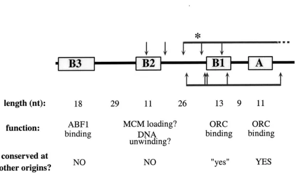
function (generally referred to as B elements; Bell, 1995). ARSI, the first
well-characterized origin, has three such elements (B 1, B2 and B3; see Figure 1). In vitro and
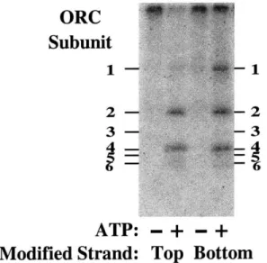
in vivo, DNase I protection assays of ARS] demonstrated that ORC protects
approximately fifty base pairs of DNA that include the ACS and B 1 sequences (Bell and
Stillman, 1992; Diffley and Cocker, 1992; Santocanale and Diffley, 1996). The DNase I
cleavage pattern also contains several sites that become hypersensitive to digestion in the
presence of ORC. These sites are spaced roughly 10 base-pairs (bp) apart, suggesting
that ORC wraps DNA around itself or that the DNA lies along a flat surface created by
ORC (Travers and Klug, 1987). The A and B 1 elements direct ORC-DNA binding at
ARS] and at ARS307 (Rao et al., 1994; Rowley et al., 1995), and together represent the
smallest functional region of either origin. I will refer to this minimal region required for
ORC-DNA binding and origin function as the Core origin.
The role of the other B elements in origin function is not yet clear. The B2
element may contribute origin melting during the initiation process (reviewed in Bell,
1995). The origin of bidirectional replication (OBR, the site at which discontinuous replication switches to continuous replication) has been mapped to a site in between the
BI and B2 elements of ARS] (Bielinsky and Gerbi, 1998; Bielinsky and Gerbi, 1999),
*
B3
-
-
B2 -
-1
1length (nt): 18 29 11 26 13 9 11
function: ABF1 MCM loading? ORC ORC
binding DNA binding binding
unwinding? conserved atcosevd t NO
NO "1yes"v YES
other origins?
Figure 1. Properties of Functional Elements at ARSJ. The ARS1 elements are as previously described (Marahrens and Stillman, 1992). The A element contains the conserved ARS Consensus Sequence (ACS) and is essential for replication, while the B elements enhance replication function. The ACS and B 1 elements together comprise the ORC binding site and form the Core origin. B1-like ele-ments are found at other yeast origins and can functionally substitute for the ARS1 B 1 element. The B2 element is required for loading MCM proteins onto the
ARS1 origin (C. Aparicio and S. P. Bell, unpublished results). B3 is the binding
site for the transcriptional activator, ABF1; it can be functionally substituted by a binding site for other transcriptional activators. The region of DNA protected from DNase I cleavage by ORC is indicated by red horizontal lines above and below the ARS1 schematic. ORC-induced sites of increased DNase I cleavage are indicated by arrows. The position of the origin of bidirectional replication (OBR) is indicated by the blue asterisk (Belinsky and Gerbi, 1999).
consistent with initial origin unwinding occurring near B2. Furthermore, a linker
substitution mutation in B2 that decreases origin function also inhibits MCM loading at
ARS1 (0. Aparicio and S. P. B., unpublished observations). If MCMs function as the
replicative helicase (see Chapter I), they would likely be loaded onto origins at sites of
initial origin unwinding. The B3 element is a binding site for the ABF1 transcriptional
activator and it can be functionally substituted by a binding site for other transcriptional
activators (Marahrens and Stillman, 1992). One possible role for these transcription
factors in origin function is to regulate chromatin structure. The ARS] origin was shown
to have a unique chromatin structure important for origin function, with a nucleosome
free region encompassing the A and B elements and precisely positioned nucleosomes to
either side (Simpson, 1990; Thoma et al., 1984). Recent work from our lab has suggested
that ORC and ABF1 are required to establish this specific chromatin structure (discussed
in Chapter V).
We have a general understanding of the DNA sequence requirements for the
association of ORC with origins (Bell and Stillman, 1992; Rao and Stillman, 1995;
Rowley et al., 1995) but previous studies have not examined the requirements of different
ORC subunits for DNA binding. A thorough understanding of ORC bound to yeast
origins of replication can address three important questions: (1) How does ORC interact
with DNA? In particular, I wanted to determine which of the approximately 50 base
pairs protected from DNase I cleavage are important for ORC-DNA binding and how the
structure of origin DNA is affected by interaction with ORC. Additionally, since none of
the six ORC subunits contain a canonical DNA binding motif, I wanted to identify the
ORC proteins are required for DNA binding. (2) How do ORC subunits interact with
each other? ORC is a pre-assembled complex in the absence of DNA and I wanted to
understand the organization of ORC subunits both in solution and in DNA-complexes.