Publisher’s version / Version de l'éditeur:
Molecular Microbiology, 2016-10-01
READ THESE TERMS AND CONDITIONS CAREFULLY BEFORE USING THIS WEBSITE.
https://nrc-publications.canada.ca/eng/copyright
Vous avez des questions? Nous pouvons vous aider. Pour communiquer directement avec un auteur, consultez la
première page de la revue dans laquelle son article a été publié afin de trouver ses coordonnées. Si vous n’arrivez pas à les repérer, communiquez avec nous à PublicationsArchive-ArchivesPublications@nrc-cnrc.gc.ca.
Questions? Contact the NRC Publications Archive team at
PublicationsArchive-ArchivesPublications@nrc-cnrc.gc.ca. If you wish to email the authors directly, please see the first page of the publication for their contact information.
This publication could be one of several versions: author’s original, accepted manuscript or the publisher’s version. / La version de cette publication peut être l’une des suivantes : la version prépublication de l’auteur, la version acceptée du manuscrit ou la version de l’éditeur.
For the publisher’s version, please access the DOI link below./ Pour consulter la version de l’éditeur, utilisez le lien DOI ci-dessous.
https://doi.org/10.1111/mmi.13544
Access and use of this website and the material on it are subject to the Terms and Conditions set forth at A novel glycan modifies the flagellar filament proteins of the oral bacterium Treponema denticola
Kurniyati, Kurni; Kelly, John F.; Vinogradov, Evgeny; Robotham, Anna; Tu, Youbing; Wang, Juyu; Liu, Jun; Logan, Susan M.; Li, Chunhao
https://publications-cnrc.canada.ca/fra/droits
L’accès à ce site Web et l’utilisation de son contenu sont assujettis aux conditions présentées dans le site LISEZ CES CONDITIONS ATTENTIVEMENT AVANT D’UTILISER CE SITE WEB.
NRC Publications Record / Notice d'Archives des publications de CNRC: https://nrc-publications.canada.ca/eng/view/object/?id=8aaeced3-0cba-4ac8-a809-935aacd7ce49 https://publications-cnrc.canada.ca/fra/voir/objet/?id=8aaeced3-0cba-4ac8-a809-935aacd7ce49
Kurni Kurniyati1, John F. Kelly3, Evgeny Vinogradov3, Anna Robotham3, Youbing Tu1, Juyu Wang4, Jun Liu4, Susan M. Logan3, and Chunhao Li1,2*
1
Department of Oral Biology, and 2Department of Microbiology and Immunology, The State University of New York at Buffalo, New York 14214, USA
3
Vaccine Program, Human Health Therapeutics, National Research Council, Ottawa, Ontario, Canada K1A 0R6
4
Department of Pathology and Laboratory Medicine, McGovern Medical School at UT Health Science Center, Houston, Texas 77030, USA
Running title: Flagellin glycosylation of Treponema denticola
Keywords (Spirochete/ Treponema denticola/ flagella/ glycosylation)
* Corresponding author. Mailing address: Department of Oral Biology, SUNY at Buffalo, 3435 Main St., Buffalo, NY 1421473092
Electronic mail address: cli9@buffalo.edu; phone: (716) 82976014; Fax: (716) 82973942
This article has been accepted for publication and undergone full peer review but has not been through the copyediting, typesetting, pagination and proofreading process which may lead to
While protein glycosylation has been reported in several spirochetes including the syphilis bacterium Treponema pallidum and Lyme disease pathogen Borrelia burgdorferi, the pertinent glycan structures and their roles remain uncharacterized. Herein, we report a novel glycan with an unusual chemical composition and structure in the oral spirochete Treponema denticola, a keystone pathogen of periodontitis. The identified glycan of mass 450.2 Da is composed of a monoacetylated nonulosonic acid (Non) with a novel extended N7 acyl modification, a 29 methoxy94,5,69trihydroxy9hexanoyl residue in which the Non has a pseudaminic acid configuration (L9glycero9L9manno) and is β9linked to serine or threonine residues. This novel glycan modifies the flagellin proteins (FlaBs) of T. denticola by O9linkage at multiple sites near the D1 domain, a highly conserved region of bacterial flagellins that interact with Toll9like receptor 5. Furthermore, mutagenesis studies demonstrate that the glycosylation plays an essential role in the flagellar assembly and motility of T. denticola. To our knowledge, this novel glycan and its unique modification sites have not been reported previously in any bacteria.
Spirochetes are a group of medically important but poorly understood bacteria, some of which cause human illnesses such as Lyme disease (Borrelia burgdorferi), leptospirosis (Leptospira interrogans), and syphilis (Treponema pallidum) [for recent reviews, see references (Radolf et al., 2012, Giacani & Lukehart, 2014, Evangelista & Coburn, 2010)]. Spirochetes are recognized for their distinct cell morphology and unusual means of motility 9 they are either flat9waved or corkscrew9shaped and swim by means of rotating flagella that are located between the outer membrane and the peptidoglycan layer (Li et al., 2000b, Charon et al., 2012). Thus, spirochetal flagella are referred to as endoflagella or periplasmic flagella (PFs). Several studies have shown that the PFs are not only essential for cell motility and morphology but are also directly linked to the pathogenicity of spirochetes [for recent review, see reference (Charon et al., 2012)]. For example, aflagellated mutants of B. burgdorferi are non9motile, rod9shaped instead of flat9 waved, and unable to establish infection in mice (Li et al., 2010, Sultan et al., 2013, Sultan et al., 2015, Li et al., 2000b, Lin et al., 2015). Flagella9deficient mutants of L. interrogans are non9 motile, lack the distinctive hook9shaped ends, and are unable to cause infection in hamsters, a standard experimental model of leptospirosis (Lambert et al., 2012, Wunder et al., 2016, Fontana et al., 2016).
PFs are structurally similar to the flagella of other bacteria, as each consists of a basal body9 motor complex, a hook, and a filament (Charon et al., 2012, Zhao et al., 2014). The filament is a long, thin, helical structure that functions as a propeller and is essential for bacterial locomotion (Berg & Anderson, 1973). In most bacteria, flagellar filaments consist of a single polymeric protein known as flagellin (FliC in Escherichia coli and Salmonella enterica) (Samatey et al.,
2001). However, the PF filament has an unusual structure and protein composition, and it is one of the most complex bacterial flagellar filaments thus far analyzed [for review, see references (Li et al., 2000b, Charon et al., 1992)]. Specifically, in most spirochete species, the PFs are comprised of at least one sheath protein (FlaA) and one to three core proteins (FlaB193). The FlaA proteins (ranging in size from 37 to 44 kDa) are similar between species, based on their amino acid sequences and antigenic cross9reactivity (Norris et al., 1988, Norris, 1993). The FlaA proteins are likely exported to the periplasmic space via the type II secretion pathway, as their N9 termini contain a typical peptidase I cleavage site (Pugsley, 1993). The FlaA proteins form a sheath around the core proteins, determine the diameter and helicity of the PFs, and are important for cell motility (Li et al., 2000a, Li et al., 2008). In contrast, the FlaB proteins are exported to the periplasmic space through the flagellar type III secretion apparatus (Erhardt et al., 2010). The FlaB proteins are generally 30 to 41 kDa in size and immunologically cross9 reactive (both within a given species and also between species) (Norris et al., 1988, Norris, 1993). For spirochetes that have multiple core proteins, such as Brachyspira hyodysenteriae (the causative agent of swine dysentery) (Koopman et al., 1992), the function of FlaB proteins overlaps such that neither one of these proteins is essential for the intact PF structure and cell motility (Li et al., 2000a, Li et al., 2008). The FlaB proteins form a core structure that is sheathed by FlaA. There is no sequence similarity or antigenic cross9reactivity between the FlaA and FlaB proteins (Norris et al., 1988, Norris, 1993).
The genus Treponema includes both commensal and pathogenic spirochetes, some of which cause human illnesses such as yaws, pinta, and syphilis (Giacani & Lukehart, 2014, Radolf, 1996). Certain Treponema species are also implicated in polymicrobial infections in humans and
animals, such as periodontitis and bovine digital dermatitis [for reviews, see (Evans et al., 2016, Giacani & Lukehart, 2014)]. Since a majority of treponemal species cannot be reliably cultivated in vitro, the oral spirochete Treponema denticola, which can be cultivated, has been used as a model organism to understand the biology and pathogenicity of the treponemes (Anand et al., 2013, Chi et al., 1999, Bian et al., 2015). T. denticola is an obligatory anaerobic, facultative, and highly motile bacterium, which is implicated in both periodontal and endodontic infections [for review, see references (Ellen & Galimanas, 2005, Dashper et al., 2011)]. As revealed by electron microscopy and cryo9electron tomography (cryo9ET), T. denticola has two PFs that arise from each end of the cell and extend toward the center of the cell where they overlap (Izard et al., 2008, Izard et al., 2009). Similar to those in other spirochetes, the T. denticola PFs are essential for cell shape and motility; flagella9deficient mutants are non9motile and less helical at the regions where the PFs interact with the cell cylinder (Li et al., 1996, Ruby et al., 1997, Limberger et al., 1999). Motility also contributes to the pathogenicity of T. denticola; non9 flagellated mutants are unable to penetrate oral epithelial cells (Lux et al., 2001).
The PF filament of T. denticola consists of at least one sheath protein, FlaA, and three core proteins designated as FlaB1, FlaB2, and FlaB3 (Ruby et al., 1997). These flagellin proteins share a high sequence similarity (> 67% identity) to their counterparts in T. pallidum (Norris et al., 1988, Norris, 1993). Wyss et al. reported that the flagellin proteins of T. pallidum and pathogenic oral Treponema spp. are glycosylated (Wyss, 1998). Flagellar glycosylation was also reported in other spirochetes, including B. hyodysenteriae (Li et al., 1993) and Spirochaeta aurantia (Brahamsha & Greenberg, 1988). However, their chemical features, structures, and roles in motility and flagellation remain uncharacterized.
Flagella glycosylation has been demonstrated in numerous motile bacteria and archaea and it has been found that such a post9translational modification affects flagellar assembly, motility, as well as virulence in some pathogenic bacteria [for reviews, see references (Logan, 2006, Merino & Tomas, 2014)]. For instance, the flagellins of Campylobacter jejuni and Campylobacter coli are glycosylated with O9linked pseudaminic acid (Pse) or its acetamidino derivative PseAm (Logan et al., 2002). Mutant strains which could no longer synthesize Pse were unable to assemble intact flagella, were non9motile and less virulent (Howard et al., 2009, Guerry et al., 2006). Similar to Campylobacter, the flagellins of Helicobacter pylori are also modified with Pse and the glycan modification is also essential for flagellar assembly, motility, and virulence (Josenhans et al., 2002, Schirm et al., 2003). In this study, we used genetic, biochemical, and structural approaches to elucidate the structure of a novel glycan that modifies T. denticola flagellins. We demonstrate that glycosylation is required for flagellar assembly and consequent motility.
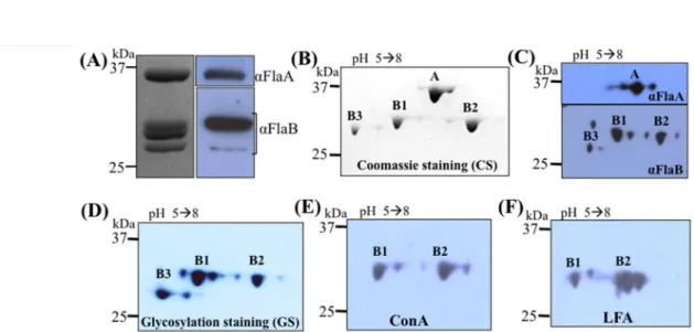
T. denticola The genome of T. denticola encodes six putative flagellin proteins – three FlaA (TDE1408, 236 aa; TDE1409, 246 aa; and TDE1712, 349 aa) and three FlaB proteins (TDE1004, 286 aa; TDE1475, 285 aa; and TDE1477, 286 aa) (Seshadri et al., 2004). However, Ruby et al., found only four of these proteins (one FlaA and three FlaBs) in the isolated flagellar filaments of T. denticola (Ruby et al., 1997). To address this discrepancy, the PFs were isolated from the wild9type strain and analyzed in detail. Four protein bands were visualized by SDS9PAGE (Fig. 1A). The top band (approximate 37.0 kDa) reacted to T. denticola FlaA antiserum and the lower bands (around 31.0 kDa) were recognized by the T. pallidum FlaB antibody. Of note, the size of FlaA (TDE1712) was smaller than its predicted
molecular weight (MW, 39.3 kDa), suggesting that it is posttranslationally processed prior to assembly. Along with this prediction, a 22 aa signal peptide was identified at its N9terminus (Fig. S1). The MWs of the three FlaB proteins are very close to each other and could not be fully separated by 1D SDS9PAGE (Fig. 1A). To decipher the filament protein composition, the isolated PFs were separated by 2D9gel electrophoresis. In addition to FlaA, three FlaB proteins were clearly visualized on the 2D gels (Fig. 1B) and could be detected by T. pallidum FlaB antiserum (Fig. 1C). These results indicate that the flagellar filament of T. denticola comprises one FlaA and three FlaB proteins, which corroborates the previous report (Ruby et al., 1997). Based on their sequences, MWs, and isoelectric points (pIs), these filament proteins were assigned: FlaA (TDE1712, pI 5.39, 39.3 kDa), FlaB1 (TDE1477, pI 5.4, 31.3 kDa), FlaB2 (TDE1004, pI 6.54, 31.6 kDa), and FlaB3 (TDE1475, pI 5.3, 30.9 kDa).
T. denticola Wyss previously reported that the flagellar proteins of pathogenic Treponema species are glycosylated (Wyss, 1998). To determine if this is the case for T. denticola, the PFs were isolated from the wild9type strain and analyzed by SDS9 PAGE and 2D9gel electrophoresis followed by glycosylation staining as well as lectin blotting analyses with ConA and LFA. The three FlaBs, but not FlaA, stained positive for glycosylation (Fig. 1D). In addition, FlaB1 and FlaB2 reacted with both ConA and LFA (Fig. 1E, F). LFA is a lectin that specifically recognizes sialic acid9like sugar moieties (Miller et al., 1982), suggesting that the FlaB proteins may be modified with sialic acid9like sugars. Of note, FlaB3 was not visibly reactive with ConA and LFA (Fig. 1E, F), but this may be due to its relatively low level in the PFs and the sensitivity of the lectin blotting analysis. We used densitometry analysis to
measure the level of FlaB3 relative to the other three flagellin proteins and found that the average stoichiometry of these four proteins is: FlaA (5.0): FlaB1 (3.5): FlaB2 (3.5): FlaB3 (1.0).
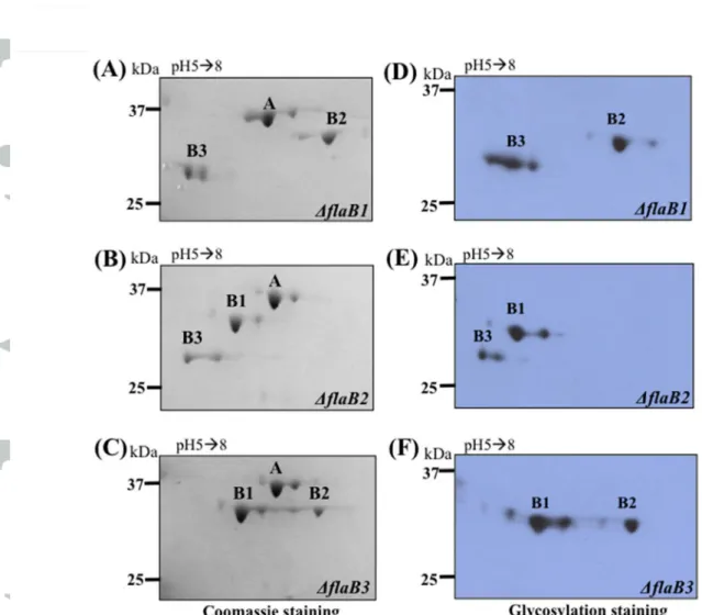
To confirm that the observed glycosylated products are indeed the filament core proteins, the genes encoding the three FlaBs were deleted by allelic exchange mutagenesis as illustrated in Fig. S2. The PFs were isolated from these individual mutants (BflaB1, BflaB2, and BflaB3) and subjected to 2D9gel electrophoresis, followed by Coomassie blue and glycosylation staining. As shown in Fig. 2, the cognate gene products were abolished in each of these mutants (Fig. 2A9C), and the pertinent glycosylated products were absent, as shown by glycosylation staining (Fig. 2D9F). For example, the glycosylated product tentatively assigned as FlaB1 was not detected in the PFs isolated from the BflaB1 mutant. The same results were observed in the BflaB2 and BflaB3 mutants. Based on these results, we concluded that the three filament core proteins (FlaB1, FlaB2, and FlaB3) are glycosylated. This deletion mutagenesis analysis also confirms the gene assignment of the observed PF proteins (e.g., FlaB1 is encoded by TDE1477).
O Protein glycosylation occurs as either O9linkage or N9 linkage. Some N9linked glycoproteins can be deglycosylated with PNGase F (Kim & Leahy, 2013) and O9linked glycans can be chemically removed through β9elimination with alkaline (Peter9Katalinic, 2005). To determine the glycan linkage in the FlaB proteins, deglycosylation assays with either PNGase F or by β9elimination were carried out using the PFs isolated from the BflaA mutant to avoid any potential influence of the outer sheath during the treatments. PNGase F treatments under different conditions had no evident impact on the flagellin glycosylation (Fig. S3A). In contrast, β9elimination appeared to remove the glycoreactive moieties so that no
glycosylated FlaB proteins were detected by glycosylation staining (lower panel, Fig. S3B). Of note, after the treatment, the FlaB proteins were moderately degraded; whereas a non9specific protein associated with the PFs remained unaffected (top band, Fig. S3B), suggesting that glycosylation may affect the stability of FlaB within the assembled filaments. Nevertheless, these degraded products were still reactive with the FlaB antiserum (middle panel, Fig. S3B). Collectively, these results suggest that the FlaB proteins are modified with O7linked glycans.
!" As shown in Fig. 1, the MWs of
three FlaB proteins are very similar. In order to separate them and prepare pure FlaB proteins for LC9MS/MS analysis, two double mutants were constructed BflaAflaB1 and BflaAflaB2, which allowed us to prepare the pure FlaB1 from BflaAflaB2 and FlaB2 and FlaB3 proteins from BflaAflaB1 (Fig. S3). The FlaB proteins were separated by SDS9PAGE, excised, digested with trypsin and then analyzed by using nLC9MS/MS, as previously described (Logan et al., 2002, Schoenhofen et al., 2006a). An initial MASCOT database search was conducted and many of the MS/MS spectra were assigned to unmodified tryptic peptides from all three of the FlaB proteins. Manual inspection of the fragment ion patterns in the remaining, unassigned MS/MS spectra identified 394 peptides from each of the FlaB proteins, all of which are modified with a novel 450.2 Da moiety (m/z 451.2 in Fig. 3 and Table 1). Moreover, many of the fragment ions in the lower region of the MS/MS spectra arise from the 450.2 Da modification (highlighted with a “♦” in Fig. 3c) and some have been observed previously in the MS/MS fragment ion spectra of the nonulosonic acids (i.e., pseudaminic acid and legionaminic acid) that decorate the flagellin of many bacteria. Therefore, it was hypothesized that the 450.2 Da moiety is a modified nonulosonic acid glycan. All of the identified glycopeptides contain a serine (S) and/or a
threonine (T) residue (insets in Fig. 3 and Table 1) and have no N9linkage sequon (NXS or NXT, where X is any amino acid other than proline) (Rao & Bernd, 2010), indicative of O7linked glycosylation. Furthermore, no evidence of the presence of any other glycan modification was found. Also, of note, glycosylation staining suggested that the sheath protein FlaA is not glycosylated (Fig. 1D). To confirm this observation, the FlaA band on the SDS9PAGE gels was excised, digested with trypsin, and then analyzed by nLC9MS/MS as described above. All MS/MS spectra were assigned to tryptic peptides from the FlaA protein sequence, and no evidence of glycosylation was observed (Fig. S5).
The distinct glycan fragment ion pattern in the glycopeptide LC9MS/MS spectra indicated that the 450.2 Da glycan consists of two components – a 274 Da sugar thought to be a monoacetylated nonulosonic acid (the m/z 275.1 oxonium ion in Fig. 3A9C) and an unknown 176 Da component. High9resolution mass spectrometry (HRMS) analysis performed with the LTQ9Orbitrap XL (Fig. 4) determined that the 274 Da sugar has the same mass as a monoacetylated nonulosonic acid (calculated m/z value for oxonium ion: m/z 275.1243; observed m/z value: m/z 275.1233 ± 0.0004). HRMS also indicated that the other component has an elemental composition of C7O5H12 (observed mass 176.0688 ± 0.0004 Da), which ruled out the possibility that it could be an uronic acid (calc. mass 176.0321 Da).
# To determine the structure of 450.2 Da
glycan, 25 mg of PFs was isolated from the flaA mutant, digested with proteinase K, and fractionated on a Biogel P10 column. 1H NMR spectra were recorded for all fractions. The fractions containing glycopeptides were further fractionated on a Zorbax C18 column and then
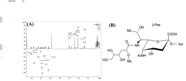
containing sub9fractions were selected for 2D NMR analysis. Each sub9fraction contained the same sugar component, but they differed in terms of peptide compositions. NMR data for one of the sub9fractions (fraction #32) are presented in Fig. 5A and Table 2. Amino acids observed in this sub9fraction correspond to the peptide sequence of VSQLV found in FlaB3 (TDE1475) and FlaB1 (TDE1477) (Table 1). In addition to the signals of amino acids, the spectra contained spin systems of a 5,79diamino93,5,7,99tetradeoxy9non929ulopyranosonic acid (Non), an acetyl group, and a component that we designated as ‘A’. The configuration of the pyranose ring of Non is identical to that of β9pseudaminic acid, as confirmed by signal positions and coupling constants of the structure’s atoms matching what was previously reported (Knirel et al., 2003). An observed coupling constant J6,7 of 11 Hz was also characteristic of the Pse, and thus C97 had the same configuration in Non as in Pse. The configuration of C98 was not clear from NMR data. The position of the Non C99 signal, which allows discrimination between Pse and 89epi9 pseudaminic acid (89epi9Pse), was right in the middle between the signal positions of both isomers. The H98 signal was quite far from that observed for Pse and closer to that reported for 89epi9Pse (observed 4.38 ppm, 4.18 ppm in Pse, 4.25 ppm in 89epi9Pse), although it should be acknowledged that the presence of a bulky acyl group on N97 (see below) could significantly influence the NMR spectra. As a consequence, we have chosen to present the structure of the flagellin glycan in a Pse configuration (Fig. 5B) but do acknowledge the possibility that it may be 89epi9Pse.
N95 of Non was acylated by an acetyl group as determined from the heteronuclear bond correlation (HMBC) between Ac C91 and Non H95. COSY, TOCSY and HSQC data for the component A indicated that this structure is a 29methoxy94,5,69trihydroxy9hexanoyl residue (Fig.
5). No signals of its carboxyl group were observed in the HMBC spectrum, but accurate mass analysis by MS confirmed the elemental composition. The configurations of the three chiral atoms (C92, C94, and C95) in A were not determined. This group has to acylate N97 of Non, since there is no other location where it can be linked that agrees with the NMR and MS data. The linkage between Non and Ser was found from the HMBC between Non C92 and Ser H93. In summary, the NMR and MS data indicate that the structure is a novel C79acylated pseudaminic acid derivative, as presented in Fig. 5B. There is a possibility that the Non sugar could be the structural isomer at C98 of Pse, known as 89epi9Pse.
$%&'(&" Pse and Pse9like glycans have been identified in several flagellated bacteria such as Campylobacter and Helicobacter species (Logan, 2006). In Campylobacter jejuni, the Pse biosynthetic genes are located within a large flagellar glycosylation locus that contains ~50 genes. In contrast, the Pse biosynthetic genes of Helicobacter pylori are sporadically distributed around the genome (Schirm et al., 2003, Logan et al., 2002). BLAST searches of the T. denticola genome identified several putative Pse biosynthesis genes that are sporadically distributed on the genome. One of those homologs is PseI (TDE0960), a putative Pse synthase. TDE0960 has a 28% identity to the PseI protein of H. pylori and contains a conserved NeuB domain that is associated with bacterial sialic acid synthases (Fig. S6). To determine if TDE0960 is involved in FlaB glycosylation, the cognate gene was deleted, and the resultant mutant (Tde960mut) was cis7complemented using the method illustrated in Fig. S2. Immunoblotting analysis using a specific antibody against TDE0960 showed that the cognate gene product was abolished in the mutant and restored in the complemented strain (named Tde960com) (Fig. 6A). Glycosylation staining analysis revealed that
the glycosylated FlaBs were detected in the wild9type and Tde960com strains but not in Tde960mut (Fig. 6B). Immunoblotting analysis with the FlaA and FlaB antibodies showed that the flagellin proteins were almost undetectable in the mutant (Fig. 6C). Interestingly, a trace amount of FlaA and FlaB proteins could still be detected in the mutant whole cell lysate when its loading amount was increased 89fold (Fig. 6D). In contrast to the FlaA and FlaB proteins, the flagellar hook protein FlgE remains unaffected in the TDE960 mutant strain (Fig. 6C). It is worth noting that the MWs of the detected FlaBs in Tde960mut were lower than in the wild type (Fig. 6D), indicative of unmodified forms of FlaBs. Collectively, these data suggest that TDE0960 is essential for flagellin production and glycosylation in T. denticola.
$%&'(& ) To elucidate the potential
mechanisms that underlie the reduction of FlaA and FlaBs that was observed in Tde960mut, the levels of flagellin gene transcripts (flaA, flaB1, flaB2, and flaB3) were measured by qRT9PCR. Using the dnaK transcript (TDE0628) as an internal control, the relative levels of the four flagellin mRNAs in Tde960mut were measured. The results showed that the levels of flaA and flaB transcripts in the mutant were decreased by 50~90% relative to those in the wild type, with the flaB1 mRNA being decreased by nearly 90% (Fig. 7A).
Protein turnover assays were also carried out to monitor the stability of flagellin proteins in the mutant (Fig. 7B). After arresting protein synthesis with addition of spectinomycin, the levels of FlaA and FlaBs remained unchanged in the wild type during a course of 24 hrs (left panels). In contrast, in the Tde960mut mutant, the FlaA protein was decreased nearly 60% at 4 h and completely abolished by 24 h; and the levels of FlaB proteins were reduced by nearly 54% by 24 h compared to their levels in the wild type (right panels). Taken together, these results indicate
that both flagellin gene transcription and protein stability are influenced by perturbation of the glycan biosynthetic pathway.
'(& To determine if the reduction of FlaA and FlaBs in Tde960mut impairs the PF structure, the mutant cells were dissected in situ using cryo9ET. In the wild9type cells, two long PFs (average length >1,500 nm, n= 18 cells) arose from each cell pole, wrap around the cell cylinder, and extended towards the central region of the cell (Fig. 8A, B and video 1). In the mutant cells, although two PFs were visible at each cell pole, the flagellar filaments were much shorter (average length = 96 ± 36.5 nm, n=14 cells) than the PFs of the wild9type cells (Fig. 8C, D and video 2). In contrast to the filaments, the flagellar basal body and hook structures were unaffected, which is consistent with the immunoblotting results of FlgE that showed no change in its expression level (lower panel, Fig. 6C). These results indicate that the deletion of TDE0960 only impairs the level of FlaA and FlaB proteins and assembly of the filaments in T. denticola but does not affect the assembly of the flagellar hook.
'(& Microscopic observation and
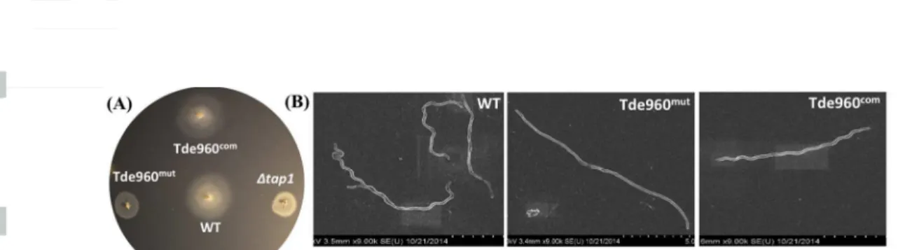
bacterial motion tracking analysis showed that Tde960mut failed to migrate in the growth medium containing 1% methylcellulose (videos 3 & 4), indicating that the mutant is non9motile. This proposition was further substantiated by bacterial swimming plate assays (Fig. 9A). The average diameter of swimming rings formed by Tde960mut (5.8 mm, n=5) was significantly smaller (P < 0.01) than that of the wild9type (12.75 mm, n=5) and Tde960com (12.80 mm, n=5) and was almost the same diameter as seen with Btap1 (5.5 mm, n=5), a non9motile mutant of T. denticola (Limberger et al., 1999). Interestingly, SEM analysis revealed that the mutant cells are rod9
shaped instead of the corkscrew9shaped wild9type and Tde960com strains (Fig. 9B). Taken together, these results indicate that the Tde960mut mutant is non9motile and has an altered cell shape, highlighting the important role of flagellar glycosylation in cell motility and morphogenesis.
$
In this study, we isolated PF filaments from T. denticola and analyzed the protein composition using SDS9PAGE, 29D electrophoresis, and immunoblotting analyses. The results clearly demonstrate that the T. denticola flagellar filament consists of one sheath protein (FlaA) and three core FlaB proteins and that these proteins are immunologically cross9reactive to antiserum raised against the corresponding T. pallidum flagellin proteins (Fig. 1). The protein composition was further confirmed by targeted mutagenesis. Deletions of individual flagellin genes abolished the cognate gene products in the PFs isolated from the mutants (Fig. 2). Two putative FlaA homologs (TDE1408 and TDE1409) were not found in the isolated PFs. A BLAST search revealed that although these two proteins belong to the superfamily of FlaA (pfam04620), they have very low (E value > 1.5) sequence similarity to the FlaA protein (TDE1712) that we identified as a component of PFs. It is possible that these two proteins are components of PFs but their levels are too low to be detected. Alternatively, they may have a distinct role to play in motility but are not components of the isolated PFs. Future studies will investigate the role of these two genes in the motility of T. denticola.
After delineating the filament protein composition, we undertook a comprehensive study to characterize the flagellin structural proteins FlaA, FlaB1, FlaB2 and FlaB3. The results from
these experiments provide conclusive proof that the FlaA sheath protein is not glycosylated while the three FlaB proteins are all modified with an identical, novel, O9linked glycan (Figs. 29 5). The glycopeptides identified for each FlaB protein were localized to two regions of the primary amino acid sequences that lie within the predicted D1 and D2/D3 domains based on the atomic model of the Salmonella flagellin protein FliC (Fig. 10) (Samatey et al., 2001). While the D2/D3 region is most likely exposed on the external surface of the assembled flagellar filament, the D1 domain is buried in polymerized flagellar filaments. Glycosylation within the D2/D3 domains has been observed in many other bacterial species that produce glycosylated flagellins (Goon et al., 2003, Schirm et al., 2003, Schirm et al., 2004, Verma et al., 2006), but this is the first identification of a glycosylation site within the conserved D1 domain (Samatey et al., 2001). A recent cryo9ET study of T. pallidum flagellum architecture suggested that the FlaA sheath forms a lattice structure covering the inner FlaB core (Liu et al., 2010). This lattice structure could provide surface access to the D2/D3 glycan modifications found on the FlaB proteins. While flagellar glycans in other bacterial pathogens have been shown to play a role in host pathogen interactions due to their location on the external surface of the assembled flagellar filaments [for reviews, see references (Logan, 2006, Nothaft & Szymanski, 2010)], it remains to be established what the biological roles of the glycans are on the T. denticola flagella and motility. It is possible that the glycosylation facilitates interactions with the FlaA sheath structure and/or increases the stability of flagellin proteins after they are secreted into the periplasmic space.
As a keystone pathogen of periodontitis, T. denticola inhabits at the forefront of subgingival biofilms and directly encounters host innate immune responses (Ellen & Galimanas, 2005,
Kurniyati et al., 2013). The innate immune response to flagellins is mediated by Toll9like receptor 5 (TLR5) (Hayashi et al., 2001, Smith et al., 2003), and structural studies have defined the precise domains and amino acid residues of both TLR5 and the D0/D1 domains of FliC that are involved in this interaction (Yoon et al., 2012). One of the identified sites of glycosylation present in all three T. denticola FlaBs (S116) lies between conserved residues E114 and Q117 of the αND1b region of this D1 domain (Fig. 10). Both residues were identified in the co9 crystallization study of TLR5 with Salmonella FliC fragment CBLB502 as residues that interact via H bonding with the LRR9 loop groove of primary interface9B of TLR5. The LRR9 loop has been described as a “hot spot” in the TLR9FliC interaction (Yoon et al., 2012). It remains to be seen if the novel glycosylation identified on the T. denticola FlaB proteins at S116 perturbs this interaction and so affects the pattern recognition mediated by TLR5. If so, this could subsequently protect the pathogen from the host immune responses. The role of glycosylation in the interplay between the T. denticola flagellins and TLR5 receptor and its subsequent impact on the host immune responses can now be explored at the molecular level.
Flagellin glycosylation and Pse biosynthesis have been extensively studied in C. jejuni and H. pylori. A set of core enzymes (i.e., PseB, C, G, H, F, and I) has been shown to be required for the biosynthesis of Pse (Schoenhofen et al., 2006a, Schoenhofen et al., 2006b, Schoenhofen et al., 2006c). Genome mining efforts to identify the biosynthetic genes in T. denticola revealed that there is no comparable flagellar genetic locus. In addition, BLAST searches using the H. pylori and C. jejuni biosynthetic enzymes as queries identified only four Pse homologs in the genome of T. denticola – TDE0714 (PseB), TDE0725 (PseC), TDE0960 (PseI), and TDE1917 (PseF). Interestingly, these 4 homologs were also found in T. pallidum (Fraser et al., 1998), (TP0077,
TP0078, TP0562, and TP0289), and they share high sequence identities (50~70%) with their counterparts in T. denticola. These findings suggest that the flagellins of T. pallidum are probably modified with the same glycan as those of T. denticola. From a biosynthetic perspective, no homologs of PseH (N9acetyltransferase) and PseG (nucleotidase) were identified in either of the Treponema genomes. PseH is responsible for the N94 acetylation of UDP949 amino94,69dideoxy9β9L9AltNAc to form UDP92,49diacetamido92,4,69trideoxy9β9L9AltNAc. PseH is responsible for removal of UDP from C1 of the PseH product to form 2,49diacetamido92,4,69 trideoxy9 β9L9altropyranose (Liu et al., 2014, McNally et al., 2006, Ud9Din et al., 2015,
Schoenhofen et al., 2006b). Although such an apparent absence of two key biosynthetic enzymes may simply be due to very low levels of sequence similarity with the Campylobacter and Helicobacter enzymes, it is tempting to speculate that a divergence in the biosynthetic pathway may have occurred between these organisms and Treponema spp., which is reflected in the novel acyl moiety identified at N7 in the final Pse sugar (N4 in the biosynthetic precursor sugar). This is currently under investigation.
Among the Pse homologs identified in Treponema genomes, TDE0960/TP0562 is the most conserved with 27~28 % sequence identity to PseI (HP0178) of H. pylori and C. jejuni (Cj1317) (Fig. S6). Previous studies in H. pylori and C. jejuni have shown that PseI functions as a Pse synthase, which condenses 2,49diacetamido92,4,69trideoxy9β9L9altropyranose with pyruvate to form Pse (McNally et al., 2006). To determine if this homolog (TDE0960) is involved in biosynthesis of the T. denticola glycan, we constructed a Tde960mut mutant and found that the deletion abolished flagellar glycosylation and filament assembly (Figs. 698). Of potential significance regarding the possible divergence in the Pse biosynthetic pathway in T. denticola is
the inability to demonstrate restoration of motility via heterologous complementation of either the T. denticola TDE0960 mutant with the PseI from H. pylori or the H. pylori PseI mutant with TDE0960 (data not shown). The substrate specificity of the T. denticola and H. pylori PseI enzymes may have prevented effective cross complementation.
While previous publications had provided indirect evidence for PF glycosylation (Wyss, 1998, Brahamsha & Greenberg, 1988, Li et al., 1993), this study provides the first direct proof that the process of prokaryotic flagellar glycosylation extends to the periplasmic flagella of spirochetes. Furthermore, it expands the catalog of diverse prokaryotic nonulosonate sugar structures by identifying a novel monoacetylated Pse that carries a unique acyl moiety at N7. Both the biosynthetic pathway and biological function of this novel glycan can now be examined and will undoubtedly reveal new and exciting roles for the glycosylation process in both the assembly of periplasmic flagella and the biology of pathogenic spirochetes, in particular in oral Treponema spp and the syphilis spirochete T. pallidum.
T. denticola ATCC 35405 (wild9type) was used in this study (Seshadri et al., 2004). Cells were grown in tryptone9yeast extract9gelatin9volatile fatty acids9serum (TYGVS) medium at 37°C in an anaerobic chamber in the presence of 85% nitrogen, 10% carbon dioxide, and 5% hydrogen (Ohta et al., 1986). T. denticola isogenic mutants were grown with appropriate antibiotic(s) for selective pressure as needed: 50 µg/ml erythromycin and/or 20 µg/ml gentamicin. E. coli 5α strain (New England Biolabs, Ipswich, MA) was used for DNA cloning, and BL219CodonPlus (DE3)9RIL (Agilent, Santa Clara, CA)
was used for preparing recombinant proteins. E. coli strains were cultivated in lysogeny broth (LB) supplemented with appropriate concentrations of antibiotics.
" The PFs of T. denticola were isolated as previously described with some modifications (Miller et al., 2014). Briefly, 500 ml of the late9logarithmic9 phase cultures (~108 cells/ml) were harvested at 8,000×g for 20 min at 4oC. The cell pellets were washed four times with phosphate9buffered saline (PBS, pH 7.4) and once with T1 buffer (0.15 M Tris9HCl, pH 6.8). The final cell pellets were resuspended in 30 ml of T1 buffer. Then, 3 ml 10% Triton X9100 was slowly added, followed by incubation for 1 h at room temperature (RT). Three milliliters of 200 µg/ml mutanolysin (Sigma9Aldrich, St. Louis, MO) were slowly added, followed by addition of 300 µl T2 buffer (0.1 M Tris9HCl, pH 6.8). The mixture was incubated for 2 h at RT and then at 4oC overnight. After the incubation, 600 µl 0.1 M MgSO4 was added, followed by addition of 600 µl T2 buffer. The mixture was incubated for 5 min at RT. The cell suspension was centrifuged at 17,000×g for 15 min at 4oC. The supernatant containing PFs was collected, and 2 ml 20% PEG 8000 (Alfa Aesar, London, UK) in 1 M NaCl was added. The resultant sample was incubated for 30 min on ice. The precipitated PFs were centrifuged at 27,000×g for 30 min at 4oC. The resultant pellets were resuspended in an alkaline buffer (0.1 M KCl, 0.5 M sucrose, 0.1% Triton X9100, 50 mM sodium bicarbonate, pH 11) and incubated for 1 h on ice. The PFs were collected by ultracentrifugation at 80,000×g for 45 min at 4oC and washed once in 20 mM Tris9HCl (pH 8). The final PF pellets were resuspended in water and stored at 4oC for further analysis.
% Two9dimensional (2D) gel electrophoresis was carried out as previously described (Veith et al., 2009). Equal amounts of purified PFs were resuspended in a rehydration buffer (5 M urea, 2 M thiourea, 2% CHAPS, 2% SB 3910, 0.2% Bio9Lyte 3/10 Ampholyte, 40 mM Tris, and 0.0002% Bromophenol Blue) and then subjected to 17 cm long pH 5→8 linear IPG strips. The first dimension of isoelectric focusing (IEF) was performed using PROTEAN IEF (Bio9Rad Laboratories, Hercules, CA), followed by equilibration according to manufacturer’s protocols. The second dimension separation was carried out using sodium9 dodecyl9sulfate polyacrylamide9gel electrophoresis (SDS9PAGE) as described previously (Li et al., 2012). The resultant gels were subjected to Coomassie blue staining, glycosylation staining, or immunoblotting analyses. The antibodies against T. pallidum FlaBs or T. denticola FlaA are described in previous publications (Ruby et al., 1997, Bian et al., 2015). The antibodies against T. denticola DnaK or TDE0960 were recently raised in rats as described below.
* Lectin blot analysis was performed as previously
described (Kurniyati et al., 2013). Equal amounts of whole cell lysates or purified PFs were separated on SDS9PAGE or 29D gels and then transferred to PVDF membranes. The blots were first blocked in 1X Carbo9Free blocking solution (Vector Laboratories, Burlingame, CA) containing 0.05% Tween920 and then incubated with biotinylated Concanavalin A (ConA, Sigma9Aldrich) or Limax Favus Agglutinin (LFA) in an incubation buffer (0.2X Carbo9Free solution and 0.05% Tween920) for 1 h at room temperature. The resulting blots were washed four times with PBS9T buffer (PBS, 0.05% Tween920), followed by incubation with streptavidin9 horseradish peroxidase conjugate. After the incubation, the blots were washed four times with PBS9T buffer and developed with the enhanced chemiluminescent luminol (ECL) detection
system (Thermo Scientific). Glycosylation staining was performed using an ECL glycoprotein detection kit (GE Healthcare, Buckinghamshire, UK), according to the manufacturer’s protocol.
% + β For enzymatic removal of glycan, the
purified PFs were treated with a cocktail of PNGase F, O7glycosidase (New England Biolabs), and Clostridium perfringens neuraminidase NanH (Sigma9Aldrich), as previously described (Kurniyati et al., 2013). Briefly, 5 µg of purified PFs was boiled for 10 min to denature the flagella and then incubated with 0.005 U NanH, 25 U PNGase F, or 2000 U O7glycosidase in different combinations for 16 h at 37ºC. The treated samples were subjected to SDS9PAGE and lectin blot analyses. β9elimination treatment of the purified PFs was carried out using GlycoProfile β9Elimination Kit (Sigma9Aldrich), according to the manufacturer’s protocol. The treated samples were subjected to SDS9PAGE, immunoblotting, and glycosylation staining analyses.
, flaA- flaB1- flaB2- flaB3" The
diagrams in Fig. S2 show how the entire open reading frames (ORFs) of flaA (TDE1712), flaB1 (TDE1477), flaB2 (TDE1004), and flaB3 (TDE1475) were deleted. Here, we used flaB1 as an example to describe how this vector (FlaB1::ermB) was constructed and used to delete the targeted gene. The same method was used to construct FlaB2::ermB, FlaB3::ermB, and FlaA::aacC1. To construct FlaB1::ermB, the flaB1 upstream region and a previously described erythromycin B resistant cassette (ermB) (Goetting9Minesky & Fenno, 2010) were PCR amplified with primers P1/P2 andP3/P4, respectively, and then fused together with primers P1/P4, generating Fragment 1. The downstream region of flaB1 was PCR amplified with primers P5/P6,
and then fused to Fragment 1 by PCR using primers P1/P6. The fused fragment was finally cloned into the pGEM9T easy vector (Promega, Madison, WI), generating FlaB1::ermB (Fig. S1A). For the FlaA::aacC1 vector, a previously modified gentamicin cassette (aacC1) (Bian et al., 2012) was used. The primers used here are listed in Table S1. These primers were synthesized by Integrated DNA Technologies (Coralville, IA). To delete flaB1, the FlaB1::ermB vector was linearized and transformed into T. denticola wild9type competent cells via electroporation, as previously described (Kurniyati et al., 2013). The deletions were confirmed by PCR and immunoblotting. The resultant mutants were designated as BflaA, BflaB1, BflaB2, and BflaB3.
, TDE0960 The same method
described above was used to construct TDE0960::ermB (Fig. S2E), which was used to replace the entire ORF of TDE0960 with ermB. The vector TDE09607aacC1 (Fig. S2F) was constructed to cis9complement the TDE0960 deletion mutant. To construct TDE09607aacC1, the full length TDE0960 gene along with its upstream promoter region and the aacC1 cassette were PCR amplified with primers P21/P25 and P17/P18, respectively, and then fused together with primers P21/P18, generating Fragment 1. The downstream region of TDE0960 was PCR amplified with primers P26/P24 and then fused to Fragment 1 by PCR using primers P21/P24, generating TDE09607aacC1 (Fig. S2F). The obtained DNA fragment was cloned into the pGEM9T Easy vector (Promega). For the complementation, TDE09607aacC1 was transformed into TDE0960 mutant competent cells via electroporation. The complemented clones were screened and characterized, as previously described (Kurniyati et al., 2013). The primers used here are listed in Table S1.
$ . $%&'(& Truncated DnaK (TDE0628) (from aa 61 to 646) and the full9length TDE0960 recombinant protein were prepared to generate polyclonal antibodies in rats, as previously described (Kurniyati et al., 2013). Briefly, the DNA fragments encoding the DnaK and TDE0960 proteins were PCR amplified with primers P27/P28 and P29/P30, respectively, using Pfx DNA polymerase (Life Technologies, Grand Island, NY). The resultant dnaK PCR product was cloned into the pET200/D9TOPO expression vector (Life Technologies), which encodes a six9histidine (6xHis) tag at the N9terminus. The resulting plasmids were transformed into BL219CodonPlus (DE3)9RIL (Agilent). For TDE0960, the resultant PCR product was cloned into the pGEMT9easy vector (Promega) and then cloned into pQE30 expression vector (Qiagen, Valencia, CA), which encodes 6xHis9tag at the N9terminus. The resulting plasmids were transformed into M15 (Qiagen). The expression of DnaK and TDE0960 were induced using 1 mM isopropyl β9D919thiogalactosidase (IPTG). The recombinant proteins were purified using Ni9NTA agarose (Qiagen) under native conditions according to the manufacturer’s protocol. The purified proteins were then dialyzed in a buffer containing 20 mM Tris9HCl buffer (pH 8) at 4°C overnight using 3.0 kDa molecular weight cut9off Spectra/Pordialysis bags (Spectrum Laboratories, Rancho Dominguez, CA). The concentrations of purified proteins were determined using a Bio9Rad Protein Assay Kit (Bio9Rad). Approximately 5 mg of purified recombinant protein was used to immunize the rats (2.5 mg for each animal). The primers used here are listed in Table S1.
*, !/ ! Purified PFs were separated using SDS9PAGE. Flagellar filament proteins were visualized by staining in 0.25 M potassium chloride, excised from the gels, and placed into 3.0 kDa molecular weight cut9off
Spectra/Pordialysis bags. PBS was then added, and the proteins were eluted from the gel matrix using SDS9PAGE running buffer. The eluted proteins were transferred into new 3.0 kDa molecular weight cut9off dialysis bags and dialyzed in PBS at 4°C overnight. The concentration of purified protein was determined using a Bio9Rad Protein Assay Kit (Bio9Rad). Approximately 100 µg purified flagellar filament proteins were digested and subjected to LC9MS/MS analysis.
*, !/ ! All analyses were performed on tryptic digests of the purified flagellin proteins (Schirm et al., 2003). Briefly, the flagellin isolates were incubated overnight at 37°C with trypsin (Promega) at an approximate vol/vol ratio of 20:1 (protein:enzyme). The digests were analyzed by nano9liquid chromatography9tandem mass spectrometry (nLC9MS/MS) using a NanoAquity UPLC system (Waters, Milford, MA) coupled to an Ultima hybrid quadruple time9of9flight (Q9Tof) mass spectrometer (Waters). The digests were injected onto an Acclaim PepMax100 C18 µ9precolumn (5 mm by 300 µm i.d.; Dionex/Thermo Scientific, Sunnyvale, CA) and resolved on a 1.7 µm BEH130 C18 column (100 µm by 100 mm i.d.; Waters, Milford, CA) using the following gradient conditions: hold at 1% mobile phase B (ACN, 0.1% formic acid) for 1 minute, 1% to 45% B in 36 min and 45% to 95% B in 2 min. The flow rate was 400 nL/min. MS/MS spectra were acquired on double, triple and quadruple charged ions and searched against the NCBInr database using the MASCOT search engine (Matrix Science, Ltd., London, United Kingdom). Glycopeptide LC9MS/MS spectra were interpreted by hand.
Exact mass analysis of the glycan oxonium ions was performed using nanoAquity UPLC coupled to a LTQ9Orbitrap XL hybrid mass spectrometer. The nanoLC conditions were
similar to those described above. The Orbitrap resolution was set to 60,000 and HCD MS/MS spectra were acquired in a data9dependent manner (lock mass: polysiloxane at m/z 593.158120, isolation width 3.0 Da, normalized collision energy: 28.5 V, default charge state: 3, activation time: 30 msec). The exact m/z values of the glycan oxonium ions were measured to 4 decimal places. The mass accuracy of the measurements was assessed using the m/z values of adjacent peptide b and y fragment ions.
* # To obtain glycan material devoid of
protein backbone for structural analysis, 25 mg purified flagellin was digested with proteinase K at a ratio of 1:1 (Sigma) in 10 mM Na2PO4 pH 7.6 at 37oC for 48 hours. The proteinase K9 digested material was lyophilized and resuspended in dH2O. The sample was then fractionated on a Biogel P10 column (2.5x80 cm, 1% acetic acid, RI detector). Each fraction was analyzed by 1
H NMR. The glycopeptide9containing fraction was then applied to a Zorbax C18 column in a 0.1% TFA980% acetonitrile gradient with UV detector at 220 nm. The fractions were collected and reexamined by 1H NMR for the presence of glycan.
# NMR experiments were carried out on a Bruker AVANCE III 600 MHz (1H) spectrometer with 5 mm Z9gradient probe with acetone internal reference (2.225 ppm for 1H and 31.45 ppm for 13C) using standard pulse sequences cosygpprqf (gCOSY), mlevphpr (TOCSY, mixing time 120 ms), roesyphpr (ROESY, mixing time 500 ms), hsqcedetgp (HSQC), hsqcetgpml (HSQC9TOCSY, 80 ms TOCSY delay) and hmbcgplpndqf (HMBC, 70 or 100 ms long range transfer delay). Resolution was kept <3 Hz/pt in F2 in proton9proton correlations and
<5 Hz/pt in F2 of H9C correlations. The spectra were processed and analyzed using the Bruker Topspin 2.1 program.
0 The swimming plate assay was conducted as previously described (Limberger et al., 1999). Briefly, 3 µL of T. denticola cultures (109 cells/ml) were inoculated onto 0.35% agarose in 1:10 PBS9diluted TYGVS medium. The plates were incubated at 37°C for 5 to 7 days. The diameters of swim ringswere measured in millimeters. Btap1, a non9 motile mutant of T. denticola (Limberger et al., 1999), was used as a negative control to determine the initial inoculum size. The average diameters of each strain were calculated from three independent plates. The results are represented as the mean of diameters ± standard error of the mean (SEM).
! !% " T. denticola cells were centrifuged, washed once with PBS, and then resuspended in fresh PBS. The resultant samples were subjected to SEM. Briefly, 10 ^L of cell suspensions was settled on poly9L9lysine9coated round cover slips (BD BioSciences, San Jose, CA). Samples were fixed statically in 0.1 M sodium cacodylate buffer (pH 7.2) containing 2.5% glutaraldehyde, 0.075% ruthenium red, and 0.075 M lysine acetate for 1h at RT followed by three 109min washes using 0.2 M sodium cacodylate buffer (pH 7.2) containing 0.075% ruthenium red at room temperature without agitation. The resultant samples were then dehydrated with a graded ethanol series (30%, 50%, 75%, 95%, and 100%) at RT for 10 min for each incubation, exchanged into 100% hexamethyl9disilazane, and allowed to air dry. The resultant samples were subjected to a Hitachi SU970 scanning electron microscope at an acceleration voltage of 2.0 kV.
, % " T. denticola cultures were mixed with 10 nm gold markers, deposited onto freshly glow9discharged holey carbon grids, and then blotted with filter paper and plunge frozen in liquid ethane. The grids were imaged at 9170ºC on a 300 kV Technai F30 Polara (FEI) equipped with a K2 Summit direct electron detector (Gatan). Tilt series were collected in dose fractionation mode at a magnification of 9,400x, resulting in a final pixel size of 4.5 Å at the specimen level. Using Serial EM (Mastronarde, 2005), low9dose tilt series were collected at 98 µm defocus with a cumulative dose of ~60 e9 /Å2 distributed over 41 images and covering an angular range of 960° to +60°, with angular increments of 3°. We used MotionCorr and IMOD (Kremer et al., 1996) for drift correction and alignment. We then used Tomo3D (Agulleiro & Fernandez, 2015) to generate 39D reconstructions. In total, 48 reconstructions from cell tips of three T. denticola strains (wild type, a TDE0960 deletion mutant and its complemented strain) were generated.
We used IMOD for 39D visualization of the cell tips and built 39D models for four objects: the outer membrane (blue), the inner membrane (green), the PFs (red) and motors (yellow). Each object is one or more groups of contours, and all contours of the same object share common attributes (e.g., the same color and line width). Except for the motors, we used open contours to represent three objects since the starting point and the ending point of the contour are not connected. The open contour was drawn in sections along the boundary of the object. The radii for contour points of the outer and inner membranes are varied because the cross sectional radii of these two objects are variable while the radius of the PFs is constant. After drawing contours, we meshed them by computing the surfaces of objects. For the motors, the scatter points were
used to display a sphere in 39D. After drawing the contours, we built the surface rendering of the whole cell tips.
This assay was carried out as previously described (Zhang et al., 2012). T. denticola strains were first grown to late9logarithmic9phase (~108 cells/ml). Following addition of 100 ^g/ml spectinomycin to arrest protein synthesis, 5 ml T. denticola cultures were sampled at the indicated time points and then subjected to immunoblotting with the DnaK, FlaA, or FlaB antibodies. Immunoblots were developed using horseradish peroxidase9labeled secondary antibodies with Pierce ECL Western Blotting Substrate Kit (Thermo Scientific).
Densitometry of immunoreactive proteins in the blots was used to determine the relative amounts of proteins. Densitometry was measured using the Molecular Imager® ChemiDoc™ XRS Imaging system (Bio9Rad).
1 , 2 , " Bacterial RNA was isolated as previously described (Kurniyati et al., 2013). Briefly, T. denticola cells were harvested in late9logarithmic phase (~108 cells/ml). Total RNA was extracted using Trizol reagent (Sigma9Aldrich), following the manufacturer's instructions. The resultant samples were treated with Turbo DNase I (Ambion, Austin, TX) at 37°C for 2 h to eliminate genomic DNA contamination. The resultant RNA samples were re9extracted using acid phenol9chloroform, precipitated in isopropanol, and washed once with 70% ethanol. The RNA pellets were resuspended in RNase9free water. cDNA was generated from 1 ^g of the purified RNA using AMV Reverse Transcriptase (Promega). qPCR was performed using iQ SYBR Green Supermix and a MyiQ thermal cycler (Bio9Rad). The dnaK (TDE0628) transcript, a house9keeping gene of T. denticola, was used as an internal
control to normalize the qRT9PCR data, as previously described (Bian et al., 2013). The results were expressed as the normalized difference of the threshold cycle (ddCT) between the wild type
and mutants. The primers used here are listed in Table S1.
3 Pairwise sequence alignments of FlaB proteins were conducted using Clustal X. Automodel module in Modeller 9v7 (Sali & Blundell, 1993) was applied to build homology models using Salmonella FliC (PDB: 1IO1) (Samatey et al., 2001) as a template. Based on the results from pairwise sequence alignments, conserved sequences were postioned to the corresponding sites of 1IO1 using an approach of homology mapping (Beaver et al., 2007).
4 0
We thank R. Limberger for providing the non9motile Tap1 mutant, C. Fenno for providing the FlaA antiserum, and L. Tessier for assistance with the mass spectrometry analysis. This research was supported by Public Health Service Grants DE023080 and AI078958 to C. Li, DE023431 to N. Charon, NIH grant AI087946 and Welch Foundation grant AU91714 to J. Liu.
5 ,
KK and YT conducted genetics, biochemistry and bioinformatics studies. JFK, EV, and AR performed LC9MS/MS and NMR experiments. JW and JJ carried out the cryo9ET experiment. SML and CL designed the study and wrote the manuscript.
,
Agulleiro, J.I. & J.J. Fernandez, (2015) Tomo3D 2.099exploitation of advanced vector extensions (AVX) for 3D reconstruction. J Struct Biol 67': 1479152.
Anand, A., A. Luthra, M.E. Edmond, M. Ledoyt, M.J. Caimano & J.D. Radolf, (2013) The major outer sheath protein (Msp) of Treponema denticola has a bipartite domain architecture and exists as periplasmic and outer membrane9spanning conformers. J Bacteriol 6'8: 206092071.
Beaver, J.E., P.E. Bourne & J.V. Ponomarenko, (2007) EpitopeViewer: a Java application for the visualization and analysis of immune epitopes in the Immune Epitope Database and Analysis Resource (IEDB). Immunome Res 9: 3.
Berg, H.C. & R.A. Anderson, (1973) Bacteria swim by rotating their flagellar filaments. Nature :;8: 3809382.
Bian, J., J.C. Fenno & C. Li, (2012) Development of a modified gentamicin resistance cassette for genetic manipulation of the oral spirochete Treponema denticola. Appl Environ Microbiol <7: 205992062.
Bian, J., X. Liu, Y.Q. Cheng & C. Li, (2013) Inactivation of cyclic Di9GMP binding protein TDE0214 affects the motility, biofilm formation, and virulence of Treponema denticola. J Bacteriol 6'8: 389793905.
Bian, J., Y. Tu, S.M. Wang, X.Y. Wang & C. Li, (2015) Evidence that TP_0144 of Treponema pallidum is a thiamine9binding protein. J Bacteriol 6'<: 116491172.
Brahamsha, B. & E.P. Greenberg, (1988) Biochemical and cytological analysis of the complex periplasmic flagella from Spirochaeta aurantia. J Bacteriol 6<&: 402394032.
Charon, N.W., A. Cockburn, C. Li, J. Liu, K.A. Miller, M.R. Miller, M.A. Motaleb & C.W. Wolgemuth, (2012) The unique paradigm of spirochete motility and chemotaxis. Annu Rev Microbiol ((: 3499370.
Charon, N.W., E.P. Greenberg, M.B. Koopman & R.J. Limberger, (1992) Spirochete chemotaxis, motility, and the structure of the spirochetal periplasmic flagella. Res Microbiol 6;9: 5979603.
Chi, B., S. Chauhan & H. Kuramitsu, (1999) Development of a system for expressing
heterologous genes in the oral spirochete Treponema denticola and its use in expression of the Treponema pallidum flaA gene. Infection and immunity (<: 365393656.
Dashper, S.G., C.A. Seers, K.H. Tan & E.C. Reynolds, (2011) Virulence factors of the oral spirochete Treponema denticola. J Dent Res '&: 6919703.
Ellen, R.P. & V.B. Galimanas, (2005) Spirochetes at the forefront of periodontal infections. Periodontol 2000 97: 13932.
Erhardt, M., K. Namba & K.T. Hughes, (2010) Bacterial nanomachines: the flagellum and type III injectisome. Cold Spring Harb Perspect Biol :: a000299.
Evangelista, K.V. & J. Coburn, (2010) Leptospira as an emerging pathogen: a review of its biology, pathogenesis and host immune responses. Future Microbiol 8: 141391425. Evans, N.J., R.D. Murray & S.D. Carter, (2016) Bovine digital dermatitis: Current concepts from
laboratory to farm. Vet J :66: 3913.
Fontana, C., A. Lambert, N. Benaroudj, D. Gasparini, O. Gorgette, N. Cachet, N. Bomchil & M. Picardeau, (2016) Analysis of a Spontaneous Non9Motile and Avirulent Mutant Shows That FliM Is Required for Full Endoflagella Assembly in Leptospira interrogans. PLoS One 66: e0152916.
Fraser, C.M., S.J. Norris, G.M. Weinstock, O. White, G.G. Sutton, R. Dodson, M. Gwinn, E.K. Hickey, R. Clayton, K.A. Ketchum, E. Sodergren, J.M. Hardham, M.P. McLeod, S. Salzberg, J. Peterson, H. Khalak, D. Richardson, J.K. Howell, M. Chidambaram, T. Utterback, L. McDonald, P. Artiach, C. Bowman, M.D. Cotton, C. Fujii, S. Garland, B. Hatch, K. Horst, K. Roberts, M. Sandusky, J. Weidman, H.O. Smith & J.C. Venter, (1998) Complete genome sequence of Treponema pallidum, the syphilis spirochete. Science :76: 3759388.
Giacani, L. & S.A. Lukehart, (2014) The endemic treponematoses. Clin Microbiol Rev :<: 899 115.
Goetting9Minesky, M.P. & J.C. Fenno, (2010) A simplified erythromycin resistance cassette for Treponema denticola mutagenesis. J Microbiol Methods 79: 66968.
Goon, S., J.F. Kelly, S.M. Logan, C.P. Ewing & P. Guerry, (2003) Pseudaminic acid, the major modification on Campylobacter flagellin, is synthesized via the Cj1293 gene. Mol Microbiol 8&: 6599671.
Guerry, P., C.P. Ewing, M. Schirm, M. Lorenzo, J. Kelly, D. Pattarini, G. Majam, P. Thibault & S. Logan, (2006) Changes in flagellin glycosylation affect Campylobacter
autoagglutination and virulence. Mol Microbiol (&: 2999311.
Hayashi, F., K.D. Smith, A. Ozinsky, T.R. Hawn, E.C. Yi, D.R. Goodlett, J.K. Eng, S. Akira, D.M. Underhill & A. Aderem, (2001) The innate immune response to bacterial flagellin is mediated by Toll9like receptor 5. Nature ;6&: 109991103.
Howard, S.L., A. Jagannathan, E.C. Soo, J.P. Hui, A.J. Aubry, I. Ahmed, A. Karlyshev, J.F. Kelly, M.A. Jones, M.P. Stevens, S.M. Logan & B.W. Wren, (2009) Campylobacter jejuni glycosylation island important in cell charge, legionaminic acid biosynthesis, and colonization of chickens. Infection and immunity <<: 254492556.
Izard, J., C.E. Hsieh, R.J. Limberger, C.A. Mannella & M. Marko, (2008) Native cellular
architecture of Treponema denticola revealed by cryo9electron tomography. J Struct Biol 6(9: 10917.
Izard, J., C. Renken, C.E. Hsieh, D.C. Desrosiers, S. Dunham9Ems, C. La Vake, L.L. Gebhardt, R.J. Limberger, D.L. Cox, M. Marko & J.D. Radolf, (2009) Cryo9electron tomography elucidates the molecular architecture of Treponema pallidum, the syphilis spirochete. J Bacteriol 6'6: 756697580.
Josenhans, C., L. Vossebein, S. Friedrich & S. Suerbaum, (2002) The neuA/flmD gene cluster of Helicobacter pylori is involved in flagellar biosynthesis and flagellin glycosylation. FEMS Microbiol Lett :6&: 1659172.
Kim, M.S. & D. Leahy, (2013) Enzymatic deglycosylation of glycoproteins. Methods Enzymol 899: 2599263.
Knirel, Y.A., A.S. Shashkov, Y.E. Tsvetkov, P.E. Jansson & U. Zahringer, (2003) 5,79diamino9 3,5,7,99tetradeoxynon929ulosonic acids in bacterial glycopolymers: chemistry and biochemistry. Adv Carbohydr Chem Biochem 87: 3719417.
Koopman, M.B., E. Baats, C.J. van Vorstenbosch, B.A. van der Zeijst & J.G. Kusters, (1992) The periplasmic flagella of Serpulina (Treponema) hyodysenteriae are composed of two sheath proteins and three core proteins. J Gen Microbiol 697: 269792706.
Kremer, J.R., D.N. Mastronarde & J.R. McIntosh, (1996) Computer visualization of three9 dimensional image data using IMOD. J Struct Biol 66(: 71976.
Kurniyati, K., W. Zhang, K. Zhang & C. Li, (2013) A surface9exposed neuraminidase affects complement resistance and virulence of the oral spirochaete Treponema denticola. Mol Microbiol 7': 8429856.
Lambert, A., M. Picardeau, D.A. Haake, R.W. Sermswan, A. Srikram, B. Adler & G.A. Murray, (2012) FlaA proteins in Leptospira interrogans are essential for motility and virulence but are not required for formation of the flagellum sheath. Infection and immunity 7&: 201992025.
Li, C., L. Corum, D. Morgan, E.L. Rosey, T.B. Stanton & N.W. Charon, (2000a) The spirochete FlaA periplasmic flagellar sheath protein impacts flagellar helicity. J Bacteriol 67:: 669896706.
Li, C., Kurniyati, B. Hu, J. Bian, J. Sun, W. Zhang, J. Liu, Y. Pan & C. Li, (2012) Abrogation of neuraminidase reduces biofilm formation, capsule biosynthesis, and virulence of
Porphyromonas gingivalis. Infection and immunity 7&: 3913.
Li, C., A. Motaleb, M. Sal, S.F. Goldstein & N.W. Charon, (2000b) Spirochete periplasmic flagella and motility. J Mol Microbiol Biotechnol :: 3459354.
Li, C., C.W. Wolgemuth, M. Marko, D.G. Morgan & N.W. Charon, (2008) Genetic analysis of spirochete flagellin proteins and their involvement in motility, filament assembly, and flagellar morphology. J Bacteriol 6'&: 560795615.
Li, C., H. Xu, K. Zhang & F.T. Liang, (2010) Inactivation of a putative flagellar motor switch protein FliG1 prevents Borrelia burgdorferi from swimming in highly viscous media and blocks its infectivity. Mol Microbiol <8: 156391576.
Li, H., J. Ruby, N. Charon & H. Kuramitsu, (1996) Gene inactivation in the oral spirochete Treponema denticola: construction of an flgE mutant. J Bacteriol 6<7: 366493667. Li, Z., F. Dumas, D. Dubreuil & M. Jacques, (1993) A species9specific periplasmic flagellar
protein of Serpulina (Treponema) hyodysenteriae. J Bacteriol 6<8: 800098007.
Limberger, R.J., L.L. Slivienski, J. Izard & W.A. Samsonoff, (1999) Insertional inactivation of Treponema denticola tap1 results in a nonmotile mutant with elongated flagellar hooks. J Bacteriol 676: 374393750.
Lin, T., L. Gao, X. Zhao, J. Liu & S.J. Norris, (2015) Mutations in the Borrelia burgdorferi Flagellar Type III Secretion System Genes fliH and fliI Profoundly Affect Spirochete Flagellar Assembly, Morphology, Motility, Structure, and Cell Division. MBio (: e00579900515.
Liu, J., J.K. Howell, S.D. Bradley, Y. Zheng, Z.H. Zhou & S.J. Norris, (2010) Cellular architecture of Treponema pallidum: novel flagellum, periplasmic cone, and cell envelope as revealed by cryo electron tomography. J Mol Biol ;&9: 5469561. Liu, Y.C., A.I. Ud9Din & A. Roujeinikova, (2014) Cloning, purification and preliminary
crystallographic analysis of the Helicobacter pylori pseudaminic acid biosynthesis N9 acetyltransferase PseH. Acta Crystallogr F Struct Biol Commun <&: 127691279. Logan, S.M., (2006) Flagellar glycosylation 9 a new component of the motility repertoire?
Microbiology 68:: 124991262.
Logan, S.M., J.F. Kelly, P. Thibault, C.P. Ewing & P. Guerry, (2002) Structural heterogeneity of carbohydrate modifications affects serospecificity of Campylobacter flagellins. Mol Microbiol ;(: 5879597.
Lux, R., J.N. Miller, N.H. Park & W. Shi, (2001) Motility and chemotaxis in tissue penetration of oral epithelial cell layers by Treponema denticola. Infection and immunity (': 62769 6283.