Assessment of chronic radiation proctopathy and
radiofrequency ablation treatment follow-up with
optical coherence tomography angiography: A pilot study
The MIT Faculty has made this article openly available.
Please share
how this access benefits you. Your story matters.
Citation
Ahsen, Osman Oguz et al. "Assessment of chronic radiation
proctopathy and radiofrequency ablation treatment follow-up with
optical coherence tomography angiography: A pilot study." World
Journal of Gastroenterology 25, 16 (April 2019): 1997-2009 © 2019
Baishideng Publishing Group
As Published
http://dx.doi.org/10.3748/wjg.v25.i16.1997
Publisher
Baishideng Publishing Group
Version
Final published version
Citable link
https://hdl.handle.net/1721.1/121440
Terms of Use
Creative Commons Attribution NonCommercial License 4.0
World Journal of
Gastroenterology
World J Gastroenterol 2019 April 28; 25(16): 1907-2018
ISSN 1007-9327 (print) ISSN 2219-2840 (online)
W J G
World Journal of
Gastroenterology
Contents
Weekly Volume 25 Number 16 April 28, 2019
OPINION REVIEW
1907 Development of Helicobacter pylori treatment: How do we manage antimicrobial resistance? Suzuki S, Esaki M, Kusano C, Ikehara H, Gotoda T
REVIEW
1913 Organoids of liver diseases: From bench to bedside Wu LJ, Chen ZY, Wang Y, Zhao JG, Xie XZ, Chen G
MINIREVIEWS
1928 Upper gastrointestinal tract involvement of pediatric inflammatory bowel disease: A pathological review Abuquteish D, Putra J
ORIGINAL ARTICLE
Basic Study
1936 Signal transducer and activator of transcription 3 promotes the Warburg effect possibly by inducing pyruvate kinase M2 phosphorylation in liver precancerous lesions
Bi YH, Han WQ, Li RF, Wang YJ, Du ZS, Wang XJ, Jiang Y
1950 Immune response pattern varies with the natural history of chronic hepatitis B
Wang WT, Zhao XQ, Li GP, Chen YZ, Wang L, Han MF, Li WN, Chen T, Chen G, Xu D, Ning Q, Zhao XP
1964 Role and mechanism of circ-PRKCI in hepatocellular carcinoma Qi SX, Sun H, Liu H, Yu J, Jiang ZY, Yan P
Retrospective Study
1975 Comparison of decompression tubes with metallic stents for the management of right-sided malignant colonic obstruction
Suzuki Y, Moritani K, Seo Y, Takahashi T
1986 Dual energy computed tomography for detection of metastatic lymph nodes in patients with hepatocellular carcinoma
Zeng YR, Yang QH, Liu QY, Min J, Li HG, Liu ZF, Li JX
Observational Study
1997 Assessment of chronic radiation proctopathy and radiofrequency ablation treatment follow-up with optical coherence tomography angiography: A pilot study
Contents
World Journal of Gastroenterology
Volume 25 Number 16 April 28, 2019
CASE REPORT
2010 Intra-abdominal desmoid tumors mimicking gastrointestinal stromal tumors — 8 cases: A case report Kim JH, Ryu MH, Park YS, Kim HJ, Park H, Kang YK
Contents
World Journal of Gastroenterology
Volume 25 Number 16 April 28, 2019
ABOUT COVER
Editorial board member of World Journal of Gastroenterology, Gwang Ha Ha Kim, MD, PhD, Full Professor, Department of Internal Medicine, Pusan National University School of Medicine, Busan 49241, South KoreaAIMS AND SCOPE
World Journal of Gastroenterology (World J Gastroenterol, WJG, print ISSN 1007-9327, online ISSN 2219-2840, DOI: 10.3748) is a peer-reviewed open access journal. The WJG Editorial Board consists of 642 experts in gastroenterology and hepatology from 59 countries.The primary task of WJG is to rapidly publish high-quality original articles, reviews, and commentaries in the fields of gastroenterology, hepatology, gastrointestinal endoscopy, gastrointestinal surgery,
hepatobiliary surgery, gastrointestinal oncology, gastrointestinal radiation oncology, etc. The WJG is dedicated to become an influential and
prestigious journal in gastroenterology and hepatology, to promote the development of above disciplines, and to improve the diagnostic and therapeutic skill and expertise of clinicians.
INDEXING/ABSTRACTING
The WJG is now indexed in Current Contents®/Clinical Medicine, Science CitationIndex Expanded (also known as SciSearch®), Journal Citation Reports®, Index
Medicus, MEDLINE, PubMed, PubMed Central, Scopus and Directory of Open Access Journals. The 2018 edition of Journal Citation Report® cites the 2017 impact
factor for WJG as 3.300 (5-year impact factor: 3.387), ranking WJG as 35th among 80
journals in gastroenterology and hepatology (quartile in category Q2).
RESPONSIBLE EDITORS
FOR THIS ISSUE
Responsible Electronic Editor: Han Song Proofing Editorial Office Director: Ze-Mao GongNAME OF JOURNAL World Journal of Gastroenterology ISSN
ISSN 1007-9327 (print) ISSN 2219-2840 (online)
LAUNCH DATE
October 1, 1995
FREQUENCY
Weekly
EDITORS-IN-CHIEF
Subrata Ghosh, Andrzej S Tarnawski
EDITORIAL BOARD MEMBERS
http://www.wjgnet.com/1007-9327/editorialboard.htm EDITORIAL OFFICE
Ze-Mao Gong, Director
PUBLICATION DATE
April 28, 2019
COPYRIGHT
© 2019 Baishideng Publishing Group Inc
INSTRUCTIONS TO AUTHORS https://www.wjgnet.com/bpg/gerinfo/204 GUIDELINES FOR ETHICS DOCUMENTS https://www.wjgnet.com/bpg/GerInfo/287
GUIDELINES FOR NON-NATIVE SPEAKERS OF ENGLISH https://www.wjgnet.com/bpg/gerinfo/240
PUBLICATION MISCONDUCT https://www.wjgnet.com/bpg/gerinfo/208 ARTICLE PROCESSING CHARGE https://www.wjgnet.com/bpg/gerinfo/242 STEPS FOR SUBMITTING MANUSCRIPTS https://www.wjgnet.com/bpg/GerInfo/239 ONLINE SUBMISSION
https://www.f6publishing.com
© 2019 Baishideng Publishing Group Inc. All rights reserved. 7041 Koll Center Parkway, Suite 160, Pleasanton, CA 94566, USA
W J G
World Journal of
Gastroenterology
Submit a Manuscript: https://www.f6publishing.com World J Gastroenterol 2019 April 28; 25(16): 1997-2009
DOI: 10.3748/wjg.v25.i16.1997 ISSN 1007-9327 (print) ISSN 2219-2840 (online)
ORIGINAL ARTICLE
Observational Study
Assessment of chronic radiation proctopathy and radiofrequency
ablation treatment follow-up with optical coherence tomography
angiography: A pilot study
Osman Oguz Ahsen, Kaicheng Liang, Hsiang-Chieh Lee, Zhao Wang, James G Fujimoto, Hiroshi Mashimo
ORCID number: Osman Oguz Ahsen (0000-0003-4811-3429); Kaicheng Liang (0000-0003-3237-4034); Hsiang-Chieh Lee (0000-0002-2976-6195); Zhao Wang (0000-0002-3628-5699); James G Fujimoto (0000-0002-0828-4357); Hiroshi Mashimo (0000-0002-6132-6771).
Author contributions: Fujimoto JG, Mashimo H, Ahsen OO designed the study; Ahsen OO, Liang K, Lee HC developed the OCT imaging technology; Ahsen OO, Liang K, Lee HC, Wang Z collected the data; Ahsen OO and Liang K analyzed the OCT and OCTA data; Fujimoto JG and Mashimo H obtained funding for the study; Ahsen OO, Fujimoto JG and Mashimo H wrote the manuscript; all authors read the manuscript; Fujimoto JG and Mashimo H are principal investigators for this study.
Supported by facility supports of the VA Boston Healthcare System, NIH grants R01-CA075289-21 (JGF and HM), Air Force Office of Scientific Research contract FA9550-15-1-0473 (JGF).
Institutional review board statement: This study was conducted under protocols at the Veteran Affairs Boston Healthcare System (VABHS) Institutional Review Board (IRB), Harvard Medical School (HMS) Office of Human Research Administration (OHRA) and Massachusetts Institute of Technology (MIT) Committee on the Use of Humans as Experimental Subjects
Osman Oguz Ahsen, Kaicheng Liang, Hsiang-Chieh Lee, Zhao Wang, James G Fujimoto, Department of Electrical Engineering and Computer Science, Massachusetts Institute of Technology, Cambridge, MA 02139, United States
Hiroshi Mashimo, Gastroenterology Section, VA Boston Healthcare System, Harvard School of Medicine, Boston, MA 02130, United States
Corresponding author: Hiroshi Mashimo, MD, MSc, PhD, Associate Professor, Research Scientist, Gastroenterology Section, VA Boston Healthcare System, Harvard School of Medicine, 150 South Huntington Ave., Boston, MA 02130, United States.
hmashimo@hms.harvard.edu
Telephone: +1-857-2035640 Fax: +1-857-2035666
Abstract
BACKGROUND
Chronic radiation proctopathy (CRP) occurs as a result of pelvic radiation therapy and is associated with formation of abnormal vasculature that may lead to persistent rectal bleeding. While incidence is declining due to refinement of radiation delivery techniques, CRP remains one of the major complications of pelvic radiation therapy and significantly affects patient quality of life.
Radiofrequency ablation (RFA) is an emerging treatment modality for eradicating abnormal vasculature associated with CRP. However, questions remain
regarding CRP pathophysiology and optimal disease management.
AIM
To study feasibility of optical coherence tomography angiography (OCTA) for investigating subsurface vascular alterations in CRP and response to RFA treatment.
METHODS
Two patients with normal rectum and 8 patients referred for, or undergoing endoscopic RFA treatment for CRP were imaged with a prototype ultrahigh-speed optical coherence tomography (OCT) system over 15 OCT/colonoscopy visits (2 normal patients, 5 RFA-naïve patients, 8 RFA-follow-up visits). OCT and OCTA was performed by placing the OCT catheter onto the dentate line and rectum without endoscopic guidance. OCTA enabled depth-resolved
(COUHES).
Informed consent statement: All study participants provided informed written consent prior to study enrollment.
Conflict-of-interest statement: We have no relevant financial relationships to disclose.
Data sharing statement: No additional data are available.
STROBE statement: Guidelines of the STROBE Statement have been adopted.
Open-Access: This article is an open-access article which was selected by an in-house editor and fully peer-reviewed by external reviewers. It is distributed in accordance with the Creative Commons Attribution Non Commercial (CC BY-NC 4.0) license, which permits others to distribute, remix, adapt, build upon this work non-commercially, and license their derivative works on different terms, provided the original work is properly cited and the use is non-commercial. See:
http://creativecommons.org/licen ses/by-nc/4.0/
Manuscript source: Unsolicited manuscript
Received: November 10, 2018
Peer-review started: November 12, 2018
First decision: December 28, 2018
Revised: February 12, 2019
Accepted: February 15, 2019
Article in press: February 16, 2019
Published online: April 28, 2019
P-Reviewer: Kamimura K, Kharlamov AN, Kupeli S
S-Editor: Yan JP
L-Editor: A
E-Editor: Song H
requiring injected dyes. OCTA features of normal and abnormal
microvasculature were assessed in the mucosa and submucosa. Blinded reading of OCTA images was performed to assess the association of abnormal rectal microvasculature with CRP and RFA treatment, and rectal telangiectasia density endoscopic scoring.
RESULTS
OCTA/OCT images are intrinsically co-registered and enabled depth-resolved visualization of microvasculature in the mucosa and submucosa. OCTA visualized normal vascular patterns with regular honeycomb patterns vs abnormal vasculature with distorted honeycomb patterns and ectatic/tortuous microvasculature in the rectal mucosa. Normal arterioles and venules < 200 μm in diameter versus abnormal heterogenous enlarged arterioles and venules > 200 μm in diameter were visualized in the rectal submucosa. Abnormal mucosal vasculature occurred in 0 of 2 normal patients and 3 of 5 RFA-naïve patients, while abnormal submucosal vasculature occurred more often, in 1 of 2 normal patients and 5 of 5 RFA-naïve patients. After RFA treatment, vascular
abnormalities decreased, with abnormal mucosal vasculature observed in 0 of 8 RFA-follow-up visits and abnormal submucosal vasculature observed in only and 2 of 8 RFA-follow-up visits.
CONCLUSION
OCTA visualizes depth-resolved microvascular abnormalities in CRP, allowing assessment of superficial features which are endoscopically visible as well as deeper vasculature which cannot be seen endoscopically. OCTA/OCT of the rectum can be performed in conjunction with, or independently from endoscopy. Further studies are warranted to investigate if OCTA/OCT can elucidate
pathophysiology of CRP or improve management.
Key words: Optical coherence tomography; Optical coherence tomography angiography;
Radiofrequency ablation; Chronic radiation proctopathy; Rectal telangiectasia density scoring system; Subsurface microvascular imaging
©The Author(s) 2019. Published by Baishideng Publishing Group Inc. All rights reserved.
Core tip: In this study we use a prototype ultrahigh-speed optical coherence tomography
(OCT) system and OCT angiography (OCTA) to perform depth-resolved visualization of microvasculature in the mucosal and submucosal layers of the rectum without requiring injected dyes. Abnormal distorted honeycomb patterns and ectatic/tortuous
microvasculature in the rectal mucosa and heterogenous enlarged arterioles and venules > 200 μm in diameter in the submucosa are associated with chronic radiation
proctopathy and resolved with radiofrequency ablation treatment. OCTA/OCT is a promising tool for investigating the pathophysiology of chronic radiation proctopathy and further studies are warranted to understand if it can help in clinical management.
Citation: Ahsen OO, Liang K, Lee HC, Wang Z, Fujimoto JG, Mashimo H. Assessment of chronic radiation proctopathy and radiofrequency ablation treatment follow-up with optical coherence tomography angiography: A pilot study. World J Gastroenterol 2019; 25(16): 1997-2009
URL: https://www.wjgnet.com/1007-9327/full/v25/i16/1997.htm
DOI: https://dx.doi.org/10.3748/wjg.v25.i16.1997
INTRODUCTION
Chronic radiation proctopathy (CRP) occurs as a result of pelvic radiation therapy and is associated with formation of abnormal vascular lesions that may lead to persistent rectal bleeding. Up to 20% of patients receiving radiation therapy for prostate and cervical cancer may develop CRP[1]. While the incidence is declining[2,3] due to
refinement of radiation delivery techniques[4-6], CRP remains one of the major
complications of pelvic radiation therapy and significantly affects patient quality of
life. For symptomatic patients with prior pelvic radiation therapy, the current clinical standard for diagnosis and assessment of disease severity for CRP is colonoscopy or sigmoidoscopy[7]; which examine rectal mucosa for CRP hallmarks, such as
hemorrhage, ulcerations and telangiectasias[8]. The rectal telangiectasia density (RTD)
scoring system was developed for endoscopic assessment of CRP and was shown to have a good correlation with clinical symptomatic assessment[9]. In this scoring
system, endoscopic appearance is evaluated based on telangiectasia density and vascular coalescence, and a score from 0 to 3 is assigned. Biopsies can be taken during the endoscopy to rule out other diseases associated with abnormal vascular lesions (such as inflammatory bowel disease), but are recommended only for select cases due to the potential of further bleeding, ulcerations, and fistulae formation[10].
Management options for CRP range from non-endoscopic treatment with topical or oral medications, to endoscopic interventions such as dilation[1 1], bipolar
electrocoagulation[12], argon plasma coagulation (APC)[13], cryotherapy[14], laser
ablation[15] and radiofrequency ablation (RFA)[16]. More severe and refractory cases of
CRP may necessitate surgical interventions such as colostomy and proctectomy[17,18].
Due to varying degrees of invasiveness and associated morbidity and mortality, it is essential to choose the optimal treatment strategy, which may begin by employing the least invasive non-endoscopic approaches such as observation or medical therapy, and escalate in accord with patient’s response to the interventions[19].
Optical coherence tomography (OCT) enables three-dimensional visualization of tissue microstructure and was recently commercialized as volumetric laser endomicroscopy (NinePoint Medical, Bedford, MA, United States)[20,21]. Previous OCT
studies have primarily focused on upper gastrointestinal pathologies such as Barrett’s esophagus (BE), and have shown utility in endoscopic surveillance for detecting dysplastic lesions that are indiscernible under white light endoscopy[22,23]. We have
recently developed an ultrahigh-speed OCT system which is more than 10 times faster than commercial instruments and can acquire volumetric images with higher transverse resolution and voxel density. The ultrahigh imaging speed also enables visualization of depth-resolved en face mucosal and microvascular features (known as OCT angiography, OCTA), in addition to cross-sectional and en face OCT imaging[24].
This pilot study investigated OCTA for assessing subsurface tissue microvasculature around the dentate line and rectum of normal patients as well as CRP patients who were RFA-naïve or had previous RFA treatments. OCT imaging was performed by directly placing the OCT catheter into the rectum of the patients without endoscopic guidance. OCTA enabled depth-resolved visualization of microvasculature, and OCTA features of normal and abnormal rectal microvasculature were described. Blinded reading of the OCTA features were performed to demonstrate association of abnormal rectal microvasculature with CRP and RFA treatment, and the RTD endoscopic scoring system.
MATERIALS AND METHODS
Study setting and patient requirement
This study was conducted at the Veteran Affairs Boston Healthcare System. Two patients with normal rectum and 8 patients referred for, or undergoing endoscopic treatment with RFA for CRP were enrolled in the study between October 2013 and November 2016 (n = 10). Five of the CRP patients were RFA-naïve at the baseline visit (time of initial OCT imaging), while 3 patients had previous RFA treatments. Five of the CRP patients imaged at the baseline visit were also imaged at RFA-follow-up visits within the study period, yielding a total of 15 OCT/colonoscopy visits 2 normal patients, 5 RFA-naïve patients, 8 RFA-follow-up visits). OCT imaging was performed during scheduled colonoscopy visits immediately before the colonoscopy procedure. Standard rectal examination with the colonoscope was conducted subsequent to OCT imaging. RFA was performed in patients with active rectal bleeding and/or based on endoscopic indication. A focal ablation catheter (Barrx 90, Medtronic, MN, United States) attached to the colonoscope (CF-HQ190L or CF-2T160, Olympus, Japan) typically in the six o’clock position and applied to the rectal mucosa with the endoscope retroflexed, as previously described[16]. Patient charts were reviewed to
obtain information about rectal bleeding status and hemoglobin concentrations before and after the colonoscopy visits.
Ultrahigh-speed OCT system
OCT imaging was performed with a prototype ultrahigh-speed OCT system operating at an axial (depth) scan repetition rate of 600 kHz and a micromotor catheter imaging at 400 frames per second[24]. The depth and lateral image resolutions were 8 μm and 20
μm (full width at half maximum, in tissue), respectively, and the imaging range was 2.4 mm (in tissue). OCT datasets from the anterior side of the dentate line and rectum were acquired by directly placing the OCT catheter onto the dentate line and rectum of the patients without endoscopic guidance, but with real-time OCT imaging guidance. OCTA images were generated by calculating the intensity changes between sequential frames, and enabled depth-resolved imaging of microvasculature using motion contrast from flowing blood, without requiring injected dyes[24]. Each
OCTA/OCT dataset covered an area of 10 mm × 16 mm (circumferential x longitudinal) and was acquired in 8 seconds. En face OCT and OCTA images at a given depth were viewed by summing over +/-50 μm depth (100 μm projection range) to improve contrast and reduce noise. Multiple acquisitions were performed by varying OCT catheter placement around the dentate line, guided by real-time OCT imaging, to ensure that the squamocolumnar junction and a distal margin of ± 2 cm were captured in the images.
Endoscopic grading of CRP severity and validation of OCTA assessment
Endoscopic RTD scores were assigned by the study physician (H.M.) during or shortly after the colonoscopy procedure, who was blinded to the OCTA assessment. All OCT and OCTA images were reviewed by a researcher experienced in OCT and OCTA interpretation (O.O.A.), who was not blinded to the clinical status of the patients. Rectal mucosal and submucosal microvasculature was assessed in normal and CRP patients, in conjunction with standard histology literature on rectal vasculature[25], to determine normal and abnormal microvascular features. All OCTA
images were then read by another researcher experienced in OCT and OCTA interpretation (K.C.L.) to assess occurrence of abnormal microvascular features in the mucosal and submucosal layers. This reader assessed depth-resolved en face OCTA images in conjunction with the cross-sectional and en face OCT images in order to visualize the structure of and delineate between the mucosal and submucosal layers. This reader was blinded to the clinical status and RTD scores of the patients.
Normal and abnormal rectal OCTA features
Examples of OCT and OCTA images of rectal microstructural and microvascular architecture of normal and CRP patients are shown in Figures 1-3. Cross-sectional OCT images show mucosal and submucosal layers, while cross-sectional OCTA shows the location of vasculature relative to structural features. Vascular features exhibit a shadowing effect because structures located below large vessels fluctuate with blood flow. En face OCT and OCTA images summed over 100 μm projection range in the mucosa vs submucosa enable separate visualization of features at varying depths. Based on review of the images, OCTA features of normal and abnormal microvasculature were defined as follows (Figure 4): Normal rectal mucosal microvasculature consisted of a honeycomb-like microvascular pattern corresponding to subsurface capillary network described in the literature[25]. Abnormal rectal mucosal
microvasculature had distortions to the honeycomb-like microvascular pattern, and had ectatic and tortuous microvasculature. Normal rectal submucosal microvasculature consisted of arterioles and venules with homogeneous vessel diameters typically of < 200 μm[25], in addition to shadowing from the superficial
mucosal microvasculature. Abnormal rectal submucosal microvasculature consisted of arterioles and venules with heterogonous and unusually high vessel diameters (> 200 μm).
Statistical analysis
MATLAB (Mathworks, Inc, MA, United States) was used to perform all statistical calculations. Quantitative metrics were represented as mean ± standard deviation. A two-tailed t-test was used to compare the quantitative metrics between the continuous variables. A p-value of < 0.05 was considered statistically significant.
RESULTS
Patient demographics and baseline clinical status
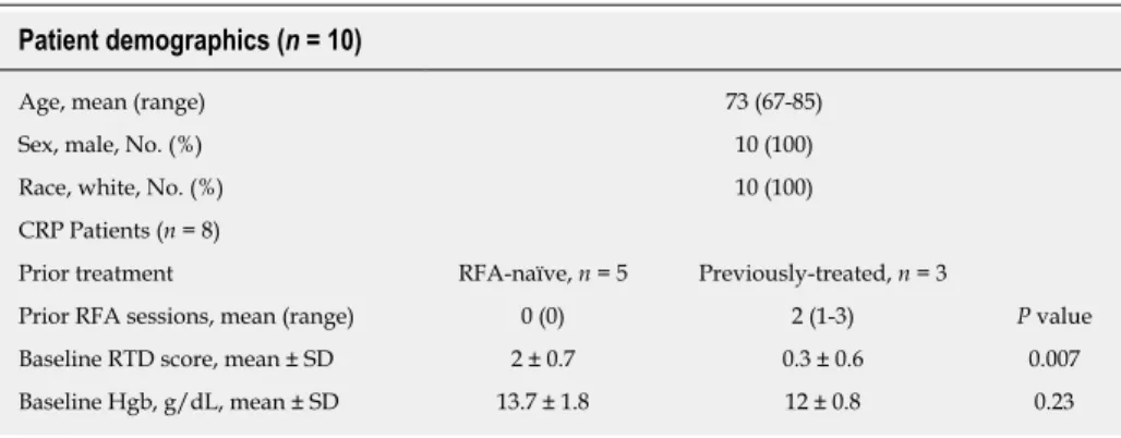
Patient demographics are summarized in Table 1. Previously-treated patients had a mean of 2 RFA sessions prior to this study (range: 1-3). Baseline RTD scores were significantly different between the RFA-naïve and the previously-treated patients (mean score: 2 ± 0.7 vs 0.3 ± 0.6, respectively, P = 0.007). While the mean hemoglobin concentrations were higher for the previously-treated patients compared to RFA-naïve patients, the difference did not reach statistical significance (mean: 13.7 ± 1.8 g/dL vs 12 ± 0.8 g/dL, respectively, P = 0.23). 4 out of 5 (80%) RFA-naïve patients presented with rectal bleeding at the baseline visit, while none of the
Figure 1
Figure 1 Depth-resolved en face optical coherence tomography and optical coherence tomography angiography images and cross-sectional optical coherence tomography and optical coherence tomography angiography images of a normal rectum. A: En face optical coherence tomography (OCT) image at
150 μm depth; B: En face OCT at 350 μm depth showing regular circular mucosal patterns characteristics of normal rectum; C: En face OCT angiography (OCTA) image at 150 μm depth showing regular honeycomb-like microvascular pattern of the subsurface capillary network in the mucosal layer; D: En face OCTA image at 350 μm depth showing rectal microvasculature corresponding to arterioles and venules in the submucosal layer, in addition to shadowing from the superficial mucosal microvasculature; E: Cross-sectional OCT image from the solid green lines in A and B, showing regular columnar architecture of normal rectum. The submucosal layer can be identified by the vertical layers traversing across the image (arrows). A dilated mucosal gland can be observed in both en face and cross-sectional image (asterisk); F: Cross-sectional OCTA image from the solid blue lines in C and D. Subsurface capillaries in the mucosal layer can be identified as horizontal structures connecting submucosal layer to the mucosal layer (solid arrows), while the arterioles and venules can be identified as vertical structures in the submucosal layer traversing across the images (dashed arrows). Cross-sectional OCTA image was averaged over a 50 µm projection range in the longitudinal direction to improve contrast and reduce noise. Dashed and solid lines in E and F indicate 150 μm and 350 μm depth levels, respectively. Scale bars are 1 mm in A-D, 500 μm in E and F. OCT: Optical coherence tomography; OCTA: Optical coherence tomography angiography.
treated patients exhibited rectal bleeding.
Rectal OCT and OCTA features of normal and CRP patients
OCT imaging was completed in less than 10 min for each patient (mean: 8 ± 4 min, range: 4-18 min). OCT and OCTA images enabled depth-resolved visualization of rectal microstructural and microvascular architecture of patients with normal rectum (Figure 1). En face OCT images showed regular circular mucosal patterns characteristics of normal rectum (Figure 1A and B). Images at superficial depths showed the outline of the mucosal crypt architecture (Figure 1A) whereas images at deeper depths delineated the mucosal architecture and patterns in greater detail and contrast (owing to optical transmission and penetration from near-vertical columnar epithelium and lumens to deeper depths) (Figure 1B). En face OCTA images at superficial depths showed regular honeycomb-like microvascular pattern corresponding to the subsurface capillary network in the mucosal layer (Figure 1C)[25].
En face OCTA images at deeper depths showed rectal microvasculature corresponding to arterioles and venules in the submucosal layer, in addition to shadowing from the mucosal microvasculature (Figure 1D). The submucosal arterioles and venules had homogeneous vessel diameters of typically < 200 μm. Cross-sectional OCT images showed regular columnar architecture of normal rectum and allowed determination of mucosal and submucosal layer depths (Figure 1E)[26]. Cross-sectional OCTA images
showed subsurface capillaries in the mucosal layer and arterioles and venules in the submucosal layer (Figure 1F).
OCT and OCTA images enabled depth-resolved visualization of rectal microstructural and microvascular architecture of CRP patients (Figures 2 and 3). OCT, OCTA and endoscopy images over the dentate line of an RFA-naïve CRP patient were shown in Figure 2. En face OCT images showed regular circular mucosal patterns on the rectal side with no apparent abnormalities (Figure 2A). En face OCTA images at superficial depths showed distortions on the honeycomb-like microvascular pattern, and had ectatic and tortuous microvasculature in the mucosal layer (Figure 2C). En face OCTA images at deeper depths showed vessels with heterogonous and unusually large diameters (> 200 μm) suggesting presence of abnormal arterioles and venules in the submucosal layer (Figure 2D). Rectal examination confirmed the OCTA
Figure 2
Figure 2 Depth-resolved en face optical coherence tomography and optical coherence tomography angiography images, cross-sectional optical coherence tomography and optical coherence tomography angiography images, and corresponding endoscopy image over the dentate line of a radiofrequency ablation-naïve chronic radiation proctopathy patient. A: En face optical coherence tomography (OCT) image at 150 μm depth showing regular
circular mucosal patterns on the rectal side and squamous epithelium with a smooth appearance on the anal canal side; B: Shows the corresponding endoscopy image at the dentate line showing ulcerations, and edematous and non-confluent telangiectasias (rectal telangiectasia density = 2). White arrows highlight areas of telangiectasias on the rectal side; C: En face OCT angiography (OCTA) image at 150 μm depth showing distortions to the honeycomb-like microvascular pattern, and ectatic and tortuous rectal microvasculature (red oval) in the mucosal layer; D: En face OCTA image at 350 μm depth showing vessels with heterogonous and unusually large diameters (red oval) suggesting presence of abnormal arterioles and venules in the submucosal layer; E: Cross-sectional OCT image from the solid green line in A showing mucosal and submucosal layers; F: Cross-sectional OCTA image from the solid blue lines in C and D. Cross-sectional OCTA image was averaged over a 50 μm projection range in the longitudinal direction to improve contrast and reduce noise. Dashed and solid lines in E and F indicate 150 μm and 350 μm depth levels, respectively. Scale bars are 1 mm in A-D, 500 μm in E and F. OCT: Optical coherence tomography; OCTA: Optical coherence tomography angiography.
findings and the corresponding endoscopy image showed ulcerations, and edematous and non-confluent telangiectasias (Figure 2B).
OCT, OCTA and endoscopy images over the dentate line of a previously-treated CRP patient are shown in Figure 3. En face OCT images showed regular circular mucosal patterns on the rectal side with no apparent abnormalities (Figure 3A). En
face OCTA images at superficial depths showed regular honeycomb-like microvascular pattern in the mucosal layer suggesting normalization of rectal mucosal microvasculature following RFA treatment (Figure 3C). En face OCTA images at deeper depths showed vessels with heterogonous and unusually large diameters (> 200 μm) that suggested presence of persistent abnormal arterioles and venules in the submucosal layer (Figure 3D). Corresponding endoscopy image showed healing of the rectal mucosa with some residual telangiectatic areas (Figure 3B).
Blinded OCTA reading results
Blinded OCTA reading results are given in Table 2. Occurrence of abnormal rectal OCTA features were higher in RFA-naïve patients compared with normal patients or CRP patients in RFA-follow-up visits. OCTA in 0 out of 2 (0%) normal patients, 3 out of 5 (60%) RFA-naïve patients and 0 out of 8 (0%) RFA-follow-up visits had abnormal rectal mucosal microvasculature. One out of 2 (50%) normal patients, 5 out of 5 (100%) RFA-naïve patients and 2 out of 8 (13%) RFA-follow-up visits had abnormal rectal submucosal microvasculature.
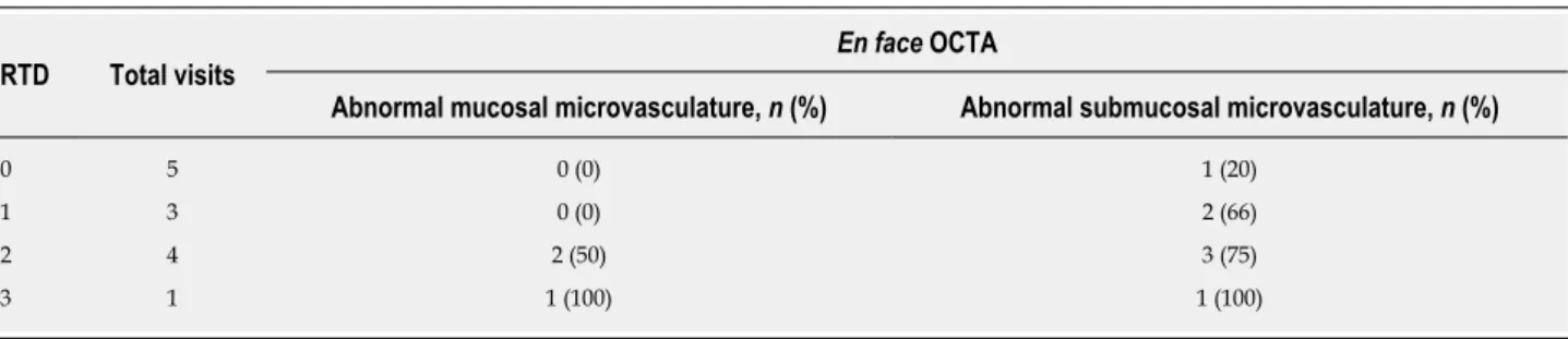
OCTA reading results for the CRP patients were also stratified according to endoscopic RTD scores (Table 3). Occurrence of abnormal rectal OCTA features increased with increasing RTD scores, suggesting a correlation of abnormal rectal microvasculature, as observed with OCTA, with the RTD scores. OCTA in 0 out of 5 (0%) visits with RTD = 0, 0 out of 3 (0%) visits with RTD = 1, 2 out of 4 (50%) visits with RTD = 2 and 1 out of 1 (100%) visit with RTD = 3 showed abnormal mucosal microvasculature. OCTA in 1 out of 5 (20%) visits with RTD = 0, 2 out of 3 (66%) visits with RTD = 1, 3 out of 4 (75%) visits with RTD = 2 and 1 out of 1 (100%) visit with RTD = 3 showed abnormal submucosal microvasculature.
Figure 3
Figure 3 Depth-resolved en face optical coherence tomography and optical coherence tomography angiography images, cross-sectional optical coherence tomography and optical coherence tomography angiography images, and corresponding endoscopy image over the dentate line of a previously-treated chronic radiation proctopathy patient. A: En face optical coherence tomography (OCT) image at 150 μm depth showing regular circular
mucosal patterns on the rectal side and squamous epithelium with a smooth appearance on the anal canal side; B: Shows the corresponding endoscopy image at the dentate line showing some residual telangiectatic areas (arrows, rectal telangiectasia density = 1); C: En face OCT angiography (OCTA) image at 150 µm depth showing regular honeycomb-like microvascular pattern in the mucosal layer; D: En face OCTA image at 350 μm depth showing vessels with heterogonous and unusually large diameters (red oval) suggesting presence of abnormal arterioles and venules in the submucosal layer; E: Cross-sectional OCT image from the solid green line in A showing mucosal and submucosal layers; F: Cross-sectional OCTA image from the solid blue lines in C and D. Cross-sectional OCTA image was averaged over a 50 μm projection range in the longitudinal direction to improve contrast and reduce noise. Dashed and solid lines in E and F indicate 150 μm and 350 μm depth levels, respectively. Scale bars are 1 mm in A-D, 500 μm in E and F. OCT: Optical coherence tomography; OCTA: Optical coherence tomography angiography.
DISCUSSION
Most OCT studies to date have been primarily focused on esophageal diseases, specifically on BE and its related dysplasia. Traditionally, OCT imaging speeds were limited and it was only possible to generate cross-sectional images which are complimentary to endoscopic forward-viewing (i.e., en face) images. With advances in OCT technology, imaging speeds more than 10 times faster than current commercially-available technology are possible[24,27]. These ultrahigh speeds enable
depth-resolved en face imaging of tissue microstructure as well as imaging microvasculature (with OCTA) which are analogous to endoscopic views and complement traditional cross-sectional OCT images. To our knowledge, this is the first study demonstrating the potential clinical utility of this next-generation OCT technology in a lower gastrointestinal tract pathology.
The depth-resolved imaging capability of OCT is a powerful advantage for assessing disease severity as well as efficacy of endoscopic treatment modalities such as RFA, where it is crucial to visualize both the treatment area and depth. A previous study on BE patients showed that presence of residual BE glands and BE epithelium thickness post-RFA predicted treatment response[28]. In addition, OCT measurements
of epithelial thickness > 333 μm prior to RFA predicted the presence of residual BE on follow-up with a 92.3% sensitivity and 85% specificity. A recent study on ex vivo swine esophageal specimens showed that OCT can monitor RFA in real time (imaging through the ablation catheter) and quantitatively assess treatment depth using optical scattering changes of the epithelial layer during ablation[29]. Another study showed
that OCT can assess the anal canal in CRP patients undergoing RFA[30], and showed
re-epithelialization by neosquamous mucosa over areas of prior hemorrhage, potentially forming a protective layer preventing recurrence of bleeding. Our current study investigated depth-resolved en face OCT and OCTA images of the rectum from normal and CRP patients, demonstrated that rectal microvasculature in the mucosal and submucosal layers can be independently assessed and presented OCTA features of normal and abnormal rectal microvasculature. Blinded reading of the OCTA features demonstrated association of abnormal rectal microvasculature in RFA-naïve patients compared with normal patients and RFA-follow-up visits (Table 2), as well as a correlation of abnormal rectal microvasculature, as observed with OCTA, with the
Figure 4
Figure 4 Summary of normal and abnormal microvasculature in the mucosal and submucosal layers of the rectum. A: Normal rectal mucosal
microvasculature consisted of a honeycomb-like microvascular pattern corresponding to subsurface capillary network; B: Abnormal rectal mucosal microvasculature had distortions to the honeycomb-like microvascular pattern, and had ectatic and tortuous microvasculature; C: Normal rectal submucosal microvasculature consisted of arterioles and venules with homogeneous vessel diameters typically of < 200 μm, in addition to shadowing from the superficial mucosal microvasculature; D: Abnormal rectal submucosal microvasculature had arterioles and venules with heterogonous and unusually high vessel diameters (> 200 μm). Scale bars are 1 mm.
endoscopic RTD scores (Table 3). Altogether, these studies suggest the potential clinical utility of depth-resolved OCT and OCTA imaging in assessing disease severity of CRP as well as efficacy of endoscopic treatment modalities such as RFA.
While it is generally acknowledged that CRP is associated with angiogenic changes and corresponding tissue remodeling, the pathophysiology of CRP with regards to its onset and progression is not well elucidated[31]. A widely recognized model
hypothesizes that CRP originates within the mucosal layer, where the hypoxemia caused by irradiation damages endothelial cells, inducing transcription factors such as hypoxia-inducible factor that promote angiogenic factors such as VEGF, angiogenin and FGF1[32,33]. This leads to neovascularization and dilatation of small vessels, and
telangiectasia formation in the mucosal layer. According to this model, inhibition of angiogenic factors such as angiogenin and FGF1 may be a valid approach to treat CRP[33].
A recent study, on the other hand, suggested a strong role of submucosal microvascular changes leading to onset and progression of CRP[34]. Researchers
collected fresh surgical rectal specimens from 30 patients with CRP and 29 patients w i t h o u t C R P t h a t a l l o w e d a s s e s s m e n t o f t h e e n t i r e r e c t a l w a l l f o r immunohistochemistry arrays of angiogenic factors. This was in contrast to most prior studies which evaluated rectal biopsy samples that only sampled the mucosal layer. This study found that angiostatin deposits that reside within rectal vessels are perfused throughout the mucosal-submucosal layer upon radiation damage of the endothelial cells. Angiostatins subsequently suppress microvessel formation, causing vessel stenosis and fibrotic vascular sclerosis, decreasing microvessel density in the submucosal layer[34]. The vascular changes and formation of telangiectasias in the
mucosal layer are hypothesized to be compensatory changes in response to the alteration of microvasculature in the submucosal layer. This study then suggested that restoration of vascular functionality by promoting angiogenesis in the submucosal layer may help in reversing the effects of CRP.
OCTA can independently assess rectal microvasculature in the mucosa and submucosa and is well suited for studying the pathophysiology of CRP in order to develop more effective prevention and treatment approaches. OCTA allows longitudinal study of rectal microvasculature in situ without injected dyes or disrupting tissue integrity, unlike taking excisional biopsies or performing surgical
Table 1 Patient demographics and baseline clinical status
Patient demographics (n = 10)
Age, mean (range) 73 (67-85)
Sex, male, No. (%) 10 (100)
Race, white, No. (%) 10 (100)
CRP Patients (n = 8)
Prior treatment RFA-naïve, n = 5 Previously-treated, n = 3
Prior RFA sessions, mean (range) 0 (0) 2 (1-3) P value
Baseline RTD score, mean ± SD 2 ± 0.7 0.3 ± 0.6 0.007
Baseline Hgb, g/dL, mean ± SD 13.7 ± 1.8 12 ± 0.8 0.23
Hgb: Hemoglobin concentration; SD: Standard deviation; CRP: Chronic radiation proctopathy; RFA: Radiofrequency ablation; RTD: Rectal telangiectasia density.
resection. The results of this study demonstrated that only 3 out of 5 (60%) RFA-naïve patients had abnormal rectal mucosal microvasculature on OCTA, but all 5 out of 5 (100%) had abnormal rectal submucosal microvasculature. This may support the aforementioned recent hypothesis that CRP originates in the submucosal layer and subsequently progress to the mucosal layer[34]. Furthermore, after RFA treatment,
patients had abnormal rectal mucosal microvasculature on OCTA in 0 out of 8 (0%) RFA-follow-up visits and abnormal rectal submucosal microvasculature in only 2 out of 8 (13%) RFA-follow-up visits. This may indicate that in a subset of patients RFA may not be effective for coagulating the abnormal vessels in the submucosal layer. Treatment approaches such as CSA or APC may be more appropriate to target deeper vasculature in this cohort of patients.
The OCTA reading and endoscopic RTD scoring were performed by independent researchers who were blinded to clinical status of the patients as well as each other’s respective readings. Inter-observer and intra-observer agreement of these two readings was not assessed. Previous studies have showed that RTD scoring system has good inter-observer agreement, but this has not yet been widely validated[9].
However, it should be noted that OCTA is extensively used in ophthalmology and multiple techniques have been developed for automatically quantitating vasculature and vascular alterations which can be adapted to this application[35]. These software
based methods will address problems of inter-observer agreement.
This study was limited in that it was retrospective and the patient enrollment was limited to the patient demographics at the clinical center and the availability of the OCT system. A fraction of patients (3 out of 8) were not imaged when they were RFA-naïve. Furthermore, RFA-follow-up patients were at different stages of treatment, and there was not a fixed interval between RFA and follow-up. Therefore, prospective studies with larger sample sizes and standardized follow-up intervals are needed to further validate the results of this study.
Typically, an endoscope is used to introduce the OCT probe into the GI tract via accessory channels. Recently, a capsule based OCT device was demonstrated for upper GI imaging which enabled wide field of view OCT imaging without requiring an endoscope[36]. In this current study, we have also demonstrated OCT imaging
without requiring an endoscope by directly placing the OCT catheter onto the dentate line and rectum. OCT imaging was rapid, taking an average of 8 min per patient.
OCTA images are generated by detecting OCT signal changes caused by moving erythrocytes. Therefore, excessive OCT catheter pressure on the bowel wall may impair vascular flow and affect the visualization of smaller microvessels[37,38]. Control
of catheter pressure was difficult and repeat acquisitions were performed over the same regions while applying minimal pressure, but maintaining tissue contact. During analysis, we discarded datasets that did not visualize microvasculature over all imaging depths as well as datasets exhibiting effacement of mucosal patterns (on the rectal side) as a surrogate marker for excessive catheter pressure. This problem can be addressed by designing OCT imaging devices specifically for anal/rectal applications.
The small diameter of the OCT catheter used in this study restricted the single acquisition field of view of to 10 mm × 16 mm. Although multiple acquisitions were performed to increase the imaging coverage in the longitudinal direction, only the anterior side of the rectal wall was captured in the circumferential direction. Imaging the entire circumference of the rectum is important in order to more comprehensively assess and map vascular alterations. We have recently demonstrated a tethered OCT
Table 2 Endoscopic rectal telangiectasia density scores and occurrence of abnormal rectal mucosal and submucosal microvasculature based on en face optical coherence tomography angiography images
Category Total patients/visits
Endoscopy En face OCTA
RTD ± SD Abnormal mucosal
microvasculature, n (%)
Abnormal submucosal microvasculature, n (%)
Normal patients 2 Not assessed 0 (0) 1 (50)
RFA-naïve patients 5 2.0 ± 0.7 3 (60) 5 (100)
RFA-follow-up visit 8 0.5 ± 0.8 0 (0) 2 (13)
SD: Standard deviation; RFA: Radiofrequency ablation; RTD: Rectal telangiectasia density; OCTA: Optical coherence tomography angiography.
capsule which enabled circumferential imaging over a 40 mm × 240 mm area in the esophagus in 20 seconds[39]. We also reported OCT and OCTA using a balloon-based
catheter imaging in living swine, imaging over a 50 mm × 26 mm area in 18 seconds[40]. Related devices can be developed to image the proximal portion of the
lower gastrointestinal tract for CRP assessment in the future[41]. OCTA/OCT imaging
can be performed with a stand-alone device independent of colonoscopy, prior to colonoscopy, or with an endoscopic attachment during colonoscopy.
In summary, this study demonstrated that OCTA/OCT can visualize depth-resolved tissue microvasculature around the dentate line and rectum, relevant to assessment and treatment of CRP. We described OCTA features of normal and abnormal rectal microvasculature in the mucosa and submucosa. Blinded reading of these OCTA features suggest that submucosal vascular abnormalities are more strongly associated with CRP than mucosal vascular abnormalities. Both mucosal and submucosal vasculature was observed to normalize after RFA treatment. However, these observations must be viewed with caution because of our small patient enrollment. Submucosal vasculature is not visible endoscopically and OCTA/OCT provides a unique modality for assessing CRP. OCTA is well suited for longitudinal studies because it does not require injected dyes and can rapidly imaging large regions of rectum to yield integrated microstructural and microvascular maps. These advantages suggest that it could be a viable tool for rapid assessment of CRP to elucidate pathophysiology as well as potentially plan treatment and assess response. Further larger scale, prospective, longitudinal studies are warranted.
Table 3 Occurrence of abnormal rectal mucosal and submucosal microvasculature in chronic radiation proctopathy patients based on
en face optical coherence tomography angiography images, stratified by endoscopic rectal telangiectasia density score
RTD Total visits
En face OCTA
Abnormal mucosal microvasculature, n (%) Abnormal submucosal microvasculature, n (%)
0 5 0 (0) 1 (20)
1 3 0 (0) 2 (66)
2 4 2 (50) 3 (75)
3 1 1 (100) 1 (100)
RTD: Rectal telangiectasia density; OCTA: Optical coherence tomography angiography.
ARTICLE HIGHLIGHTS
Research background
Chronic radiation proctopathy (CRP) occurs as a result of pelvic radiation therapy and is associated with formation of abnormal vasculature that may lead to persistent rectal bleeding. While incidence is declining due to refinement of radiation delivery techniques, CRP remains one of the major complications of pelvic radiation therapy and significantly affects patient quality of life. Radiofrequency ablation (RFA) is an emerging treatment modality for eradicating abnormal vasculature associated with CRP. However, questions remain regarding CRP pathophysiology and optimal disease management.
Research motivation
This pilot study utilizes ultrahigh-speed optical coherence tomography (OCT) and OCT angiography (OCTA) to investigate microvascular features of normal versus CRP patients and how they respond under endoscopic RFA treatment.
Research objectives
We utilized OCT and OCTA for assessing subsurface depth-resolved microvasculature around the dentate line and rectum of normal patients as well as CRP patients who were RFA-naïve or under treatment. OCTA can image normal and abnormal microvasculature in the mucosal and submucosal layers of the rectum, providing information not available by endoscopy. Blinded reading of vascular features were performed to assess incidence of abnormal features in RFA treatment naïve CRP as well as response under treatment. Association with endoscopic rectal telangiectasia density scoring was also investigated.
Research methods
Two patients with normal rectum and 8 patients referred for, or undergoing endoscopic treatment with RFA for CRP were imaged with ultrahigh-speed OCTA/OCT over a total of 15 OCT/colonoscopy visits (2 normal patients, 5 RFA-naïve CRP patients, 8 RFA-follow-up visits). Imaging was performed using a prototype ultrahigh-speed OCT instrument at 600 kHz axial scan rate using a small imaging catheter. OCTA enabled depth-resolved microvasculature imaging using motion contrast from flowing blood, without requiring injected dyes.
Research results
OCTA visualized normal vasculature with regular honeycomb patterns versus abnormal distorted honeycomb patterns with ectatic and tortuous microvasculature in the rectal mucosa. Normal arterioles and venules < 200 μm in diameter versus abnormal heterogenous enlarged arterioles and venules > 200 μm in diameter were visualized in the submucosa. Abnormal mucosal vasculature occurred in 0 of 2 normal patients and 3 of 5 RFA-naïve patients, while abnormal submucosal vasculature occurred more often, in 1 of 2 normal patients and 5 of 5 RFA-naïve patients. After RFA treatment, vascular abnormalities decreased, with abnormal mucosal vasculature observed in 0 of 8 RFA-follow-up visits and abnormal submucosal vasculature observed in only and 2 of 8 RFA-follow-up visits.
Research conclusions
This study demonstrated that OCTA can visualize depth-resolved normal and abnormal microvasculature around the dentate line and rectum associated with CRP and treatment response. Submucosal vascular abnormalities seemed more strongly associated with CRP than mucosal vascular abnormalities. Both mucosal and submucosal abnormal vasculature was observed to normalize after RFA treatment. However, these observations must be viewed with caution since this was a retrospective study with a small patient enrollment. Further larger scale, prospective, longitudinal studies are warranted.
Research perspectives
The role of mucosal vs submucosal vascular alterations in CRP remains an open question in pathophysiology. Submucosal vasculature is not visible endoscopically and therefore
OCTA/OCT provides a unique modality for assessing CRP. Furthermore, OCTA does not require injected dyes and is well suited for longitudinal studies. It can rapidly imaging large regions of rectum to yield integrated microstructural and microvascular maps. These advantages suggest that OCTA/OCT could be a viable tool for investigation of CRP to elucidate pathophysiology as well as potentially plan treatment and assess response.
REFERENCES
1 Tagkalidis PP, Tjandra JJ. Chronic radiation proctitis. ANZ J Surg 2001; 71: 230-237 [PMID: 11355732
DOI: 10.1046/j.1440-1622.2001.02081.x]
2 Garg AK, Mai WY, McGary JE, Grant WH, Butler EB, Teh BS. Radiation proctopathy in the treatment of
prostate cancer. Int J Radiat Oncol Biol Phys 2006; 66: 1294-1305 [PMID: 17126204 DOI:
10.1016/j.ijrobp.2006.07.1386]
3 Grodsky MB, Sidani SM. Radiation proctopathy. Clin Colon Rectal Surg 2015; 28: 103-111 [PMID:
26034407 DOI: 10.1055/s-0035-1547337]
4 Zelefsky MJ, Levin EJ, Hunt M, Yamada Y, Shippy AM, Jackson A, Amols HI. Incidence of late rectal
and urinary toxicities after three-dimensional conformal radiotherapy and intensity-modulated radiotherapy for localized prostate cancer. Int J Radiat Oncol Biol Phys 2008; 70: 1124-1129 [PMID: 18313526 DOI:
10.1016/j.ijrobp.2007.11.044]
5 Staffurth J; Radiotherapy Development Board. A review of the clinical evidence for intensity-modulated
radiotherapy. Clin Oncol (R Coll Radiol) 2010; 22: 643-657 [PMID: 20673708 DOI:
10.1016/j.clon.2010.06.013]
6 Bazan JG, Hara W, Hsu A, Kunz PA, Ford J, Fisher GA, Welton ML, Shelton A, Kapp DS, Koong AC,
Goodman KA, Chang DT. Intensity-modulated radiation therapy versus conventional radiation therapy for squamous cell carcinoma of the anal canal. Cancer 2011; 117: 3342-3351 [PMID: 21287530 DOI:
10.1002/cncr.25901]
7 Leiper K, Morris AI. Treatment of radiation proctitis. Clin Oncol (R Coll Radiol) 2007; 19: 724-729
[PMID: 17728120 DOI: 10.1016/j.clon.2007.07.008]
8 O'Brien PC, Hamilton CS, Denham JW, Gourlay R, Franklin CI. Spontaneous improvement in late rectal
mucosal changes after radiotherapy for prostate cancer. Int J Radiat Oncol Biol Phys 2004; 58: 75-80 [PMID: 14697423 DOI: 10.1016/S0360-3016(03)01445-7]
9 Chi KD, Ehrenpreis ED, Jani AB. Accuracy and reliability of the endoscopic classification of chronic
radiation-induced proctopathy using a novel grading method. J Clin Gastroenterol 2005; 39: 42-46 [PMID: 15599209 DOI: 10.1067/mge.2003.107]
10 Theodorescu D, Gillenwater JY, Koutrouvelis PG. Prostatourethral-rectal fistula after prostate
brachytherapy. Cancer 2000; 89: 2085-2091 [PMID: 11066049 DOI:
10.1002/1097-0142(20001115)89:10<2085::AID-CNCR8>3.0.CO;2-Q]
11 Yates MR, Baron TH. Treatment of a radiation-induced sigmoid stricture with an expandable metal stent.
Gastrointest Endosc 1999; 50: 422-426 [PMID: 10462671 DOI: 10.1053/ge.1999.v50.97950] 12 Lenz L, Tafarel J, Correia L, Bonilha D, Santos M, Rodrigues R, Gomes G, Andrade G, Martins F,
Monaghan M, Nakao F, Libera E, Ferrari AP, Rohr R. Comparative study of bipolar eletrocoagulation versus argon plasma coagulation for rectal bleeding due to chronic radiation coloproctopathy. Endoscopy 2011; 43: 697-701 [PMID: 21611944 DOI: 10.1055/s-0030-1256467]
13 Weiner J, Schwartz D, Martinez M, Safdieh J, Aytaman A, Schreiber D. Long-term results on the efficacy
of argon plasma coagulation for patients with chronic radiation proctitis after conventionally fractionated, dose-escalated radiation therapy for prostate cancer. Pract Radiat Oncol 2017; 7: e35-e42 [PMID:
27663931 DOI: 10.1016/j.prro.2016.07.009]
14 Moawad FJ, Maydonovitch CL, Horwhat JD. Efficacy of cryospray ablation for the treatment of chronic
radiation proctitis in a pilot study. Dig Endosc 2013; 25: 174-179 [PMID: 23362977 DOI:
10.1111/j.1443-1661.2012.01355.x]
15 Taylor JG, Disario JA, Bjorkman DJ. KTP laser therapy for bleeding from chronic radiation proctopathy.
Gastrointest Endosc 2000; 52: 353-357 [PMID: 10968849 DOI: 10.1067/mge.2000.107726] 16 Rustagi T, Corbett FS, Mashimo H. Treatment of chronic radiation proctopathy with radiofrequency
ablation (with video). Gastrointest Endosc 2015; 81: 428-436 [PMID: 24973172 DOI:
10.1016/j.gie.2014.04.038]
17 Jao SW, Beart RW, Gunderson LL. Surgical treatment of radiation injuries of the colon and rectum. Am J
Surg 1986; 151: 272-277 [PMID: 3946764 DOI: 10.1016/0002-9610(86)90086-3]
18 Turina M, Mulhall AM, Mahid SS, Yashar C, Galandiuk S. Frequency and surgical management of
chronic complications related to pelvic radiation. Arch Surg 2008; 143: 46-52; discussion 52 [PMID:
18209152 DOI: 10.1001/archsurg.2007.7]
19 Weiner JP, Wong AT, Schwartz D, Martinez M, Aytaman A, Schreiber D. Endoscopic and
non-endoscopic approaches for the management of radiation-induced rectal bleeding. World J Gastroenterol 2016; 22: 6972-6986 [PMID: 27610010 DOI: 10.3748/wjg.v22.i31.6972]
20 Swager A, Boerwinkel DF, de Bruin DM, Weusten BL, Faber DJ, Meijer SL, van Leeuwen TG, Curvers
WL, Bergman JJ. Volumetric laser endomicroscopy in Barrett's esophagus: A feasibility study on histological correlation. Dis Esophagus 2016; 29: 505-512 [PMID: 25951873 DOI: 10.1111/dote.12371] 21 Wolfsen HC, Sharma P, Wallace MB, Leggett C, Tearney G, Wang KK. Safety and feasibility of
volumetric laser endomicroscopy in patients with Barrett's esophagus (with videos). Gastrointest Endosc 2015; 82: 631-640 [PMID: 25956472 DOI: 10.1016/j.gie.2015.03.1968]
22 Evans JA, Poneros JM, Bouma BE, Bressner J, Halpern EF, Shishkov M, Lauwers GY, Mino-Kenudson
M, Nishioka NS, Tearney GJ. Optical coherence tomography to identify intramucosal carcinoma and high-grade dysplasia in Barrett's esophagus. Clin Gastroenterol Hepatol 2006; 4: 38-43 [PMID: 16431303 DOI:
10.1016/S1542-3565(05)00746-9]
23 Leggett CL, Gorospe EC, Chan DK, Muppa P, Owens V, Smyrk TC, Anderson M, Lutzke LS, Tearney G,
Wang KK. Comparative diagnostic performance of volumetric laser endomicroscopy and confocal laser endomicroscopy in the detection of dysplasia associated with Barrett's esophagus. Gastrointest Endosc 2016; 83: 880-888.e2 [PMID: 26344884 DOI: 10.1016/j.gie.2015.08.050]
24 Tsai TH, Ahsen OO, Lee HC, Liang K, Figueiredo M, Tao YK, Giacomelli MG, Potsaid BM, Jayaraman
V, Huang Q, Cable AE, Fujimoto JG, Mashimo H. Endoscopic optical coherence angiography enables
dimensional visualization of subsurface microvasculature. Gastroenterology 2014; 147: 1219-1221 [PMID: 25172015 DOI: 10.1053/j.gastro.2014.08.034]
25 Araki K, Furuya Y, Kobayashi M, Matsuura K, Ogata T, Isozaki H. Comparison of mucosal
microvasculature between the proximal and distal human colon. J Electron Microsc (Tokyo) 1996; 45: 202-206 [PMID: 8765715 DOI: 10.1093/oxfordjournals.jmicro.a023433]
26 Adler DC, Zhou C, Tsai TH, Schmitt J, Huang Q, Mashimo H, Fujimoto JG. Three-dimensional
endomicroscopy of the human colon using optical coherence tomography. Opt Express 2009; 17: 784-796 [PMID: 19158891 DOI: 10.1364/OE.17.000784]
27 Tsai TH, Lee HC, Ahsen OO, Liang K, Giacomelli MG, Potsaid BM, Tao YK, Jayaraman V, Figueiredo
M, Huang Q, Cable AE, Fujimoto J, Mashimo H. Ultrahigh speed endoscopic optical coherence tomography for gastroenterology. Biomed Opt Express 2014; 5: 4387-4404 [PMID: 25574446 DOI:
10.1364/BOE.5.004387]
28 Tsai TH, Zhou C, Tao YK, Lee HC, Ahsen OO, Figueiredo M, Kirtane T, Adler DC, Schmitt JM, Huang
Q, Fujimoto JG, Mashimo H. Structural markers observed with endoscopic 3-dimensional optical coherence tomography correlating with Barrett's esophagus radiofrequency ablation treatment response (with videos). Gastrointest Endosc 2012; 76: 1104-1112 [PMID: 22831857 DOI:
10.1016/j.gie.2012.05.024]
29 Lee HC, Ahsen OO, Liu JJ, Tsai TH, Huang Q, Mashimo H, Fujimoto JG. Assessment of the
radiofrequency ablation dynamics of esophageal tissue with optical coherence tomography. J Biomed Opt 2017; 22: 76001 [PMID: 28687822 DOI: 10.1117/1.JBO.22.7.076001]
30 Zhou C, Adler DC, Becker L, Chen Y, Tsai TH, Figueiredo M, Schmitt JM, Fujimoto JG, Mashimo H.
Effective treatment of chronic radiation proctitis using radiofrequency ablation. Therap Adv Gastroenterol 2009; 2: 149-156 [PMID: 20593010 DOI: 10.1177/1756283X08103341]
31 Frazzoni L, La Marca M, Guido A, Morganti AG, Bazzoli F, Fuccio L. Pelvic radiation disease: Updates
on treatment options. World J Clin Oncol 2015; 6: 272-280 [PMID: 26677440 DOI:
10.5306/wjco.v6.i6.272]
32 Liu Y, Kudo K, Abe Y, Hu DL, Kijima H, Nakane A, Ono K. Inhibition of transforming growth
factor-beta, hypoxia-inducible factor-1alpha and vascular endothelial growth factor reduced late rectal injury induced by irradiation. J Radiat Res 2009; 50: 233-239 [PMID: 19346676 DOI: 10.1269/jrr.08112] 33 Takeuchi H, Kimura T, Okamoto K, Aoyagi E, Miyamoto H, Kaji M, Takenaka H, Okamura S, Sato Y,
Kato J, Okahisa T, Takayama T. A mechanism for abnormal angiogenesis in human radiation proctitis: Analysis of expression profile for angiogenic factors. J Gastroenterol 2012; 47: 56-64 [PMID: 22081051
DOI: 10.1007/s00535-011-0470-2]
34 Wu P, Li L, Wang H, Ma T, Wu H, Fan X, Yang Z, Chen D, Wang L. Role of Angiogenesis in Chronic
Radiation Proctitis: New Evidence Favoring Inhibition of Angiogenesis Ex Vivo. Dig Dis Sci 2018; 63: 113-125 [PMID: 29080145 DOI: 10.1007/s10620-017-4818-1]
35 Spaide RF, Fujimoto JG, Waheed NK, Sadda SR, Staurenghi G. Optical coherence tomography
angiography. Prog Retin Eye Res 2018; 64: 1-55 [PMID: 29229445 DOI:
10.1016/j.preteyeres.2017.11.003]
36 Gora MJ, Sauk JS, Carruth RW, Gallagher KA, Suter MJ, Nishioka NS, Kava LE, Rosenberg M, Bouma
BE, Tearney GJ. Tethered capsule endomicroscopy enables less invasive imaging of gastrointestinal tract microstructure. Nat Med 2013; 19: 238-240 [PMID: 23314056 DOI: 10.1038/nm.3052]
37 Holloway GA, Daly CH, Kennedy D, Chimoskey J. Effects of external pressure loading on human skin
blood flow measured by 133Xe clearance. J Appl Physiol 1976; 40: 597-600 [PMID: 931880 DOI:
10.1152/jappl.1976.40.4.597]
38 Choi WJ, Wang H, Wang RK. Optical coherence tomography microangiography for monitoring the
response of vascular perfusion to external pressure on human skin tissue. J Biomed Opt 2014; 19: 056003 [PMID: 24810259 DOI: 10.1117/1.JBO.19.5.056003]
39 Liang K, Ahsen OO, Lee HC, Wang Z, Potsaid BM, Figueiredo M, Jayaraman V, Cable AE, Huang Q,
Mashimo H, Fujimoto JG. Volumetric Mapping of Barrett's Esophagus and Dysplasia With en face Optical Coherence Tomography Tethered Capsule. Am J Gastroenterol 2016; 111: 1664-1666 [PMID: 27808130
DOI: 10.1038/ajg.2016.419]
40 Lee HC, Ahsen OO, Liang K, Wang Z, Cleveland C, Booth L, Potsaid B, Jayaraman V, Cable AE,
Mashimo H, Langer R, Traverso G, Fujimoto JG. Circumferential optical coherence tomography angiography imaging of the swine esophagus using a micromotor balloon catheter. Biomed Opt Express 2016; 7: 2927-2942 [PMID: 27570688 DOI: 10.1364/BOE.7.002927]
41 Trindade AJ, Sultan K, Vamadevan AS, Fan C, Sejpal DV. Successful use of volumetric laser
endomicroscopy in imaging a rectal polyp. Therap Adv Gastroenterol 2016; 9: 128-131 [PMID: 26770274
DOI: 10.1177/1756283X15615309]