Publisher’s version / Version de l'éditeur:
Molecular Plant-Microbe Interactions, 22, 12, pp. 1601-1610, 2009-12
READ THESE TERMS AND CONDITIONS CAREFULLY BEFORE USING THIS WEBSITE. https://nrc-publications.canada.ca/eng/copyright
Vous avez des questions? Nous pouvons vous aider. Pour communiquer directement avec un auteur, consultez la
première page de la revue dans laquelle son article a été publié afin de trouver ses coordonnées. Si vous n’arrivez pas à les repérer, communiquez avec nous à PublicationsArchive-ArchivesPublications@nrc-cnrc.gc.ca.
Questions? Contact the NRC Publications Archive team at
PublicationsArchive-ArchivesPublications@nrc-cnrc.gc.ca. If you wish to email the authors directly, please see the first page of the publication for their contact information.
NRC Publications Archive
Archives des publications du CNRC
This publication could be one of several versions: author’s original, accepted manuscript or the publisher’s version. / La version de cette publication peut être l’une des suivantes : la version prépublication de l’auteur, la version acceptée du manuscrit ou la version de l’éditeur.
For the publisher’s version, please access the DOI link below./ Pour consulter la version de l’éditeur, utilisez le lien DOI ci-dessous.
https://doi.org/10.1094/MPMI-22-12-1601
Access and use of this website and the material on it are subject to the Terms and Conditions set forth at
Analysis of a Blumeria graminis-secreted lipase reveals the importance
of host epicuticular wax components for fungal adhesion and
development
Feng, Jie; Wang, Feng; Liu, Guosheng; Greenshields, David; Shen,
Wenyun; Kaminskyj, Susan; Hughes, Geoff R.; Peng, Youliang; Selvaraj,
Gopalan; Zou, Jitao; Wei, Yangdou
https://publications-cnrc.canada.ca/fra/droits
L’accès à ce site Web et l’utilisation de son contenu sont assujettis aux conditions présentées dans le site LISEZ CES CONDITIONS ATTENTIVEMENT AVANT D’UTILISER CE SITE WEB.
NRC Publications Record / Notice d'Archives des publications de CNRC:
https://nrc-publications.canada.ca/eng/view/object/?id=92244bc4-edac-496c-bf19-c4932cc1e235 https://publications-cnrc.canada.ca/fra/voir/objet/?id=92244bc4-edac-496c-bf19-c4932cc1e235MPMI Vol. 22, No. 12, 2009, pp. 1601–1610. doi:10.1094 / MPMI -22-12-1601. © 2009 The American Phytopathological Society
Analysis of a Blumeria graminis-Secreted Lipase Reveals
the Importance of Host Epicuticular Wax Components
for Fungal Adhesion and Development
Jie Feng,1 Feng Wang,1 Guosheng Liu,1 David Greenshields,1 Wenyun Shen,2 Susan Kaminskyj,1
Geoff R. Hughes,3 Youliang Peng,4 Gopalan Selvaraj,2 Jitao Zou,2 and Yangdou Wei1
1Department of Biology, University of Saskatchewan, 112 Science Place, Saskatoon, SK S7N 5E2, Canada; 2National Research Council of Canada, Plant Biotechnology Institute, Saskatoon, SK S7N 0W9, Canada;3Department of Plant Sciences, University of Saskatchewan, 51 Campus Drive, Saskatoon, SK S7N 5A8, Canada;4State Key Laboratory of Agrobiotechnology, Department of Plant Pathology, China Agricultural University, Yuanmingyuan West Road 2, Beijing 100094, P. R. China
Submitted 28 March 2009. Accepted 1 July 2009.
The biotrophic powdery mildew fungus Blumeria graminis releases extracellular materials to the surface of fungal in-fection structures that facilitate anchoring them to hydro-phobic plant surfaces prior to infection; however, the chemistry of fungal adhesives and the mechanism of adhe-sion remain largely unclear. Expressed sequence tag analy-sis led to identification of a secreted lipase, Lip1, from B.
graminis. Expression of LIP1 is dramatically upregulated
during the early stages of fungal development. Lip1, se-creted to the surface of fungal cell walls, possesses lipolytic activity against a broad range of glycerides and releases alkanes and primary fatty alcohols from the epicuticular wax of wheat leaves. Of the epicuticular wax components released by Lip1 activity, long-chain alkanes are the most efficient cues for triggering appressorium formation. Pre-treatment of wheat leaves with Lip1, thereby removing leaf surface wax, severely compromises components of fungal pathogenicity, including conidial adhesion, appressorium formation, and secondary hypha growth. Our data suggest that Lip1 activity releases cues from the host surface to promote pathogen development and infection.
Hydrophobic epicuticular waxes minimize the adhesion of water and other particles on the surface of leaves, thereby keep-ing leaves clean and enhanckeep-ing their ability to trap light for photosynthesis (Samuels et al. 2008). Adhesion to host surfaces represents an initial strategy for successful infection by many phytopathogenic fungi (Jones 1994; Carver and Gurr 2006). Furthermore, components of the plant surface or cuticle and their breakdown products can be taken up by the pathogen as part of a signaling system triggering spore germination and infection structure differentiation (Pascholati et al. 1992; Francis et al. 1996; Gilbert et al. 1996; Hegde and Kolattukudy 1997;
Skamnioti and Gurr 2007). It is generally accepted that the pathogen firmly attaches to the host surface by release of extracellular materials (ECM) from fungal spores and germ-lings. ECM-mediated adhesion occurs in a wide range of patho-genic fungi, including the rice blast Magnaporthe grisea (Bourett and Howard 1990); the anthracnose Colletotrichum spp. (Mercure et al. 1994, 1995; Sugui et al. 1998); the rusts
Puccinia and Uromyces spp. (Chaubal et al. 1991; Clement et
al. 1993, 1994, 1997); and the powdery mildews Uncinuliella
australiana (Mims et al. 1995), Erysiphe pisi (Carver et al.
1996; Carver and Gurr 2006), and Blumeria graminis (Carver et al. 1996, 1999; Wright et al. 2002a; Zhang et al. 2005).
Powdery mildew fungi are a group of obligate biotrophic pathogens that cause destructive foliar diseases of many plant species. The cereal powdery mildew B. graminis only infects epidermal cells and colonizes the host surface. B. graminis infection processes and the early interaction with its cereal hosts have been well-documented (Green et al. 2002; Zhang et al. 2005). Adhesives of the powdery mildew fungi B. graminis and U. viciae-fabae contain nonspecific esterases and cuti-nases (Nicholson et al. 1988; Deising et al. 1992; Pascholati et al. 1992), and the isolated B. graminis ECM is able to degrade host surface features (Kunoh et al. 1988), suggesting that lipolytic enzymes such as esterases and cutinases are released into the fungal ECM and lead to adhesion (Walton 1994).
Esterase (EC 3.1.1.1), cutinase (EC 3.1.1.74), and lipase (EC 3.1.1.3) belong to the α/β-hydrolase family, with a signa-ture catalytic triad Ser-Glu-His (Schrag et al. 1991). Distinc-tion between these enzymes has been proposed based on their substrate specificity and their capacity to hydrolyze esters in solution or emulsion (Chahinian et al. 2002; Schrag et al. 1991). Esterases act on solutions of short-chain fatty acyl esters, whereas lipases have the capacity to act on water-in-soluble long-chain fatty acyl groups. Cutinases, considered a link between esterases and lipases for substrate preference (Chahinian et al. 2002), are generally small enzymes of ap-proximately 22 to 25 kDa (Kolattukudy 1984; Longhi and Cambillau 1999) that are active in both soluble-phase and emulsified interface and do not show so-called interfacial acti-vation, a phenomenon commonly observed in lipases (Verger 1997). Although cutinases are also able to efficiently hydro-lyze triglycerides (Martinez et al. 1994), they exhibit a higher specificity for short-chain acyl groups, and this feature has been used as an indicator of cutinase activity (Kolattukudy
J. Feng and F. Wang contributed equally to this research.
Corresponding author: Y. Wei; Telephone: 9664447; Fax: +1-306-9664461; E-mail: yangdou.wei@usask.ca
Sequence data is available in EMBL and GenBank databases under acces-sion number EU583448.
* The e-Xtra logo stands for “electronic extra” and indicates that a sup-plemental table is published online and also that Figures 2, 3, 4, and 7 ap-pear in color online.
1985). In contrast, lipases are larger lipolytic enzymes and can hydrolyze substrates with long-chain fatty acids. The N-termi-nal sequence of lipases contains a significant hydrophobic re-gion corresponding to the open lid that is responsible for inter-facial activation and for determining the substrate specificity (Chahinian et al. 2002).
The roles for cutinases in early stages of fungal pathogene-sis have been investigated intensively in some pathogenic fungi for decades, yet many of the conclusions remain contro-versial. Recently, gene disruption studies on the cutinase gene Pbc1 in Pyrenopeziza brassicae and CUT2 in M. grisea pro-vided genetic evidence that cutinase is a crucial pathogenicity or virulence determinant involved in host penetration (Li et al. 2003; Skamnioti and Gurr 2007). However, disruption of CUT2 has no effect on pathogen adhesion to the host surface (Skamnioti and Gurr 2007). Secreted lipases also play a key role in the infection processes of some necrotrophic fungi such as Botrytis cinerea (Comménil et al. 1998, 1999; Reis et al. 2005), Alternaria brassicicola (Berto et al. 1997, 1999), and
Fusarium graminearum (Nasser-Eddine et al. 2001; Voigt et
al. 2005). When lipase-specific antibodies were added to co-nidial suspensions of B. cinerea (Comménil et al. 1998) and A.
brassicicola (Berto et al. 1999) prior to inoculation, disease
development was largely suppressed, suggesting that the fun-gal surface-bound lipase is involved in interacting with epicu-ticular leaf waxes for adhesion or penetration of the fungal propagules. However, when the corresponding gene (lip1) for the B. cinerea lipase was disrupted, the knock-out mutants showed no reduction of virulence (Reis et al. 2005). We previ-ously identified a similar lip1 gene in F. graminearum that was responsible for lipid utilization but dispensable for fungal pathogenicity (Feng et al. 2005). Further analyses of available genomic data revealed that many members of a large lipase gene family are present in necrotrophic fungal pathogens, whereas very few members of the gene family are present in biotrophic pathogens (Supplementary Table S1). This promoted us to investigate the role of lipases in the pathogenesis of biotrophic fungi. In this study, we characterize a secreted lipase from the obligate biotroph Blumeria graminis f. sp. tritici and demonstrate that Lip1-mediated epicuticular wax liquefaction is required for fungal adhesion and development during early stages of infection.
RESULTS
Lip1 is structurally a typical triglyceride lipase.
To probe the landscape of gene expression during the infec-tion process of B. graminis f. sp. tritici on wheat, a cDNA library was constructed from mitochondrial RNA of epidermis
of T. monococcum leaves 20 to 48 h after inoculation (hai) with B. graminis f. sp. tritici (Liu et al. 2005). Expressed se-quence tag analysis identified a cDNA similar to extracellular lipase genes in other fungal species. Multiple alignments be-tween the deduced amino acid sequence of this cDNA and lipases from Candida rugosa (Lotti et al. 1993), F. graminea-rum (Feng et al. 2005), and Geotrichum candidum (Shimada et al. 1990) suggested that the predicted protein of this cDNA is a triglyceride lipase belonging to the C. rugosa lipase family (data not shown). This gene was named LIP1.
Polymerase chain reaction (PCR) amplification of B. graminis f. sp. tritici genomic DNA with LIP1-specific primers produced a fragment with sequences identical to the cDNA, indicating no introns in the LIP1 gene. Southern blot analysis using the full-length LIP1 cDNA to probe the digested B. graminis f. sp. tritici genomic DNA indicates that LIP1 is present as a single copy in the genome of B. graminis f. sp. tritici (Fig. 1A).
The deduced amino acid sequence of LIP1 contains a pre-dicted N-terminal signal peptide, suggesting that Lip1 is a secreted protein. The predicted mature Lip1 protein has an estimated molecular mass of 60.8 kDa. Using Lip1, CrLip2 from C. rugosa (Lotti et al. 1993), and Lip1 from F. graminea-rum (Feng et al. 2005) to query the Broad Institute Whole Ge-nome Database identified putative Lip1 orthologues from other fungal species. Any Blast hit that has a molecular mass at 50 to 80 kDa and contains the two fungal lipase signatures was con-sidered to be a member of the C. rugosa lipase-like lipase fam-ily. Of the fungal genomes investigated, 12 to 23 Lip1-like sequences were found in most of the pathogenic and the sapro-phytic filamentous fungi but only a few in the biotrophic fungi, including Ustilago maydis and Puccinia graminis. This reduc-tion in Lip1-like sequences is consistent with a biotrophic life-style, in which damage to the host and releasing of cell wall fragments by hydrolysis enzymes should be minimized (Mendgen and Hahn 2002).
Expression of LIP1 gene is differentially induced during infection process.
Northern analyses were performed to investigate the expres-sion profile of B. graminis f. sp. tritici transcripts during the infection process. Transcripts of B. graminis f. sp. tritici
CAP20 started to accumulate at 3 hai, peaked at 6 to 12 hai
(correlating with appressorium formation), and decreased after 12 hai (Fig. 1B), whose expression profile essentially agrees with that described in B. graminis f. sp. hordei (Both et al. 2005). High levels of transcripts at 96 to 144 hai were associ-ated with conidiation (Fig. 1B). We also tracked the expression pattern of the TEF1, a constitutive expression control, to moni-tor fungal biomass during the development of fungi (Dean et
Fig. 1. Southern and Northern analyses of LIP1. A, Genomic DNA of Blumeria graminis f. sp. tritici was digested with the indicated restriction enzymes and
the Southern blot was hybridized with 32P-labeled LIP1 cDNA. The LIP1 sequence contains one recognition site for KpnI and none for other restriction
enzymes. B, Total RNA was extracted from conidia of B. graminis f. sp. tritici (Co), noninoculated wheat leaves (CK), and inoculated leaves harvested at 0 to 144 h after inoculation. The blot was probed successively with LIP1, CAP20, and TEF1 cDNAs.
al. 2002). TEF1 transcripts were detected at low but steady levels among samples from 0 to 48 hai, consistent with mini-mal biomass changes within this period. The LIP1 transcript was consistently expressed at higher levels at all time points compared with marker genes that represented biomass and showed a conspicuous expression peak at 6 to 12 hai, a phase corresponding to the formation of the appressorial germ tube.
B. graminis f. sp. tritici biomass accumulated between 72 and
144 hai, as indicated by the increase of TEF1 transcripts. Ac-cordingly, the LIP1 transcripts increased during this period. However, enhanced expression of LIP1 started at 48 to 72 hai and remained high until 96 hai, correlating with secondary elongation hypha development. There was also a dramatic in-crease in LIP1 expression at 96 to 120 hai in association with conidiation, which was more striking than the progressive change observed in the TEF1 transcript at the same time point. These results suggest that the expression level of LIP1 was upregulated at several stages tightly associated with fungal pri-mary and appressorial germ tube formation, and secondary hypha development and conidiation.
Lip1 shows a broad substrate activity.
High-level expression of LIP1 during B. graminis f. sp.
tritici development led us to investigate its possible function
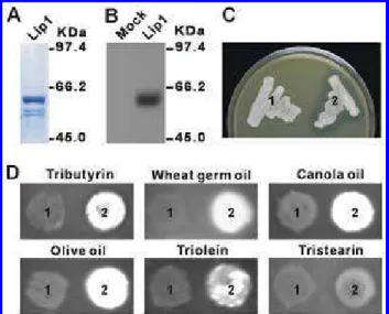
during fungal pathogenesis. Recombinant Lip1 protein was produced in Escherichia coli M15 cells harboring plasmid pQE60-Lip1 (Fig. 2A). However, no lipolytic activity was de-tected from the purified protein. We then expressed Lip1 in
Pichia pastoris X-33 cells using the EasySelect Pichia
expres-sion vector pPICZαA. Western analysis revealed a single band at approximately 60 kDa, which confirmed not only the pres-ence of Lip1 in or on the P. pastoris transformant but also the specificity of the primary antibody we prepared (Fig. 2B).
To examine lipolytic activity of Lip1, we performed both tributyrin emulsion and rhodamine-B fluorescence assays. Hy-drolysis of tributyrin produces a clear zone around the colony on tributyrin emulsion plates whereas, in rhodamine B assay, free fatty acids produced by hydrolysis of lipid substrates form
a fluorescent complex with rhodamine B (Feng et al. 2005). In the tributyrin plate assay, a clear zone was observed only at the colony peripheral of the P. pastoris transformant containing pPICZαA-Lip1, whereas the empty vector-transformed P.
pas-toris strain did not produce any secretory lipolytic activity
under the condition (Fig. 2C). The most appropriate way to categorize an enzyme as a lipase is to test whether it can hy-drolyze long-chain acyl glycerols (Schmid and Verger 1998). This was confirmed by the rhodamine-B assay, in which fluo-rescence was detected from the pPICZαA-Lip1 transformant on plates containing tributyrin (glyceryl tributyrate, C4), triolein (glyceryl trioleate, C18), tristearin (glyceryl tristearate, C18), wheat germ oil, canola oil, or olive oil as lipid substrate (Fig. 2D). These results provide direct evidence that Lip1 is a triglyc-eride lipase and that it possesses lipolytic activity on a broad range of lipid substrates regardless the length and the satura-tion of the fatty acid moieties.
Secreted Lip1 protein predominately localizes to the surface of fungal infection structures.
To reveal the localization of Lip1 during B. graminis f. sp.
tritici infection and development, an immunolocalization assay
was conducted with anti-Lip1 IgG as the primary antibody and FluoroLink Cy 3 as the secondary antibody. Under a confocal laser-scanning microscope, a strong immunofluorescence signal was observed on the surface of B. graminis f. sp. tritici conidia (Fig. 3A). Labeling of individual conidia was stronger on both poles than on the side face, with a dome-shaped pattern of
Fig. 2. Production and lipolytic activity of recombinant Lip1. A, Protein
purified from Escherichia coli M15 cells harboring pQE60-Lip1 was sepa-rated on acrylamide gel and visualized with Coomassie blue staining. B, Western blot analysis of proteins extracted from Pichia pastoris cells har-boring pPICZαA (Mock) or pPICZαA-Lip1 (Lip1). C and D, Analysis of lipolytic activity of Lip1 expressed in P. pastoris. The P. pastoris cells har-boring pPICZαA (1) or pPICZαA-Lip1 (2) were grown on BMMY (1% yeast extract, 2% peptone, 100mM KH2PO4 [pH 6.0]) plates containing C,
1% (wt/vol) tributyrin or D, 0.0001% (wt/vol) rhodamine-B and 1% (wt/vol) lipid substrates as indicated.
Fig. 3. Lip1 localization during development of Blumeria graminis f. sp.
tritici on wheat leaf surface. Conidia or infected leaf samples were incu-bated sequentially with A, C, and D, the primary antibody from Lip1-im-munized rabbit or B, preimmune serum and FluoroLink CyTM3-labeled goat anti-rabbit immunoglobulin G. Images were obtained by confocal laser-scanning microscopy. Bar = 10 µm. Mature conidia A, with or B, without Lip1-antiserum preincubation. C, Mature conidium after plasmo-lysis. The arrowhead indicates the plasma membrane separated away from cell wall. D, Primary (pgt) and appressorial germ tubes (agt) on the leaf surface at 6 h after inoculation (hai). E, Secondary hyphae on the leaf sur-face at 168 hai. Arrowheads indicate secondary penetration sites.
rings. A similar distribution pattern of extracellular mucilage has been observed on fresh conidia of the powdery mildew fungus Uncinuliella australiana (Mims et al. 1995). In the control treatment, when preimmune rabbit serum was used instead of the primary antibody, no labeling signal could be detected (Fig. 3B). Plasmolysis of conidia in 0.8 M NaNO3 for
30 min before fixation revealed that Lip1 is localized to the cell wall rather than the plasma membrane (Fig. 3C). On ger-minated conidia from the surface of wheat leaves, fluorescence was abundant at the tip of the primary germ tube and slightly weaker on the appressorial germ tube (Fig. 3D). Fluorescence was also detected on secondary hyphae growing on the leaf
surface, with the secondary infection sites showing the most prominent accumulation (Fig. 3E). Taken together, the immu-nolocalization data suggest that regional and temporal Lip1 regulation plays a role in B. graminis f. sp. tritici infection-related development on its wheat host.
Lip1 mediates liquefaction of wheat leaf epicuticular wax.
To investigate the effect of Lip1 activity on leaf epicuticular wax, we dipped wheat leaves into 48-h culture supernatants of
P. pastoris transformants containing either pPICZαA-Lip1
(Lip1) or pPICZαA (mock treatment) for 20 or 30 min. Water droplets (5 µl) containing trace amounts of aniline blue readily
Fig 4. Effect of Lip1 treatment on wheat leaf surface wax. A, Hydrophobicity assays for the leaves pretreated with 48-h culture supernatant of Pichia
pas-toris transformants containing pPICZαA (Mock) or pPICZαA-Lip1 (Lip1) for 20 or 30 min. B, Scanning electron microscopy for the leaves pretreated with mock or Lip1 for 30 min. Bar = 100 µm. C and D, Gas chromatography (GC) analysis of wheat surface wax components liquefied by Lip1. E, Selected peaks from GC identified by mass spectrometry analysis. F, Alkanes and primary fatty alcohols in the wax extract from mock-treated leaves (blank), Lip1-treated leaves (black), and Lip1 supernatant (hatched) were calculated into micrograms of component per leaf area to illustrate the quantitative potential of Lip1 on wax liquefaction.
spread out on the leaf surface treated with Lip1 compared with the mock-treated leaves, suggesting a reduction in leaf surface hydrophobicity (Fig. 4A). To further investigate the structural changes on the leaf surface, leaves of both treatments were freeze dried and examined with scanning electron microscopy (SEM). Protruding epicuticular wax crystalloids were evident on the surface of the mock-treated leaf but completely absent from the leaf treated with Lip1 (Fig. 4B), indicating that Lip1 possesses the ability to liquefy leaf epicuticular wax.
To determine which wax components were liquefied by Lip1 treatment, we analyzed total lipids liquefied into the superna-tants as well as what remained on the surface from the leaves after Lip1 or mock treatment. The lipid extracts were separated through gas chromatography (GC) and subjected to mass spec-trometry (MS) analyses. Alkanes, including docosane (C22), tetracosane (C24), and pentacosane (C25), and primary fatty alcohols, including hexacosanol (C26) and octacosanol (C28), were identified from the Lip1 supernatant but absent in the mock supernatant (Fig. 4C and E). These molecules were de-rived from the leaf surface because similar components were identified from both Lip1- and mock-treated leaves (Fig. 4D). Octacosanol was found to be the most abundant component of epicuticular wax on wheat leaves. Calculation of the amount of wax components per leaf surface area (micrograms per square centimeter) indicated that Lip1 treatment could remove ap-proximately half of the chloroform- or methanol-extractable surface wax from wheat leaves (Fig. 4F).
Wax components are required
for conidial germination and differentiation.
The major wax components identified from GC-MS, includ-ing dodecane, docosane, tetracosane, pentacosane, hexaco-sanol, and octacohexaco-sanol, were coated as a thin-layer film on glass slides. B. graminis f. sp. tritici germination, indicated by the presence of primary germ tubes (PGT), and appressorium development were investigated at 24 hai on these coated slides (Fig. 5). On slides without wax coating (CK), less than 30% conidia could form PGT and very few could develop appres-soria or elongation hyphae. Conidial germination was promoted by crude leaf surface wax or wax components, and long-chain alkanes showed the strongest induction, especially with respect
Fig. 5. Effect of wax components on germination of Blumeria graminis f.
sp. tritici conidia. Glass slides were coated with the indicated commercial alkanes and alcohols and inoculated with B. graminis f. sp. tritici conidia. Germ tube formation and appressorium development were investigated at 24 h after inoculation; u.g., ungerminated; pgt, primary germ tube; pgt + h, pgt and hypha; pgt + agt, pgt and appressorial germ tube.
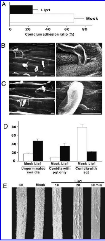
Fig. 6. Effect of Lip1-mediated surface wax liquefaction on early
develop-ment of Blumeria graminis f. sp. tritici. A, Adhesion of B. graminis f. sp. tritici conidia on wheat leaf surface was inhibited by Lip1 treatment. Numbers of conidia on the same leaf area were counted before and after wash and the ratios were plotted. B and C, B. graminis f. sp. tritici devel-opment on leaf surface at 24 h after inoculation (hai) after a 12-h pretreat-ment with culture supernatant of B, pPICZαA (Mock) or C, pPICZαA-Lip1 transformant. Right panels of scanning electron microscopy images are the magnified areas on left panels. Bar = 10 µm. D, Analysis of conid-ial germination and primary germ tube (pgt)/appressorconid-ial germ tube (agt) formation at 24 hai. Ungerminated conidia and conidia with only pgt as well as those with agt were plotted as percentages against total conidium numbers. Approximately 100 conidia were counted for each treatment. Mock and Lip1 treatments were performed as the above. E, Symptom de-velopment of powdery mildew on untreated (CK), mock-, or Lip1-treated leaves at 10 days postinoculation.
to the formation of mature appressorium, a structure critical for penetration. Furthermore, we observed a significant pro-gressive increase (r = 0.99, P < 0.01) in the percentage of ma-ture appressorium-forming conidia (5.0, 30.2, 32.7, and 39.3%) with the increased length of the alkane carbon chain (C12, C22, C24, and C25).
Lip1-mediated epicuticular wax liquefaction is involved in fungal adhesion and development.
To determine the effect of Lip1-mediated epicuticular wax liquefaction on development and pathogenesis of B. graminis f. sp. tritici, wheat leaves treated with the 48-h culture super-natant of P. pastoris transformants containing either pPICZαA-Lip1 (pPICZαA-Lip1) or pPICZαA (mock) for 10 to 30 min were incu-bated in a growth chamber for 12 h and then inoculated with
B. graminis f. sp. tritici conidia. At 6 hai, conidia could be
more readily washed away from Lip1-treated leaves than from mock-treated leaves (Fig. 6A), indicating that Lip1 treatment removes wax components that aid in B. graminis f. sp. tritici adhesion.
We then investigated the effect of Lip1 on B. graminis f. sp.
tritici infection-related development by SEM. At 24 hai, on the
surface of mock-treated leaves, most conidia produced a pri-mary germ tube and an appressorial germ tube (Fig. 6B). In
contrast, the majority of conidia on Lip1-treated leaves either failed to germinate or produced only the primary germ tube (Fig. 6C and D). After incubation in a growth chamber for 10 days, extensive powdery mildew developed on B. graminis f. sp. tritici-inoculated untreated (CK) and mock-treated leaves, whereas Lip1-treated leaves had few B. graminis f. sp. tritici colonies (Fig. 6E), indicating a reduction of disease severity.
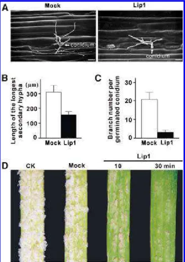
Powdery mildew grows on the surface of hosts by extending secondary elongation hyphae. By 48 hai, primary infection by
B. graminis f. sp. tritici establishes functional haustoria
associ-ated with appressorial germ tube-mediassoci-ated penetrations in the leaf epidermal cells, and secondary elongation hyphae begin to develop on the leaf surface. To investigate whether Lip1-medi-ated leaf epicuticular wax liquefaction was important for sec-ondary hypha development, wheat leaves were inoculated with
B. graminis f. sp. tritici conidia and, at 48 hai, treated with the
culture supernatant of the P. pastoris transformants containing either pPICZαA-Lip1 (Lip1) or pPICZαA (mock treatment) for 10 or 30 min. At 60 hai, SEM revealed that Lip1 treatment impaired secondary hypha growth, with reduced secondary hy-pha elongation and branching (Fig. 7A through C). Late in the infection time course, powdery mildew symptom development on the leaf surface was significantly impaired by the Lip1 treatment (Fig. 7D).
DISCUSSION
Through characterization of a potent lipase, Lip1, from B.
graminis f. sp. tritici, we provide several lines of evidence
sup-porting the idea that lipase is critical for fungal adhesion and development on the host surface. First, LIP1 is one of the abundantly expressed Blumeria genes during pathogenesis, and its expression patterns coincide with conidial germination, germ tube development, and conidial formation and matura-tion. These data are also consistent with the observation that a LIP1 orthologue in B. graminis f. sp. hordei was among the 20 most highly expressed genes at the early stages of barley pow-dery mildew development (Thomas et al. 2002). Second, in situ immunolocalization revealed that Lip1 is secreted to the cell surface and predominantly present on the surface of spore tips, germ tubes, and secondary hyphae, possibly associated with the fungal ECM. Third, the recombinant Lip1 possesses a broad spectrum of substrate specificities on both short- and long-chain fatty acids, which distinguishes it from cutinases that prefer short-chain acyl groups. Finally, Lip1 effectively hydrolyzes leaf epicuticular wax and the hydrolyzed products, especially alkanes, are required for adhesion of conidia and development of germ tubes and secondary hyphae.
Two basic strategies for fungal adhesion to the host surface have been postulated: “passive attachment” resulting from the presence of preformed materials on the fungus surface and “active attachment” requiring the de novo production of fungal adhesives (Carver and Gurr 2006; Jones 1994). Whether the passive or active mechanism is involved in Blumeria spp. ad-hesion deserves further study but the long-chain fatty acid lay-ers coating Blumeria conidia are expected to lead to immediate passive adhesion through hydrophobic interactions (Senior et al. 1993; Cooper et al. 2000; Carver et al. 2006; Wright et al. 2002b). The adhesion between host plant and powdery mildew could be facilitated via a direct physical interaction between the host and the pathogen in the merit of hydrophobic long-chain fatty acids and their derivatives present on the surfaces of both encounters (Fig. 5) (Senior et al. 1993). Furthermore, the secreted Lip1 may enhance the interaction by forming co-valent ester bonds among these hydrophobic molecules through its transesterification activities (Gandhi et al. 2000; Schmid and Verger 1998). Immunolocalization analyses
to-Fig. 7. Effect of Lip1-mediated surface wax liquefaction on secondary
hy-pha development of Blumeria graminis f. sp. tritici. A, Secondary hyhy-pha development on leaf surface was examined by scanning electron micros-copy at 60 h after inoculation (hai). Bar = 100 µm. B, The longest secon-dary hypha was measured and C, the number of hyphal branches was counted at 60 hai from each of 30 germinating conidia. The average values were plotted to illustrate the significant differences between Mock and Lip1 treatments. D, Symptom development of powdery mildew on un-treated (CK), mock-, or Lip1-un-treated leaves at 10 days after inoculation.
gether with transcript profiling showed that Lip1 is not only present on the surface of mature conidia but also during spore germination, germ tube differentiation, and secondary hypha development. These results suggest that production of Lip1 is constitutive in conidia and inducible upon contact with the plant surface during the infection process, and support the view that both passive and active processes underpin powdery mildew adhesion.
Aerial plant surfaces are covered with a noncellular cuticle, which serves as the initial site of interaction between plants and pathogens. The cuticle is composed of an insoluble poly-mer of mainly C16 and C18 hydroxy and epoxy fatty acids (cutin) (Kolattukudy 2001) and associated solvent-soluble ubiquitous aliphatic compounds (epicuticular wax, mainly C20-C60 alkanes, alcohols, and ketones) (Samuels et al. 2008). Due to their close association, it is difficult to distin-guish between the relative contribution of the cutin matrix and epicuticular wax to the physical properties and the biological functions of the cuticle. This study supports previous observa-tions that octacosanol is the most abundant chemical compo-nent of wheat epicuticular wax (Bianchi et al. 1980; Koch et al. 2006). Octacosanol is responsible for induction of infection structures of wheat stem rust P. graminis f. sp. tritici (Reisige et al. 2006) and barley powdery mildew B. graminis f. sp. hor-dei(Zabka et al. 2008). Other long-chain fatty alcohols (>C24) from avocado surface wax components also induce appressor-ium formation in Colletotrichum gloeosporioides(Podila et al. 1993). However, we found that long-chain alkanes, rather than alcohols, are the most efficient wax component for triggering germination and appressorium development of B. graminis f. sp. tritici. It is possible that alkanes are more hydrophobic than alcohols and, therefore, create a more hydrophobic surface that facilitates fungal development and differentiation. Previous studies showed that germinating conidia and germ tubes were able to gain nutrients and signaling molecules from the host sur-face (Senior et al. 1993; Carver et al. 1999; Kinane et al. 2000; Nielsen et al. 2000). During infection processes, alkanes re-leased from the epicuticular wax by Lip1 activity may serve as better nutrients or stronger signaling molecules to trigger devel-opment and differentiation. The importance of plant epicuticular wax or wax monomers in the induction of fungal infection struc-tures has been addressed from Colletotrichum gloeosporioides (Podila et al. 1993), M. grisea (Hegde and Kolattukudy 1997),
B. graminis (Zabka et al. 2008), and Erysiphe pisi (Gniwotta et
al. 2005). In the corn smut fungus, Ustilago maydis, a mixture of triacylglycerides from corn oil activates the signaling path-way dependent on both the cAMP pathpath-way and a Ras/mitogen-activated protein kinase pathway to promote and maintain the filamentous growth of the fungus in the host environment (Klose et al. 2004). These results support the view that deriva-tives of epicuticular wax, perhaps generated by fungal lipolytic activities, could serve as plant-derived signaling molecules.
Lipases in hydrophobic media possess the ability to carry out esterification activity in addition to hydrolysis (Schmid and Verger 1998; Gandhi et al. 2000). It is also possible that Lip1 generates covalent ester bonds between hydrophobic molecules present on the conidium and the host leaf surface through a transesterification reaction. Indeed, hydrophobic lay-ers particularly rich in long-chain fatty acids coat both the co-nidium and host leaf surface (Senior et al. 1993; Cooper et al. 2000; Samuels et al. 2008). However, the biotrophic nature of
B. graminis disables gene-disruption strategies to obtain a
LIP1-null mutant, thus hindering further investigation into its role in powdery mildew pathogenicity. Nevertheless, our re-sults show that the obligate biotroph B. graminis secretes a hydrolytic lipase to facilitate its adhesion and pathogenesis on the host plant.
MATERIALS AND METHODS Plant and fungal materials.
The B. graminis f. sp. tritici strain was maintained and used for inoculation as described (Wei et al. 1998; Liu et al. 2005). Diploid wheat (Triticum monococcum L.) line TM441 inocu-lated with B. graminis f. sp. tritici was used to construct a cDNA library (Liu et al. 2005). All other experiments were conducted on spring wheat (T. aestivum L.) cv. CDC Conway as the susceptible host.
Nucleic acid isolation and Northern and Southern hybridizations.
Nucleic acid isolation and Northern and Southern hybridiza-tions followed the standard protocols (Sambrook and Russell 2001). Total RNA was extracted from ungerminated conidia and infected leaves at 0, 1, 3, 6, 12, 24, 48, 72, 96, 120, and 144 hai. In Northern analysis, 20 µg of total RNA was frac-tionated in 1.2% formaldehyde gels and transferred to Nytran membranes (Whatman, Florham Park, NJ, U.S.A.). The blots were hybridized successively with the 32P-labeled cDNAs of
LIP1, CAP20, and TEF1. Both CAP20 and TEF1 serve as
con-trols to monitor the B. graminis f. sp. tritici transcript profiles during infection. Transcripts of CAP20 homolog in B. grami-nis f. sp. hordei accumulated during formation of appressoria (Both et al. 2005), whereas TEF1 constitutively expressed in ungerminated and germinated conidia, appressoria, and elonga-tion hyphae (Thomas et al. 2002). In Southern analysis, the ge-nomic DNA was digested with BamHI, EcoRV, NcoI, BglII, or KpnI before being subjected to electrophoresis, and the blot was hybridized with the 32P-labeled cDNA of LIP1.
Database searching and computational analysis.
Deduced amino acid sequences of LIP1 orthologues in other fungal species were obtained from the Broad Institute Whole Genome Database by running BlastP with a threshold value of E ≤ 1 × 10⁻⁵. Multiple alignments were conducted using
Clus-talW. Signal peptides of lipases were predicted with SignalP 3.0. Other analyses of Lip1 were performed using appropriate software hosted by the ExPASy Proteomics Server.
Expression of recombinant Lip1 in bacteria and yeast.
Expression and purification of recombinant Lip1 from
Escherichia coli was performed with the Qiaexpressionist kit
(Qiagen, Valencia, CA, U.S.A.). The open-reading frame of LIP1, excluding the predicted signal peptide and stop codon, was obtained from LIP1 cDNA by PCR and inserted into the pQE60 vector. The resulted construct, designated pQE60-Lip1, was used to transform the E. coli M15 cells. Expres-sion of recombinant Lip1 was induced with 1 mM isopropyl β-D-1-thiogalactopyranoside. Preparation of cell lysate and purification of recombinant Lip1 under native conditions were performed according to the manufacturer’s protocol.
For Lip1 expression in the nonlipolytic yeast P. pastoris, the LIP1 open reading frame excluding the predicted signal peptide was amplified by PCR and inserted into the EasySe-lect Pichia expression vector pPICZαA (Invitrogen, Carls-bad, CA, U.S.A.) to generate plasmid pPICZαA-Lip1. Plas-mid DNA (50 µg) of pPICZαA-Lip1 or the empty vector pPICZαA was linearized with PmeI and used to transform P.
pastoris X-33 cells by the lithium chloride method described
in the manufacturer’s instructions. Transformants were cul-tured on or in plates or broth of buffered methanol-complex medium (BMMY; 1% yeast extract, 2% peptone, 100 mM KH2PO4 [pH 6.0], 1.34% yeast nitrogen base, 4 × 10⁻⁵ of
bio-tin) containing 0.5% methanol as the recombinant protein in-ducer.
Lipase activity assay.
Functional expression of the recombinant lipase was con-ducted by the emulsified tributyrin (Rapp and Backhaus 1992) and rhodamine B assays (Kouker and Jaeger 1987). For the tributyrin assay, 1% tributyrin (vol/vol), and, for the rhoda-mine B assay, 2 µM rhodarhoda-mine B and 1% (vol/vol) appropriate substrate, were added into the medium.
Preparation of the primary antibody.
Rabbits were immunized with the purified recombinant Lip1 from E. coli. Immunoglobulin G (IgG) was purified from the antiserum using the Econo-Pac serum IgG purification kit (Bio-Rad, Richmond, CA, U.S.A.).
Western analysis.
Recombinant Lip1 purified from E. coli M15 cells or crude proteins extracted from P. pastoris X33 culture were separated by sodium dodecyl sulfate polyacrylamide gel electrophoresis with 12% resolving gel. The gel containing proteins from bac-teria or yeast was either stained with Coomassie blue to visual-ize the protein bands or used for Western blotting, respectively. In the Western analysis, proteins were electroblotted onto a polyvinylidene difluoride membrane (Millipore, Billerica, MA, U.S.A.), blocked with 4% skim milk and 0.1% Tween 20 in Tris-buffered saline (TBS) (25 mM Tris-HCl, pH 7.5, and 200 mM NaCl) for 2 h, and then incubated with a 1:500 dilu-tion of the primary antibody for 2 h, followed by incubadilu-tion with a 1:1,000 dilution of peroxidase-conjugated donkey anti-rabbit IgG (GE Healthcare, Piscataway, NJ, U.S.A.) for 1 h. After washing, immunoreactive bands were detected by the enhanced chemiluminescence reagents using luminol and p-coumaric acid (Sigma-Aldrich, St. Louis).
Immunofluorescence of Lip1 in fungal tissues.
Immunofluorescence of Lip1 was performed modified after Kemen and associates (2005). Briefly, conidia or leaf sections from inoculated wheat were fixed in a mixture of acetic acid and ethanol (1:3, vol/vol) at room temperature for 2 h and then rehydrated successively in 100, 70, and 50% ethanol. The sam-ples were blocked with 0.1% bovine serum albumin for 20 min followed by incubation with the primary antibody (1:100) at 4°C overnight. For the control treatment, samples were incu-bated with preimmune rabbit serum instead of the primary antibody. For fluorescence detection, samples were incubated with 1:1,000 FluoroLink Cy 3 IgG (GE Healthcare) for 30 min and, after washing with TBS (10 mM Tris-HCl, 150 mM NaCl, pH 7.6) (four times for 5 min each), mounted on slides with 80% glycerol and examined under a confocal laser-scan-ning electron microscope (LSM510; Carl Zeiss, Thornwood, NY, U.S.A.) with excitation and emission wavelengths of 543 and 570 nm, respectively.
Liquefaction of leaf epicuticular wax and pathogenicity assays.
P. pastoris transformants harboring pPICZαA-Lip1 or
pPICZαA were cultured in liquid BMMY media for 48 h and the supernatants were collected by centrifugation. The first leaves of 11-day-old wheat were dipped into the culture super-natants for 10, 20, or 30 min. The effect of leaf treatment on the leaf surface wax was examined by SEM (SEM505; Philips, New York) immediately after the leaves were air dried. Plants were inoculated with B. graminis f. sp. tritici 12 h after Lip1 treatment. The conidial adhesion assay was performed fol-lowing Rumbolz and associates (2000) at 6 hai. The early de-velopment of B. graminis f. sp. tritici was examined at 24 hai with SEM and powdery mildew symptoms were assessed at 120 hai.
The effect of Lip1 treatment on development of secondary hyphae was assayed on leaves 48 hai by dipping into the culture supernatant of P. pastoris transformants containing pPICZαA-Lip1 or pPICZαA for 30 min. The secondary hypha devel-opment was examined by SEM at 60 hai and the symptom development was assessed at 120 hai.
Profiling of leaf epicuticular wax compositions.
To investigate which wax components were liquefied by Lip1 treatment, 20 leaf fragments, 0.8 cm long, were treated for 30 min with 5 ml of the culture supernatant of P. pastoris transformants containing pPICZαA-Lip1 or pPICZαA. Wax was extracted from untreated and treated leaves by dipping into chloroform/methanol (1:1, vol/vol) for 15 s. Wax compo-nents released to the culture supernatant after leaf treatment were extracted with the chloroform/methanol method described by Lalman and Bagley (2004). After drying undera stream of nitrogen, all wax extracts were derivatized using 50 µl of pyri-dine containing N,O-bis (trimethylsilyl) fluoroacetamide and 1% (vol/vol) trimethylchlorosilane. GC and MS analyses were performed with a HP 6890 Series GC System (Global Medical Instrumentation, Ramsey, MN, U.S.A.) and a Trio 2000 gas chromatograph mass spectrometer (Agilent Technologies, Santa Clara, CA, U.S.A.), respectively, by following the protocol de-scribed by Millar and associates (1999). In the GC analysis, heptadecanoic acid (C17:0) and tricosanoic acid (C23:0) were used as the internalstandards. Wax component quantification was based on the comparison of flame ionization detector peak areas between the component and heptadecanoic acid.
Coating glass slides with wax components and
B. graminis f. sp. tritici germination assay.
Commercial long-chain fatty alcohols and alkanes, and wax extracted from untreated wheat leaves, were used to create a thin film on the surface of a cover slide (2.2 × 5.0 cm2)
follow-ing Reisige and associates (2006). The coated slides were inoculated with B. graminis f. sp. tritici conidia and conidial germination and differentiation were investigated at 24 hai. The experiments were conducted three times and, for each treat-ment, approximately 100 conidia were counted.
ACKNOWLEDGMENTS
We thank R. Hirji for technical support. This work was supported by the Natural Sciences and Engineering Research Council of Canada and the Canadian Wheat Board in the form of grants to Y. Wei and scholarships to J. Feng.
LITERATURE CITED
Berto, P., Belingheri, L., and Dehorter, B. 1997. Production and purifica-tion of a novel extracellular lipase from Alternaria brassicicola. Bio-technol. Lett. 19:533-536.
Berto, P., Comménil, P., Belingheri, L., and Dehorter, B. 1999. Occurrence of a lipase in spores of Alternaria brassicicola with a crucial role in the infection of cauliflower leaves. FEMS (Fed. Eur. Microbiol. Soc.) Mi-crobiol. Lett. 180:183-189.
Bianchi, G., Lupotto, E., Borghi, B., and Corbellini, M. 1980. Cuticular wax of wheat. Planta 148:328-331.
Both, M., Eckert, S. E., Csukai, M., Müller, E., Dimopoulos, G., and Spanu, P. D. 2005. Transcript profiles of Blumeria graminis develop-ment during infection reveal a cluster of genes that are potential viru-lence determinants. Mol. Plant-Microbe Interact. 18:125-133. Bourett, T. M., and Howard, R. J. 1990. In vitro development of
penetra-tion structures in the rice blast fungus Magnaporthe grisea. Can. J. Bot. 68:329-342.
Carver, T. L. W., and Gurr, S. J. 2006. Filamentous fungi on plant surfaces. Pages 368-397 in: Biology of the Plant Cuticle. M. Riederer and M. Müller, eds. Blackwell Publishing Ltd., Oxford.
Carver, T. L. W., Ingerson, S. M., and Thomas, B. J. 1996. Influences of host surface features on development of Erysiphe graminis and
Erysi-phe pisi. Pages 255-256 in: Plant Cuticles: An Integrated Functional Approach. G. Kerstiens, ed. BIOS Scientific Publishers Ltd., Oxford. Carver, T. L. W., Kunoh, H., Thomas, B. J., and Nicholson, R. L. 1999.
Release and visualization of the extracellular matrix of conidia of Blumeria graminis. Mycol. Res. 103:547-560.
Chahinian, H., Nini, L., Boitard, E., Dubes, J. P., Comeau, L. C., and Sarda, L. 2002. Distinction between esterases and lipases: A kinetic study with vinyl esters and TAG. Lipids 37:653-662.
Chaubal, R., Wilmot, V. A., and Wynn, W. K. 1991. Visualization, adhe-siveness and cytochemistry of the extracellular matrix produced by urediniospore germ tubes of Puccinia sorghi. Can. J. Bot. 69:2044-2054.
Clement, J. A., Porter, R., Butt, T. M., and Beckett, A. 1993. Germination and the role of extracellular matrix in adhesion of urediniospores of Uromyces viciae-fabae to synthetic surfaces. Mycol. Res. 97:585-593. Clement, J. A., Martin, S. G., Porter, R., Butt, T. M., and Beckett, A. 1994.
The role of hydrophobicity in attachment of urediniospores and sporelings of Uromyces. Germination and the role of extracellular ma-trix in adhesion of urediniospores of Uromyces viciae-fabae. Mycol. Res. 98:1217-1228.
Clement, J. A., Porter, R., Butt, T. M., and Beckett, A. 1997. Characteris-tics of adhesion pads formed during imbibition and germination of urediniospores of Uromyces viciae-fabae on host and synthetic surfaces. Mycol. Res. 101:1445-1458.
Comménil, P., Belingheri, L., and Dehorter, B. 1998. Antilipase antibodies prevent infection of tomato leaves by Botrytis cinerea. Physiol. Mol. Plant Pathol. 52:1-14.
Comménil, P., Belingheri, L., Bauw, G., and Dehorter, B. 1999. Molecular characterization of a lipase induced in Botrytis cinerea by components of grape berry cuticle. Physiol. Mol. Plant Pathol. 55:37-43.
Cooper, L. L. D., Oliver, J. E., De Vilbiss, E. D., and Doss, R. P. 2000. Lipid composition of the extracellular matrix of Botrytis cinerea germ-lings. Phytochemistry 53:293-298.
Dean, J. D., Goodwin, P. H., and Hsiang, T. 2002. Comparison of relative RT-PCR and northern blot analyses to measure expression of beta-1,3-glucanase in Nicotiana benthamiana infected with Colletotrichum de-structivum. Plant Mol. Biol. Rep. 20:347-356.
Deising, H., Nicholson, R. L., Haug, M., Howard, R. J., and Mengden, K. 1992. Adhesion pad formation and the involvement of cutinase and esterases in the attachment of uredospores to the host cuticle. Plant Cell 4:1101-1111.
Feng, J., Liu, G., Selvaraj, G., Hughes, G. R., and Wei, Y. 2005. A secreted lipase encoded by LIP1 is necessary for efficient use of saturated tri-glyceride lipids in Fusarium graminearum. Microbiology 151:3911-3921.
Francis, S. A., Dewey, F. M., and Gurr, S. J. 1996. The role of cutinase in germling development and infection by Erysiphe graminis f. sp. hordei. Physiol. Mol. Plant Pathol. 49:201-211.
Gandhi, N. N., Patil, N. S., Sawant, S. B., Joshi, J. B., Wangikar, P. P., and Mukesh, D. 2000. Lipase-catalyzed esterification. Catal. Rev. Sci. Eng. 42:439-480.
Gilbert, R. D., Johnson, A. M., and Dean, R. A. 1996. Chemical signals re-sponsible for appressorium formation in the rice blast fungus Mag-naporthe grisea. Physiol. Mol. Plant Pathol. 48:335-346.
Gniwotta, F., Vogg, G., Gartmann, V., Carver, T. L. W., Riederer, M., and Jetter, R. 2005. What do microbes encounter at the plant surface? Chemical composition of pea leaf cuticular waxes. Plant Physiol. 139:519-530.
Green, J. R., Carver, T. L. W., and Gurr, S. J. 2002. The formation and function of infection and feeding structures. Pages 66-82 in: The Pow-dery Mildews—A Comprehensive Treatise. R. R. Bélanger, W. Bushnell, A. J. Dik, and T. L. W. Carver, eds. American Phytopathological Society Press, St. Paul, MN, U.S.A.
Hegde, Y., and Kolattukudy, P. E. 1997. Cuticular waxes relieve self-inhi-bition of germination and appressorium formation by the conidia of Magnaporthe grisea. Physiol. Mol. Plant Pathol. 51:75-84.
Jones, E. B. G. 1994. Fungal adhesion. Mycol. Res. 98:961-981.
Kemen, E., Kemen, A. C., Rafiqi, M., Hempel, U., Mendgen, K., Hahn, M., and Voegele, R. T. 2005. Identification of a protein from rust fungi transferred from haustoria into infected plant cells. Mol. Plant-Microbe Interact. 18:1130-1139.
Kinane, J., Dalvin, S., Bindslev, L., Hall, A., Gurr, S., and Oliver, R. P. 2000. Evidence that the cAMP pathway controls emergence of both pri-mary and appressorial germ-tubes of barley powdery mildew. Mol. Plant-Microbe Interact. 13:494-502.
Klose, J., Moniz de Sá, M., and Kronstad, J. W. 2004. Lipid-induced fila-mentous growth in Ustilago maydis. Mol. Microbiol. 52:823-835. Koch, K., Barthlott, W., Koch, S., Hommes, A., Wandelt, K., Mamdouh,
H., De-Feyter, S., and Broekmann, P. 2006. Structural analysis of wheat wax Triticum aestivum, cv. ‘Naturastar’ L: From the molecular level to
three dimensional crystals. Planta 223:258-270.
Kolattukudy, P. E. 1984. Cutinases from fungi and pollen. Pages 471-504 in: Lipases. B. Borgstrom and H. L. Brockman, eds. Elsevier, New York.
Kolattukudy, P. E. 1985. Enzymatic penetration of the plant cuticle by fun-gal pathogens. Annu. Rev. Phytopathol. 23:223-250.
Kolattukudy, P. E. 2001. Polyesters in higher plants. Adv. Biochem. Eng. Biotechnol. 71:1-49.
Kouker, G., and Jaeger, K. E. 1987. Specific and sensitive plate assay for bacterial lipases. Appl. Environ. Microbiol. 53:211-213.
Kunoh, H., Yamaoka, N., Yoshioka, H., and Nicholson, R. L. 1988. Prepa-ration of the infection court by Erysiphe graminis. I, Contact-mediated changes in morphology of the conidium surface. Exp. Mycol. 12:325-335.
Lalman, J. A., and Bagley, D. M. 2004. Extracting long chain fatty acids from a fermentation medium. J. Am. Oil Chem. Soc. 81:105-110. Li, D., Ashby, A. M., and Johnstone, K. 2003. Molecular evidence that the
extracellular cutinase Pbc1 is required for pathogenicity of Pyrenope-ziza brassicae on oilseed rape. Mol. Plant-Microbe Interact. 16:545-552.
Liu, G., Sheng, X., Greenshields, D. L., Ogieglo, A., Kaminskyj, S., Selvaraj, G., and Wei, Y. 2005. Profiling of wheat class-III peroxidase genes derived from powdery mildew attacked epidermis reveals distinct sequence-associated expression patterns. Mol. Plant-Microbe Interact. 18:730-741.
Longhi, S., and Cambillau, C. 1999. Structure-activity of cutinase, a small lipolytic enzyme. Biochim. Biophys. Acta 1141:185-196.
Lotti, M., Grandori, R., Fusetti, F., Longhi, S., Brocca, S., Tramontano, A., and Alberghina, L. 1993. Cloning and analysis of Candida cylindracea lipase sequences. Gene 124:45-55.
Martinez, C., Nicolas, A., van Tilbeurgh, H., Egloff, M. P., Cudrey, C., Verger, R., and Cambillau, C. 1994. Cutinase, a lipolytic enzyme with a preformed oxyanion hole. Biochemistry 33:83-89.
Mendgen, K., and Hahn, M. 2002. Plant infection and the establishment of fungal biotrophy. Trends Plant Sci. 7:352-356.
Mercure, E. W., Leite, B., and Nicholson, R. L. 1994. Adhesion of unger-minated conidia of Colletotrichum graminicola to artificial hydrophobic surfaces. Physiol. Mol. Plant Pathol. 45:421-440.
Mercure, E. W., Kunoh, H., and Nicholson, R. L. 1995. Visualization of materials released from adhered ungerminated conidia of Colleto-trichum graminicola. Physiol. Mol. Plant Pathol. 46:121-135.
Millar, A. A., Clemens, S., Zachgo, S., Giblin, E. M., Taylor, D. C., and Kunst, L. 1999. CUT1, an Arabidopsis gene required for cuticular wax biosynthesis and pollen fertility encodes a very-long-chain fatty acid condensing enzyme. Plant Cell 11:825-838.
Mims, C. W., Liljebjelke, K. A., and Richardson, E. A. 1995. Surface mor-phology wall structure and initial adhesion of conidia of the powdery mildew fungus Uncinuliella australiana. Phytopathology 85:352-358. Nasser-Eddine, A., Hannemann, F., and Schäfer, W. 2001. Cloning and
ex-pression analysis of Nhi, a gene encoding an extracellular lipase from the fungal pea pathogen Nectria haematococca MP VI Fusarium solani f. sp. pisi that is expressed in planta. Mol. Genet. Genomics 265:215-224.
Nicholson, R. L., Yoshioka, H., Yamaoka, N., and Kunoh, H. 1988. Prepa-ration of the infection court by Erysiphe graminis. II, Release of esterase enzyme from conidia in response to a contact stimulus. Exp. Mycol. 12:336-349.
Nielsen, K. A., Nicholson, R. L., Carver, T. L. W., Kunoh, H., and Oliver, R. P. 2000. First touch: An immediate response to surface recognition in conidia of Blumeria graminis. Physiol. Mol. Plant Pathol. 56:63-70. Pascholati, S. F., Yoshioka, H., Kunoh, H., and Nicholson, R. L. 1992.
Preparation of the infection court by Erysiphe graminis f. sp. hordei— cutinase is a component of the conidial exudates. Physiol. Mol. Plant Pathol. 41:53-59.
Podila, G. K., Rogers, L. M., and Kolattukudy, P. E. 1993. Chemical sig-nals from avocado surface wax triggers germination and appressorium formation in Colletotrichum gloeosporioides. Plant Physiol. 103:267-272.
Rapp, P., and Backhaus, S. 1992. Formation of extracellular lipases by filamentous fungi yeasts and bacteria. Enzyme Microb. Technol. 14:938-943.
Reis, H., Pfiffi, S., and Hahn, M. 2005. Molecular and functional charac-terization of a secreted lipase from Botrytis cinerea. Mol. Plant Pathol. 6:257-267.
Reisige, K., Gorzelanny, C., Daniels, U., and Moerschbacher, B. M. 2006. The C28 aldehyde octacosanal is a morphogenetically active component involved in host plant recognition and infection structure differentiation in the wheat stem rust fungus. Physiol. Mol. Plant Pathol. 68:33-40. Rumbolz, J., Kassemeyer, H. H., Steinmetz, V., Deising, H. B., Mendgen,
infection structures of the powdery mildew fungus Uncinula necator and adhesion to the host cuticle. Can. J. Bot. 78:409-421.
Sambrook, J., and Russell, D. W. 2001. Molecular Cloning: A Laboratory Manual. Cold Spring Harbor Laboratory Press, Cold Spring Harbor, NY, U.S.A.
Samuels, L., Kunst, L., and Jetter, R. 2008. Sealing plant surfaces: Cuticu-lar wax formation by epidermal cells. Annu. Rev. Plant Biol. 59:683-707.
Schmid, R. D., and Verger, R. 1998. Lipases: Interfacial enzymes with attractive applications. Angew. Chem. Int. Ed. Engl. 37:1608-1633. Schrag, J. D., Li, Y., Wu, S., and Cygler, M. 1991. Ser-His-Glu triad forms
the catalytic site of the lipase from Geotrichum candidum. Nature 351:761-764.
Senior, I. J., Hollomon, D. W., and Holloway, P. J. 1993. Unusual long chain monoenoic fatty acids in conidia of Erysiphe graminis. Phyto-chemistry 34:65-68.
Shimada, Y., Sugihara, A., Iizumi, T., and Tominaga, Y. 1990. cDNA clon-ing and characterization of Geotrichum candidum lipase II. J. Biochem. 107:703-707.
Skamnioti, P., and Gurr, S. J. 2007. Magnaporthe grisea cutinase2 medi-ates appressorium differentiation and host penetration and is required for full virulence. Plant Cell 19:2674-2689.
Sugui, J. A., Leite, B., and Nicholson, R. L. 1998. Partial characterization of the extracellular matrix released onto hydrophobic surfaces by co-nidia and germlings of Colletotrichum graminicola. Physiol. Mol. Plant Pathol. 52:411-425.
Thomas, S. W., Glaring, M. A., Rasmussen, S. W., Kinane, J. T., and Oliver, R. P. 2002. Transcript profiling in the barley mildew pathogen Blumeria graminis by serial analysis of gene expression SAGE. Mol. Plant-Microbe Interact. 15:847-856.
Verger, R. 1997. ‘Interfacial activation’ of lipases: Facts and artifacts. Trends Biotechnol. 15:32-38.
Voigt, C. A., Schäfer, W., and Salomon, S. 2005. A secreted lipase of
Fusarium graminearum is a virulence factor required for infection of cereals. Plant J. 42:364-375.
Walton, J. D. 1994. Deconstructing the cell wall. Plant Physiol. 104:1113-1118.
Wei, Y., Zhang, Z., Andersen, C. H., Schmelzer, E., Gregersen, P. L., Collinge, D. B., Smedegaard-Petersen, V., and Thordal-Christensen, H. 1998. An epidermis/papilla-specific oxalate oxidase-like protein in the defence response of barley attacked by the powdery mildew fungus. Plant Mol. Biol. 36:101-112.
Wright, A. J., Thomas, B. J., and Carver, T. L. W. 2002a. Early adhesion of Blumeria graminis to plant and artificial surfaces demonstrated by cen-trifugation. Physiol. Mol. Plant Pathol. 61:217-226.
Wright, A. J., Thomas, B. J., Kunoh, H., Nicholson, R. L., and Carver, T. L. W. 2002b. Release and visualization of the extracellular matrix of conidia of Blumeria graminis. Physiol. Mol. Plant Pathol. 61:163-178. Zabka, V., Stang, M., Bringmann, G., Vogg, G., Riederer, M., and
Hildebrandt, U. 2008. Host surface properties affect prepenetration processes in the barley powdery mildew fungus. New Phytol. 177:251-263.
Zhang, Z., Henderson, C., Perfect, E., Carver, T. L. W., Thomas, B. J., Skamnioti, P., and Gurr, S. J. 2005. Of genes and genomes, needles and haystacks: Blumeria graminis and functionality. Mol. Plant Pathol. 6:561-575.
AUTHOR-RECOMMENDED INTERNET RESOURCES
Broad Institute Whole Genome database: www.broad.mit.edu/annotation ClustalW: www.ebi.ac.uk/clustalw
ExPASy Proteomics Server, http://expasy.org
ExPASy PROSITE Carboxylesterases type-B signatures webpage: expasy.org/cgi-bin/nicedoc.pl?PDOC00112