Publisher’s version / Version de l'éditeur: NeuroImage, 58, 1, pp. 269-274, 2011-06-13
READ THESE TERMS AND CONDITIONS CAREFULLY BEFORE USING THIS WEBSITE. https://nrc-publications.canada.ca/eng/copyright
Vous avez des questions? Nous pouvons vous aider. Pour communiquer directement avec un auteur, consultez la
première page de la revue dans laquelle son article a été publié afin de trouver ses coordonnées. Si vous n’arrivez pas à les repérer, communiquez avec nous à [email protected].
Questions? Contact the NRC Publications Archive team at
[email protected]. If you wish to email the authors directly, please see the first page of the publication for their contact information.
NRC Publications Archive
Archives des publications du CNRC
This publication could be one of several versions: author’s original, accepted manuscript or the publisher’s version. / La version de cette publication peut être l’une des suivantes : la version prépublication de l’auteur, la version acceptée du manuscrit ou la version de l’éditeur.
For the publisher’s version, please access the DOI link below./ Pour consulter la version de l’éditeur, utilisez le lien DOI ci-dessous.
https://doi.org/10.1016/j.neuroimage.2011.06.004
Access and use of this website and the material on it are subject to the Terms and Conditions set forth at
Emotion-dependent responses in spinal cord neurons: a spinal fMRI study
Smith, Stephen D.; Kornelsen, Jennifer
https://publications-cnrc.canada.ca/fra/droits
L’accès à ce site Web et l’utilisation de son contenu sont assujettis aux conditions présentées dans le site LISEZ CES CONDITIONS ATTENTIVEMENT AVANT D’UTILISER CE SITE WEB.
NRC Publications Record / Notice d'Archives des publications de CNRC:
https://nrc-publications.canada.ca/eng/view/object/?id=6b3e9093-47a3-47b1-b183-9aaacc23583e https://publications-cnrc.canada.ca/fra/voir/objet/?id=6b3e9093-47a3-47b1-b183-9aaacc23583e
Emotion-dependent responses in spinal cord neurons: A spinal fMRI
study
Stephen D. Smith
Jennifer Kornelsen
National Research Council Institute for Biodiagnostics, Winnipeg, Canada
Department of Psychology, University of Winnipeg, Canada
Abstract
Previous research has demonstrated that emotional stimuli receive preferential processing in the brain. In the current study, functional magnetic resonance imaging was utilized to determine if emotion-specific responses are detectable in the cervical spinal cord. During the passive (i.e., non-motoric) perception of images, activity was detected in the left dorsal and right ventral spinal cord in response to negative emotional stimuli; however, this pattern was reversed in response to neutral and positive stimuli. Critically, during active motoric responses to images, there was greater activity in the ventral cervical spinal cord in response to negative emotional stimuli than to neutral stimuli. These results demonstrate preferential motor responses to negative emotional images by the spinal cord, likely indicating an enhancement of activity in response to threat.
Research highlights
Activity in the cervical spinal cord is influenced by emotional images.
Positive and negative images elicit opposite patterns of activity during perception. Threatening images increase activity in the cervical spinal cord during
movements.
Activity occurs in the ventral aspect of the cord during the negative condition. Activity is lateralized to the side producing movement in the negative condition.
Keywords: Emotion; Affect; Threat; Movement; Spinal cord; Spinal fMRI Introduction
Emotional stimuli receive preferential processing in the nervous system (LeDoux, 2000). Indeed, neuroimaging studies have shown that emotional stimuli elicit greater levels of activity in the extrastriate cortex of the brain than do neutral stimuli (Surguladze et al., 2003) thus allowing individuals to prioritize important information in the environment (e.g., a threat) at the expense of less important information (Pessoa, 2005). This emotional modulation of attention is quite rapid, occurring as early as 160 ms after the appearance of an emotional stimulus (Pizzagalli et al., 2002), and is likely dependent
upon input from the amygdala, a group of nuclei in the medial portions of the temporal lobe (Morris et al., 1996).
However, an emotional experience is not limited to a cognitive representation of that affective state. Instead, most emotions are associated with some form of action (i.e., to approach or withdraw from an emotional stimulus; Davidson and Irwin, 1999). For example, when a person is frightened, this fear is often accompanied by a protective movement such as raising one's hands in front of one's face when threatened or jumping away when startled. Given the instinctive nature and clear survival value of such actions, it is reasonable to assume that movements linked to emotional responses would receive preferential processing in brain areas related to motoric activity much in the same way that emotional stimuli receive prioritized attentional processing (Pereira et al., 2010).
Recent studies suggest that the neural link between emotion and movement does in fact exist. Using transcranial magnetic stimulation (TMS), Hajcak et al.(2007) found greater motor cortex excitability in response to emotionally arousing positive and negative images than to neutral, non-emotional images. This excitability is likely due to activity in the supplementary motor area in the frontal lobes. Activity in this brain region has been linked to emotional responses in the primary motor cortex, suggesting that an ‗emo-motoric‘ system exists in the cortex (Oliveri et al., 2003). Subsequent studies have extended this network to include descending white-matter pathways connecting the cortex to the brainstem and spinal cord. Schutter et al. (2008; see also [Coombes et al., 2009] and [van Loon et al., 2010]) demonstrated that photographs of faces expressing fear elicited greater excitability in the corticospinal tract than did faces expressing happiness or no emotion (i.e., neutral faces). Similar corticospinal excitability has been detected in response to negative emotional scenes (Coelho et al., 2010). As pointed out by these authors, greater responsivity of this white-matter tract suggests that emotional reactions often include some form of action preparedness.
One limitation of these TMS studies is that the neural substrates of motoric responses were inferred from motor evoked potentials in which the activity of specific muscle groups was assessed, rather than being observed directly. TMS studies are also traditionally limited to cortex and dense white-matter connections; although this
neuroanatomy encompasses important components of the emo-motoric system, it does omit portions of the central nervous system that may be critical to emotional responses. Specifically, current conceptualizations of emotion and movement generally discount the role of the spinal cord in these behaviors, despite the fact that spinal-cord neurons are the final link between the central nervous system's motoric programs and the peripheral nervous system that will execute them.
The spinal cord is composed of 31 segments (8 cervical, 12 thoracic, 5 lumbar, 5 sacral, and 1 coccygeal). Each segment consists of (1) nerves that carry afferent sensory information that comes from the periphery to synapse in the dorsal horns of the spinal cord and (2) nerves that originate in the ventral horns that convey efferent motor information to the periphery. The cervical spinal cord segments C5–C8 consist of
nerves that are associated with the upper limbs and hands. To produce a movement, the upper motor neurons which originate in the primary motor cortex terminate in the ventral horns of the appropriate spinal cord segments and synapse with the lower motor neurons which project to the periphery, eventually leading to observable behavior
(Kiernan, 1998).
Until recently, it had not been possible to assess functional activity within the spinal cord, thus limiting the ability of researchers to directly test theories of spinal cord functioning. This difficulty in acquiring spinal cord images was due to a number of related factors. First, the spinal cord has a very small cross-section (approximately 16 mm by 10 mm in the cervical enlargement), making it a very small target for functional imaging. This issue is compounded by the anatomy surrounding the spinal cord, as the differences in the magnetic susceptibilities of the different tissue types (cord, cartilage, bone) cause differences in the magnetic fields within the tissues and in the field gradients at their boundaries. This causes distortion and loss of signal and can result in poor image quality. In addition, the cerebrospinal fluid flows around the spinal cord and causes both physical motion of the spinal cord in the canal and motion artifacts in the image.
However, recent advances in functional MRI scanning sequences and methodology have allowed researchers to overcome many of these barriers, thus allowing for the non-invasive imaging of activity in the spinal cord using a variation of the conventional blood-oxygen-level-dependent (BOLD) imaging typically used in brain fMRI ( [Ng et al., 2006] and [Stroman, 2005]). For instance, Stroman (2005) demonstrated that a spin echo sequence with a short echo time, which is less affected by magnetic susceptibility differences, can be used to acquire a higher quality spinal cord image that remains sensitive to signal change. Subsequent studies noted that using a thin sagittal slice orientation can increase the volume of spinal cord that can be imaged (Stroman et al., 2005). Additionally, by reformatting the data into a three dimensional volume and smoothing only along uniform tissue types, one can increase the signal to noise ratio while minimizing partial volume effects (Stroman et al., 2005).
Functional MRI of the spinal cord (spinal fMRI) has now been used to investigate sensory and motor activity in the lumbar (Kornelsen and Stroman, 2004) and cervical spinal cord ( [Ghazni et al., 2010], [Lawrence et al., 2008], [Maieron et al., 2007], [Piché et al., 2009], [Stroman et al., 2005] and [Xie et al., 2007]), and has been utilized in studies of several patient populations ( [Agosta et al., 2008], [Kornelsen and Stroman, 2007] and [Stroman et al., 2004]). These studies provide an important ‗proof of principle‘ for the technique: spinal fMRI detects activity that is consistent with known physiology in the spinal cord. For example, in response to cold thermal stimulation of the hand,
activity in the dorsal horn ipsilateral to the side of stimulation was observed in the lower cervical spinal cord segments (Stroman et al., 2005) and similarly, in response to the same stimulation applied to the inner leg, activity was observed in the dorsal horn ipsilateral to the side of stimulation in the lower lumbar spinal cords segments (Stroman et al., 2002). During a motor task of the lower limbs, activity was detected in the lower
lumbar spinal cord segments in the ventral and dorsal horns bilaterally (Kornelsen and Stroman, 2004).
Although these studies have established spinal fMRI as a valid method of measuring neural activity, no previous studies have used this technique to investigate whether cognitive and emotional processes in the brain can influence activity in spinal cord neurons. The current research utilized spinal fMRI to determine (1) if the cervical spinal cord is active during passive viewing of emotional images, thereby suggesting an action preparedness, and (2) if the ventral horns of the cervical spinal cord show different patterns of activation during the execution of motoric responses to emotional, as compared to neutral, stimuli.
Materials and methods Participants
Fourteen healthy volunteers (8 female, mean age = 22.75; 6 male, mean age = 21.33, all right hand dominant) with no neurological or psychiatric history participated in this study. All participants underwent MR screening at the National Research Council prior to scanning to ensure that they could be safely scanned. Ethical approval was obtained from the National Research Council Canada's Institute for Biodiagnostics (IBD) Human Research Ethics Board and the University of Winnipeg Senate Committee on Ethics in Human Research and Scholarship; all participants provided written informed consent prior to their participation in these studies.
Stimulus materials
Emotional and neutral pictures consisted primarily of images taken from the International Affective Picture Systems (IAPS), a standardized image database commonly used in emotion studies (Lang et al., 2008). This set of photographs was supplemented with additional images taken from publicly available sources on the internet, as the IAPS did not have enough highly arousing stimuli for our experimental design. Pilot testing was conducted with all images to ensure that they were properly categorized and that the positive and negative stimulus sets were of equal arousal levels; the valence and arousal ratings for all three stimulus types are listed in Table 1.
Table 1. Stimuli were rated on a scale from 1 to 9 for both valence (positive vs. negative) and arousal (high vs. low). Values in parentheses indicate standard deviations. There were no differences for the arousal ratings between positive and negative image sets. The valence ratings for positive and negative images were significantly different from each other, as well as from the valence ratings for neutral images.
Stimulus type Arousal rating Valence rating
High-arousal negative 7.23 (1.98) 2.01 (1.84) High-arousal positive 7.12 (1.67) 7.15 (2.45) Neutral 2.17 (1.31) 5.20 (2.10)
Negative photographs consisted of fear-related scenes (e.g., people being attacked, vicious animals) and disgust-related scenes (e.g., close-up images of people with skin diseases). These pictures were designed to elicit avoidance responses. Positive photographs consisted of erotic images; these images were chosen so that positive items were as emotionally arousing as negative items. Neutral scenes consisted of photographs of landscapes or of the interiors of houses (e.g., kitchens). The three groups of stimuli were equated for visual complexity.
Study design and procedure
Participants completed six experimental runs lasting 5 min and 40 s each. This design allowed for the examination of each of the two research questions – Perception and Movement – using emotionally negative, emotionally positive, and neutral images, with each emotion type assessed in separate fMRI runs to reduce emotional contamination. Each run utilized a blocked design in which 60-second blocks of images were preceded and followed by 40-second rest periods. Due to the tendency for emotional responses to images to habituate rapidly ( [Stark et al., 2004] and [Wright et al., 2001]), each 60-second block of images consisted of 15 individual stimuli presented for four 60-seconds each. This manipulation increased the likelihood that participants would remain in a given affective state for the full 60 s, thereby facilitating the acquisition of signals from the cervical spinal cord.
In the Perception conditions (runs 1–3), participants viewed the images on the screen and were not required to make any response. During Movement conditions (runs 4–6), participants viewed the images on the screen and were required to make a response via key press on a finger pad. In order to make the motoric responses equally relevant across all three stimulus types (negative, positive, and neutral), participants were asked to indicate if each stimulus was an indoor or an outdoor scene. All participants made responses using their right hand, using either the index finger or middle finger,
depending on the response. In both the Perception and the Movement conditions, the order of the runs – positive, negative, and neutral – were counterbalanced across participants.
Functional magnetic resonance imaging parameters
Spinal-cord images were acquired using a Siemen's neck coil and body matrix coil on a 3-Tesla whole-body Siemens scanner (TRIO, Siemens Medical Solutions, Erlangen). Images were acquired using a single-shot fast spin-echo scanning sequence with partial Fourier sampling (HASTE) with the following parameters: TE = 38 ms, TR = 1000 ms per slice, 1.04 mm × 1.04 mm resolution, FOV 200 mm × 100 mm, 50% phase encode FOV. Nine 2 mm thick contiguous slices were centered rostro-caudally on the C5 vertebra and spanned the cervical spinal cord segments and the lower brainstem. Spatial saturation pulses were applied to eliminate signal from surrounding areas to avoid aliasing, and to reduce motion artifacts arising from regions anterior to the spine.
Statistical analyses
Data were preprocessed and analyzed with Matlab custom written software (Mathworks, Inc., Natick, MA); the series of steps involved with these analyses are described in detail here. First, to realign each spinal fMRI data series, a raw data image was used to define the location and curvature of the spinal cord, according to methods published elsewhere (Stroman et al., 2005). Specifically, a reference line was drawn manually along the anterior edge of the spinal cord in the sagittal plane, and the intersection with the bottom of the pons and the C7/T1 disc were manually marked. The data at each time point was combined into a three dimensional volume and linearly interpolated to 0.5 × 0.5 × 0.5 mm3 cubic voxels. The volume was then resliced transverse to the manually drawn reference line. The reference line was used to realign the slice data to correct for motion between time points and to apply spatial smoothing in the rostro-caudal direction. Next, each realigned and smoothed data series was analyzed using the general linear model as described in Stroman (2006) with the basis set generated for the stimulation paradigm described above, consisting of a block paradigm of 40 second rest before and after each 60 second block of image presentation. A threshold of t = 2.0 (Bonferroni corrected for multiple comparisons at p = 0.05) was chosen to identify active voxels for display in color on a gray-scale reference image. This T-threshold is consistent with previous published work ( [Stroman et al.,
2005] and [Kornelsen and Stroman, 2007]). The average percent signal change for active voxels was calculated for each series.
The analyzed individual results were then spatially normalized by reslicing the spinal cord every 1 mm along the length of the reference line between the bottom of the pons and the C7/T1 disc, scaling to a 140 mm length, and fine-tuning the alignment in each transverse slice (Stroman et al., 2008). The analyzed individual results were compiled by task (passive or active) and stimulus type (neutral, positive, or negative) for all participants and a grouped analysis was conducted for each of the resulting six
conditions. The grouped analysis consisted of a random effects analysis that calculated the magnitude of the response as determined in the general linear model analyses across subjects for each voxel, and the standard error of the mean across subjects to produce a t-value. The significant grouped results (T-threshold = 2.5, df = 13,
p = 0.0135) were displayed on a normalized cervical spinal cord template
(Fig. 1 and Fig. 2). The number of active voxels for each condition was manually counted. The emotional stimulus conditions (negative and positive) were analyzed against the control condition (neutral) for each task (Perception and Movement) with a Dunnett's multiple comparisons test (Statistica, StatSoft, Tulsa, OK). The Dunnett's test is a post hoc test that is used to determine significant differences between a control group mean and treatment group means in an analysis of variance; thus, this is an appropriate multiple comparisons test for our research question.
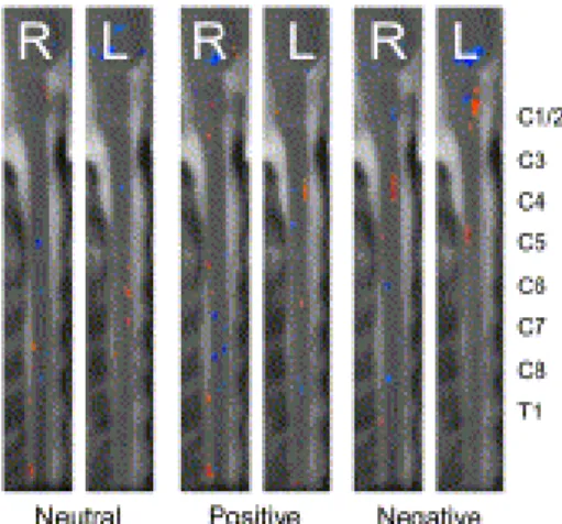
Fig. 1. A group analysis map of activity elicited during the Perception condition in response to the neutral, positive and negative stimuli. Sagittal slices from the right (R) and left (L) sides of the cervical spinal cord are shown with the dorsal aspect of the spinal cord to the left of each image and rostral toward the top of each image. Each slice spans the brainstem and cervical spinal cord. Increases in signal intensity are shown in the orange color scale and decreases in signal intensity are shown in the blue color scale (T
threshold = 2.5).
Fig. 2. A group analysis map of activity elicited during the Movement condition in response to the neutral, positive and negative stimuli. Sagittal slices from the right (R) and left (L) sides of the cervical spinal cord are shown with the dorsal aspect of the spinal cord to the left of each image and rostral toward the top of each image. Each slice spans the brainstem and cervical spinal cord. Increases in signal intensity are shown in orange color scale and decreases in signal intensity are shown in blue color scale (T threshold = 2.5).
Results
Behavioral results
Analysis of participants' responses in the Movement condition demonstrated that reaction times did not differ across conditions: F < 1. It should be noted, however, that participants were not instructed to respond as quickly as possible as it was important that movement was the only difference between the Movement and Perception
conditions. To include a speeded reaction time instruction would have added additional elements to the Movement condition, specifically the effort and the stress of making a rapid response. For this initial attempt at imaging emotion-dependent responses in the
spinal cord, we felt it was important to keep the conditions as ‗process-pure‘ as possible.
Functional MRI results
The analyses of cervical spinal cord activity during the Perception condition
demonstrated activity in response to all stimulus types (Fig. 1). Although the negative emotional stimuli produced more active voxels than the neutral, or the positive, stimuli, the comparison did not reach significance (p = 0.22 for negative vs. neutral, p = 0.99 for positive vs. neutral). The neutral and positive stimulus conditions elicited activity
primarily in the right dorsal and left ventral cervical spinal cord segments. In both conditions, the dorsal activity occurred at more caudal spinal cord segments (C7, C8) than the ventral activity (C5, C6). The negative stimuli elicited activity in the right ventral and left dorsal spinal cord (i.e., a pattern opposite to the other conditions), in the more rostral spinal cord segments (C3, C4, and C5).
The analyses of the cervical spinal cord during the Movement condition also
demonstrated activity in response to all stimulus types (Fig. 2). The analysis of the voxel counts for the negative emotional stimuli revealed that significantly more voxels are active during this condition than during the control (neutral) condition (p = 0.02) (see Table 2). Although the positive emotional stimuli condition also had higher voxel counts than the control condition, this comparison did not reach significance (p = 0.07). During the Movement condition (Fig. 2), the neutral stimuli elicited activity primarily in the right medial dorsal spinal cord in the lower cervical spinal cord segments (C6, C7). The positive stimuli also elicited activity in the right dorsal spinal cord in the same lower cervical spinal cord region, C6 and C7. In contrast, the negative stimulus condition elicited right ventral spinal cord activity centered on the C5 spinal cord segment but spread rostro-caudally to include the C4 and C6 segments (Fig. 3). Activity was also elicited at the lower segments C6 and C7 in the right medial spinal cord and at C8 in the right ventral spinal cord.
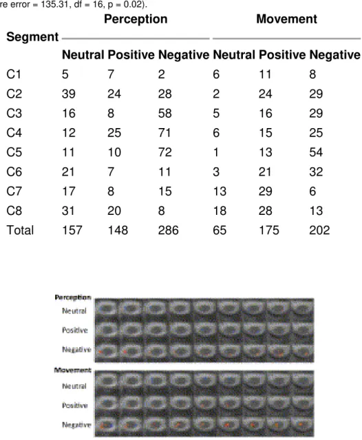
Table 2. Active voxels (t-threshold = 2.5) from each of the approximate spinal cord segments were counted for each of the six conditions. During the Movement condition only, the negative stimuli elicited significantly more active voxels than the neutral stimuli (Dunnett's post hoc test for multiple comparisons, 2-sided, mean square error = 135.31, df = 16, p = 0.02).
Segment
Perception Movement
Neutral Positive Negative Neutral Positive Negative
C1 5 7 2 6 11 8 C2 39 24 28 2 24 29 C3 16 8 58 5 16 29 C4 12 25 71 6 15 25 C5 11 10 72 1 13 54 C6 21 7 11 3 21 32 C7 17 8 15 13 29 6 C8 31 20 8 18 28 13 Total 157 148 286 65 175 202
Fig. 3. The distribution of activity elicited during the Perception (top three rows) and Movement (bottom three rows) conditions displayed on C5 transverse spinal cord slices. This subset of axial slices is from the group analysis and is shown in the identical anatomical location for each condition. Images are oriented in
radiological convention with dorsal toward the bottom of each frame, left side of the cord on the right of each frame, and is displayed rostral-to-caudal from left-to-right of each row (T threshold = 2.5).
The average percent signal change did not significantly differ between conditions (Perception neutral 6.95 +/− 1.55%, Perception positive 7.23 +/− 1.63%, Perception negative 6.93 +/− 1.13%, Movement neutral 7.00 +/− 1.55%, Movement positive 7.28 +/− 1.38%, Movement negative 6.82 +/− 1.56% (mean +/− standard deviation)). These values are consistent with those in the published literature for spinal cord fMRI studies (Stroman, 2005); particularly relevant to the current task, in a cervical spinal cord study involving finger tapping, similar average percent signal changes of 5.5% to 7.6% were observed (Xie et al., 2009).
Our grouped analysis activity maps also reveal a distribution of significantly decreased activity in the cervical spinal cord for all conditions. In the Perception condition,
decreases in activity were observed primarily on the right side of the spinal cord for all tasks. In the Movement condition, decreases in activity were observed bilaterally during the neutral and positive stimulus presentation, and were primarily on the left side of the spinal cord during the negative stimulus presentation. Possible explanations for these data will be discussed below.
Discussion
The key result of the current study is the significant difference in spinal cord activity during motoric responses to negative stimuli than to neutral stimuli. This increased activity along the ventral aspect of the spinal cord, unilaterally, likely represents activity directing movement of the right hand to press the response key. The upper limb is innervated by the brachial plexus that consists of the lower cervical and first thoracic spinal cord nerves, corresponding to spinal cord segments C5–T1. The neurons responsible for movement synapse in the ventral horns of the spinal cord, ipsilateral to the side of the body moved. Therefore, in our study, we would expect to see activity elicited in the right, ventral, C5–T1 spinal cord segments. However, the fact that the physical movements were identical across stimulus types (i.e., a button press) suggests that the different patterns of activation were due to the emotional valence of the
photographs. Thus, negative emotional images elicited enhanced activity in spinal cord neurons during motoric processing, just as they do in the extrastriate cortex during attentional processing (e.g., Surguladze et al., 2003). This valence-dependent firing pattern likely reflects the motoric imperative associated with these different classes of stimuli. From a survival perspective, it is much more important to be able to escape from something that is threatening or noxious than it is to approach something pleasant. In the current study, the negative images were related to threat and disgust, both of which elicit robust avoidance responses in laboratory and electrophysiological studies. Future research is required to determine whether the observed effects vary across different types of negative emotions, or instead reflect a more generalized avoidance system.
Negative emotional stimuli also elicited a qualitatively different pattern of activations in the Perception (passive viewing) condition. Specifically, viewing negative photographs led to significant activity in the left dorsal and right ventral spinal cord, whereas viewing positive or neutral photographs led to activity in the right dorsal and left ventral spinal cord (i.e., opposite patterns). This functional asymmetry in the spinal cord may be related to asymmetrical activity in the amygdala. Substantial data indicate that the left amygdala shows greater, or more consistent, activation in response to fear-relevant stimuli than does the right amygdala (see [Baas et al., 2004] and [Zald, 2003], for reviews). The same stimuli that elicit amygdalar responses also activate the
corticospinal tract ( [Coelho et al., 2010], [Coombes et al., 2009], [Hajcak et al., 2007], [Schutter et al., 2008] and [van Loon et al., 2010]). The corticospinal tract originates in the primary motor cortex, the fibers cross to the other side of the body at the level of the medulla, and travel down the spinal cord in the lateral corticospinal tract to synapse in
the ventral horn in the appropriate spinal cord segment. Therefore, assuming this
corticospinal tract excitability is mediated (to some degree) by the amygdala, the current findings of right ventral spinal cord responses to passively viewed negative stimuli are reasonable.
The idea that negative, potentially threatening, images are processed differently than are other types of stimuli is not novel; indeed, the brain circuitry associated with threat perception is well-established (e.g., LeDoux, 2000). However, this study is the first to suggest that the perception of negative stimuli can cause differential activity as low as the spinal cord. But, while interesting, this result must be viewed with caution. As the participants did not receive specific instructions for how the images should be
perceived, it is possible that the different types of images were processed differently. For instance, threat-related images may have led participants to mentally simulate potential defensive movements, whereas such responses would not be triggered by neutral items. While it is unclear if imagined movements would elicit activity in the spinal cord, this potential qualification is worth noting. This issue will be addressed in future studies in which different – and specific – types of instructions are provided to
participants prior to each experimental run.
Although negative images produced the most robust effects in both conditions of this study, it is interesting to note that positive stimuli also showed enhanced – albeit not significant – activation in comparison to neutral stimuli. This difference is likely due to the emotional arousal associated with the positive images. Indeed, a network involving the amygdala, the parabrachial nucleus of the brainstem, and the vestibular nuclei (see Carmona et al., 2009 for a review) is a possible candidate for an arousal-dependent modulator of the cervical spinal cord. However, given that the effect of positive stimuli merely approached significance, this point remains speculative. Further research involving manipulations of both valence and arousal is necessary to clarify this issue.
A potentially interesting outcome of the current study was the detection of decreased activity during some experimental conditions. It is possible that the decreases observed are due to local spinal circuitry and that we are observing the effect of inhibitory
modulation of interneurons within or across spinal cord segments. Changes in supraspinal neural activity could also account for these decreases at the spinal cord level via changes in descending modulation. It is also possible that we may be observing regions that are tonically active at rest and demonstrate reduced activity during stimulation or during the performance of an experimental task. This may suggest that the spinal cord regions whose activity was anti-correlated to our task are part of a spinal cord default mode network. However, without further investigation into activity anti-correlated to a study paradigm and corroboration via resting state functional scans, these potential explanations remain speculative.
One limitation of the current study was that it was not possible to counterbalance the Perception and Movement conditions—Perception always preceded Movement. This design was necessary in order ensure that the Perception condition was ‗process-pure‘. If participants had responded to emotional images in previous blocks, it would be
impossible to determine if the Perception data reflected perception in isolation, or memory for having responded (i.e., moved) earlier in the experiment. Whether the pattern of activity found in the Movement condition would occur upon subsequent repetitions of the stimulus-movement pairing, or in presentations of the images without requiring a motoric response, is uncertain. Based upon the studies of amygdala
habituation discussed above, one would predict an attenuation of this effect over multiple presentations. However, from a survival perspective, it would be useful for an individual to remember previous emo-motoric responses so that subsequent
experiences with the stimulus elicited more efficient motoric responses (i.e., emotional learning). Although these perspectives appear to predict divergent patterns of data, they may in fact be complementary. We would predict that upon repeated pairing of a
specific stimulus and a motor response, the activity in the lower cervical spinal cord observed in the Movement conditions would decrease. However, this decrease in motor-specific responses would likely be paired with changes in upper cervical spinal cord neurons, thereby demonstrating a change from an immediate response to a preparedness for future emotion-specific actions.
The current results suggest that the role of the spinal cord is more nuanced than was previously thought; it also extends previous theories of spinal cord function which generally limited the spinal cord to a receptive role influencing subjective experiences (e.g., Hohmann, 1966). Although we are not claiming that this structure actually
performs cognitive or emotional functions, its activity does appear to be sensitive to top-down inputs from the brain. Recent evidence also suggests that the spinal cord is involved in bottom-up modulation of brain activity as well. Nicotra et al. (2006) used functional MRI to compare brain activity in patients with spinal cord damage to healthy controls. Spinal cord patients showed reduced activation in brain areas related to the subjective experience of emotions—the subgenual anterior cingulate gyrus and ventromedial prefrontal cortex. Reductions also occurred in an area related to motor responses, the posterior cingulate gyrus. In contrast, these patients showed increased activity in areas related to attention and neural conflict resolution (dorsal anterior cingulate gyrus), auditory and language processing (superior temporal gyrus), and the ―freezing‖ emotional response (periaqueductal gray area). Therefore, entirely different neural networks are activated during emotional responses when the spinal cord is, or is not, able to convey information from the peripheral nervous system to the brain. When paired with the results of the current study, these data suggest that the spinal cord is part of one or more neural networks involved with emotional behaviors and that more studies should be conducted to further characterize its emotional functions.
Conclusions
The aim of the current study was to examine whether the spinal cord itself is uniquely activated during emotional responses and, if so, to delineate these potential spinal cord responses. Consistent with this goal, our analyses showed a spinal cord response to passive viewing of emotional and neutral images. In addition, our analyses detected a greater number of active voxels in the cervical spinal cord segments during active motor responses to emotional images than to neutral images. Critically, significantly more
activity was detected in response to the negative emotional images as compared to the control images. These data provide the first ever empirical evidence of spinal cord neuron responses to emotions, and suggest a preferential action tendency in response to negative, threatening stimuli. This action tendency likely serves an evolutionary purpose, enhancing the ability to perform rapid protective gestures when threatened. Future studies will attempt to link these emotion-dependent spinal cord activations to emotion-specific responses in the brain (e.g., enhanced activity in the extrastriate cortex; Morris et al., 1998) to create a more complete model of the nervous system's responses to emotional stimuli.
Acknowledgments
The authors contributed equally to this research; order of authorship was decided by a coin toss. This research was supported by funding from the Manitoba Medical Services Foundation (MMSF) and the Natural Sciences and Engineering Research Council (NSERC) of Canada. The authors wish to thank Ms. Theresa McIver for research assistance, Drs. Uta Sboto-Frankenstein, Boguslaw Tomanek, Patricia Gervai, and Patrick Stroman for technical assistance, and Randy Summers for statistical assistance.
References
1. F. Agosta, P. Valsasina, D. Caputo, P.W. Stroman, M. Fillippi Tactile-associated recruitment of the cervical cord is altered in patients with multiple sclerosis Neuroimage, 39 (2008), pp. 1542–1548 http://dx.doi.org/10.1016/j.neuroimage.2007.10.048
2. D. Baas, A. Aleman, R.S. Kahn Lateralization of amygdala activation : a systematic review of functional neuroimaging studies Brain Res. Rev., 45 (2004), pp. 96–103 http://dx.doi.org/10.1016/j.brainresrev.2004.02.004
3. J.E. Carmona, A.K. Holland, D.W. Harrison Extending the functional cerebral systems theory of emotion to the vestibular modality: a systematic and integrative approach Psychol. Bull., 135 (2009), pp. 286–302
4. C.M. Coelho, O.V. Lipp, W. Marinovic, G. Wallis, S. Riek Increased corticospinal excitability induced by unpleasant visual stimuli Neurosci. Lett., 481 (2010), pp. 135–138 http://dx.doi.org/10.1016/j.neulet.2010.03.027
5. S.A. Coombes, C. Tandonnet, H. Fujiyama, C.M. Janelle, J.H. Cauraugh, J.J. Summers Emotion and motor preparation: a transcranial magnetic stimulation study of corticospinal motor tract excitability Cogn. Affect. Behav. Neurosci., 9 (2009), pp. 380–388 http://dx.doi.org/10.3758/CABN.9.4.380
6. R.J. Davidson, W. Irwin The functional neuroanatomy of emotion and affective style Trends Cogn. Sci., 3 (1999), pp. 11–21 http://dx.doi.org/10.1016/S1364-6613(98)01265-0 7. N.F. Ghazni, C.C. Cahill, P.W. Stroman Tactile sensory transmission in the human
spinal cord and brainstem Am. J. Neuroradiol., 31 (2010), pp. 661–667 http://dx.doi.org/10.3174/ajnr.A1909
8. G. Hajcak, C. Molnar, M.S. George, K. Bolger, J. Koola, Z. Nahas Emotion facilitates action: a transcranial magnetic stimulation study of motor cortex excitability during picture viewing Psychophysiology, 44 (2007), pp. 91–97
9. G.W. Hohmann Some effects of spinal cord lesions on experienced emotional feelings Psychophysiology, 3 (1966), pp. 143–155 http://onlinelibrary.wiley.com/doi/10.1111/j.1469-8986.1966.tb02690.x/pdf
10. J.A. Kiernan Barr's The Human Nervous System: An Anatomical Viewpoint (seventh ed.)Lippincott-Raven Publishers, Philadelphia, PA, US (1998)
11. J. Kornelsen, P.W. Stroman fMRI of the lumbar spinal cord during a lower limb motor task Magn. Reson. Med., 52 (2004), pp. 411–414 http://dx.doi.org/10.1002/mrm.20157 12. J. Kornelsen, P.W. Stroman Detection of the neuronal activity occurring caudal to the
site of spinal cord injury that is elicited during lower limb movement tasks Spinal Cord, 45 (2007), pp. 485–490 http://dx.doi.org/10.1038/sj.sc.3102019
13. P.J. Lang, M.M. Bradley, B.N. Cuthbert International affective picture system (IAPS): affective ratings of pictures and instruction manual Technical Report A-8, University of Florida, Gainesville, FL (2008)
14. J.M. Lawrence, P.W. Stroman, S.S. Kollias Functional magnetic resonance imaging of the human cervical spinal cord with vibration stimulation of different dermatomes
Neuroradiology, 50 (2008), pp. 273–280 http://dx.doi.org/10.1007/s00234-007-0338-6 15. J.E. LeDoux Emotion circuits in the brain Annu. Rev. Neurosci., 23 (2000), pp. 155–184
http://dx.doi.org/10.1146/annurev.neuro.23.1.155
16. M. Maieron, G.D. Iannetti, J. Bodurka, I. Tracey, P.A. Bandettini, C.A. Porro
Functional responses in the human spinal cord during willed motor actions: evidence for side- and rate-dependent activity J. Neurosci., 27 (2007), pp. 4182–4190
http://dx.doi.org/10.1523/JNEUROSCI.3910-06.2007
17. J.S. Morris, K.J. Friston, C. Büchel, C.D. Frith, A.W. Young, A.J. Calder, R.J. Dolan
A neuromodulatory role for the human amygdala in processing emotional facial expressions Brain, 121 (1998), pp. 47–57
18. J.S. Morris, C.D. Frith, D.I. Perrett, D. Rowland, A.W. Young, A.J. Calder, R.J. Dolan A differential neural response in the human amygdala to fearful and happy facial expressions Nature, 383 (1996), pp. 812–815 http://dx.doi.org/10.1038/383812a0 19. M.C. Ng, K.K. Wong, G. Li, S. Lai, E.S. Yang, Y. Hu, K.D. Luk
Proton-density-weighted spinal fMRI with sensorimotor stimulation at 0.2 T Neuroimage, 29 (2006), pp. 995–999 http://dx.doi.org/10.1016/j.neuroimage.2005.08.011
20. A. Nicotra, H.D. Critchley, C.J. Mathias, R.J. Dolan Emotional and autonomic consequences of spinal cord injury explored using functional brain imaging Brain, 129 (2006), pp. 718–728 http://dx.doi.org/10.1093/brain/awh699
21. M. Oliveri, C. Babiloni, M.M. Filippi, C. Caltagirone, F. Babiloni, P. Cicinelli, R. Traversa, M.G. Palmieri, P.M. Rossini Influence of the supplementary motor area on primary motor cortex excitability during movements triggered by neutral or emotionally unpleasant visual cues Exp. Brain Res., 149 (2003), pp. 214–221
http://dx.doi.org/10.1007/s00221-002-1346-8
22. M.G. Pereira, L. de Oliveira, F.S. Erthal, M. Joffily, I.F. Mocaiber, E. Volchan, L. Pessoa Emotion affects action: midcingulate cortex as a pivotal node of interaction
between negative emotion and motor signals Cogn. Affect. Behav. Neurosci., 10 (2010), pp. 94–106 http://dx.doi.org/10.3758/CABN.10.1.94
23. L. Pessoa To what extent are emotional visual stimuli processed without attention and awareness Curr. Opin. Neurobiol., 15 (2005), pp. 188–196
http://dx.doi.org/10.1016/j.conb.2005.03.002
24. M. Piché, J. Cohen-Adad, M.K. Nejad, V. Perlbarg, G. Xie, G. Beaudoin, H. Benali, P. Rainville Characterization of cardiac-related noise in fMRI of the cervical spinal cord Magn. Reson. Imaging, 27 (2009), pp. 300–310 http://dx.doi.org/10.1016/j.mri.2008.07.019
25. D.A. Pizzagalli, D. Lehmann, A.M. Hendrick, M. Regard, R.D. Pascual-Marqui, R.J. Davidson Affective judgments of faces modulate early activity (~ 160 ms) within the fusiform gyri Neuroimage, 16 (2002), pp. 663–677
http://dx.doi.org/10.1006/nimg.2002.1126
26. D.J.L.G. Schutter, D. Hofman, J. van Honk Fearful faces selectively increase corticospinal motor tract excitability: a transcranial magnetic stimulation study Psychophysiology, 45 (2008), pp. 345–348 http://dx.doi.org/10.1111/j.1469-8986.2007.00635.x
27. R. Stark, A. Schienle, B. Walter, P. Kirsch, C. Blecker, U. Ott, A. Schafer, G.
Sammer, M. Zimmermann, D. Vaitl Hemodynamic effects of negative emotional pictures — a test–retest analysis Neuropsychobiology, 50 (2004), pp. 108–118
http://dx.doi.org/10.1159/000077948
28. P.W. Stroman Magnetic resonance imaging of neuronal function in the spinal cord: spinal FMRI Clin. Med. Res., 3 (2005), pp. 146–156 http://dx.doi.org/10.3121/cmr.3.3.146
29. P.W. Stroman Discrimination of errors from neuronal activity in functional MRI of the human spinal cord by means of general linear model analysis Magn. Reson. Med., 56 (2006), pp. 452–456
30. P.W. Stroman, B. Tomanek, V. Krause, U.N. Frankenstein, K.L. Malisza Mapping of neuronal function in the healthy and injured human spinal cord with spinal fMRI
NeuroImage, 17 (2002), pp. 1854–1860
31. P.W. Stroman, J. Kornelsen, A. Bergman, V. Krause, K. Ethans, K.L. Malisza, B. Tomanek Noninvasive assessment of the injured human spinal cord by means of functional magnetic resonance imaging Spinal Cord, 42 (2004), pp. 59–66
http://dx.doi.org/10.1038/sj.sc.3101559
32. P.W. Stroman, J. Kornelsen, J. Lawrence An improved method for spinal fMRI with large volume coverage of the spinal cord J. Magn. Reson. Imaging, 21 (2005), pp. 520–526 http://dx.doi.org/10.1002/jmri.20315
33. P.W. Stroman, C.R. Figley, C.M. Cahill Spatial normalization and motion correction for functional magnetic resonance imaging of the spinal cord and brainstem Magnetic
Resonance Imaging, 26 (2008), pp. 809–814
34. S.A. Surguladze, M.J. Brammer, A.W. Young, C. Andrew, M.J. Travis, S.C.R. Williams, M.L. Phillips A preferential increase in the extrastriate response to signals of danger Neuroimage, 19 (2003), pp. 1317–1328
http://dx.doi.org/10.1016/S1053-8119(03)00085-5
35. A.M. van Loon, W.P.M. van den Wildenberg, A.H. van Stegeren, G. Hajcak, R.J. Ridderinkhof Emotional stimuli modulate readiness for action: a transcranial magnetic stimulation study Cogn. Affect. Behav. Neurosci., 10 (2010), pp. 174–181
36. C.I. Wright, H. Fischer, P.J. Whalen, S.C. McInerney, L. Shin, S.L. Rauch
Differential prefrontal cortex and amygdala habituation to repeatedly presented emotional stimuli Neuroreport, 12 (2001), pp. 379–383
37. C.H. Xie, K.M. Kong, J.T. Guan, J.X. Chen, R.H. Wu Functional MR imaging of the cervical spinal cord by use of 20 Hz functional electrical stimulation to median nerve Conf. Proc. IEEE Eng. Med. Biol. Soc., 3392–3395 (2007)
http://dx.doi.org/10.1109/IEMBS.2007.4353059
38. C.H. Xie, K. Kong, J. Guan, Y. Chen, J. He, W. Qi, X. Wang, Z. Shen, R. Wu
SSFSE sequence functional MRI of the human cervical spinal cord with complex finger tapping Eur. J. Radiol., 70 (2009), pp. 1–6
39. D.H. Zald The human amygdala and the emotional evaluation of sensory stimuli Brain Res. Rev., 41 (2003), pp. 88–123