Suppressor mutations in Rpf2
–Rrs1 or Rpl5 bypass
the Cgr1 function for pre-ribosomal 5S
RNP-rotation
Matthias Thoms
1
, Valentin Mitterer
1
, Lukas Kater
2
, Laurent Falquet
3
, Roland Beckmann
2
,
Dieter Kressler
3
& Ed Hurt
1
During eukaryotic 60S biogenesis, the 5S RNP requires a large rotational movement to
achieve its mature position. Cryo-EM of the Rix1-Rea1 pre-60S particle has revealed the
post-rotation stage, in which a gently undulating
α-helix corresponding to Cgr1 becomes wedged
between Rsa4 and the relocated 5S RNP, but the purpose of this insertion was unknown.
Here, we show that cgr1 deletion in yeast causes a slow-growth phenotype and reversion of
the pre-60S particle to the pre-rotation stage. However, spontaneous extragenic suppressors
could be isolated, which restore growth and pre-60S biogenesis in the absence of Cgr1.
Whole-genome sequencing reveals that the suppressor mutations map in the Rpf2
–Rrs1
module and Rpl5, which together stabilize the unrotated stage of the 5S RNP. Thus, mutations
in factors stabilizing the pre-rotation stage facilitate 5S RNP relocation upon deletion of Cgr1,
but Cgr1 itself could stabilize the post-rotation stage.
DOI: 10.1038/s41467-018-06660-w
OPEN
1Biochemistry Centre, University of Heidelberg, Heidelberg 69120, Germany.2Gene Center, University of Munich, Munich 81377, Germany.3University of
Fribourg and Swiss Institute of Bioinformatics, Fribourg 1700, Switzerland. These authors contributed equally: Matthias Thoms, Valentin Mitterer. Correspondence and requests for materials should be addressed to M.T. (email:matthias.thoms@bzh.uni-heidelberg.de)
or to E.H. (email:ed.hurt@bzh.uni-heidelberg.de)
123456789
E
ukaryotic ribosome synthesis is a complex and highly
spa-tially and temporally coordinated process that requires the
consecutive action of more than 200 trans-acting assembly
factors to meet the enormous cellular demand for accurately
assembled mature ribosomal subunits
1–5. The biogenesis pathway
starts in the nucleolus with RNA-polymerase-I-catalysed
tran-scription of ribosomal DNA into a large 35S precursor rRNA,
which, upon concomitant and hierarchical joining of ribosome
assembly factors and ribosomal proteins, is embedded into the
huge 90S particle
6–10. Endonucleolytic cleavage of the 35S
pre-RNA subsequently generates the pre-40S and pre-60S particles,
which from that point on undergo individual maturation and
quality-control steps to
finally join again in the cytoplasm
forming translation-competent ribosomes.
The large 60S ribosomal subunit is composed of three rRNA
species (25S/28S, 5.8S and 5S rRNA) and 46 (in yeast) or 47 (in
human) ribosomal proteins
11,12. Once separated from the pre-40S
particles, the
first individual precursors of the 60S subunit are
formed within the nucleolus. Upon binding of ribosomal
pro-teins, the nucleolar pre-60S maturation pathway is initiated by the
appearance of the 27SA
2pre-rRNA that is further processed to
the 27SB pre-rRNA. Concomitantly, the intertwined rRNA
domains are shaped into the developing 60S core in a consecutive
order in which
first the solvent-exposed side, followed by the
polypeptide exit tunnel (PET) and
finally the inter-subunit side
are formed
13–16. At the stage of nucleolar maturation
inter-mediates, the 5S ribonucleoprotein particle (5S RNP), consisting
of the 5S rRNA and ribosomal proteins Rpl5 (also known as
uL11) and Rpl11 (also known as uL18), is already recruited, and
the characteristic pre-60S
‘foot’ structure surrounding the internal
transcribed spacer 2 (ITS2) RNA fragment has already
formed
14,17–20. Crucial pre-60S remodelling events, such as the
removal of the Erb1–Ytm1 complex by the AAA–ATPase Rea1,
facilitate the transition of the particle to the nucleoplasm
14,21–23.
A hallmark structure on early nucleoplasmic maturation
inter-mediates, isolated via Arx1 or Nog2 (also known as Nug2), is the
twisted 5S RNP, which adopts a conformation rotated ~180°
compared to mature 60S subunits
18–20. The recruitment of the
Rix1 subcomplex, which allows stable docking of Rea1, and
the removal of assembly factors Rpf2 and Rrs1 occur during the
rotation of the 5S RNP into a near-mature conformation
20,24.
Subsequently, Rea1 performs its second restructuring role by
triggering the release of Rsa4
25. Prior to nuclear export,
con-formational proofreading of the particle takes places that links the
removal of Rsa4 with activation and release of the GTPase Nog2,
which in turn allows the recruitment of the export adaptor
Nmd3
26. After nuclear export, the AAA–ATPase Drg1 initiates
the cytoplasmic maturation cascade by releasing the placeholder
protein Rlp24, thus permitting the recruitment of Rpl24 (also
known as eL24)
27,28. Subsequent cytoplasmic pre-60S maturation
steps include the Rei1–Jjj1–Ssa1-dependent dissociation of the
export factor Arx1
29,30, assembly of the P-stalk and incorporation
of Rpp0 (also known as uL10)
31,32, removal of Nmd3 by the
GTPase Lsg1 coupled to the incorporation of Rpl10 (also known
as uL16)
33–35, and release of the anti-association factor Tif6
promoted by Efl1 and Sdo1
30,36, which
finally activates the 60S
subunit to enter the pool of functionally translating ribosomes.
Whereas the 35S pre-rRNA is the common precursor of three
of the four rRNA species (18S, 5.8S, 25S/28S), the 5S rRNA
precursor is transcribed separately by RNA polymerase III. The
5S rRNA subsequently associates with the ribosomal proteins
Rpl5 and Rpl11 to form the 5S RNP that is incorporated as a
prefabricated complex adopting an immature conformation on
the pre-60S particle
17,19. Nuclear import of Rpl5 and Rpl11 is
coordinated by the adaptor protein Syo1, which, in a second
function, chaperones the 5S RNP until its pre-ribosomal assembly
by shielding exposed RNA-binding sites on Rpl11
37,38. In
addi-tion, the heterodimer Rpf2–Rrs1 is thought to guide 5S RNP
incorporation by providing a docking platform that anchors the
5S RNP in a network of interactions around the central
protu-berance (CP) involving the 25S rRNA and assembly factor
Rsa4
39–41. Therefore, the Rpf2–Rrs1 complex has to dissociate
from the pre-60S particle, a reaction that appears to be necessary
for 5S RNP relocation. However, to date, the mechanistic details
of the events that trigger 5S RNP rotation have remained
unexplored.
Here, we show that the small and conserved protein Cgr1,
which was implicated in 60S biogenesis
20,42,43, plays a role in the
relocation of the 5S RNP during 60S biogenesis. We found that
yeast cells with a chromosomal cgr1 deletion (cgr1Δ)—resulting in
a slow-growth phenotype—exhibit a 5S RNP maturation defect
on pre-60S particles. However, specific suppressor mutations
could be isolated that map in genes encoding Rpf2, its binding
partner Rrs1, and the ribosomal protein Rpl5. Owing to the
nature of these suppressor mutations, which bypass Cgr1’s
function in this process, we were able to gain insight into the
mechanism of 5S RNP rotation, revealing how untying of the
twisted 5S RNP from its surrounding assembly factor network
can drive 5S RNP rotation.
Results
Cgr1 marks pre-60S particles during 5S RNP rotation.
Cryo-EM analysis of the Rix1–Rea1 pre-60S particle showed that the 5S
RNP had already rotated ~180° to its near-mature position
24,
whereas in the
‘upstream’ pre-60S particles, such as the early Arx1
particle or Nog2 particle, the 5S RNP was still in the unrotated
topology
19,20. Among the many other structural peculiarities, the
Rix1–Rea1 particle exhibited a 114 Å long, slightly undulating,
α-helix inserted between the
β-propeller domain of Rsa4 and the
rotated 5S RNP, thereby clamping H38 of the 25S rRNA (A-site
finger) at a new position (Fig.
1
a)
24. We suspected that this
α-helix corresponds to the small, 120-amino-acid-long protein Cgr1
(Fig.
1
b), which has been suggested to perform a role in pre-60S
biogenesis
42–44. Consistent with this interpretation, Gao and
colleagues identified this long α-helix as Cgr1 in the early
(unrotated 5S RNP) and late (rotated 5S RNP) states of their
Nog2 pre-60S particles that resemble the early Arx1 and
Rix1-Rea1 particles, respectively
20.
To
find out with which pre-60S particles Cgr1 interacts, we first
affinity purified both N- and C-terminally tagged Cgr1 from
whole yeast cell lysates via TAP–Flag or Flag–TEV–ProtA
(FTpA), respectively. Consistent with a predominantly
nucleo-lar/nuclear localization of GFP–Cgr1 (Fig.
1
c), the two different
Cgr1 purifications were co-enriched for ribosome assembly
factors that are typically present on intermediate pre-60S particles
(i.e. Nog2, Rix1 and Arx1), and, accordingly, Cgr1 was not found
on early nuclear (Ssf1 and Nsa1) or later cytoplasmic (Lsg1)
particles (Fig.
1
d, e, Supplementary Fig. 1a, b).
Cgr1 depletion stalls the pre-60S prior to 5S RNP rotation. To
study the in vivo role of CGR1 during 60S maturation, a cgr1Δ
null strain was generated. Earlier data indicated that CGR1 is
either essential or non-essential for cell growth, depending on the
strain background
42,43. In our laboratory yeast strain, W303
45,
CGR1 is a non-essential gene, but displays an extreme
slow-growth phenotype at all tested temperatures (23, 30 and 37 °C)
(Fig.
2
a). To analyse such a near-essential phenotype in a
con-trolled way, we generated an auxin-inducible degron (AID)
46allele of CGR1, which efficiently targeted Cgr1 for proteasomal
degradation within 30–45 min of auxin addition (Supplementary
Fig. 2a). This CGR1–HA–AID strain did not display an obvious
growth defect when incubated in the absence of auxin
(Supple-mentary Fig. 2b), but exhibited a very mild half-mer phenotype,
which could be due to the HA–AID-tag at the C-terminus
(Fig.
2
b). However, the polysome profile of the cells after
auxin-dependent Cgr1–HA–AID depletion showed a drastic increase of
the half-mer phenotype, consistent with previous
findings
42and
indicative of a severe 60S biogenesis defect (Fig.
2
b). Moreover,
robust nuclear accumulation of the 60S reporter Rpl25–GFP was
observed upon Cgr1 depletion, suggesting that the 60S
matura-tion defect occurs prior to nuclear export (Fig.
2
c).
Next, we wished to
find out where exactly Cgr1 participates in
the nuclear pre-60S maturation pathway. Since Cgr1 is closely
intertwined with the interaction network around the CP,
adopting considerably different conformations depending on
the rotation state of the 5S RNP
20, we hypothesized that the
protein could function at a maturation step during 5S RNP
relocation. To assess whether 5S RNP maturation might be
affected in absence of Cgr1, we compared the assembly factor
profile of Arx1-derived pre-60S particles, isolated from
non-depleted (−auxin) versus Cgr1-non-depleted (+auxin) cells (Fig.
2
d).
Since Arx1 is associated with a broad range of pre-60S
intermediates, from nuclear to cytoplasmic particles
18,19,47, it
can serve as a bait to define the stage of pre-60S arrest by
biochemical means. To allow monitoring of the 5S RNP
maturation stage of the isolated particles, we used a strain
expressing a chromosomal Rpf2–3xHA fusion, which is
c
GFP-Cgr1 DIC TAP-Flag-Cgr1Cgr1-FTpA Rea1d
kDa 200 150 120 100 85 70 60 50 40 30 25 20 15e
Cgr1-3xHASsf1 Nsa1 Nog2 Rix1 Arx1 Lsg1
1 2 3 4 5 6 α Nog2 α Rpl3 α HA (Cgr1) α Nog1 kDa 200 150 120 100 85 70 60 50 40 30 25 20 15 Sda1 Rix1 Nop7 Nog1 Arx1, Nug1 Nop53, Nog2, Rsa4 Nsa3 Rpl3 Rpf2, Rlp7 Rpl4 Rpl5 Nsa2, Nop15 Mrt4, Rlp24 r-proteins M M
b
Sc / 1-120 Sp / 1-111 Kl / 1-121 Yl / 1-123 Ct / 1-123 Nc / 1-123 Sc / 1-120 Sp / 1-111 Kl / 1-121 Yl / 1-123 Ct / 1-123 Nc / 1-123a
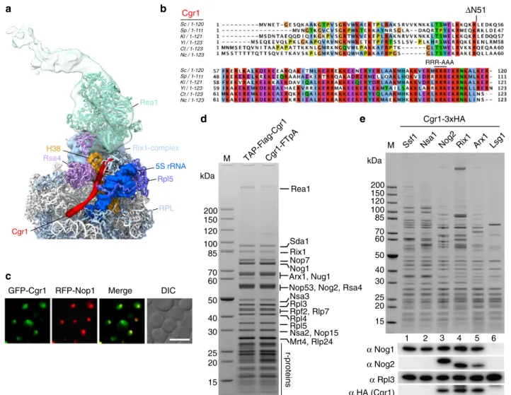
Cgr1 RFP-Nop1 Merge Rsa4 Rea1 Rix1-complex Rpl5 RPL Cgr1 5S rRNA H38 ΔN51 RRR-AAAFig. 1 The shortα-helical protein Cgr1 is wedged on nucleoplasmic pre-60S particles close to the rotated 5S RNP. a Cryo-EM position of Cgr1 wedged between theβ-propeller domain of Rsa4 and the rotated 5S RNP on the Rix1–Rea1 pre-ribosomal particle (PDB: 5jcs,24). The 5S rRNA (blue) and H38 of the 25S rRNA (orange) are shown as a surface models, Cgr1 (red), Rsa4 (purple), Rea1 (cyan), Rix1-complex (light blue), Rpl5 (dark blue), and other ribosomal proteins (RPL, light blue) are depicted.b Multiple sequence alignment of Cgr1 orthologues from different fungal species: Saccharomyces cerevisiae (Sc), Schizosaccharomyces pombe (Sp), Kluyveromyces lactis (Kl), Yarrowia lipolytica (Yl), Chaetomium thermophilum (Ct) and Neurospora crassa (Nc); for the sequence alignment with higher eukaryotic orthologues including human Cgr1, see Supplementary Fig. 9. Two mutant constructs, Cgr1ΔN51 and Cgr1RRR > AAA, used for genetic interaction studies, are indicated above the alignment.c Subcellular distribution in yeast cells of GFP-tagged Cgr1 and RFP-Nop1 was monitored byfluorescence microscopy. The localization of GFP–Cgr1 is distributed over the nucleus, with the tendency to show a slightly stronger signal in the nucleolus. Scale bar is 5µm. d, e Cgr1 is co-enriched on intermediate pre-60S particles typically found in the nucleus. d Cgr1 tagged either N-terminally (TAP–Flag) or C-terminally (FTpA) were isolated from yeast lysates in two affinity-purification steps. The final Flag eluates were analysed by SDS-PAGE followed by Coomassie staining. The bands identified by mass spectrometry are indicated. e Different pre-60S particles affinity purified via bait proteins Ssf1–FTpA (early nucleolar), Nsa1–FTpA (early nucleolar), Nog2–FTpA (intermediate nucleoplasmic), Rix1–FTpA (intermediate nucleoplasmic), Arx1–FTpA (intermediate nucleoplasmic to late cytoplasmic) and Lsg1–FTpA (late cytoplasmic) were affinity purified from yeast strains, which expressed Cgr1 carrying 3xHA (Cgr1–3xHA). Final eluates were analysed by SDS-PAGE and Coomassie staining (upper panel) or western blotting, using the indicated antibodies detecting Nog1, Nog2, Rpl3 and Cgr1 (lower panels). M: molecular weight marker
functional based on growth (Supplementary Fig. 1c), as it was
suggested that the presence of the assembly factor Rpf2 in
complex with its binding partner Rrs1 hinders 5S RNP
rotation
17,20,39–41. Indeed, western blot analyses revealed that
Rpf2–3xHA became significantly enriched on Arx1 particles
isolated from Cgr1-depleted cells in comparison to non-depleted
cells (Fig.
2
d), indicating Cgr1 might facilitate 5S RNP relocation.
Consistent with this
finding, Cgr1 depletion caused a significant
reduction of late-acting pre-60S factors (e.g. Yvh1, Rei1, Nmd3),
whereas earlier assembly factors (e.g. Rsa4, Nog2, Nsa2, Mrt4,
Rlp24) became more enriched (Fig.
2
d). In contrast, the foot
factors Nop7 and Nsa3 (also known as Cic1) were reduced on
cgr1Δ 23°C 30°C 37°C CGR1 Vectora
CGR1-HA-AID CGR1-HA-AID +auxin 40S60S 80S Polysomes Top Bottomb
DIC Rpl25-GFP RFP-Nop1 Merge
DIC Rps3-GFP RFP-Nop1 Merge +Auxin CGR1-HA-AID +Auxin
c
SDC SDC+FOA SDC SDC+FOA nug1-1 nug1-1 NUG1 NUG1 NUG1 nug1-1 NOP7 NOP7 NOP7 nop7-1 nop7-1 nop7-1 cgr1ΔN51 CGR1 cgr1ΔN51 cgr1Δ nug1Δ CGR1 CGR1 NSA2 NSA2 NSA2 nsa2-1 cgr1ΔN51 cgr1RRR>AAA CGR1 cgr1Δ nsa2Δ nsa2-1 nsa2-1 cgr1ΔN51 cgr1RRR>AAA SDC+FOAe
CGR1 RIX1 RIX1 RIX1 rix1-1 cgr1ΔN51 cgr1RRR>AAA CGR1 cgr1Δ rix1Δ rix1-1 rix1-1 cgr1ΔN51 cgr1RRR>AAA SDC SDC+FOA CGR1 CGR1 cgr1ΔN51 cgr1RRR>AAA cgr1ΔN51 cgr1RRR>AAA cgr1Δ nop7Δ cgr1RRR>AAA cgr1RRR>AAA Arx1-FTpA Cgr1-HA-AID Rpf2-3xHAKap121 Sda1 Rix1 Nop7 Nog1 Arx1-Flag Nsa3 Rpl4 Rpl5 Nsa2 Nug1 Nmd3, Nog2, Rsa4, Nop53 Rei1 Yvh1, Rlp7 Ipi1 Rpf2-3xHA Rpl3, Cgr1-HA-AID 200 150 120 100 85 70 60 50 40 30 25 20 15 10 kDa Eluate Lysate Arx1-Flag HA Short exposure Cgr1-HA-AID Rpf2-3xHA Cgr1-HA-AID Rpf2-3xHA Long exposure Rsa4 Nog2 Mrt4 Nsa2 Nop7 Nog1 Rlp24 Nug1 Yvh1 Nmd3 Rei1 Rpl3 Rpl5 Rps3 Arc1 – + Auxin – + Rea1 Eluate Rpp0
d
SDC Auxin–
+*
Cgr1-depleted particles, indicating that ITS2 processing and
removal of the foot structure could proceed uncoupled of 5S RNP
rotation.
Genetic interactions between cgr1 and pre-60S factors. Next, we
performed genetic analyses to further elucidate the in vivo
function of Cgr1. For this purpose, we generated
‘milder’ cgr1
mutant alleles compared to the cgr1 null by truncating either the
N-terminus (cgr1ΔN51) or mutating a cluster of positively
charged residues at the C-terminus (R108A, R109A, R110A,
cgr1RRR > AAA). The latter motif contacts a part of the 5S rRNA
in the pre-rotation state
20. Both of these cgr1 mutants grew well at
30 °C compared to the cgr1-null, but exhibited a
temperature-sensitive phenotype at 37 °C (Supplementary Fig. 3a, b).
Com-bining cgr1ΔN51 or cgr1RRR > AAA with mutant alleles of other
pre-60S assembly factors revealed a synthetic lethal phenotype at
30 °C in the case of rix1-1, nsa2-1 and nug1-1, but not with
nop7-1 (Fig.
2
e). The Rix1 subcomplex is implicated in the initiation of
5S RNP rotation
24, and an
α-helix in the Nug1 N-terminal
domain is in direct contact with and the Nsa2 N-domain in close
proximity to Cgr1
19,20, whereas Nop7 is located far away at the
‘foot’ of the pre-60S particle
20. Thus, the observed genetic
rela-tionships correlate well with the biochemical and cryo-EM data,
reinforcing Cgr1’s role in 5S RNP relocation.
Speci
fic suppressor mutations bypass the function of Cgr1.
During the course of growing the cgr1Δ strain on plates, we
consistently noticed a few fast-growing colonies in the
high-cell-density streak-out, which among other possibilities could be
spontaneous suppressors that bypass the requirement for CGR1
(Fig.
3
a). To further elaborate on this possibility, we performed
clarifying genetic tests with these putative suppressors. First, we
backcrossed a few of these suppressor strains to a haploid cgr1Δ
strain of opposite mating type, which harboured wild-type CGR1
on a URA3-containing plasmid. After sporulation and tetrad
dissection, the four germinated cgr1Δ spores containing
URA3-CGR1 plasmid showed a 2
+:2
−segregation regarding slow versus
fast growth on 5-fluoroorotic acid (5-FOA) plates (Fig.
3
b).
Apparently, the fast-growth-suppressor phenotype points to a
single mutated gene locus responsible for the extragenic
suppression.
This
finding prompted us to perform whole-genome DNA
sequencing of two selected suppressor strains that upon
back-crossing showed a 2:2 segregation (see above). Strikingly, in both
strains a single missense mutation (G227A and C84F) in the open
reading frame of the RPF2 gene was found. The G227A mutation
mapped to the conserved sigma-70-like motif found in all
members of the Brix protein family
48, whereas the C84F mutation
is found in a conserved region known to be involved in the
interaction with Rrs1
40. Thus, the identified mutations, together
with the observed accumulation of Rpf2 on pre-ribosomes after
Cgr1 depletion, establish a direct link between Cgr1 and 5 S RNP
maturation.
To
find out whether suppressor mutations in genes other than
RPF2 exist, we systematically analysed the remaining cgr1Δ
suppressors in a different way. For this purpose, we expressed the
wild-type allele of RPF2 and other factors suspected to
functionally interact with Cgr1 on the pre-60S particles (i.e.
RRS1, RPL5 and RPL11; all placed under GAL1-10 control) in all
41 cgr1Δ suppressor strains (39 remained uncharacterized) and
tested for reversion of the fast-growing phenotype. Strikingly,
overexpression of RPF2 changed 30 suppressors, RRS1 six
suppressors and RPL5
five suppressors into a slow-growth
phenotype, suggesting that all of our isolated suppressor strains
were hit in only three genes (Fig.
3
c). Cloning and DNA
sequencing of these suppressor genes revealed single point
mutations in RPF2 (25 unique exchanges), RRS1 (four unique
exchanges) and RPL5 (four unique exchanges) (Table
1
).
To confirm that the cloned suppressor alleles behave like
anticipated, double-shuffle strains were generated, in which cgr1Δ
was
finally combined with the given cloned suppressor allele. This
genetic analysis revealed that all identified suppressor alleles
complemented the severe growth defect of cgr1Δ mutant cells,
although wild-type growth levels were not reached (Fig.
3
d–f,
Supplementary Fig. 4).
cgr1Δ suppressor mutations within the pre-60S structure. We
sought to localize the suppressor mutations within the cryo-EM
structure of pre-60S particles, where the 5S RNP is still unrotated
and in direct contact with the Rpf2–Rrs1 heterodimer
19,20(Fig.
4
,
Supplementary Figs. 5–7 and Table
1
). For Rpf2, where a total of
25 different suppressor mutations were isolated, three mutations
(A10E, R14I, K18T) map in an N-terminal
α-helix interacting
with H83 and H87 of 25S rRNA, whereas the remaining ones are
distributed throughout the Brix-fold, which broadly participates
in the interaction with both the 5S RNP and Rrs1 (Fig.
4
,
Sup-plementary Fig. 5). Several of these mutations showed
substitu-tions of surface-exposed basic residues that change the
electrostatic surface potential. Notably, surface-exposed basic
amino acid clusters within Rpf2 were recently analysed in vitro,
demonstrating that highly conserved R236, the R62–K63 cluster
and the KKR loop (residues 94–96) are important for 5S rRNA
binding
39. Strikingly, the Rpf2 R62L/S, K63T and R236G/I
Fig. 2 Cgr1 plays a crucial role in ribosome biogenesis of pre-60S particles. a Chromosomal CGR1 deletion in wild-type yeast strain W303 yields viable cells with an extreme slow-growth phenotype. The cgr1Δ shuffle strains transformed with empty plasmid or plasmid carrying wild-type CGR1 were shuffled on SDC+ FOA plates, before representative colonies were spotted in 10-fold serial dilutions on YPD plates. They were grown at the indicated temperatures for 2 days.b Cgr1 depletion impairs 60 S subunit synthesis. Polysome-profiles of CGR1–HA–AID (i.e. Cgr1-depletion strain) were recorded for untreated or auxin-treated (for 120 min) cells. Arrows denote ribosomal half-mers, indicating a specific 60S biogenesis defect. c pre-60S export is inhibited in cells depleted of Cgr1. Subcellular localization of the 60S reporter Rpl25–GFP, the 40S reporter Rps3–GFP and the nucleolar marker RFP–Nop1 was analysed in untreated or auxin-treated (for 120 min) CGR1-HA-AID cells. Arrows indicate nuclear accumulation of Rpl25–GFP. Scale bar is 5 µm. d Depletion of Cgr1 shifts Arx1 pre-60S particles to the early pool typical for the unrotated 5S RNP. Arx1–FTpA particles were affinity purified from untreated or auxin-treated (for 120 min) CGR1–HA–AID cells expressing a chromosomally integrated RPF2–3xHA variant. Lysates serving as input for the purifications and final eluates were analysed by SDS-PAGE and Coomassie staining. Indicated bands were identified by mass spectrometry (left panel, asterisk indicates Rpf2-3xHA) and western blotting based on specific antibodies (right panel). Rpf2 carries a 3xHA tag, whereas only one HA epitope is fused to Cgr1, explaining the different signal intensities of the HA western blots.e Synthetic lethal relationship between cgr1 mutant alleles and distinct pre-60S assembly factors. Double-shuffle strains of cgr1Δ in combination with nsa2Δ, rix1Δ, nug1Δ and nop7Δ, respectively, were co-transformed with indicated plasmid-based wild type and mutant constructs. Transformants were spotted in 10-fold serial dilutions and growth on SDC-Leu-Trp (SDC) and SDC+ FOA plates at 30 °C was monitored after 2 and 6 days, respectively. The cgr1RRR > AAA and cgr1ΔN51 mutants are shown in Fig.1b and Supplementary Fig. 3. Published mutant alleles nsa2-1, rix1-1, nug1-1 and nop7-1 are listed in Supplementary Table 3pGAL1-10-RRS1 RPF2 RPL5 RPL11 Vector Vector RRS1 RPF2 Glucose Galactose Wild-type Supp #19 Supp #3 Supp #1 2 days 3 days
c
SDC SDC+FOA CGR1 CGR1 CGR1 RPF2 RPF2 rpf2 G227A rpf2 G227A CGR1 CGR1 CGR1 RRS1 RRS1 rrs1 E102D rrs1 E102D CGR1 CGR1 CGR1 RPL5 RPL5 rpl5 I190F rpl5 I190F rpf2Δ cgr1Δ cgr1Δ rrs1Δ cgr1Δ cgr1Δ cgr1Δ cgr1Δ rpl5Δ CGR1 CGR1 RPF2 rpf2 G227A rpf2 G227A 23°C 30°C 37°C CGR1 CGR1 RRS1 rrs1 E102D rrs1 E102D CGR1 CGR1 RPL5 rpl5 I190F rpl5 I190F cgr1Δ cgr1Δ cgr1Δ G227A ES loop3 I190F E102D Rpf2 Rrs1 Rpl5b
Tetrad Spore 1 2 3 4 Spore 1 Spore 2 Spore 3 Spore 4SDC SDC+FOA -Ura -His Nat
a
cgr1Δ CGR1 Empty vector YPD 23°C 30°C 37°C YPD 23°C 30°C 37°C YPDe
f
d
RRS1 RPF2 RPL5 RPL11 Vector Vector RRS1 RPF2 Rpf2 Rrs1 Rpl5 240 263 246 240 235 229 231 231 128 S.c. C.t. N.c. K.l. Y.l. C.e. M.m. H.s. S.c. C.t. N.c. K.l. Y.l. C.e. M.m. H.s. S.c. C.t. N.c. K.l. Y.l. C.e. M.m. H.s. P.h. H.h. 119 118 128 126 104 128 128 210 213 213 210 210 209 210 210 155 154 198 224 207 198 169 190 192 192 86 77 76 86 84 62 86 86 168 171 171 168 168 167 168 168 126 125mutations were all among our cgr1Δ suppressors. Although no
mutations in the highly conserved KKR motif were found, the
suppressor mutation Rpf2 S93F is within this KKR loop as well,
which may be destabilized by the S93F change (Fig.
4
,
Supple-mentary Fig. 5), and hence could be the cause of a reduced
interaction with the 5S rRNA. Consistent with this data, specific
point mutations in the 5S rRNA tip, mediating the interaction
with the Rpf2 KKR loop, strongly impaired the interaction
between Rpf2–Rrs1 and the 5S rRNA
41. Other suppressor
mutations in Rpf2, such as D48Y, D112Y or H180N, are found
within the Brix-domain fold and eventually destabilize the Rpf2–
Rrs1 interaction (Fig.
4
, Supplementary Fig. 5).
In the case of Rrs1, all identified mutations are clustered in a
highly conserved, proline-rich unstructured region (residues
92–108), which protrudes from the Rpf2 interaction-domain
and continues into the carboxy-terminal sequence that contacts
the 25S rRNA at multiple sites (Fig.
4
, Supplementary Fig. 6),
thereby also stabilizing the unrotated 5S RNP
20,40. In vitro, both
the proline-rich region and the C-terminal end of Rrs1 are not
required for complex formation with Rpf2
39, but, in the cryo-EM
structure, the proline-rich region is in contact with the Brix1-fold
domain of Rpf2 (Fig.
4
, Supplementary Fig. 6). Therefore, it is
conceivable that our identified suppressor mutations in the
proline-rich Rrs1 loop might change the position of the Rrs1
C-terminus, and thereby destabilize the unrotated 5S RNP.
Interestingly, three of our discovered suppressor mutations
map to the ribosomal protein Rpl5 (E126K, E128K, I190F),
specifically in two of the three eukaryote-specific loop regions
required for 60S biogenesis, which if deleted cause trapping of
Rpf2–Rrs1 on pre-60S particles
49. In particular, the I190F
mutation is located in eukaryotic-specific loop 3 (residues 185–
198) that bridges Rpl5 with the Rsa4
β-propeller and the twisted
5S rRNA, whereas the mutations E126K and E128K map to the
neighbouring eukaryote-specific loop 2 (residues 122–138), which
is not resolved in the cryo-EM structure, but most likely connects
the
β-propeller of Rsa4 and Rpf2 (Fig.
4
, Supplementary Fig. 7).
In contrast, the fourth suppressor mutation within Rpl5 (V73F)
maps to a conserved short loop motif sandwiched between the 5S
rRNA and Rpf2 (Fig.
4
, Supplementary Fig. 7).
Thus, considering all these different suppressor mutations in
the structural context of the pre-60S particle, they likely
destabilize the intricate interaction network between Rpf2–Rrs1,
Rsa4 and the unrotated 5S RNP, which consequentially could
allow driving the equilibrium towards the rotated state of the 5S
RNP, thus compensating for the absence of Cgr1.
Suppressor mutants promote 5S RNP rotation in cgr1
Δ strains.
Based on the structural interpretation of the various cgr1Δ
sup-pressors, we examined the impact of a few of these mutations on
pre-60S maturation. First, we determined the localization of the
60S export reporter Rpl25–GFP in the mutants rpf2V203F,
rrs1E102D and rpl5I190F, which all confer a strong suppression
Table 1 Comparison of the isolated cgr1
Δ suppressor
mutations
Protein Point mutation Interaction/role Rpf2 (30 isolated
suppressor strains)
A10E (2 strains) 25S rRNA (CP H87) R14I (2 strains) 25S rRNA (CP H87) K18T 25S rRNA (CP H87) D48Y Folding K53R Folding K54E 5S rRNA R62L 5S rRNA/25S rRNA R62S 5S rRNA/25S rRNA K63T 5S rRNA/25S rRNA N64K 5S rRNA/25S rRNA K81N 25S rRNA K81T (2 strains) 25S rRNA C84F Folding C84W Folding S93F 5S rRNA (KKR loop) R104L Rrs1, folding D112Y (3 strains) Folding
M117V 5S rRNA (KKR loop), folding
G177R Rpl5/Rrs1, folding
H180N Folding
V203F Rrs1, folding
G227A Rrs1 (sigma70-like motif), folding G227V Rrs1 (sigma70-like motif), folding R236G 5S rRNA R236I 5S rRNA Rrs1 (6 isolated suppressor strains) L92H Rpf2 E102D Rpf2 K103N (3 strains) Rpf2 P106Q Rpf2 Rpl5 (5 isolated suppressor strains) V73F (2 strains) 5S rRNA/Rpf2 E126K Rsa4/Rpf2 (ES loop 2) E128K Rsa4/Rpf2 (ES loop 2) I190F Rsa4/5S rRNA (ES loop 3)
CP= central protuberance; ES = eukaryote-specific
Fig. 3 Suppressor mutations in RPF2, RRS1 and RPL5 bypass the requirement for CGR1. a Dot spot growth analyses of the cgr1Δ strain, harbouring plasmid-borne CGR1 (left panel) or empty plasmid (middle and right panels), incubated on YPD plates at 30 °C for 3 days. The dot spot on the right, but not the middle, exhibits faster-growing colonies, which are suppressors of cgr1Δ. b cgr1Δ (cgr1::natNT2) suppressor strain was crossed with a cgr1Δ (cgr1:: HIS3MX6) strain containing CGR1 on a URA3 plasmid (pRS316-CGR1). After sporulation and tetrad dissection (upper panel shows a representative tetrad), the four haploid spores were tested for growth in the absence of pRS316-CGR1 on SDC+ FOA plates, for the presence of pRS316-CGR1 on SDC-Ura, and for the presence of the CGR1 gene disruption markers on SDC-His and YPD+ clonNat. Cells were spotted in 10-fold serial dilutions and incubated at 30 °C for 2 days (lower panel).c–f Suppressor mutations are located in genes encoding RPF2, RRS1 and RPL5. c Wild type and different cgr1Δ suppressor strains (suppressor #1, #3 and #19) were transformed with plasmids expressing RPF2, RRS1, RPL5 or RPL11 under the control of the galactose-inducible GAL1-10 promoter. Representative transformants were spotted in 10-fold serial dilutions on SDC plates containing glucose (GAL repression) and galactose (GAL induction) and growth was assessed after incubation at 30 °C for 2 and 3 days, respectively.d, e Double-shuffle strains of cgr1Δ ( + pURA3-CGR1) combined with rpf2Δ ( + pURA3-RPF2), rrs1Δ ( + pURA3-RRS1) and rpl5Δ ( + pURA3-RPL5), respectively, were transformed with plasmids harbouring the suppressor allele or the respective wild-type gene combined with plasmids harbouring wild-type CGR1 or empty plasmid. Transformants were spotted in 10-fold serial dilutions on SDC+ FOA plates (d) and after plasmid shuffling on YPD plates (e). Growth was analysed after incubation for 2 days at the indicated temperatures.f Multiple sequence alignment of Rpf2, Rrs1 and Rpl5 orthologues from Saccharomyces cerevisiae (S.c.), Chaetomium thermophilum (C.t.), Neurospora crassa (N.c.), Kluyveromyces lactis (K.l.), Yarrowia lipolytica (Y.l.), Caenorhabditis elegans (C.e.), Mus musculus (M.m.), Homo sapiens (H.s.), Pyrococcus horikoshii (P.h.) and Halobacterium hubeiense (H.h.). The respective suppressor alleles analysed ind and e are indicated
phenotype on cgr1Δ. In contrast to wild-type RPF2, RRS1 and
RPL5 cells, which display nuclear accumulation of Rpl25–GFP
after auxin-dependent Cgr1–HA–AID depletion, efficient nuclear
export of Rpl25–GFP was re-established in the respective
sup-pressor strains, which is clear-cut evidence for resuming pre-60S
biogenesis (Fig.
5
a). Moreover, we analysed the assembly factor
composition of Arx1-affinity purified pre-60S particles derived
from the rpf2V203F, rrs1E102D and rpl5I190F suppressor
mutants, before and after Cgr1 depletion (Fig.
5
b). In all cases, the
pattern of factor enrichment on and removal from the Arx1
particles was consistent with our previous interpretation that
nuclear export of pre-60S subunits was re-established in cgr1Δ
cells by specific mutations in Rpf2, Rrs1 and Rpl5 (Fig.
5
b).
Notably, the assembly factor Rpf2, which became enriched on
Arx1 pre-60S particles upon Cgr1 depletion (see also above),
co-purified similar to the wild-type condition in the suppressor
mutants (Fig.
5
b). This
finding further strengthens the hypothesis
that the bypassing function of the suppressors could be
specifi-cally connected to a step in the course of 5S RNP relocation.
To directly assess whether 5S RNP rotation is inhibited in
pre-60S particles when Cgr1 is depleted, but restored in the
suppressor mutants, we performed cryo-EM analysis (Fig.
6
,
Supplementary Fig. 8 and Supplementary Table 1). This method
showed that in the Cgr1 non-depleted strain (Arx1–FTpA Cgr1–
HA–AID, no auxin), which served as control, the 5S RNP was
rotated in ~40% of the Arx1 particles, whereas ~60% of particles
exhibited the non-rotated stage (Fig.
6
a). This ratio is typical for
the distribution of rotated (mature) versus non-rotated
(imma-ture) 5 S RNP in Arx1 or Nog2 pre-60S particles
19,20. Strikingly,
the 5S RNP remained to 100% non-rotated in the cgr1-depletion
mutant (Fig.
6
b; Arx1–FTpA Cgr1–HA–AID, + auxin). However,
5S RNP relocation was significantly restored in the rrs1E102D
suppressor strain, showing 23% of the Arx1 pre-60S particles in
the post-rotation stage (Fig.
6
c; Arx1–FTpA Cgr1–HA–AID
rrs1E102D,
+ auxin). Thus, structural analysis also supports the
view that the suppressor mutations facilitate 5S RNP rotation in
the absence of Cgr1, which explains well why the suppressor
strains can re-export pre-60S particles and regain better cell
growth. However, suppressor mutants did not reach optimal
growth (see also Fig.
3
e), which may correlate with the degree of
the 5S RNP relocation. Notably, the cryo-EM analysis further
revealed that the foot structure, carrying the ITS2 fragment of the
7S pre-rRNA and associated assembly factors, was absent from
the Cgr1-depleted Arx1 particles (Fig.
6
b). This
finding is in line
with the biochemical data demonstrating a strong decrease of foot
factors Nop7 and Nsa3 on these particles (see Figs.
2
d and
5
b),
which suggests that maturation of the foot can proceed
independent of 5S RNP maturation.
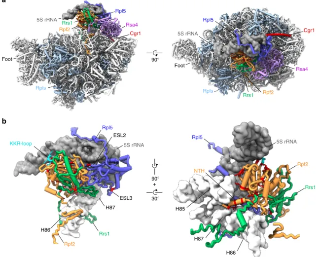
90° Foot Cgr1 Rpls Rsa4 Rpl5 Rpf2 Rpl5 Rsa4 5S rRNA Foot Rpls Rrs1 5S rRNA 30° 90° + H86 H87 KKR-loop Rpl5 Rpf2 ESL2 H85 H86 H87 5S rRNA Rpf2 Rrs1 Rpl5 NTH ESL3 Cgr1 Rpf2 Rrs1 5S rRNA Rrs1a
b
Fig. 4 cgr1Δ suppressor mutations in RPF2, RRS1 and RPL5 destabilize the unrotated 5S RNP on the pre-60S particle. a Overview of biogenesis factors Rpf2 (orange), Rrs1 (green), Rsa4 (purple), Cgr1 (red) and the ribosomal protein Rpl5 (dark blue) on the Nog2 particle (PDB: 3jct,20) in the front and top view. Ribosomal proteins are shown in light blue and 5S rRNA as a dark grey surface modelfiltered at 6 Å resolution. b Positions of cgr1Δ null suppressor mutations (red) displayed with surface models of the 5S rRNA (dark grey) and helices H83 to H87 (nucleotides 2650–2754) of the 25S rRNA (light grey). The KKR-loop of Rpf2 is highlighted in cyan. ESL2 and ESL3 mark the eukaryotic-specific loops 2 and 3 of Rpl5, NTH marks the N-terminal helix of Rpf2
Discussion
In this study, we unveiled a function of the small conserved
α-helical protein Cgr1 (Supplementary Fig. 9) in 5S RNP rotation
during 60S biogenesis, which occurs in the nucleus prior to
nuclear export. Previous
findings have shown that Cgr1 decorates
nuclear pre-60S particles, which are in the process of 5S RNP
rotation
20,24. Due to its topological positioning, Cgr1 can ideally
influence progression through this maturation step, by either
affecting the transition stage to overcome the rotational block or
by stabilizing the rotated stage. Consistent with this
interpreta-tion, pre-60S particles are shifted back to the pre-rotational stage
in cgr1Δ cells, thus identifying the arrest of 60S maturation as a
possible cause of the severe slow-growth phenotype of the
cgr1-null mutant. However, this defect can be overcome by second-site
revertants (i.e. extragenic suppressor mutations), which allow
resumption of cell growth. Strikingly, all the isolated suppressor
mutations map in only three factors—Rpf2, Rrs1 and Rpl5—
which normally under wild-type conditions keep the 5S RNP on
pre-60S particles in the pre-rotation stage. Thus, the identified
suppressor mutations hint to the mechanism by which cells can
re-locate the 5S RNP during 60S biogenesis in the absence of
Cgr1. Accordingly, depletion of Cgr1 results in inhibition of 5S
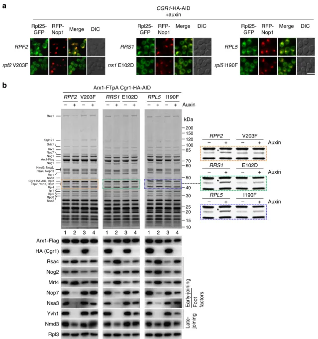
Arx1-FTpA Cgr1-HA-AID RPF2 V203F – + – + 200 150 120 100 85 70 60 50 40 30 25 20 15 10 RPL5 I190F Auxin – + – + RRS1 E102D – + – + kDa Kap121 Rea1 Sda1 Rix1 Nop7 Nog1 Arx1-Flag Nsa3 Cgr1-HA-AID, Rpl3 Rlp7, Yvh1, Rpf2 Rpl4 Rpl5 Nsa2 Nug1 Nmd3, Nog2, Rsa4, Nop53 Rei1 Ipi1 Arx1-Flag HA (Cgr1) Rpl3 Nmd3 Mrt4 Rsa4 Nog2 1 2 3 4 1 2 3 4 1 2 3 4 Rpl25-GFPRFP-Nop1 Merge DIC
RPF2 rpf2 V203F CGR1-HA-AID +auxin RRS1 rrs1 E102D RPL5 rpl5 I190F Rpl25-GFP RFP-Nop1 Merge DIC Rpl25-GFP RFP-Nop1 Merge DIC RPF2 V203F Auxin RPL5 I190F Auxin RRS1 E102D Auxin – + – + – + – + – + – + Rpp0 Nop7 Nsa3 Yvh1 Foot factors Late- joining Early-joining
*
*
*
a
b
Fig. 5 Suppressor mutations in Rpf2, Rrs1 and Rpl5 rescue the 60S biogenesis defect in Cgr1-depleted cells. a Nuclear pre-60S export is restored in suppressor mutants after Cgr1 depletion. Subcellular location of Rpl25–GFP and RFP–Nop1 (nucleolar marker) was examined in CGR1–HA–AID cells expressing either wild-type RPF2, RRS1 and RPL5 or the respective mutant alleles rpf2V203F, rrs1E102D and rpl5I190F, after incubation with auxin for 120 min. Scale bar is 5µm. b Biochemical maturation of Arx1 pre-60S particles is restored in cgr1Δ suppressor mutants. Arx1–FTpA particles were affinity purified from CGR1–HA–AID cells expressing either wild-type RPF2, RRS1 or RPL5, or the indicated suppressor mutants before and after treatment with auxin for 120 min. Final eluates were analysed by SDS-PAGE and Coomassie staining (indicated bands were identified by mass spectrometry; upper panels) or by western blotting using the antibodies shown on the left (lower panels). The area of the Coomassie-stained SDS-polyacrylamide gel to which Rpf2 migrates is enlarged on the right to better reveal how the intensity of co-enriched Rpf2 changes, depending on Cgr1 depletion in the various suppressor mutants
RNP rotation, but suppressor mutations mapping in factors
sta-bilizing the pre-rotational stage of the 5S RNP allow to partly
overcome this defect.
As previously observed in the cryo-EM structure of the Rix1–
Rea1 particle, the gently undulating C-terminal
α-helix of Cgr1 is
wedged between the rotated 5S RNP, the relocated A-site
finger
H38 and the
β-propeller domain of Rsa4, thereby stabilizing the
rotated 5S in a position that hinders back rotation
24. In the
‘early’
state 1 of Nog2 pre-60S particles (resembling the
‘early’ pool of
Arx1 pre-60S particles), in which the 5S RNP is non-rotated, the
binding sites for Cgr1 are very different compared to those of
the rotated stage
19,20. Specifically, Cgr1 is located on the solvent
side in the pre-rotation stage, contacting H38 as well as one tip of
the unrotated 5S RNP, whereas after 5S RNP rotation, the Cgr1
α-helix adopts a more straightened conformation and is clamped
between Rsa4 and the 5S RNP, thereby holding the relocated H38
in a bent position on the inter-subunit side. This rearranged
topology suggests that Cgr1 accompanies or even facilitates H38
relocation from the solvent to the inter-subunit side. In addition,
by snapping in after relocation, Cgr1 could stabilize the rotated 5S
RNP position. It is tempting to speculate that upon initiation of
the 5S RNP rotational movement, potentially induced by the
recruitment of the Rix1 subcomplex
24, the contact between the 5S
RNP and Cgr1’s C-terminal α-helix gets temporarily
dis-connected, which could allow H38 to slide under the detached
Cgr1 C-terminus. During the subsequent 5S RNP rotation, the
straightened and co-rotating Cgr1
α-helix could continuously
exert pressure on H38, which helps to bring it into the new
position at the inter-subunit interface. Interestingly, in bacteria, a
key role for H38 in the maturation of the CP was postulated
50.
Depletion of the circularly permuted GTPase YlqF allowed the
isolation of late ribosome assembly intermediates with an
immature CP, which was highly disordered with no obvious
structured intermediate, in contrast to the stable arrangement of
the 5S RNP in the
‘early’ Arx1 particle. Nevertheless, it was shown
that in these particles, H38 also adopts different orientations, and
it was suggested that re-orientation of the A-site
finger is a
pre-requisite for stable CP formation.
In summary, this study provided mechanistic insight into the
5S RNP rotation during large subunit biogenesis and its coupling
to pre-60S nuclear export. This could be achieved by combining
classical yeast genetic methods with modern whole-genome
high-throughput sequencing, which appears to be an effective
approach to further unravel the complicated process of eukaryotic
ribosome assembly.
Methods
Yeast strains and plasmids. The Saccharomyces cerevisiae strains used in this study were derived from W30345and are listed in Supplementary Table 2. Strains were constructed by using established gene disruption, genomic tagging51,52, mating and tetrad dissection methods. Shuffle strains were constructed by knocking out an essential gene in a diploid yeast strain, transformation with a URA3 plasmid containing the respective wild-type gene and sporulation to gen-erate haploids harbouring the gene knockout and the complementing URA3 plasmid. Subsequently, double-shuffle strains containing knockouts of cgr1 and an essential gene (as indicated in the respectivefigures) complemented by two URA3 plasmids harbouring the corresponding wild-type genes were generated by crossing
Foot Ø Ø Foot Ø Ø 5S 5S 5S 5S 5S Ø Pre-rotation 38.9% Pre-rotation 38.1% Post-rotation 23.0% CGR1 cgr1Δ cgr1Δ rrs1E102D Ø Ø 5S 5S Pre-rotation 100% Post-rotation 22.9% Post-rotation 16.7% Pre-rotation 60.4%
a
b
c
Fig. 6 Cryo-EM reveals inhibition of 5S RNP rotation in cgr1Δ cells but restoration in rrs1E102D suppressor mutant. a–c 3D cryo-EM reconstructions of pre-60S particles affinity purified via the Arx1 bait protein from the indicated yeast strains. a CGR1 control strain: rrs1Δ [YCplac111–RRS1] Arx1–FTpA Cgr1–HA–AID; -auxin. b cgr1Δ depleted: rrs1Δ [YCplac111–RRS1] Arx1–FTpA Cgr1–HA–AID; + auxin (2 h). c cgr1Δ depleted in the presence of the rrs1E102D suppressor: rrs1Δ [YCplac111–rrs1E102D] Arx1–FTpA Cgr1–HA–AID + auxin (2 h). For each obtained class of the respective data set, the rotation state of the 5S is indicated by afit model of 5S rRNA taken from PDB: 3jct (green: pre-rotation,20) or PDB: 5jcs (blue: post-rotation,24). Also, the presence or absence of the ITS2-harbouring foot structure is indicated
of two in thefirst step generated shuffle strains with opposing mating types and subsequent sporulation and identification of haploids containing both knockouts and both URA3 plasmids (i.e. spores containing both selection markers used for the two knockouts, fast-growing on plates lacking uracil, and non-viable on 5-FOA containing plates).
The plasmids used in this study are listed in Supplementary Table 3 and were constructed according to standard DNA cloning techniques and verified by sequencing.
Identification of suppressors by high-throughput sequencing. The two sup-pressor mutants and the CGR1 shuffle strain (parental control strain) were grown in YPD medium to an OD600value of around 1, and cells corresponding to 20
OD600units were harvested by centrifugation. Genomic DNA was extracted
essentially as described in Current Protocols in Molecular Biology53. After washing in dH2O, cells were transferred to a 2.2 ml safe-seal Eppendorf tube, centrifuged
again and resuspended in 200 µl breaking buffer [100 mM NaCl, 10 mM Tris-HCl (pH 8), 1 mM EDTA (pH 8), 2% Triton X-100, 1% SDS]. After addition of 0.3 g glass beads and 200 µl phenol–chloroform–isoamyl alcohol (49.5:49.5:1; Sigma), cells were broken by vigorous vortexing for 3 min. Then, 200 µl of TE buffer [10 mM Tris-HCl (pH 7.5), 1 mM EDTA (pH 8)] was added and the tubes were briefly vortexed. Tubes were centrifuged for 5 min at 13,500 rpm in an Eppendorf centrifuge and the aqueous upper phase was transferred to a 1.5 ml Eppendorf tube. Then, 1 ml of absolute ethanol was added and the contents of the tubes were mixed by inversion. Following centrifugation for 3 min at 13,500 rpm, the supernatant was removed and the pellet was resuspended in 400 µl of TE buffer. To digest the RNA, 30 µl of a 1 mg/ml DNase-free RNase A solution (Sigma) was added and the tubes were incubated for 5 min at 37 °C. Genomic DNA was then precipitated upon addition of 10 µl of 5 M ammonium acetate and 1 ml of absolute ethanol. After mixing by inversion, the tubes were centrifuged for 3 min at 13,500 rpm and the supernatant was discarded. Finally, the air-dried pellet was resuspended in 100 µl of TE buffer. To estimate the integrity of the isolated genomic DNA, 2.5 µl of the preparation was migrated on a 1% agarose gel. The concentration of the genomic DNA was determined with a Qubit 2.0fluorimeter (Invitrogen).
Libraries were generated from 1 µg of genomic DNA and high-throughput sequencing was performed on a HiSeq 3000 instrument (Illumina). Library preparation and Illumina sequencing were carried out by the Next Generation Sequencing (NGS) Platform of the University of Bern. The raw reads (paired-end reads of 150 bp) were processed according to the following procedure: after performing a quality check with FastQC v0.11.2 (https://www.bioinformatics. babraham.ac.uk/projects/fastqc/), all the reads werefiltered for quality (minimum of 20), truncated to 100 bp with Sickle v1.2954and then mapped with BWA-MEM v0.7.1055to the S. cerevisiae reference genome R64-1-1.79 (strain S288C) obtained from Ensembl56. The SAMfiles were sorted and converted to BAM files with SAMtools v1.257. Single-nucleotide variants (SNVs), as well as small insertions and deletions (Indels), were identified with SAMtools and BCFtools v1.2757. Variant annotation was added with SnpEff v4.358. Then, variants werefiltered with SnpSift59to retain homozygous variants that are not found in the parental control strain and that are not‘synonymous’ or ‘intergenic’, leading to an annotated and curated Variant Call Format (VCF)file. Results were viewed with the Integrative Genomics Viewer (IGV) software60. Deletion of the CGR1 gene was verified visually using IGV. Our sequence analysis revealed 13 variants for the three genomes and unambiguously identified one single-nucleotide change within the RPF2 gene in each suppressor strain.
Yeast affinity purification. Two-step affinity purifications were performed with either N-terminally TAP–Flag- or C-terminally Flag–TEV–proteinA (FTpA)-tag-ged bait proteins. The respective yeast strains were grown in 2 l of YPD medium at 30 °C, harvested in the logarithmic growth phase,flash frozen in liquid nitrogen and stored at−80 °C. Where indicated in the figures, cultures were incubated in the presence of 0.5 mM auxin (3-indoleacetic acid, Sigma–Aldrich) for 120 min prior to harvesting the cells. Cell pellets were resuspended in‘lysis buffer’ [50 mM Tris-HCl (pH 7.5), 100 mM NaCl, 5 mM MgCl2, 0.05% NP-40, 1 mM DTT,
sup-plemented with 1 mM PMSF, 1 × SIGMAFAST protease inhibitor (Sigma– Aldrich)], and cells were ruptured by shaking in a bead beater (Fritsch) in the presence of glass beads. Lysates were cleared by two subsequent centrifugation steps at 4 °C for 10 and 30 min at 5000 and 14,000 rpm, respectively. Supernatants were incubated with immunoglobulin G (IgG) Sepharose 6 Fast Flow beads (GE Healthcare) on a rotating wheel at 4 °C for 90 min. Beads were transferred into Mobicol columns (Mobitec) and, after washing with 10 ml of lysis buffer, cleavage with tobacco etch virus (TEV) protease was performed at 16 °C for 120 min. In a second purification step, TEV eluates were incubated with Flag agarose beads (ANTI-FlagM2 Affinity Gel, Sigma–Aldrich) for 60 min at 4 °C. After washing with 5 ml of lysis buffer, bound proteins were eluted with lysis buffer containing 300 µg/ml Flag peptide at 4 °C for 45 min. Buffer lacking NP-40 was used for the last purification step in samples used for cryo-EM. Flag eluates were analysed by SDS-PAGE on 4–12% polyacrylamide gels (NuPAGE, Invitrogen) with colloidal Coomassie staining (Roti-blue, Roth) or by western blotting with antibodies, as indicated in the respectivefigures. Uncropped gel and western blot images are shown in Supplementary Fig. 10.
Cryo electron microscopy. Cryo electron microscopy was performed for three different purifications: (1) rrs1Δ [YCplac111–RRS1] Arx1–FTpA Cgr1–HA–AID; -auxin. (2) cgr1Δ depleted: rrs1Δ [YCplac111–RRS1] Arx1–FTpA Cgr1–HA–AID; + auxin (2 h). (3) cgr1Δ depleted in the presence of the rrs1E102D suppressor: rrs1Δ [YCplac111–rrs1E102D] Arx1–FTpA Cgr1–HA–AID + auxin (2 h).
For each purification, Quantifoil holy carbon grids (R3/3, +2 nm carbon) were glow discharged at 2.2*10^-1 torr for 20 s. Then for each grid, 3.5 µl of sample concentrated to 1.8 OD260/ml was applied and plunge frozen in liquid ethane using
a vitrobot mark IV (FEI), operating at 5 °C and 90% humidity, blotting for 2 s after a 45 s incubation. For each sample 400 micrograph were recorded on a Tecnai Spirit (FEI) operating at 120 kV, equipped with a TEMCam F216 (TVIPS, Germany). Semi-automated micrograph acquisition was performed using the EM-Tools software suite (TVIPS, Germany).
Image processing. GCTF61was used to estimate the contrast transfer function parameters. Micrographs with a defocus in the range of 0.8–3.2 μm were used for further processing. Template free particle picking was performed with Gautomatch (http://www.mrc-lmb.cam.ac.uk/kzhang). All further image processing (classifica-tions, refinements, and post processing) was performed using Relion-2.162, ana-logously for all data sets as described in the following. First, the particle sets were cleaned using reference free 2D classification to eliminate falsely picked particles. Then, a consensus refinement was performed using EMD-319924as a reference. To address structural heterogeneity, multiple subsequent steps of alignment free 3D classification was performed. After every classification step, similar classes were joined and all remaining classes were refined and subsorted to check for additional heterogeneity (see Supplementary Fig. 8). For the cgr1Δ depleted sample, all clas-sification attempts failed to separate the particles into subsets with structurally distinguishable features, resulting in onefinal class.
Western blotting. Western blot analysis was performed using the following antibodies: anti-Nog1 antibody (1:5000), anti-Nog2 antibody (1:20,000), anti-Arx1 antibody (1:2000), anti-Rei1 antibody (1:10,000), anti-Nsa2 antibody (1:10,000), anti-Rlp24 antibody (1:2000), provided by Micheline Fromont-Racine, anti-Nug1 antibody (1:10,000), Yvh1 antibody (1:4000), provided by Vikram Panse, anti-Nmd3 antibody (1:10,000), anti-Rpl10 antibody (1:10,000), provided by Arlen Johnson, anti-Rpl3 antibody (1:5000), provided by Jonathan Warner, anti-Rpl5 antibody (1:10.000), provided by John Woolford, anti-Nop7 antibody (1:50,000), provided by Bruce Stillman, anti-Rsa4 antibody (1:10,000), provided by Miguel Remacha, anti-Mrt4 antibody (1:1000), provided by Juan Pedro Ballesta, anti-Arc1 antibody (1:5000), raised in our lab, anti-HA antibody (1:10,000, Covance Research Products, MMS-101R), horseradish-peroxidase-conjugated anti-Flag antibody (1:15,000, Sigma–Aldrich, A8592), secondary horseradish-peroxidase-conjugated goat anti-rabbit antibody (1:2000, Bio-Rad-170-6515), secondary horseradish-peroxidase-conjugated goat anti-mouse antibody (1:2000, Bio-Rad-170-6516). Polysome profile analyses. Cells expressing chromosomal C-terminal fusions of Cgr1 tagged with HA–AID (CGR1–HA–AID) were grown in YPD medium to early logarithmic growth phase. Prior to harvesting, cultures were incubated with 0.5 mM auxin for 120 min to induce proteasomal degradation of Cgr1–HA–AID or left untreated. Subsequently, 100μg/ml cycloheximide was added and after incu-bation for 10 min on ice, cells were pelleted and washed once with lysis buffer [50 mM Tris-HCl (pH 7.5), 100 mM KCl, 12 mM MgCl2, 100μg/ml cycloheximide].
After resuspension in lysis buffer and cell lysis with glass beads, 6 A260units of the
cell extracts were loaded onto 10–50% sucrose gradients [dissolved in 50 mM Tris-HCl (pH 7.5), 100 mM KCl, 12 mM MgCl2] and centrifuged with a SW40 rotor
(Beckman Coulter) at 39,000 rpm for 2 h 45 min at 4 °C. Gradients were analysed on a Foxy Jr. fraction collector (Teledyne ISCO) with continuous monitoring at 254 nm.
Fluorescence microscopy. Living yeast cells expressing GFP- or RFP-tagged proteins were grown to the logarithmic growth phase and imaged byfluorescence microscopy using a Zeiss Imager Z1 microscope. As indicated, auxin was added to afinal concentration of 0.5 mM and cells were subsequently incubated for 120 min prior to imaging.
Data availability
All relevant data supporting thefindings of this study can be found in the results or the supplementary information section and are available from the corresponding authors upon request. All experiments were performed at least twice with similar outcome. Cryo-EM densities of maps 1-7 of the Arx1 particles have been deposited in the Electron Microscopy Data Bank and can be retrieved using the following accession codes, respectively: EMDB-0218, EMDB-0219, EMDB-0220, EMDB-0221, EMDB-0222, EMDB-0223, EMDB-0224.
References
1. de la Cruz, J., Karbstein, K. & Woolford, J. L. Functions of ribosomal proteins in assembly of eukaryotic ribosomes in vivo. Annu. Rev. Biochem. 84, 93–129 (2015).
2. Kressler, D., Hurt, E. & Bassler, J. Driving ribosome assembly. Biochim. Biophys. Acta 1803, 673–683 (2010).
3. Kressler, D., Hurt, E. & Baßler, J. A puzzle of life: crafting ribosomal subunits. Trends Biochem. Sci. 42, 640–654 (2017).
4. Peña, C., Hurt, E. & Panse, V. G. Eukaryotic ribosome assembly, transport and quality control. Nat. Struct. Mol. Biol. 24, 689–699 (2017).
5. Woolford, J. L. & Baserga, S. J. Ribosome biogenesis in the yeast Saccharomyces cerevisiae. Genetics 195, 643–681 (2013).
6. Chaker-Margot, M., Hunziker, M., Barandun, J., Dill, B. D. & Klinge, S. Stage-specific assembly events of the 6-MDa small-subunit processome initiate eukaryotic ribosome biogenesis. Nat. Struct. Mol. Biol. 22, 920–923 (2015). 7. Chaker-Margot, M., Barandun, J., Hunziker, M. & Klinge, S. Architecture of
the yeast small subunit processome. Science 355, eaal1880 (2017). 8. Cheng, J., Kellner, N., Berninghausen, O., Hurt, E. & Beckmann, R.
3.2-Å-resolution structure of the 90S preribosome before A1 pre-rRNA cleavage. Nat. Struct. Mol. Biol. 24, 954–964 (2017).
9. Kornprobst, M. et al. Architecture of the 90S pre-ribosome: a structural view on the birth of the eukaryotic ribosome. Cell 166, 380–393 (2016). 10. Sun, Q. et al. Molecular architecture of the 90S small subunit pre-ribosome.
Elife 6, e22086 (2017).
11. Ben-Shem, A. et al. The structure of the eukaryotic ribosome at 3.0 Å resolution. Science 334, 1524–1529 (2011).
12. Khatter, H., Myasnikov, A. G., Natchiar, S. K. & Klaholz, B. P. Structure of the human 80S ribosome. Nature 520, 640–645 (2015).
13. Gamalinda, M. et al. A hierarchical model for assembly of eukaryotic 60S ribosomal subunit domains. Genes Dev. 28, 198–210 (2014).
14. Kater, L. et al. Visualizing the assembly pathway of nucleolar Pre-60S ribosomes. Cell 171, 1599–1610 (2017). e14.
15. Sanghai, Z. A. et al. Modular assembly of the nucleolar pre-60S ribosomal subunit. Nature 556, 126–129 (2018).
16. Zhou, D. et al. Cryo-EM structure of an early precursor of large ribosomal subunit reveals a half-assembled intermediate. Protein Cell.https://doi.org/ 10.1007/s13238-018-0526-7(2018).
17. Zhang, J. et al. Assembly factors Rpf2 and Rrs1 recruit 5S rRNA and ribosomal proteins rpL5 and rpL11 into nascent ribosomes. Genes Dev. 21, 2580–2592 (2007).
18. Bradatsch, B. et al. Structure of the pre-60S ribosomal subunit with nuclear export factor Arx1 bound at the exit tunnel. Nat. Struct. Mol. Biol. 19, 1234–1241 (2012).
19. Leidig, C. et al. 60S ribosome biogenesis requires rotation of the 5S ribonucleoprotein particle. Nat. Commun. 5, 3491 (2014).
20. Wu, S. et al. Diverse roles of assembly factors revealed by structures of late nuclear pre-60S ribosomes. Nature 534, 133–137 (2016).
21. Bassler, J. et al. The AAA-ATPase Rea1 drives removal of biogenesis factors during multiple stages of 60S ribosome assembly. Mol. Cell 38, 712–721 (2010).
22. Thoms, M., Ahmed, Y. L., Maddi, K., Hurt, E. & Sinning, I. Concerted removal of the Erb1-Ytm1 complex in ribosome biogenesis relies on an elaborate interface. Nucleic Acids Res. 44, 926–939 (2016).
23. Wegrecki, M., Rodríguez-Galán, O., de la Cruz, J. & Bravo, J. The structure of Erb1-Ytm1 complex reveals the functional importance of a high-affinity binding between twoβ-propellers during the assembly of large ribosomal subunits in eukaryotes. Nucleic Acids Res. 43, 11017–11030 (2015). 24. Barrio-Garcia, C. et al. Architecture of the Rix1-Rea1 checkpoint machinery
during pre-60S-ribosome remodeling. Nat. Struct. Mol. Biol. 23, 37–44 (2016). 25. Ulbrich, C. et al. Mechanochemical removal of ribosome biogenesis factors
from nascent 60S ribosomal subunits. Cell 138, 911–922 (2009).
26. Matsuo, Y. et al. Coupled GTPase and remodelling ATPase activities form a checkpoint for ribosome export. Nature 505, 112–116 (2014).
27. Kappel, L. et al. Rlp24 activates the AAA-ATPase Drg1 to initiate cytoplasmic pre-60S maturation. J. Cell Biol. 199, 771–782 (2012).
28. Pertschy, B. et al. Cytoplasmic recycling of 60S preribosomal factors depends on the AAA protein Drg1. Mol. Cell. Biol. 27, 6581–6592 (2007).
29. Greber, B. J. et al. Insertion of the biogenesis factor Rei1 probes the ribosomal tunnel during 60S maturation. Cell 164, 91–102 (2016).
30. Lo, K.-Y. et al. Defining the pathway of cytoplasmic maturation of the 60S ribosomal subunit. Mol. Cell 39, 196–208 (2010).
31. Kemmler, S., Occhipinti, L., Veisu, M. & Panse, V. G. Yvh1 is required for a late maturation step in the 60S biogenesis pathway. J. Cell Biol. 186, 863–880 (2009).
32. Lo, K.-Y., Li, Z., Wang, F., Marcotte, E. M. & Johnson, A. W. Ribosome stalk assembly requires the dual-specificity phosphatase Yvh1 for the exchange of Mrt4 with P0. J. Cell Biol. 186, 849–862 (2009).
33. Hedges, J., West, M. & Johnson, A. W. Release of the export adapter, Nmd3p, from the 60S ribosomal subunit requires Rpl10p and the cytoplasmic GTPase Lsg1p. EMBO J. 24, 567–579 (2005).
34. Ma, C. et al. Structural snapshot of cytoplasmic pre-60S ribosomal particles bound by Nmd3, Lsg1, Tif6 and Reh1. Nat. Struct. Mol. Biol. 24, 214–220 (2017).
35. Malyutin, A. G., Musalgaonkar, S., Patchett, S., Frank, J. & Johnson, A. W. Nmd3 is a structural mimic of eIF5A, and activates the cpGTPase Lsg1 during 60S ribosome biogenesis. EMBO J. 36, 854–868 (2017).
36. Weis, F. et al. Mechanism of eIF6 release from the nascent 60S ribosomal subunit. Nat. Struct. Mol. Biol. 22, 914–919 (2015).
37. Calviño, F. R. et al. Symportin 1 chaperones 5S RNP assembly during ribosome biogenesis by occupying an essential rRNA-binding site. Nat. Commun. 6, 6510 (2015).
38. Kressler, D. et al. Synchronizing nuclear import of ribosomal proteins with ribosome assembly. Science 338, 666–671 (2012).
39. Asano, N. et al. Structural and functional analysis of the Rpf2-Rrs1 complex in ribosome biogenesis. Nucleic Acids Res. 43, 4746–4757 (2015).
40. Kharde, S., Calviño, F. R., Gumiero, A., Wild, K. & Sinning, I. The structure of Rpf2-Rrs1 explains its role in ribosome biogenesis. Nucleic Acids Res. 43, 7083–7095 (2015).
41. Madru, C. et al. Chaperoning 5S RNA assembly. Genes Dev. 29, 1432–1446 (2015).
42. Moy, T. I., Boettner, D., Rhodes, J. C., Silver, P. A. & Askew, D. S. Identification of a role for Saccharomyces cerevisiae Cgr1p in pre-rRNA processing and 60S ribosome subunit synthesis. Microbiol. (Read., Engl.) 148, 1081–1090 (2002).
43. Sun, J. et al. Cgr1p, a novel nucleolar protein encoded by Saccharomyces cerevisiae orf YGL0292w. Curr. Microbiol. 42, 65–69 (2001).
44. Tarassov, K. et al. An in vivo map of the yeast protein interactome. Science 320, 1465–1470 (2008).
45. Thomas, B. J. & Rothstein, R. Elevated recombination rates in transcriptionally active DNA. Cell 56, 619–630 (1989).
46. Nishimura, K., Fukagawa, T., Takisawa, H., Kakimoto, T. & Kanemaki, M. An auxin-based degron system for the rapid depletion of proteins in nonplant cells. Nat. Methods 6, 917–922 (2009).
47. Nissan, T. A., Bassler, J., Petfalski, E., Tollervey, D. & Hurt, E. 60S pre-ribosome formation viewed from assembly in the nucleolus until export to the cytoplasm. EMBO J. 21, 5539–5547 (2002).
48. Wehner, K. A. & Baserga, S. J. The sigma(70)-like motif: a eukaryotic RNA binding domain unique to a superfamily of proteins required for ribosome biogenesis. Mol. Cell 9, 329–339 (2002).
49. Baßler, J. et al. A network of assembly factors is involved in remodeling rRNA elements during preribosome maturation. J. Cell Biol. 207, 481–498 (2014). 50. Li, N. et al. Cryo-EM structures of the late-stage assembly intermediates of the
bacterial 50S ribosomal subunit. Nucleic Acids Res. 41, 7073–7083 (2013). 51. Janke, C. et al. A versatile toolbox for PCR-based tagging of yeast genes: new
fluorescent proteins, more markers and promoter substitution cassettes. Yeast 21, 947–962 (2004).
52. Longtine, M. S. et al. Additional modules for versatile and economical PCR-based gene deletion and modification in Saccharomyces cerevisiae. Yeast 14, 953–961 (1998).
53. Ausubel, F. M. et al. Current Protocols in Molecular Biology 2 13.11.1–13.11.4 (John Wiley & Sons, Inc, New York, NY, 1994).
54. Joshi, N. A. and Fass, J. N. (2011). Sickle: A sliding-window, adaptive, quality-based trimming tool for FastQfiles (Version 1.33) [Software]. 26 April 2018 Available athttps://github.com/najoshi/sickle
55. Li, H. & Durbin, R. Fast and accurate long-read alignment with Burrows-Wheeler transform. Bioinformatics 26, 589–595 (2010).
56. Yates, A. et al. Ensembl 2016. Nucleic Acids Res. 44, D710–D716 (2016). 57. Li, H. A statistical framework for SNP calling, mutation discovery, association
mapping and population genetical parameter estimation from sequencing data. Bioinformatics 27, 2987–2993 (2011).
58. Cingolani, P. et al. A program for annotating and predicting the effects of single nucleotide polymorphisms, SnpEff: SNPs in the genome of Drosophila melanogaster strain w1118; iso-2; iso-3. Fly. (Austin) 6, 80–92 (2012). 59. Cingolani, P. et al. Using Drosophila melanogaster as a model for genotoxic
chemical mutational studies with a new program, SnpSift. Front. Genet. 3, 35 (2012).
60. Thorvaldsdóttir, H., Robinson, J. T. & Mesirov, J. P. Integrative Genomics Viewer (IGV): high-performance genomics data visualization and exploration. Brief. Bioinforma. 14, 178–192 (2013).
61. Zhang, K. Gctf: Real-time CTF determination and correction. J. Struct. Biol. 193, 1–12 (2016).
62. Kimanius, D., Forsberg, B. O., Scheres, S. H. & Lindahl, E. Accelerated cryo-EM structure determination with parallelisation using GPUs in RELION-2. Elife 5, e18722 (2016).
Acknowledgements
We would like to thank Marén Gnädig for excellent technical support. We are grateful to Micheline Fromont-Racine, Vikram Panse, Arlen Johnson, Jonathan Warner, John Woolford, Bruce Stillman, Miguel Remacha and Juan Pedro Ballesta for the generous gift of antibodies. The research was supported by the Deutsche Forschungsgemeinschaft DFG (grants HU363/10-5, HU363/12-1 to E.H.) and by the Swiss National Science Foundation (grants 31003A_156764 and 31003A_175547 to D.K.).
Author contributions
M.T. and E.H. conceived the study. M.T. and V.M. constructed yeast strains and plas-mids. M.T., V.M., L.K., L.F. and D.K. performed the experiments. M.T., V.M., L.K., L.F., R.B., D.K. and E.H. analysed the data and discussed the results. M.T., V.M. and E.H. wrote the manuscript and all authors commented on the manuscript.
Additional information
Supplementary Informationaccompanies this paper at https://doi.org/10.1038/s41467-018-06660-w.
Competing interests:The authors declare no competing interests.
Reprints and permissioninformation is available online athttp://npg.nature.com/ reprintsandpermissions/
Publisher's note:Springer Nature remains neutral with regard to jurisdictional claims in published maps and institutional affiliations.
Open Access This article is licensed under a Creative Commons Attribution 4.0 International License, which permits use, sharing, adaptation, distribution and reproduction in any medium or format, as long as you give appropriate credit to the original author(s) and the source, provide a link to the Creative Commons license, and indicate if changes were made. The images or other third party material in this article are included in the article’s Creative Commons license, unless indicated otherwise in a credit line to the material. If material is not included in the article’s Creative Commons license and your intended use is not permitted by statutory regulation or exceeds the permitted use, you will need to obtain permission directly from the copyright holder. To view a copy of this license, visithttp://creativecommons.org/ licenses/by/4.0/.