Title: First fossil frog from Antarctica: implications for Eocene high latitude climate conditions and Gondwanan cosmopolitanism of Australobatrachia
Authors
Thomas Mörs1,2,*, Marcelo Reguero3, Davit Vasilyan4,4
Affiliations
1Department of Palaeobiology, Swedish Museum of Natural History, P.O, Box 50007, SE-104 05 Stockholm, Sweden
2Bolin Centre for Climate Research, Stockholm University, Stockholm, Sweden
3Instituto Antártico Argentino, Campus Miguelete, 25 de Mayo 1151, 3° piso B1650HMK, San Martín, Buenos Aires, Argentina
4JURASSICA Museum, Fontenais 21, CH-2900 Porrentruy, Switzerland
5Department of Geosciences, University of Fribourg, Chemin du musée 6, 1700 Fribourg, Switzerland
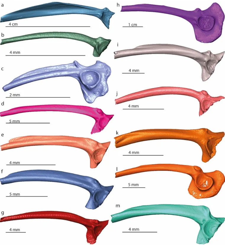
Figure S1. 3D models of ilia of Pipa parva (a), Syncope antenori (b), Brachycephalus albolineatus (c),
Eleutherodactylus glaphycompus (d), Hypodactylus araiodactylus (e), Craugastor brocci (f), Gastrotheca peruana (g), Ceratophrys aurita (h), Proceratophrys boiei (i), Rhinoderma darwinii (j), Telmatobius marmoratus (k), Cycloramphus asper (l), Hylodes asper (m). Collection numbers of each specimen are listed in Table S1.
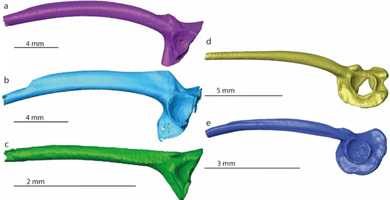
Figure S2. 3D models of ilia of Alsodes nodosus (a), Leptodactylus validus (b), Allophryne ruthveni (c), Centrolene buckleyi (d), Dendrobates tinctorius (e). Collection numbers of each specimen are listed in Table S1.
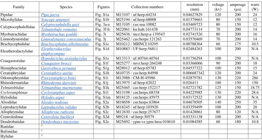
Table S1. List of South American and Australian frog species and families used for comparison with the Antarctic fossil. The Latin name of each species is linked to the corresponding website of Morphosource. Collection numbers and CT-scanning configurations (resolution, voltage, amperage, watts) are indicated according to the information in www.morphosource.com1.
Family Species Figures Collection numbers resolution
(mm) voltage (kv) amperage (µa) watts (W)
Pipidae Pipa parva Fig. S1a M13107 / uf:herp:64234 0.04627829 120 200 24
Microhylidae Syncope antenori Fig. S1b M25194 / uf:herp:68008 0.01379663 80 150 12
Calyptocephalellidae Calyptocephalella gayi Fig. 3a-e M13105 / cas:sua:10082 0.03469723 80 150 12 Telmatobufo venustus Fig. 3f-h M22061 / ku:kuh:161438 0.04773114 70 200 14
Myobatrachidae Myobatrachus gouldii Fig. 3i M25636 / mcz:herp:a-139543 0.02741526 80 200 16
Limnodynastidae Limnodynastes convexiusculus Fig. 3j M25462 / cas:herps:121263 0.03570469 70 200 14 Brachycephalidae Brachycephalus albolineatus Fig. S1c M10212 / MHNCI:10295 0.00788364 60 175 10.5 Eleutherodactylidae Eleutherodactylus
glaphycompus
Fig. S1d M10883 / UF:herp:56811 0.02484263 100 200 N/A
Craugastoridae Hypodactylus araiodactylus Fig. S1e M13313 / uf:40764:40764 0.01756294 100 250 N/A Craugastor brocci Fig. S1f M25277 / mvz:herp:264248 0.03366066 90 200 18
Hemiphractidae Gastrotheca peruana Fig. S1g M28912 / uf:herp:65783 0.04537322 100 150 15
Ceratophryidae Ceratophrys aurita Fig. S1h M10735 / cas:herp:84998 0.006687342 120 200 24
Odontophrynidae Proceratophrys boiei Fig. S1i M13988 / CM-H-45986 0.02879781 130 220 286
Rhinodermatidae Rhinoderma darwinii Fig. S1j M13975 / uf:herp:62022 0.026411 100 150 150
Telmatobiidae Telmatobius marmoratus Fig. S1k M25465 / cas:herp:152217 0.02721782 125 150 18.75 Cycloramphinae Cycloramphus asper Fig. S1l M13198 / cm:herps:68338 0.04225985 130 220 28.6
Hylodidae Hylodes asper Fig. S1m M18125 / cm:herps:45975 0.03172522 130 200 N/A
Alsodidae Alsodes nodosus Fig. S2a M18658 / cm:herps:63864 0.04678505 140 250 35
Leptodactylidae Leptodactylus validus Fig. S2b M18245 / uf:herp:103920 0.03359499 100 200 20
Allophrynidae Allophryne ruthveni Fig. S2c M12630 / cas:herp:257677 0.01428861 80 120 N/A
Centroledinae Centrolene buckleyi Fig. S2d M9218 / uf:herp:30579 0.03331138 100 200 N/A
Dendrobatidae Dendrobates tinctorius Fig. S2e M25692 / ypm:vz:ypm hera 010610 0.01084585 60 180 10.8
Ranidae 2
Bufonidae 2
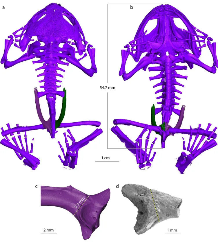
Figure S3. a-c 3D reconstruction of Calyptocephalella gayi (M13105 / cas:sua:10082 from
www.morphosourse.com1). Body of C. gayi with indication of the measured value of the snout-vent length in dorsal (a) and ventral (b) views, as well as the left ilium of the same individual in lateral view (c). (d) Antarctic ilium (NRM-PZ B282) in lateral view. The measured distance of the height of the transition from the iliac shaft and ilial body is indicated by a black-yellow line with the corresponding numerical value.
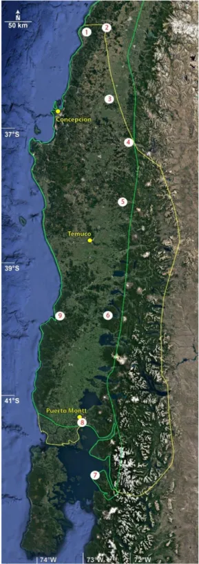
Figure S4. Map showing the area of the sympatric occurrences of Calyptocephalella gayi (green line)3 and Dromiciops gliroides (yellow line)4 with the locations of the climatic stations (white circles). The climatic station numbers correspond to those in Table 2. Map redrawn from an original generated using ArcGIS 10.17.1 (www.esri.com) software, based on the Satellite base map layer in google Maps (Map data ©2019 Google).
References
1. unknown. Morphosource. Available at https://www.morphosource.org/ (2020).
2. Bailon, S. Différenciation ostéologique des Anoures (Amphibia, Anura) de France (Centre de Recherches Archéologiques du CNRS, Antibes, 1999).
3. Veloso, A., Formas, R. J. & Gerson, H. Calyptocephalella gayi. The IUCN Red List of Threatened Species (2010).