HAL Id: tel-02062314
https://tel.archives-ouvertes.fr/tel-02062314
Submitted on 8 Mar 2019
HAL is a multi-disciplinary open access archive for the deposit and dissemination of sci-entific research documents, whether they are pub-lished or not. The documents may come from teaching and research institutions in France or abroad, or from public or private research centers.
L’archive ouverte pluridisciplinaire HAL, est destinée au dépôt et à la diffusion de documents scientifiques de niveau recherche, publiés ou non, émanant des établissements d’enseignement et de recherche français ou étrangers, des laboratoires publics ou privés.
Roles of astroglial cannabinoid type 1 receptors (CB1)
in memory and synaptic plasticity
Laurie Robin
To cite this version:
Laurie Robin. Roles of astroglial cannabinoid type 1 receptors (CB1) in memory and synaptic plasticity. Neurons and Cognition [q-bio.NC]. Université de Bordeaux, 2018. English. �NNT : 2018BORD0283�. �tel-02062314�
THÈSE PRÉSENTÉE POUR OBTENIR LE GRADE DE
DOCTEUR DE L’UNIVERSITÉ DE BORDEAUX
École doctorale des Sciences de la Vie et de la Santé Spécialité - Neurosciences
Laurie ROBIN
Sous la direction de : Giovanni MARSICANO
Soutenue le 30 novembre 2018 MEMBRES DU JURY FERREIRA G., Dr. WOTJAK C., Dr. HENNEBERGER C., Pr. NADJAR A., Dr. ……… BONVENTO G., Dr.
ROLES OF ASTROGLIAL CANNABINOID TYPE 1
RECEPTORS (CB
1) IN MEMORY AND SYNAPTIC
PLASTICITY.
Président Rapporteur Rapporteur Examinateur Examinateur Université de Bordeaux, FranceMax Planck Institute of Psychiatry Munich, Allemagne University of Bonn Medical Center, Bonn, Allemagne Université de Bordeaux, France
ROLES DU RECEPTEUR AUX CANNABINOÏDES DE TYPE 1
DES ASTROCYTES DANS LA MEMOIRE ET LA PLASTICITE
SYNAPTIQUE.
Le système endocannabinoïde est un important modulateur des fonctions physiologiques. Il est composé des récepteurs aux cannabinoïdes, de ses ligands lipides endogènes (les endocannabinoïdes) et de la machinerie enzymatique pour
leur synthèse et leur dégradation. Les récepteurs aux cannabinoïdes de type 1 (CB1)
sont exprimés dans différents types cellulaires dans le cerveau et sont connus pour être impliqués dans les processus mnésiques. Les endocannabinoïdes sont mobilisés dépendamment de l’activité notamment dans les régions cérébrales impliquées dans la mémoire telle que l’hippocampe. Dans cette région, les
récepteurs CB1 sont exprimés au niveau des terminaisons neuronales
pré-synaptiques où leur stimulation inhibe la libération de neurotransmetteurs, modulant ainsi différentes formes d’activité synaptique.
Outre leur expression sur les neurones, les récepteurs CB1 sont également exprimés
par les astrocytes. Avec l’élément pré- et post-synaptique, les astrocytes font partis de la « synapse tripartite » où ils participent à la plasticité synaptique et les processus mnésiques associés. De manière intéressante, la stimulation des
récepteurs CB1 astrocytaires facilite la transmission glutamatergique dans
l’hippocampe. Dans cette région, les astrocytes régulent l’activité des N-methyl-D-aspartate receptors (NMDARs) à travers le contrôle des niveaux synaptiques de leur co-agoniste, la D-serine, modulant ainsi la plasticité synaptique à long-terme. Cependant, le mécanisme entrainant la libération de D-serine par les astrocytes n’est pas identifié. De manière intéressante, notre laboratoire a montré que les effets délétères des cannabinoïdes exogènes sur la mémoire de travail spatial sont médiés
par les récepteurs CB1 astrocytaires à travers un mécanisme dépendant des
NMDARs dans l’hippocampe. Cependant, le rôle physiologique des récepteurs CB1
astrocytaires restent méconnus.
Une des formes de mémoire impliquant le récepteurs CB1 est la mémoire de
reconnaissance d’objet (NOR). La stimulation exogène des récepteurs CB1
hippocampique inhibe la consolidation de la NOR mais la délétion constitutive des
récepteurs CB1 n’affecte pas la NOR, suggérant que la signalisation des récepteurs
CB1 endogènes n’est pas nécessaire. Cependant, de récentes études soulignent que
la délétion globale du gène CB1 pourrait masquer le rôle des récepteurs CB1 des
différents types cellulaires. Ceci indique la nécessité de nouveaux outils plus sophistiqués afin de totalement comprendre le rôle physiologique du système endocannabinoïde dans des comportements complexes.
Dans cette étude, nous avons étudié le rôle physiologique des récepteurs CB1
astrocytaires dans la formation de la NOR et la plasticité synaptique. En utilisant une combinaison d’approches génétiques, comportementales, électro-physiologiques, d’imagerie et de biochimie, nous avons montré que l’activation endogène des
récepteurs CB1 astrocytaires est nécessaire pour la consolidation de la NOR à
long-terme, et ceci à travers un mécanisme impliquant l’apport en D-sérine, afin de stimuler l’activité des NMDARs synaptiques de l’hippocampe dorsal.
Cette étude révèle un mécanisme inattendu à la base de la libération de D-sérine, entrainant l’activité des NMDARs et la formation de la mémoire à long-terme.
ROLES OF ASTROGLIAL CANNABINOID TYPE 1
RECEPTORS IN MEMORY AND SYNAPTIC PLASTICITY.
The endocannabinoid system is an important modulator of physiological functions. It is composed of cannabinoid receptors, their endogenous lipid ligands (the endocannabinoids) and the enzymatic machinery for endocannabinoid synthesis and degradation. The type-1 cannabinoid receptors (CB1) are expressed in different cell types of the brain and are known to be involved in memory processes. Endocannabinoids are mobilized in an activity-dependent manner in brain areas involved in the modulation of memory such as the hippocampus. In this brain region, CB1 receptors are mainly expressed at neuronal pre-synaptic terminals where their stimulation inhibits the release of neurotransmitters, thereby modulating several forms of synaptic activity.Besides their expression in neurons, CB1 receptors are also expressed in astrocytes. Along with the pre- and post-synaptic neurons, astrocytes are part of the “tripartite synapse”, where they participate in synaptic plasticity and associated memory processes. Interestingly, modulation of astroglial CB1 receptors has been proposed to facilitate glutamatergic transmission in the hippocampus. In this brain area, astrocytes regulate the activity of N-methyl-D-aspartate receptors (NMDARs) through the control of the synaptic levels of their co-agonist D-serine, thereby mediating long-term synaptic plasticity. However, the mechanisms inducing D-serine release by astrocytes are still not identified. Interestingly, our laboratory showed that the negative effect of exogenous cannabinoids on spatial working memory is mediated by astroglial CB1 receptors through a NMDAR-dependent mechanism in the hippocampus, but the physiological role of astroglial CB1 remains unknown.
One of the forms of memory involving CB1 receptors is novel object recognition (NOR) memory. The exogenous stimulation of hippocampal CB1 receptors inhibits the consolidation of long-term NOR formation. Constitutive global deletion of CB1 receptors in mice leaves NOR memory intact, suggesting that endogenous CB1 receptor signaling is not necessary for long-term NOR. However, recent studies pointed-out that, likely due to compensatory mechanisms, the global deletion of the
CB1 gene might mask cell type-specific roles of CB1 receptors, indicating that more
sophisticated tools are required to fully understand the physiological roles of the endocannabinoid system in complex behavioral functions.
In this work, we investigated the physiological role of the astroglial CB1 receptors on NOR memory formation and synaptic plasticity. By using a combination of genetic, behavioral, electrophysiological, imaging and biochemical techniques, we showed that endogenous activation of astroglial CB1 receptors is necessary for the consolidation of long-term NOR memory, through a mechanism involving the supply of D-serine to enhance synaptic NMDARs-dependent plasticity in the dorsal hippocampus.
This study uncovers an unforeseen mechanism underlying D-serine release, triggering NMDARs activity and long-term memory formation.
Key words: Astroglial CB1 receptors, D-serine, NMDA receptors, Memory, Synapse,
Key words: Astroglial CB receptors, D-serine, NMDA receptors, Memory, Synapse,
UNITE DE RECHERCHE
NEUROCENTRE MAGENDIE - U1215
146, rue Léo saignat
33077 Bordeaux cedex
France
A ma sœur Emilie
une des femmes les plus fortes que je connaisse.
« Le principal fléau de l'humanité n'est pas l'ignorance, mais le refus de savoir»
(Simone de Beauvoir)
LIST OF PUBLICATIONS
Robin, L.M.*, Oliveira da Cruz, J.F.*, Langlais, V.C.*, Martin-Fernandez, M., Metna-Laurent, M., Busquets-Garcia, A., Bellocchio, L., Soria-Gomez, E., Papouin, T., Varilh, M., Sherwood MW., Belluomo I., Balcells G., Matias I., Bosier B., Drago F., Van
Eeckhaut A., Smolders I., Georges F., Araque A., Panatier A., Oliet SHR.#,
Marsicano G.# (2018). Astroglial CB1 Receptors Determine Synaptic D-Serine
Availability to Enable Recognition Memory. Neuron 98, 935-944.e5.
Gutiérrez-Rodríguez, A., Bonilla-Del Río, I., Puente, N., Gómez-Urquijo, S.M., Fontaine, C.J., Egaña-Huguet, J., Elezgarai, I., Ruehle, S., Lutz, B., Robin, L.M., Soria-Gómez E., Bellocchio L., Padwal JD., van der Stelt M., Mendizabal-Zubiaga J., Reguero L., Ramos A., Gerrikagoitia I., Marsicano G., Grandes P. (2018). Localization of the cannabinoid type-1 receptor in subcellular astrocyte compartments of mutant mouse hippocampus. Glia 66, 1417–1431
Martin-Fernandez, M., Jamison, S., Robin, L.M, Zhao, Z., Martin, E.D., Aguilar, J., Benneyworth, M.A., Marsicano, G., and Araque, A. (2017). Synapse-specific astrocyte gating of amygdala-related behavior. Nature Neuroscience 20, 1540– 1548.
Hebert-Chatelain, E., Desprez, T., Serrat, R., Bellocchio, L., Soria-Gomez, E., Busquets-Garcia, A., Pagano Zottola, A.C., Delamarre, A., Cannich, A., Vincent, P., Varilh M., Robin LM., Terral G., García-Fernández MD., Colavita M., Mazier W., Drago F, Puente N., Reguero L., Elezgarai I., Dupuy JW., Cota D., Lopez-Rodriguez ML., Barreda-Gómez G., Massa F., Grandes P., Bénard G., Marsicano G. (2016). A cannabinoid link between mitochondria and memory. Nature 539, 555–559
Oliveira da Cruz, J.F.*, Robin, L.M.*, Drago, F., Marsicano, G., and Metna-Laurent, M.
(2016). Astroglial type-1 cannabinoid receptor (CB1): A new player in the
tripartite synapse. Neuroscience 323, 35–42. *: equal contribution, #: equal supervision
LIST OF COMMUNICATIONS
O
RAL PRESENTATIONSAstroglial CB1 receptors determine synaptic D-serine availibity to enable
recognition memory. (Salamanca, June 2017). International Symposium on
Metabolic and redox interactions between neurons and astrocytes in health and disease
Role of hippocampal astroglial cannabinoid type-1 receptors in memory and synaptic plasticity. (Bertinoro, April 2014) International astrocytes school.
P
OSTER COMMUNICATIONSInternational conferences
L.M Robin, J. F. Oliveira Da Cruz, V. C. Langlais, M. Metna-Laurent , A.Busquets-Garcia , E. Soria-Gomez , T. Papouin , B. Bosier , F. Drago, A. Van Eeckaut, I.
Smolders , F. Georges , A. Panatier , S. H. R. Oliet , G. Marsicano. Astroglial CB1
receptors control memory via D-serine (2016) Society for Neurosciences (San Diego, USA)
Laurie M. Robin, José F. Oliveira da Cruz, Valentin C. Langlais, Arnau Busquets-Garcia, Edgar Soria-Gomez, Filippo Drago, Aude Panatier, François GeorgeS, Mathilde Metna-Laurent, Stéphane Oliet, Giovanni Marsicano. Astroglial Type-1
cannabinoid receptors (CB1R) are necessary for long-term object recognition memory.
(2015) Cannabinoid conference 2015 (Sestri-Levante, Italy)
National conferences
Robin LM., Langlais V. Metna-Laurent M., Busquets-Garcia A., Papouin T.,
Leste-Lasserre T., Oliet S.H.R., Marsicano G. Astroglial CB1 receptors control long-term
memory formation and NMDAR-dependent synaptic plasticity (2015) Neurocampus conference, the quadripartite synapse.
Robin LM., Langlais V. Metna-Laurent M., Busquets-Garcia A., Papouin T.,
Leste-Lasserre T., Oliet S.H.R., Marsicano G. Astroglial CB1 receptors control long-term
memory formation and NMDAR-dependent synaptic plasticity (2014). LabeX day Robin LM., Langlais V. Metna-Laurent M., Busquets-Garcia A., Papouin T.,
Leste-Lasserre T., Oliet S.H.R., Marsicano G. Astroglial CB1 receptors control object
TABLE OF CONTENTS
CHAPTER I
Glial cells and the tripartite synapse
I.
History of the discovery of glial cells……….
1.3 .5 .10
II.
Glial cells and their functions
……… A. MICROGLIAL CELLS………...…………..……….3B. MACROGLIAL CELLS………...…………..……….3
i. Oligodendrocytes ………...…………..…...3
ii. NG2 cells………...………..……….3
iii. Schwann cells………...…………..………….4
iv. Ependymal Cells………...…………..……..4
v. Astrocytes………...…………..………...4
III.
Cell biology of astrocytes………
A. MORPHOLOGY………..……….5i. Processes and astrocyte territories……….…6
B. ASTROCYTIC NETWORK………..7
C. MAIN PHYSIOLOGICAL FUNCTIONS OF ASTROCYTES………8
i. Glutamate uptake……….…….8
ii. Potassium buffering………8
iii. Neuro-Vascular coupling………10
IV. The
tripartite
synapse……….……
A. CANONICAL VIEW OF THE GLUTAMATERGIC SYNAPSE……….………10B. ANATOMY OF THE TRIPARTITE SYNAPSE………12
C. SYNAPTIC SIGNAL DETECTION……….…..14
i. Membrane transporters……….…..14
TABLE OF CONTENTS
D. SIGNAL INTEGRATION:CA2+ SIGNALING IN ASTROCYTES………..15
i. Mechanism of Ca2+ excitability………..………....14
E. GLIOTRANSMISSION………..………....17
i. Vesicular release……….……..…....17
ii. Non vesicular release……….………...…18
F. GLIOTRANSMITTERS………...………...…19 i. Glutamate………...………...…19 ii. GABA……….……...………....…20 iii. ATP……….…….……...………....…20 iv. Taurine……….……….……...………....…20 v. D-serine………..……….……...………....…20
CHAPTER II
D-serine, a gliotransmitter and a co-agonist of NMDARs
I. D-serine
metabolism
………..………..……...…………22A. DE NOVO SYNTHESIS: SERINE RACEMASE (SR) ………..………22
B. DEGRADATION OF D-SERINE: DAAO (D-AMINO OXIDASE) ………..………..……23
II.
D-serine distribution in the brain
………..………..……24A. BRAIN DISTRIBUTION OF D-SERINE IN THE CNS………..………..……24
B. CELLULAR DISTRIBUTION OF D-SERINE IN THE CNS………..……25
III. D-serine
release
………..……25d
A. VESICULAR RELEASE OF D-SERINE BY ASTROCYTES………..………25B. CA2+-DEPENDENT RELEASE OF D-SERINE ………..………26
C. ACTIONS OF D-SERINE………..………..………26
IV.
NMDARs, composition, expression, functions an
regulation
………..………27A. NMDARS COMPOSITION………..………27
i. Subunit and genes……….………..………27
TABLE OF CONTENTS
A. ACTIVATION OF NMDARS AND PERMEABILITY……….…...28
i. Agonist binding at the GluN2 subunit………...……….28
ii. (Co)-agonist binding at the GluN1 subunit……….………..28
iii. Activation of the NMDARs……….……28
iv. Permeability of NMDARs……….………..28
v. …29 …32 36 ....36
B. LOCALISATION OF NMDARS WITHIN THE BRAIN……….…………29
i. Regional brain distribution……….………… ii. Subcellular distribution of NMDARs………..……..30
C. FUNCTIONAL CONSEQUENCES OF THE NMDAR ACTIVITY: NMDAR-DEPENDENT LTP………..32
i. Induction ……… ii. Expression………32
iii. Maintenance………33
CHAPTER III
The endocannabinoid system
I.
A bit of history: from China to pharmaceutical industrie…….
34A. ASIA………...34
B. EUROPE………..34
C. THE BEGINNING OF SCIENTIFIC STUDIES………...34
D. IDENTIFICATION OF THE PSYCHOACTIVE COMPONENT OF CANNABIS SATIVA…………35
E. SHORT OVERVIEW OF CANNABINOID RESEARCH...35
II.
The endocannainoid system
………..………… A. CANNABINOID RECEPTORS...36i. Structure and general distribution………. ii. Brain distribution………..38
B. THE ENDOCANNABINOIDS………..39
i. Synthesis and degradation of 2-AG and anandamide………..40
III.
Methods to dissect CB
1functions……….
41A. PHARMACOLOGICAL TOOLS………42
TABLE OF CONTENTS
IV.
Signaling of CB
1receptors and modulation of
n
d
synaptic plasticity
………...45A. INTRACELLULAR PATHWAYS………..……….45
B. SUPRESSION OF NEUROTRANSMITTER RELEASE………47
C. CB1 RECEPTOR-MEDIATED SYNAPTIC TRANSMISSION AND PLASTICITY………47
i. DSI and DSE...47
ii. Long-term forms of endocannabinoid-mediated plasticity………48
V. Astroglial
CB
1receptor at the tripartite synapse………...
50A. LOCALISATION AND METABOLISM………50
B. PHYSIOLOGICAL IMPACT OF ASTROGLIAL CB1 RECEPTORS………..51
CHAPTER IV
Memory
I. Neuroanatomical
and
neurological substrates of memory..
53II.
A model to study memory: The object recognitio
Memory
………55III.
Modulation of memory by the ECS, NMDARs an
Astrocytes………
56A. ROLES OF THE ENDOCANNABINOID SYSTEM IN MEMORY……….56
B. ROLES OF NMDARS IN MEMORY………...58
i. Pharmacological blockade of NMDARs……….…….58
ii. Genetic models……….……….………..58
C. ROLES OF ASTROCYTES IN MEMORY………59
i. Gliotransmission……….59
ii. Metabolic functions of astrocytes………..……….60
iii. New tool to study the role of astrocytes in memory……….60
iv. Astroglial CB1 receptors………..60
RESEARCH GOALS
RESEARCH GOALS………..62TABLE OF CONTENTS
RESULTS
Astroglial CB1 Receptors Determine Synaptic D-Serine Availability to
Enable Recognition Memory………63
DISCUSSION
Supplemental discussion on Astroglial CB1 Receptors Determine Synaptic
D-Serine Availability to Enable Recognition memory………..…………89
REFERENCES
References related to CHAPTER I-IV and supplemental discussion.………95
ANNEX
1. Localization of the cannabinoid type-1 receptor in subcellular astrocyte
compartments of mutant mouse hippocampus.………..……….……135
2. Synapse-specific astrocyte gating of amygdala-related behavior.………150
LIST OF ABBREVIATIONS
2-AG 2-Arachidonoylglycerol
AAV Adeno-Associated Virus
ABD Agonist Binding Site
AC Adenylate Cyclase
ACEA Arachidonyl-2′-chloroethylamine
AEA Arachidonoylethanolemide; Anandamide
AMPAR α-Amino-3-hydroxy-5-Methyl-4-isoxazole propionic acid receptors
ANLS Astrocyte-neuron lactate shuttle
AP5 2-amino-5-phosphonovalérique acid
ASCT Alanine-Serine-Cysteine-Transporter
ATP Adenosine Tri Phosphate
b.C. before Christ
Best-1 Bestrophin 1
CaMKII Calmodulin-dependent protein kinase II
CA1 Cornu Ammonis area 1
CA3 Cornu Ammonis area 3
cAMP Cyclic adenosine monophosphate
Ca2+ Calcium
CB-LTD Cannabinoid-induced Long-Term Depression
CB1R Cannabinoid type 1 receptor
CB2R Cannabinoid type 2 receptor
CeM Central amygdala
CNS Central Nervous System
CSF Cerebro-Spinal Fluid
Cx Connexin
DAAO D-Amino Acid Oxidase
DAG Diacylglycerol
DAGL Diacylglycerol Lipase
DREADDs Designer Receptor Exclusively Activated by Designer Drugs
DSE Depolarization-induced Suppression of Excitation
DSI Depolarization-induced Suppression of Inhibition
EAAT Excitatory Amino Acid Transporter
eCB Endocannabinoid
eCB-LTD Endocannabinoid-mediated LongTerm Depression
eCB-STD Endocannabinoid-mediated Short-Term Depression
ECS Endocannabinoid System
EM Electron Microscopy
EPSC Excitatory Post-Synaptic Currents
LIST OF ABBREVIATIONS
ERT Estrogen Receptor
FAD Flavine Adenine Dinucleotide
FAAH Fatty Acid Amid Hydrolase
GABA Gamma-Aminobutyric Acid
GAT GABA Transporters
GFAP Glial Fibrillary Acidic Protein
GLAST Glutamate Aspartate Transporter
GLT-1 Glutamate Transporter
Glu Glutamate
Gln Glutamine
GlyRs Glycine Receptors
GPCR G-Protein Coupled Receptors
I1 Phosphate Protein 1 Inhibitor 1
iGluR Ionotropic Glutamate Receptor
IP3 Inositol 1,4,5-trisphosphate
IP3R IP3 Receptor
IPSCs Inhibitory Post-Synaptic Currents
JNK c-Jun N-terminal Kinase
K+ Potassium
kDA Kilo Dalton
Kir Inward-rectifying potassium channels
KO Knock-Out
LTD Long Term Depression
LTDi Long-Term Depression of inhibition
LTM Long Term Memory
LTP Long-term Potentiation
MAGL Monoacylglycerol Lipase
MAGUK Membrane-Associated Guanylate Kinases
MAPK Mitogen Activated Protein Kinase
MCTs MonoCarboxylate Transporters
mGluR Metabotropic Glutamate receptor
Mg2+ Magnesium
Mhb Medial Habenula
mM Millimolar
mRNA Messenger Ribonucleic Acid
mtCB1 Mitochondrial CB1 receptors
Na+ Sodium
NAT N-acetyltransferase
NAPE N-Arachidonoyl Phosphatidyl Ethanol
nm nanometer
NMDAR N-Methyl-D-Asparte Receptor
NO Nitric Oxide
NOR Novel Object Recognition
LIST OF ABBREVIATIONS
NT Neurotransmitter
NTD N-terminal Domain
PAP Perisynaptic Astrocytic Processes
PCP Phencyclidine
PKA Protein Kinase A
PLA Phospholipase A
PLC Phospholipase C
PNS Peripheral Nervous System
PP1 Phosphate Protein 1
PTPN22 Protein Tyrosine Phosphatase non-receptor type-22
PSD Post-synaptic Density
SIC Slow Inward Current
SLMV Synaptic-Like Microvesicles
SNARE Soluble -ethylmaleimide-sensitive fusion protein attachment protein receptor
SR Serine Racemase
STED Stimulation-Emission-Depletion
STM Short-Term Memory
TCA cycle TriCarboxylic Acid cycle
TDM Transmembrane Domain
THC ∆9-tetrahydrocannabinol
t-LTD time-dependent Long Term Depression
TM TransMembrane
TRPA1 Transient Receptor Potential Ankyrin 1
TRPV1 Transient Receptor Potential Vanilloid type 1 ion channel
VAMP2 Synaptobrevin
VAMP3 Cellubrevin
VRAC Volume-Regulated Anion Channel
VGCC Voltage Gated Calcium Channel
WT Wild-Type
LIST OF FIGURES
Figure 1
Cajal's drawing showing neuroglia”……….………...….………1
Figure 2
Classification, main functions and structures of glial cells ……….……….………5
Figure 3
Example of different types of astrocytes………….……….……….………….………6
Figure 4
The discrete region of interaction across the fine processes of astrocytes………...….….7
Figure 5
Astrocytes maintain synaptic homeostasis………....…….…..………...9
Figure 6
The glutamatergic synapse………..………..……….………...…..12
Figure 7: Anatomy of the tripartite synapse………..………..13
Figure 8
Schematic representation of the main mechanisms of Ca2+ excitability in astrocytes...16
Figure 9
Proteins proposed to mediate exocytosis from neurons and astrocytes……….18
Figure 10
Potential non exocytotic mechanisms for gliotransmitter release……….……….19
Figure 11
Summary scheme of gliotransmission processes……….……….21
Figure 12
Simplified reaction of racemisation and A,β-Elimination by serine-racemase…………..…….23
Figure 13
Enzymatic degradation of D-serine by the enzyme DAAO………...24
Figure 14
D-serine, serine-racemase and DAAO distribution in the rodent brain……….24
Figure 15
LIST OF FIGURES
Figure 16
Distribution of NMDAR subunits in the rat brain during development………30
Figure 17
Subcellular distribution of NMDARs……….……..……….31
Figure 18
Schematic representation of the NMDAR-dependent long-term potentiation………33
Figure 19
Number of publications, in relation to cannabinoid research……….……….36
Figure 20
Brain distribution of the mouse CB1 receptor………..………39
Figure 21
Scheme of the synthesis and degradation pathways of AEA and 2-AG………...41
Figure 22
Chemical structures of natural and synthetic ligands cannabinoid receptors……….……42
Figure 23
Cell type-specific deletion of the CB1 gene by the Cre/loxP system in mice………….………43
Figure 24
Main intracellular CB1 receptor signaling pathways……….46
Figure 25
Molecular mechanisms of eCB-STD……….………..48
Figure 26
Established mechanisms of eCB-mediated long-term synaptic plasticity………...……...49
Figure 27
CB1 receptor is present in astrocytes……….50
Figure 28
Summary and putative scheme of endocannabinoid-induced gliotransmission………..51
Figure 29
Schematic representation of the different types of memory……….…53
Figure 30
Illustration of the hippocampal circuitry and the hippocampal neural network…………..……54
Figure 31
CHAPTER I
Glial Cells and the tripartite synapse
CHAPTER I: Glial cells and the tripartite synapse
Glial cells are non-neuronal cells in the brain, in the spinal cord and also in the peripheral nervous system (PNS). Rudolf Virchow described them as a connective substance and a “kind of glue” in which nervous elements are embedded (Die Cellularpathologie, 1858). Glial cells are diverse in shape and functions, and they have been historically classified in oligodendrocytes, microglia and astrocytes. Classically, they are considered to play supportive roles to neurons, by several mechanisms. Thus, oligodendrocytes provide myelin isolation to neuronal axons, microglia represent "immune cells" in the brain, and astrocytes deliver nutritional structural support to neurons (Del Río-Hortega, 1922; Ramon y Cajal, 1903; Ramon y Cajal, 1904). As they are not electrically excitable, the potential roles of glial cells in brain information processing have been long underestimated. Nowadays, however, we know that glial cells are directly and actively involved in synaptic transmission in the central and peripheral nervous system. In particular, new theories propose that
astrocytes can encode information through calcium (Ca2+) signaling and cooperate
with neurons to exert all brain functions. In this frame, modern hypotheses postulate that, as members of the "tripartite synapse", astrocytes directly participate in synaptic transmission and information exchange (Araque et al., 1999).
I.
History of the discovery of glial cells
Glial cells were discovered in the miD-1800s by a group of scientists including Robert
Remak, Theodor Schwann and Rudolf Virchow. We consider that Robert Remak was the first to mention a type of glial cells in his thesis published in 1836 where he described “nerve fibers and their surrounding sheats”, that will be later called Schwann cells. However, discovery of glial cells is more often assigned to Rudolf Virchow, who introduced the concept of neuroglia in 1856 as a “kind of connective tissue in which the nervous elements are planted”. We owe the first drawings of a star-shaped glial cell to Otto Deiters whose work was published posthumously in 1865. These cells were called later astrocytes by Michael von Lenhossek (Parpura and Verkhratsky, 2012) Other drawings of glial networks were produced some years later by Jacob Henle and Friedrich Merkel (Henle & Merkel, 1864).
In contrast to extensive studies on various neuronal functions, glial cells remained poorly studied. This is likely due to the fact that glial cells are electrically non-excitable (Orkand et al., 1966; Sontheimer, 1994), as they do not fire action potentials, with the exception of a recently discovered particular subpopulation, NG2 cells (Lin and Bergles, 2002). Thus, glial cells would not be able to be part of the genesis, exchange and integration of fast electrical information and neurophysiologists kept the idea of glia as passive cells supporting neurons (Orkand et al., 1966; Seifert and Steinhäuser, 2001; Sontheimer, 1994; Verkhratsky and Steinhäuser, 2000).
Thanks to histological studies, Camillo Golgi (1889) and Santiago Ramon y Cajal proposed that glial cells could play a role in metabolic support to neurons (Ramon y Cajal, 1903; Ramon y Cajal, 1911). In cortical slices, they observed cells with thin and long processes in contact with both blood vessels and neurons (Figure 1, page 2), and proposed a “physiological importance of these cells in the regulation of brain 1
CHAPTER I: Glial cells and the tripartite synapse
microcirculation”, establishing the so-called “Neuron Nutrition Theory” for their function. Finally, although Ramon y Cajal proposed for a long time that axon secreted myelin, he published with Del Río-Hortega in 1922 a bold hypothesis: “We are
inclined to believe, however, that both kinds of cells (oligodendroglia and Schwann cell) carry out identical functions of support, isolation, and nutrition connected with nerve conduction”(Del Rio Hortega, 1922).
Although research on glial cells started 200 years ago, it is only recently that brain scientists showed a real craze for those cells. Through the development of tools and imaging techniques, it was shown that these cells could actively participate in synaptic communication (Allen and Barres, 2005; Haydon, 2001). These observations started a new wave of interest for glial cells which seems essential since they represent 50% of the volume of the brain (Laming, 2000). Nowadays, it is generally accepted that glia-neurons ratio is 1:1 in adult human brain (Azevedo et al., 2009) However, this ratio can vary in different brain regions and species. For example, it is more accurate to say that glia-neuron ratio in the mouse brain is about 3:1(Herculano-Houzel et al., 2006).
Firstly described as a "cement" for neurons, a wide number of studies showed that glial cells are not only supportive cells for neurons, but they also display a large repertoire of properties and active functions.
A
2
Figure 1: Cajal's drawing showing “neuroglia”.
Glial cells of the pyramidal layer and stratum radiatum of the Ammon horn (from adult man autopsied 3h after death). Original labels: A, large astrocyte embracing a pyramidal neuron; B, twin astrocytes forming a nest around a cell, C, while one of them sends two branches forming another nest, D; E, cell with signs of “autolysis”; F, capillary vessel.
CHAPTER I: Glial cells and the tripartite synapse
II.
Glial cells and their functions
Based on their morphology and origin, glial cells are currently classified into 2 groups: microglial and macroglial cells. Microglia contains microglial cells only, whereas macroglia contains oligodendrocytes, NG2 cells, Schwann cells, ependymal cells and astrocytes (Figure 2, page 5). The present PhD Thesis deals with synaptic active roles of astrocytes. In the following, I will briefly describe the general functions of most glial cells subtypes, for which active roles in information processing have been proposed. Besides certain macroglial subtypes such as ependymal and Schwann cells (Jiménez et al., 2014; Kidd et al., 2013), active roles in synaptic information processes have been suggested for all glial cells in the central nervous system (CNS). In the following, I will briefly introduce the functions of each glial subtype, indicating their "classical" and "novel" proposed roles.
A. MICROGLIAL CELLS
Microglial cells are considered to form the immune system of the CNS. Indeed, they have common properties with macrophages and are essential to protect neurons. At the basal state, they sense the environment thanks to their highly mobile processes. In case of damages in the CNS, they are “activated”(Ransohoff and Cardona, 2010). They multiply and migrate to the lesion (Davalos et al., 2005), where they can act as phagocytes. They are also able to secrete cytokines that can modulate cell survival and fate (Ransohoff and Cardona, 2010). Apart from their role as immune cells, microglia seems to participate in active information processing (Bessis et al., 2007). Indeed, they are able to communicate with neurons and astrocytes to modulate
synaptic transmission and could be the 4th partner of a putative “quadripartite”
synapse (Hristovska and Pascual, 2015; Panatier and Robitaille, 2012). B. MACROGLIAL CELLS
i. Oligodendrocytes
The main function of oligodendrocytes is to provide support and insulation to axons in the CNS by creating the myelin sheath (Gill and Binder, 2007; Nave, 2010). Along neuronal axons, myelin sheaths are separated by 1-2 µm from each other, forming the so-called Ranvier nodes (also known as myelin-sheath gaps), where the neuronal membrane is exposed to the extracellular space. Because Nodes of Ranvier are
uninsulated and enriched in ion channels such as sodium (Na+) channels, they are
key elements of ions exchanges to generate action potentials and to allow their so-called "saltatory" propagation (Kaplan et al., 1997, 2001; Salzer, 1997). Recent evidence pointed out the existence of a sort of "axomyelinic neurotransmission" involving several oligodendrocyte-dependent novel mechanisms (Micu et al., 2018).
ii. NG2 cells
NG2 cells are the most recently identified glial cell subtypes. They are characterized by the expression of the proteoglycan NG2 and are considered as
CHAPTER I: Glial cells and the tripartite synapse
oligodendrocyte precursor cells in the CNS (Nishiyama et al., 2009). These cells are still poorly studied, but accumulating evidence indicates that they have many specific properties and functions in the CNS. For instance, they can interact with synapses, representing a likely additional functional component (Bergles et al., 2000; Nishiyama et al., 2009).
iii. Schwann cells
Schwann cells are the principal glia of the PNS, analogue to oligodendrocytes in the CNS. They produce the myelin sheaths around neuronal axons to ensure protection to neurons and the propagation of action potentials (Voyvodic, 1989). Schwann cells also assist in neuronal survival and signal the formation of various structures within the PNS. The Schwann cell offers trophic support to developing neurons whose axons have not yet reached their targets (Riethmacher et al., 1997). Finally, recent studies suggest that Schwann cells could be active synaptic partners, particularly at the neuromuscular junction, where these cells regulate synaptic efficacy (Rousse and Robitaille, 2006; Sugiura and Lin, 2011).
iv. Ependymal Cells
Ependymal cells are part of CNS glia. Ependymal cells forms the epithelial lining of the ventricles in the brain and the central canal of the spinal cord. They are involved in the production of cerebrospinal fluid (CSF) (de Reuck and Vanderdonckt, 1986)
and they have been shown to serve as a reservoir for neuroregeneration (Carlén et
al., 2009). One type of ependymal cells, known as tanycytes are found only on the floor of the third ventricle and play an important role in facilitating the transport of hormones and other substances in the brain (Langlet et al., 2013).
v. Astrocytes
Firstly described as star-shaped glial cells, astrocytes represent following recent evaluations approximately 20-40% of total glia in the brain, depending on brain regions and species (Herculano-Houzel, 2014). We know now that they are not all star-shaped but they have thin and ramified processes that interact with both neurons and blood vessels, allowing them to put these elements in communication (Pellerin and Magistretti, 2012). After neurons, they are the most studied CNS cells. Indeed, simple PubMed searches using cell names as key words reveal the following numbers of published papers: > 600.000 for neuron, > 53.000 papers for astrocyte, > 29.000 for oligodendrocyte, > 28.000 for microglia, > 16.000 for Schwann cells, > 4.000 for ependymal cells and in the order of some hundreds for NG2 cells. In particular, astrocytes have been recently shown to participate in many physiological functions such as sleep, reproduction and memory (Ben Achour and Pascual, 2012; Halassa et al., 2009). Ongoing research focuses on determining novel specific cellular and functional properties of astrocytes, and new subpopulations of these cell types are continuously proposed (Oberheim et al., 2012). In the following, I will briefly describe established general properties of astrocytes providing useful information for the experimental part of this Thesis.
4
CHAPTER I: Glial cells and the tripartite synapse
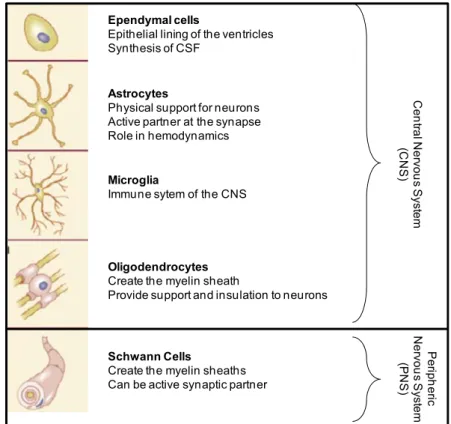
Ependymal cells
Epithelial lining of the ventricles Synthesis of CSF
Astrocytes
Physical support for neurons Active partner at the synapse Role in hemodynamics
Microglia
Immune sytem of the CNS
Oligodendrocytes Create the myelin sheath
Provide support and insulation to neurons
Schwann Cells
Create the myelin sheaths Can be active synaptic partner
Ce n tra l Ne rv o u s S ys te m (C N S ) P er iph er ic Ne rv o u s S ys te m (P N S )
Figure 2: Classification, main functions and structures of glial cells. Adapted from the book Neuroscience, Dale Purves & George J. Augustine. 2015
III.
Cell biology of astrocytes
A. MORPHOLOGY
Following classical morphological classifications, there are 3 types of astrocytes (Figure 3, page 6): protoplasmic, fibrous and radial astrocytes (Miller and Raff, 1984). Protoplasmic astrocytes are located in the grey matter and have numerous and short ramifications. On the contrary, fibrous astrocytes are in the white matter, they have less but longer and thinner ramifications (Zhang and Barres, 2010). Radial glia, with the exception of Müller cells of the retina and cerebellar Bergmann glia, are generally present only during development. They are placed perpendicular to ventricles and their main function is to help the migration of developing neurons (Rakic, 2003). Within these types, astrocyte morphology is heterogeneous since it depends on the species and the brain regions (Matyash and Kettenmann, 2010; Oberheim et al., 2012). In general, there are no clear parameters to define astrocytes. However, some features, such as the star-like shape, the presence of thin and ramified processes in contact with blood vessels and neurons and the expression of the intermediate filament of the cytoskeleton Glial Fibrillary Acidic Protein (GFAP) and/or of specific transporter proteins, are used to identify these cell types. Because they represent the main subject of the present Thesis work, we will mainly focus on protoplasmic astrocytes of the hippocampus in the following sections.
CHAPTER I: Glial cells and the tripartite synapse
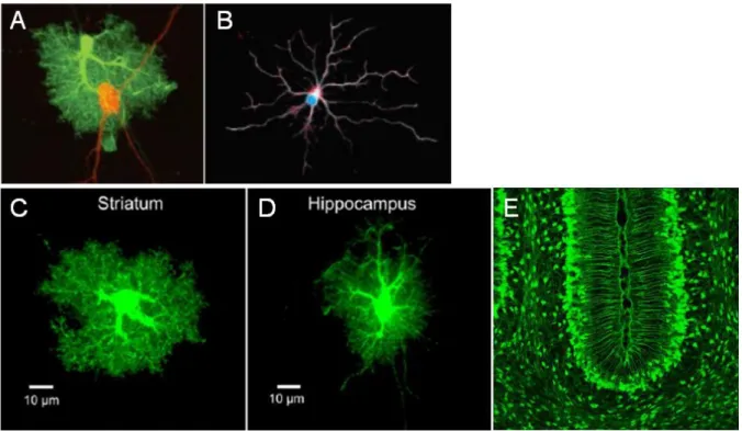
Figure 3: Example of different types of astrocytes.
(A) Example of protoplasmic astrocyte (green) in close association with neuronal cell body and processes (red). (B) Example of Fibrous astrocyte. (C-D) Representative pictures of protoplasmic astrocytes from the mouse striatum and hippocampus, respectively. (E): Example of Bergmann glia.
(A) Adapted from Allen and Barres, 2009.
(B) Adapted from https://fhs.mcmaster.ca/fxar/astrocytes_gallery.html (C-D) Adapted from Chai et al., 2017.
(E) Adapted from http://www.gensat.org/imagenavigator.jsp?imageID=10578
i. Processes and astrocyte territories
Primary processes of hippocampal protoplasmic astrocytes are quite thick and rich in GFAP while the distal processes are very small, containing a limited amount of cytoplasm, lacking most of the organelles as well as GFAP (Theodosis et al., 2008). It has been calculated that cell body and primary processes represent only 15% of the total volume of astrocytes (Bushong et al., 2002), implicating that 85% of the cellular volume is formed by very tiny elements that are extremely difficult to observe and study (Bushong et al., 2002; Freeman, 2010).
Therefore, one aim of recent studies is to develop precise methods to observe astrocyte processes. For example, filling astrocytes with appropriate markers such as Lucifer Yellow or Alexa Fluor 568 revealed that astrocytes processes extend from 50 to 100 µm from the cell body and that different astrocytes display a very limited spatial overlap (~5%; Figure 4, page 7), suggesting that each cell occupies a distinct territory in the hippocampus (Wilhelmsson et al., 2006) and, importantly, that 2 astrocytes are unlikely to contact the same synapse (Bushong et al., 2004; Oberheim et al., 2006).
6
CHAPTER I: Glial cells and the tripartite synapse
B. ASTROCYTIC NETWORK
strocytes were thought for a long time that astrocytes are organized as a global Figure 4: The discrete region of interaction across the fine processes of
rotoplasmic astrocytes.
-C) Respective x–y, y–z and x–z views of hippocampal astroglial processes occupying pecific territories called “domains”. Domains of different astrocytes (in green and in red) isplay a very limited spatial overlap (~5 %; in yellow). Scale bar, 20 µm.
dapted from Bushong et al., 2002. p (A s d A A
syncytium, which is a mass of cytoplasm that has many nuclei and an enclosing membrane but no individual cells (Brightman and Reese, 1969). Indeed, astrocytes are connected through gap-junctions and some molecules can freely move from one cell to another (Pappas et al., 1996). However, it is now clearly admitted that astrocytes are organized as delimited networks. Using some markers that can diffuse through those gap-junctions, it was shown that the number of connected astrocytes is different depending on the brain regions and physiological states of the animals (Blomstrand et al., 2004; D’Ambrosio et al., 1998; Giaume and Theis, 2010; Konietzko and Müller, 1994; Rouach et al., 2008). For instance, in the CA1 region of
CHAPTER I: Glial cells and the tripartite synapse
the hippocampus, recent data proposed that astrocytes' networks are likely composed of hundreds of cells (Rouach et al., 2008). Gap-junction coupling is an efficient way to maintain intercellular communication and coordinate information exchanges between, because these junctions allow the exchange of ions and small signaling molecules (under 1,5 kDA) (Giaume and Liu, 2012; Giaume and Theis, 2010).
Gap junctions are composed of 2 juxtaposed hemichannels called connexons. Each
C. MAIN PHYSIOLOGICAL FUNCTIONS OF ASTROCYTES
strocytes play major roles in the CNS. Some of these functions are described below.
i. Glutamate uptake
of these connexons is composed of 6 transmembrane proteins called connexins (Cx). In the astrocytes the main 2 types of Cx are Cx30 and Cx43. Moreover, when alone, connexons act as hemichannels allowing the entry or exit of molecules. For instance, it was proposed that hemichannels can participate in the release of some gliotransmitters such as glutamate (Ye et al., 2003).
A
Astrocytic processes surrounding synaptic elements express transporters for a variety of neurotransmitters and neuromodulators including glutamate (Gadea and López-Colomé, 2001). These transporters participate in the rapid removal of neurotransmitters released into the synaptic cleft (Figure 5, page 9), which is essential for the termination of synaptic transmission and maintenance of neuronal excitability, with important implications in protecting neurons against glutamate-induced excitotoxicity (Yi et al., 2005).
At rest, extracellular glutamate levels are maintained in the low micromolar range (2 µM) but they increase dramatically during glutamatergic neurotransmission, reaching up to 1 mM for a few milliseconds in the synaptic cleft (Clements et al., 1992). This concentration of glutamate would cause extensive neuronal injury in the absence of highly efficient mechanisms for its removal at the synapse. This is primarily achieved by the astrocyte-specific sodium-dependent high affinity Glutamate Transporters GLT-1 and the astroglial Glutamate Aspartate Transporter GLAST (corresponding to human EAAT2 and EAAT1, respectively) and to a lesser extent by the neuronal glutamate transporters EAAC1 (human EAAT3) and EAAT4 (Danbolt, 2001).
ii. Potassium buffering
Propagation of action potentials causes substantial local increases of extracellular
potassium ions (K+) in the extracellular space, going from 3 µM at rest to around 10
µM. Such an increase can alter the neuronal membrane potential and lead to
neuronal hyperexcitability (Walz, 2000). This is prevented by the buffering of K+ by
astrocytes (Holthoff and Witte, 2000), which occurs mainly through by 2 types of mechanisms.
he first one is passive absorption of K+ through the numerous K+ channels such as
T
the inwarD-rectifying potassium channels (Kir) (Chen and Nicholson, 2000; Sibille et
8
CHAPTER I: Glial cells and the tripartite synapse
al., 2015). K+ ions are then distributed in the astrocytic network (Bellot-Saez et al.,
2017).
A second, active and energy-costing mechanism involves the Na /K /ATPase pump. + +
For every Adenosine-tri-Phosphate (ATP) molecule that the pump uses, three sodium
(Na+) ions are exported and two K+ ions are imported into astrocytes (Figure 5, page
9). Thus the pump enables astrocytes to accumulate the excessive extracellular K+, which can then travel in the astrocytes through gap junctions, following its concentration gradient (D’Ambrosio et al., 2002). This allows the spatial dispersion of
K+ from areas of high concentration to areas of lower concentration where it can be
extruded either into the extracellular space or the circulation, thus maintaining the overall extracellular K+ concentration within the physiological range.
Neuron Astrocyte
Blood vessel
Figure 5
(1)
: Astrocytes maintain synaptic homeostasis.
During excitatory synaptic transmission, glutamate (Glu) is released by neurons. Then, nsporters (EAATs), such as (1)
(2)
(3)
Glu is uptaken by astrocytes through Excitatory Amino Acid Tra
GLT-1. Glu is converted to glutamine (Gln), which is uptaken by neuron to support Glu production. (2) Astrocytes uptake the extracellular K+, mainly through the Na+/K+ ATPase
exchanger (3) Finally, astrocytes take glucose from blood and transform it into lactate, which is then released and taken up by neurons via monocarboxylate transporters (MCTs) to fuel neuronal metabolism.
Adapted from Bélanger et al., 2011.
CHAPTER I: Glial cells and the tripartite synapse
iii. Neuro-Vascular coupling
Astrocytes are positioned between neurons and blood vessels. They have a particularly intimate relationship with blood vessels in the brain through their endfoots processes that completely envelop all cerebral blood vessels and can uptake glucose (Abbott et al., 2006). Although neurons can import glucose directly from the extracellular space, astrocytes have been proposed to play a key role in coupling neuronal activity and brain glucose uptake, through a mechanism called the astrocyte-neuron lactate shuttle (ANLS; Figure 5, page 9) (Pellerin et al., 2007; Simpson et al., 2007). This hypothesis originally proposed by Pellerin and Magistretti (1994) is based on the observation that glutamate uptake by astrocytes following synaptic release by neurons stimulate glycolysis in nearby astrocytes, and the lactate so produced is then released by astrocytes into the extracellular space and taken up by neurons via monocarboxylate transporters (MCTs) to fuel neuronal metabolism (Pellerin and Magistretti, 1994; Simpson et al., 2007). Once in neurons, lactate can be used as an energy substrate via its conversion to pyruvate by the action of lactate dehydrogenase and subsequent oxidation in the mitochondrial tricarboxylic acid cycle (TCA cycle). Over the years, experiments from several laboratories have supported the ANLS hypothesis (for reviews see (Magistretti and Allaman, 2015; Weber and Barros, 2015), and more recent in vivo experiments showed a lactate gradient between astrocytes and neurons (Mächler et al., 2016). Electrophysiological evidence also indicates that lactate released by astrocytes and taken up by neurons is necessary to sustain neuronal activity (Morgenthaler et al., 2006; Rouach et al., 2008).
The astrocytic functions described here are essential to maintain physiological neuronal activity (Bélanger et al., 2011). However, we know now that astrocytes are
ot only essential to maintain but they can also actively participate in synaptic
supports the presence of dynamic and idirectional interactions between astrocytes and synaptic neuronal elements. This vidence indicated that astrocytes can detect synaptic neuronal activity and respond
APSE
n
transmission (Araque et al., 1999; Bains and Oliet, 2007).
IV. The
tripartite
synapse
Since the 1990’s, accumulating evidence b
e
by modulating synaptic transmission. These findings suggested that astrocytes are an active protagonist of synaptic transmission and plasticity, and they established the concept of the “tripartite synapse” formed by pre- and post-synaptic elements and the surrounding astroglial processes (Araque et al., 1999). However, before describing the tripartite synapse in more details, it is important to introduce the notion of synapse and more particularly the excitatory synapse.
A. CANONICAL VIEW OF THE GLUTAMATERGIC SYN
he human brain contains approximately 1011-1012 of neurons (Kandel et al., 2000).
T
Brain functions rely mainly on a functional unit: the synapse, a structure that permits
10
CHAPTER I: Glial cells and the tripartite synapse
a neuron to pass an electrical or chemical signal to another cell and enables rapid signal transmission.
Chemical synapses are composed of the pre- and the post-synaptic element that transmits and receives the information, respectively. They are separated by the
in excitatory neurotransmitter in the CNS and the glutamatergic ynapse represents the major type of synapses in the brain. Glutamate released from
ic synapses” (Figure 6, age 12) because of their characteristic post-synaptic density (PSD), which faces the synaptic cleft, and connected through trans-synaptic protein-based nanocolumns (Tang et al., 2016).
Glutamate is the ma s
pre-synaptic terminals binds to the post-synaptic glutamate receptors. Glutamate receptors are transmembrane proteins that specifically bind to glutamate on the extracellular side of the membrane. Upon binding of glutamate, glutamate receptors transduce the signal into intracellular responses. Glutamate receptors are divided in two groups: metabotropic receptors (mGluR), which are G protein-coupled receptors (GPCR), and ionotropic receptors (iGluR), which are glutamate-gated ion channels. iGluRs are the main responsible of fast synaptic transmission and can be divided into
three main groups: N-Methyl-D-Aspartate receptors (NMDARs),
α-Amino-3-hydroxy-5-Methyl-4-isoxazole Propionic Acid receptors (AMPAR) and Kainate receptors (Dingledine et al., 1999a; Hollmann and Heinemann, 1994).
Excitatory glutamatergic synapses are defined as “asymmetr p
pre-synaptic active zone (Gray, 1959). PSD is identified by electron microscopy (EM) as an electron-dense region formed by protein complexes binding the post-synaptic membrane.
PSD is a disc-like structure measuring in average 30-60 nm thick and a few hundred nanometers wide (Carlin et al., 1980). The size of the PSD correlates with the size of
the PSD (Maglione and Sigrist, 2013). Proteins within the PSD are the dendritic spine and with the number of postsynaptic glutamate receptors (Kasai et al., 2003).
With electron and super-resolution microscopy imaging, recent studies dissected the anatomy of
located in different sites along the axo-dendritic axis of synapses, in this order : 1) Membrane receptors and cell adhesion molecules
2) membrane-associated guanylate kinases (MAGUK)
interior face of the PSD
pport, but also cilitates rapid and efficient synaptic transmission by gathering various signaling
3) SAPAP, SH3 and SHANK scaffolds 4) The actin cytoskeleton contacting the
This dense protein complex does not only provide structural su fa
components and pathways (Boeckers, 2006; Kennedy, 1997; Kim and Sheng, 2004). The modulation of synapse activity constitutes a major strategy to control brain physiology and functions. However, a third partner is necessary to the proper function of brain physiology and synaptic activity, the astrocyte.
CHAPTER I: Glial cells and the tripartite synapse
Figure 6: The glutamatergic synapse.
(A) Golgi impregnated hippocampal neurons and a representative dendrite covered by dendritic spines (arrows), 3D reconstruction materializes the presence of post-synaptic densities (PSD) on dendritic protrusions and spines (arrows). (B) EM morphology of a glutamatergic synapse. Dendritic elements appear in yellow, axons in turquoise, astrocytes in blue and PSD in pink. The pre-synaptic element containing synaptic vesicles loaded with glutamate (SV) faces the post-synaptic element with the typical electron-dense PSD. (C) Schematic protein organization of the glutamatergic synapse. Cell adhesions proteins, transmbrane proteins, scaffold proteins and component of the cytoskeleton help maintaining the synaptic architecture. Receptors and ion channels at the cell surface as well as scaffold and signaling protein mediate synaptic transmission.
Figure by Julie Jezequel, 2016 and adapted from Stewart et al. 2014; Sheng & Hoogenraad, 2007; Feng & Zhang, 2009.
B. ANATOMY OF THE TRIPARTITE SYNAPSE
The tripartite synapse is composed of the pre- and the post-synaptic elements along with the astroglial processes ensheathing them (Araque et al., 1999). In the CNS, astrocytes are closely associated with synapses thanks to their intricate cellular prolongations, known as perisynaptic astrocytic processes (PAPs; Figure 7, page 13), which facilitate transmitter exchange (Dallérac et al., 2013; Ghézali et al., 2016). PAPs are found in all brain regions, but the proportion of synapses bearing them and the level of synaptic coverage may vary within the brain regions, synapses and conditions (For review, see Bernardinelli et al., 2014). In the neocortex, astrocytes processes unsheath partially pre- or post-synaptic glutamatergic synapses (Ventura and Harris, 1999), while in the Bergmann glia, processes enwrap them completely (Grosche et al., 1999).
12
CHAPTER I: Glial cells and the tripartite synapse
In vivo studies in the barrel cortex demonstrated that 24 hours of whisker stimulation
and associated synaptic activity trigger the movement of astrocyte processes to
potentiation (LTP) also induces changes of the coverage f synapses by PAPs within minutes (Bernardinelli et al., 2014). Using live-imaging of increase PAP synaptic coverage (Genoud et al., 2006). In the hippocampus, around 60% of synapses are covered by PAPs (Araque et al. 1999), but 20% would not have any coverage (Witcher et al., 2007). Independently of the brain region, the coverage by PAPs also depends on the size of dendritic spines and synapses (Witcher et al., 2007). It is importance to notice that these results were often obtained by electron microscopy, which reflects a steady-state and does not always snapshot dynamic physiological events. Indeed, we know that astrocyte processes display particular dynamic morphological properties (Haber et al., 2006). The first studies on PAPs revealed the high mobility of PAPs in astrocytic culture (Cornell-Bell et al., 1990). More recent studies in brain slices support those results, demonstrating spontaneous remodeling within the time range of a minute (Haber et al., 2006; Hirrlinger et al., 2004). Moreover, their motility can depend on the physiological state of the subject (Theodosis et al. 2008).
The induction of long-term o
astrocytes-synapses interactions during the induction of LTP in hippocampal brain slices and in the barrel cortex in vivo, Bernardinelli and colleagues (2014) showed that PAPs are highly mobile. During the induction of LTP, PAPs' motility occurs mainly towards the active synapses, leading to their enhanced astrocytic coverage (Bernardinelli et al., 2014).
In the close future, thanks to the development of high resolution live microscopy such s STED (Stimulation-emission-depletion), studies of the tripartite synapse at the a
nanoscale level will bring a better understanding of neuron-glia interactions (Heller et al., 2017; Panatier et al., 2014).
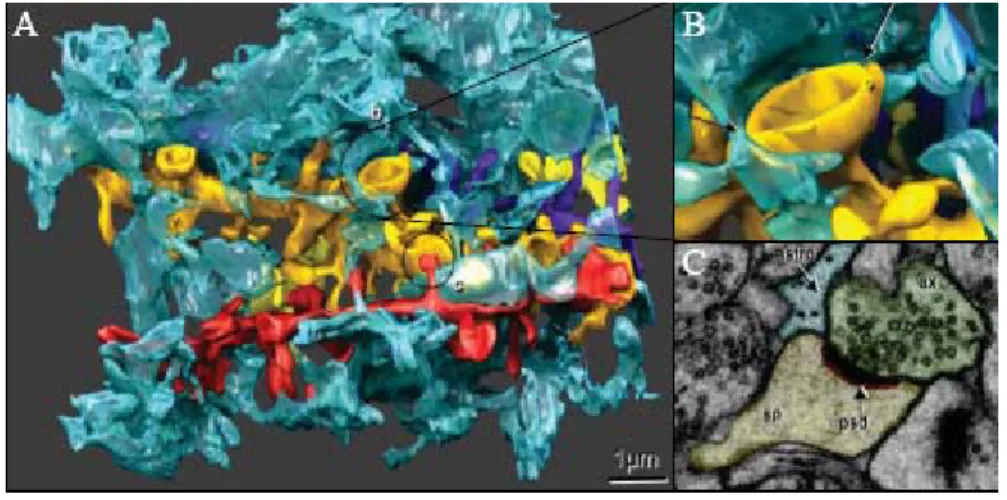
Figure 7: Anatomy of the tripartite synapse.
A) Fine processes (blue) enwrap four dendrites (orange, yellow, purple and red). (B) apse. (C) Electron microscopy image (
Enlargement of (A), fine processes enwrap the syn
showing tight interactions between neurons and astrocytes at the synapse. sp, dendritic spine; astro, astrocyte ; ax, axon ; psd, post-synaptic density.
Adapted from Witcher et al., 2007.
CHAPTER I: Glial cells and the tripartite synapse
C. SYNAPTIC SIGNAL DETECTIONlassical scheme of release of transmitter by e pre-synaptic neuron, binding on post-synaptic receptors and clearance from the late
Membrane transporters
Astro ar concentration of neurotransmitters,
hich is essential for the integrity of synaptic transmission.
e synaptic cleft is mainly ptaken by glutamate transporters in astrocytes (Bergles and Jahr, 1998; Rothstein Synaptic transmission is based on the c
th
synaptic cleft by diffusion, uptake and/or degradation in the extracellular space. Astrocytes play an important role in the transmission since they express transporters, ionotropic and metabotropic receptors that can detect signals and in turn modu synaptic activity.
i.
glial transporters regulate the extracellul w
As we previously saw, glutamate released by neurons in th u
et al., 1994). In the brain, 95 % of glutamate uptake is performed by GLT-1 which is the most prominently expressed glutamate transporter in the mammalian forebrain (Danbolt, 2001) and found almost exclusively on astrocytes. GLAST is also contributing to synaptic glutamate clearance (Rothstein et al., 1994). GLT-1-mediated glutamate removal from the tripartite synapse is conducted in an activity-regulated manner, through the membrane diffusion of GLT-1 (Murphy-Royal et al., 2015). By quickly removing glutamate from the synaptic cleft, astrocytes control the inappropriate diffusion of the neurotransmitter and limit the communication between neurons. Thus, they preserve the specificity and quality of the synaptic communication (Piet et al., 2004; Theodosis et al., 2008). Moreover, glutamate
uptake is concomitant with a Na+ co-transport (Meeks and Mennerick, 2007). This
large entrance of Na+ is important because it plays a signaling role. Indeed, it triggers
the entry of glucose into astrocytes from blood vessels to provide energy to neurons in an activity-dependent manner (Pellerin and Magistretti, 2004): a bigger neuronal
activity triggers high amount of glutamate released, higher Na+ entrance into
astrocytes and higher recruitment of glucose.
Besides glutamate transporters, astrocytes also express γ-aminobutyrique acid ABA) transporters (GAT-1 to 3), which can also influence synaptic activity. It was
the ew hypothesis that astrocytes play active roles in synaptic transmission (Kofuji and
Receptors
The role of astrocytes is not limited to uptake of neurotransmitters. Indeed astrocytes an also detect synaptic activity through the expression of a wide variety of iono- and (G
recently shown that activation of GAT-3 in the hippocampus leads to the release of ATP/adenosine by astrocytes, which then diffusely inhibits neuronal glutamate release via activation of pre-synaptic adenosine receptors (Boddum et al., 2016). The discovery of the control of neurotransmitter uptake by astrocytes suggested n
Newman, 2004).
ii.
c
metabotropic neurotransmitter receptors (Hamilton and Attwell, 2010; Perea and Araque, 2005a; Volterra and Meldolesi, 2005), which do not seem much different than the ones expressed by neurons. Receptors for neuropeptides, for purines, for 14
CHAPTER I: Glial cells and the tripartite synapse
lipids, as well as iono- and metabo-tropic GABA and glutamate receptors are all present in both neurons and astrocytes. The differences between astroglial and neuronal receptors seem to rely on the levels of expression as well as their subunit composition. Interestingly, the same receptors expressed either in neurons or in astrocytes could activate different intracellular cascades, as, for instance, is the case
for cannabinoid type 1 receptors (CB1) (Navarrete and Araque, 2008) (see also
Chapter III & Chapter IV).
Through the expression of transporters and receptors, astrocytes can detect neuronal ctivity at the level of the network. However it was recently shown that astrocytes can
D. SIGNAL INTEGRATION:CA2+ SIGNALING IN ASTROCYTES
re, these cells cannot se action potentials to propagate fast information like neurons. Indeed, they do not
or endogenous signals, such s neuronal activity within a range of milliseconds. Astrocytes express a variety of ion
anisms of Ca excitability
Ca2+ erent from the ones in neurons.
hile Ca increase in neuronal cell bodies happen in a range of milliseconds, it is a a
also detect activity at the level of a unique synapse. In the CA1 region of the hippocampus, astroglial mGluRs can be activated by glutamate release by the pre-synaptic neuron (Panatier and Robitaille, 2016; Panatier et al., 2011) and can detect the intensity and the frequency of the message (Panatier et al., 2011; Pasti et al., 1997; Perea and Araque, 2005b).
Astrocyte membranes are not electrically excitable and, therefo u
express voltage-dependent Na+ channels, they express numerous leakage channels
and have low membrane potential (Barres et al., 1990). Thus, they are electrically silent and they were considered as non excitable cells.
Nevertheless, astrocytes are able to detect exogenous a
channels and membrane receptors to react to environmental changes by slight
variation of membrane potentials and mostly by increase of intracellular Ca2+
concentrations (Barres et al., 1990; MacVicar and Tse, 1988; Marrero et al., 1989; McCarthy and Salm, 1991; Usowicz et al., 1989). These discoveries were essential to reconsider astrocytes, and glial cells in general as active partners of information processing in the CNS.
i. Mech 2+
increase mechanisms in astrocytes are diff
2+
W
hundred to thousand times slower in astrocyte somas (Parpura and Haydon, 2000).
This can be explained by the fact that Ca2+ increase in neurons is largely due to fast
opening of plasma membrane voltage-dependent ion channels, whereas it requires slower and molecularly more complex intracellular signaling cascades in astrocytes (Golovina and Blaustein, 2000; Haydon, 2001; Petravicz et al., 2008; Scemes, 2000; Scemes and Giaume, 2006; Sheppard et al., 1997).
The most widely accepted mechanism for astrocytic Ca increase is the canonical 2+
phospholipase C (PLC)/inositol 1,4,5-trisphosphate (IP3) pathway (Figure 8, page
16). Upon activation of GPCRs (seven-transmembrane membrane proteins coupled to G protein signaling), PLC hydrolyzes the membrane lipid phosphatidylinositol
4,5-bisphosphate to generate diacylglycerol (DAG) and IP3, leading to the activation of
CHAPTER I: Glial cells and the tripartite synapse
IP3 receptor (IP3R) and Ca2+release from intracellular organelles, such as the
endoplasmic reticulum (ER).
2
Figure 8: Schematic representation of the main mechanism of Ca + excitability
astrocytes. NT: Neurotransmitter; GPCR: G-Protein Coupled Receptor; PLC:
in
Phospholipase C; Ins(1,4,5); phosphatidylinositol 4,5-bisphosphate; DAG: diacylglycerol; IP3: Inositol tri-phosphate; ER: Endoplasmic reticulum; IP3R2: IP3 receptor type 2; Ca2+:
Calcium.
IP3Rs are a family of 3 different receptor isoforms that are permeable to Ca2+ and are
expressed at the ER membrane. We thought for a long time that astrocytes only
expressed the IP3R2 isoform (Petravicz et al., 2008; Sheppard et al., 1997; Weerth et
al., 2007). However, whilst functional IP3R1 and IP3R3 have not been reported in
hippocampal astroglia, mRNA for IP3R1 and IP3R3 were detected in acutely isolated
mouse astrocytes (Cahoy et al., 2008), and IP3R3 immunoreactivity was detected in
rodent brain sections (Sharp et al., 1999; Yamamoto-Hino et al., 1995). The
expression of multiple IP3R isoforms in astrocytes is particularly interesting, because
recent studies revealed that Ca2+ activity in large and finer processes of astrocytes from null mutant mice lacking the most abundant receptor (IP3R2-KO mice) is still
present (Di Castro et al., 2011; Haustein et al., 2014; Kanemaru et al., 2014; Rungta
et al., 2016; Srinivasan et al., 2015). These observations indicate the existence of
unidentified Ca2+ sources in astrocytic processes, raising the possibility that IP3R1
and IP3R3 subtypes could be functional in astrocytes. This hypothesis was recently
confirmed by Sherwood et al., who, by using genetic deletion of the different IP3R
isoforms in organotypic slices, demonstrated that multiple IP3R subtypes contribute to
Ca2+ signaling in CA1 hippocampal astrocytes (Sherwood et al., 2017).
16