HAL Id: hal-00552695
https://hal.archives-ouvertes.fr/hal-00552695
Submitted on 6 Jan 2011HAL is a multi-disciplinary open access
archive for the deposit and dissemination of sci-entific research documents, whether they are pub-lished or not. The documents may come from teaching and research institutions in France or abroad, or from public or private research centers.
L’archive ouverte pluridisciplinaire HAL, est destinée au dépôt et à la diffusion de documents scientifiques de niveau recherche, publiés ou non, émanant des établissements d’enseignement et de recherche français ou étrangers, des laboratoires publics ou privés.
Molecular analysis of Pericentrin gene (PCNT) in a
series of 24 Seckel/ MOPD II families
M. Willems, D Geneviève, G Borck, Clarisse Baumann, G Baujat, Eric Bieth,
P. Edery, C Farra, M Gérard, Delphine Héron, et al.
To cite this version:
M. Willems, D Geneviève, G Borck, Clarisse Baumann, G Baujat, et al.. Molecular analysis of Pericentrin gene (PCNT) in a series of 24 Seckel/ MOPD II families. Journal of Medical Genetics, BMJ Publishing Group, 2009, 47 (12), pp.jmg.2009.067298v2. �10.1136/jmg.2009.067298�. �hal-00552695�
Molecular analysis of Pericentrin gene (PCNT) in a series of 24 Seckel/ MOPD II families
M. Willems1, D. Geneviève1, G. Borck1, C. Baumann2, G. Baujat1, E. Bieth3, P. Edery4, C. Farra5, M. Gerard2, D. Héron6, B. Leheup7, M. Le Merrer1, S. Lyonnet1, D. Martin-Coignard8, M Mathieu9 , C. Thauvin-Robinet10, A. Verloes2, L. Colleaux1, A. Munnich1, V. Cormier-Daire1*
1. Université Paris Descartes, INSERM U781, Department of Genetics, Hôpital Necker, Paris, France
2. Department of Genetics, Hôpital Robert Debré, Paris, France 3. Department of Genetics, Hôpital Purpan, Toulouse, France
4. Hospices Civils de Lyon, Groupement Hospitalier Est, Service de Cytogénétique Constitutionnelle, Bron cedex F-69677 et Université Lyon 1, Lyon, F-69003
5.Department of Pathology and Laboratory Medicine American University of Beirut Medical Center, Beyrouth, Lebanon
6. Department of Genetics, Hôpital de la Pitié-Salpêtrière, Paris, France 7. Department of Genetics, CHU de Nancy, Nancy-Université, Nancy, France 8. Department of Genetics, Hôpital du Mans, France
9. Department of Pediatrics, CHU d’Amiens, France
10. Centre de Référence Anomalies du Développement et Syndromes Malformatifs Grand Est, Centre de Génétique, Hôpital d’Enfants, CHU Dijon, France
ABSTRACT
Microcephalic osteodysplastic primordial dwarfism type II (MOPD II, MIM 210720) and Seckel syndrome (SCKL, MIM 210600) belong to the primordial dwarfism group characterized by intrauterine growth retardation, severe proportionate short stature and marked microcephaly. MOPD II is distinct from SCKL by more severe growth retardation, radiological abnormalities and absent or mild mental retardation. Seckel syndrome is associated with defective ATR-dependent DNA damage signalling.
In 2008, loss-of-function mutations in the pericentrin gene (PCNT) have been identified in 28 patients, including 3 SCKL and 25 MOPDII cases [6, 7]. This gene encodes a centrosomal protein which plays a key role in the organization of mitotic spindles.
The aim of our study was to analyze PCNT in a large series of SCKL-MOPD II cases to further define the clinical spectrum associated with PCNT mutations. Among eighteen consanguineous families (13 SCKL and 5 MOPDII) and 6 isolated cases (3 SCKL and 3 MOPD II), we identified thirteen distinct mutations in 5/16 SCKL and 8/8 MOPDII including five stop mutations, five frameshift mutations, two splice site mutations and one apparent missense mutation affecting the last base of exon 19. Moreover, we demonstrated that this latter mutation leads to an abnormal splicing with a predicted premature termination of translation. The clinical analysis of the 5 SCKL cases with PCNT mutations showed that they all presented minor skeletal changes and clinical features compatible with MOPDII diagnosis. We therefore conclude that, despite variable severity, MOPDII is a genetically homogeneous condition due to loss-of function of pericentrin.
. KEY WORDS Seckel syndrome MOPDII Skeletal manifestations PCNT
INTRODUCTION
Among the primordial dwarfisms, microcephalic osteodysplastic primordial dwarfism type II (MOPD II, MIM 210720) and Seckel syndrome (SCKL, MIM 210600) are both characterized by intrauterine growth retardation, severe proportionate short stature and microcephaly [1, 2]. MOPDII is distinct from SCKL by the severity of the growth retardation, the presence of skeleton abnormalities and the mild/ absent mental retardation [3]. SCKL is a genetically heterogeneous condition associated with defective ATR-dependent DNA damage signalling [4]. The only reported genetic defect so far is a hypomorphic mutation in the ATR gene (Sckl1, 3q22.1-q24) [5]. In 2008, mutations in the pericentrin gene (PCNT) have been identified in 28 patients, including 3 patients with SCKL [6] and 25 with MOPDII [7].This gene encodes a centrosomal protein, which acts both at structural and regulatory levels. First, pericentrin recruits several structural centrosomal proteins, particularly gamma tubulin ring complex which initiates microtubular nucleation and spindle organization [8, 9, 10, 11]. Second, it plays a role in cell cycle regulation through its interaction with the ATR pathway [6]. All mutations identified so far lead to premature translation termination and are responsible for pericentrin loss of function as demonstrated in PCNT-mutated cell lines issued from patients with SCKL or MOPDII.
To further define the clinical spectrum of patients with PCNT mutations, we analyzed PCNT in 24 families diagnosed either with SCKL or with MOPD II, including 18 consanguineous families and 6 cases from unrelated parents.
PATIENTS AND METHODS
Patients
Criteria for inclusion in the study were:
- Intrauterine and postnatal growth retardation with birth weight < -2 SD and postnatal height < -4 SD
- Microcephaly with an occipitofrontal circumference (OFC) < -4 SD - Diagnosis of MOPDII or SCKL made by a clinical geneticist.
Diagnostic assessment was performed for all patients by their clinicians (Table 1). Seven of these families were previously clinically reported, by Faivre et al [12] (families 1,3,7, 8, 11 and 12) and Verloes et al [13] (family 19). Written informed consent was obtained from all subjects included in this study.
Microsatellites marker analysis
In all consanguineous families, microsatellites analysis of the PCNT locus was first performed and PCNT was sequenced only in compatible cases. The PCNT sequence analysis was performed in all cases from unrelated parents. Blood samples were obtained with informed consent from affected children, parents and unaffected siblings. Genomic DNA was extracted using Nucleospin® Blood XL kit (Macherey-Nagel). We established lymphoblastoid cell lines by EBV transformation and we performed a primary skin fibroblasts culture. Genotyping was performed using 4 flanking (D21S1903, D21S1897, D21SpolyATT, D21S1446) and one intragenic-PCNT (PCNT-IG) microsatellite markers in all consanguineous families and non-consanguineous families with at least two siblings.
Mutation analysis
PCNT exon and flanking intron sequences were amplified from patient DNA by PCR using 49 couples of primers designed with the Primer 3 software (Sequence of primers available on request). Sequencing reactions were performed on both strands using the BigDye Terminator Cycle Sequencing Kit v3.1 (Applied Biosystems, Foster City, California) according to the manufacturer’s instructions.
Functional consequences of c.3840G>C( p.Q1280H) mutation
RNA was extracted from cultured fibroblasts and cDNA was synthetized using primers designed to overlap 2 consecutive exons (Sequence of primers: JUNCTION17/18-5': CCTGTCCCACAGCGAAAGAG; JUNCTION18/19-5': ACAGCTCCGGCGCTGGAG; JUNCTION20/21-3': GGGTACCCTGAGTCTTGTGCAGC). RT-PCR products were expected to span exons 18 to 20 and exons 19 to 20. GAPDH expression was used as positive controls.
RESULTS
Genotyping analysis showed that 9/18 consanguineous families (4/13 SCKL and 5/5 MOPDII) were compatible with linkage to the PCNT locus.
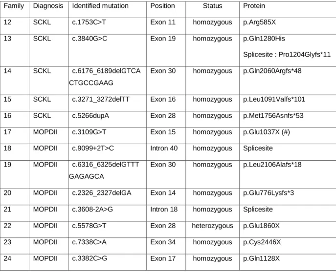
The PCNT sequence analysis performed in these 9 consanguineous families and in 6 additional cases (from unrelated parents) allowed the identification of 13 distinct mutations in 13 patients (Table 2). All but one mutation were homozygous and cosegregate with the disease. In one patient (case 22), a single heterozygous mutation, inherited from the father, was detected. The 13 mutations were located throughout the gene and among them, five were nonsense mutations (exons 11, 15, 17, 28 and 34), five were frameshift mutations (exons 14, 16, 28A, 30), two were splice site mutations (intron 18 and intron 40) and one was an apparent missense mutation affecting the last base of exon 19 (c.3840G>C, p.Q1280H). None
of the mutations were identified in 400 control chromosomes. The c.3840G>C mutation was predicted to alter splicing. This was further confirmed by sequence analysis of RT-PCR products which demonstrated exon 19 skipping, predictive of a premature termination of translation (p.P1204GfsX11).
Among the 13 patients, 5 were clinically diagnosed as SCKL (patients12-16) and 8 were diagnosed as MOPDII (patients 17-24, Table 1). The identification of PCNT mutations in 5 SCKL and 8 MOPDII patients prompted us to re-analyze the clinical features of all the patients with PCNT mutations and compare them to SCKL patients without mutation. We first observed that the five SCKL patients with PCNT mutations (families 12-16) presented a more severe growth retardation than the SCKL patients without PCNT mutation (-6 to -8 SD versus -4 to -5 SD), but less severe than the MOPDII patients with PCNT mutation (-7 to -13 SD). For two of them, the adult height is 120 cm and 140 cm respectively (Figure 1, from Hall JG). These patients presented also skeletal anomalies including gracile long bones, metaphyseal flaring, carpal condensation, and moderate hip dysplasia (Figures 2, 3, 4). These anomalies were not present during the first year of life, became more pronounced with time but were often less severe than those classically described in MOPDII patients. Importantly, we did not observe similar skeletal anomalies in 6 patients without PCNT mutation and with skeletal survey available. Finally, these patients have either normal intelligence or mild mental retardation. No difference was observed with respect to age of walking (normal to slightly delayed) and developmental course. Early developmental milestones were considered as normal with excellent social skills. Learning disability was noted after the age of 5 years. None of the 4 adult patients can live independently and they all perform “adapted work”.
Other features suggestive of MOPDII diagnosis were present in the clinically diagnosed SCKL patients with PCNT mutations including truncal obesity and death at 20 years of age of rupture of CNS vessels (patient 15), initial feeding difficulties and development of typical pigmentation anomalies with time (patients 13 and 14), polycystic ovaries (patient 14), subglottic stenosis (patient 13), microdontia (patient 12), high-pitched voice, stridor and upper respiratory tract infections (patient 16).
Finally, facial features were highly suggestive of MOPD diagnosis for 11 patients with PCNT mutations including a broad nose with hypoplastic tip, thin alae nasi, with columella lying below the alae nasi, long midface, prominent cheeks, small jaw and large eyes in the youngest children. Facial features were also changing with time, variable with the ethnic origin (patient 18) and less characteristic in the eldest patient of our series (patient15). By contrast facial features of the patients with no PCNT mutation (1-11) were quite variable
(Figure S1) mainly dominated by the microcephaly with receiding or short forehead and relatively large ears.
DISCUSSION
We report here the identification of 13 distinct mutations in 8 MOPDII and 5 SCKL patients. As previously reported by Griffith [6] and Rauch [7], mutations are distributed throughout the gene. We did not find any recurrent mutations in our series. However, the c.3109G>T mutation (exon 15) was previously reported by Rauch et al in a patient also originating from Turkey (patient 1). In one patient (patient 22), one mutation only was detected by direct sequencing but unfortunately RNA was not available. This might be due to the limit of our screening and a partial deletion of PCNT gene cannot be excluded. Our study provides also the first example of a “missense” mutation (c.3840G>C) but we demonstrated that this mutation impairs exon 19 splicing, leading to premature termination of translation. We conclude, as previously suggested, that all identified mutations are loss of function mutations.
We identified PCNT mutations in all MOPDII cases, confirming the genetic homogeneity of this disorder. Moreover, the retrospective analysis of the 5 SCKL patients with PCNT mutation also suggests that they all belong to the MOPDII spectrum. However, they were diagnosed as SCKL, based on the absence of severe skeletal manifestations and on their final stature >110 cm, which usually excludes the diagnosis of MOPDII [3]. Our study also supports that SCKL spectrum is heterogeneous and suffers from variable definition in the literature and from clinicians in practice. Indeed, Seckel syndrome has often been used as a generic term used for primordial dwarfism, without more specific diagnosis. Recently, D’Angelo and Di Bartolomeo reported two cases of SCKL with intracranial anomalies, suggestive of MOPDII diagnosis [16, 17]. Similarly, SCKL patients with bone dysplasia suggestive of MOPDII have been reported [18, 19]. From our study, we suggest that MOPDII spectrum is wider than previously defined. However, in all patients with PCNT mutations we have consistently observed 1) distinct facial features 2) growth retardation <-5SD and microcephaly <-4SD, 3) mild to absent mental retardation 4) skeletal manifestations including hip dysplasia ranging from short femoral neck to severe coxa vara; carpal condensation, and gracile long bones with metaphyseal flaring. Other suggestive features occasionally observed included 1) vascular anomalies and cutis marmorata 2) high pitched voice 3) microdontia, 4) hyperinsulinism, 5) subglottic stenosis, 6) pigmentation anomalies with areas of hypo- and hyperpigmentation.
We also observed in two patients with PCNT mutation a liver involvement varying in severity from cytolysis to cirrhosis, with the same histological features than those described in the literature for patients with MOPDI diagnosis, consisting in ductular cholestasis, inflammatory infiltrate, and giant multinucleate hepatocytes. Although theses findings are not specific, they may suggest the existence of biliar epithelium anomalies in MOPDII spectrum [20].
While this study further demonstrates that MOPDII is caused by PCNT mutations, the pathogenic mechanisms underlying the clinical features observed in these patients remain unclear [21]. First, microcephaly could be related to structural centrosomal abnormalities similar to those observed in primary microcephaly [22]. Second, defect in ATR-dependent DNA damage signalling has been demonstrated in other conditions characterized by short stature and microcephaly and may thereby also account for short stature observed in MOPDII cases [23, 24]. Other specific clinical features like vessels anomalies, generalized bone dysplasia and hepatitis remain unexplained so far, since they have not described in patients with ATR mutations or other centrosomal genes such as ASPM.
Finally, O’Driscoll and collaborators suggested that the ATR signalling pathway was unusually sensitive to haploinsufficiency and established a correlation between ATR-pathway dysfunction and growth retardation [23]. Similarly, Rauch et al reported a significant reduction of the mean height of heterozygous MOPDII parents [7]. We did not observe such a reduction in the mean of parental heights (ranging from -1 to +2 SD) but ethnical variability of growth charts may interfere with parental height analysis.
In conclusion, we identified 13 PCNT loss of function mutations in 13 patients who all presented diagnostic criteria for MOPDII. However, we observed a wider variability in the severity of the short stature and skeletal manifestations than previously admitted, modifying the MOPDII clinical spectrum. The distinction between SCKL and MOPDII appears to be crucial for the appropriate management, keeping in mind the risk of vascular anomalies in MOPDII.
ACKNOWLEDGEMENTS
We thank all the patients and their families for their contribution to this work. We also thank Professor Maroteaux for reviewing the skeleton X-rays.
REFERENCES
1. Majewski, F., Ranke, M. & Schinzel, A. Studies of microcephalic primordial dwarfism II: the osteodysplastic type II of primordial dwarfism. Am J Med Genet 12, 23-35 (1982).
2. Seckel, H. Bird-Headed Dwarfs: Studies in Developmental Anthropology Including
Human Proportions. Springfield, IL:Charles C Thomas (1960).
3. Hall, J.G., Flora, C., Scott, C.I., Jr., Pauli, R.M. & Tanaka, K.I. Majewski osteodysplastic primordial dwarfism type II (MOPD II): natural history and clinical findings. Am J Med Genet A 130A, 55-72 (2004).
4. Alderton, G.K. et al. Seckel syndrome exhibits cellular features demonstrating defects in the ATR-signalling pathway. Hum Mol Genet 13, 3127-38 (2004).
5. O'Driscoll, M., Ruiz-Perez, V.L., Woods, C.G., Jeggo, P.A. & Goodship, J.A. A splicing mutation affecting expression of ataxia-telangiectasia and Rad3-related protein (ATR) results in Seckel syndrome. Nat Genet 33, 497-501 (2003).
6. Griffith, E. et al. Mutations in pericentrin cause Seckel syndrome with defective ATR-dependent DNA damage signaling. Nat Genet 40, 232-6 (2008).
7. Rauch, A. et al. Mutations in the pericentrin (PCNT) gene cause primordial dwarfism. Science 319, 816-9 (2008).
8. Miyoshi, K. et al. DISC1 localizes to the centrosome by binding to kendrin. Biochem Biophys Res Commun 317, 1195-9 (2004).
9. Doxsey, S.J., Stein, P., Evans, L., Calarco, P.D. & Kirschner, M. Pericentrin, a highly conserved centrosome protein involved in microtubule organization. Cell 76, 639-50 (1994). 10. Dictenberg, J.B. et al. Pericentrin and gamma-tubulin form a protein complex and are organized into a novel lattice at the centrosome. J Cell Biol 141, 163-74 (1998).
11. Zimmerman, W.C., Sillibourne, J., Rosa, J. & Doxsey, S.J. Mitosis-specific anchoring of gamma tubulin complexes by pericentrin controls spindle organization and mitotic entry. Mol Biol Cell 15, 3642-57 (2004).
12. Faivre, L. et al. Clinical and genetic heterogeneity of Seckel syndrome. Am J Med Genet 112, 379-83 (2002).
13. Verloes, A., Lambrechts, L., Senterre, J. & Lambotte, C. Microcephalic osteodysplastic dwarfism (type II-like) in siblings. Clin Genet 32, 88-94 (1987).
14. Brancati, F., Castori, M., Mingarelli, R. & Dallapiccola, B. Majewski osteodysplastic primordial dwarfism type II (MOPD II) complicated by stroke: clinical report and review of cerebral vascular anomalies. Am J Med Genet A 139, 212-5 (2005).
Genet 22, 192-201 (1985).
16. Di Bartolomeo, R. et al. Malignant hypertension and cerebral haemorrhage in Seckel syndrome. Eur J Pediatr 162, 860-2 (2003).
17. D'Angelo, V.A., Ceddia, A.M., Zelante, L. & Florio, F.P. Multiple intracranial aneurysms in a patient with Seckel syndrome. Childs Nerv Syst 14, 82-4 (1998).
18. Kjaer, I., Hansen, N., Becktor, K.B., Birkebaek, N. & Balslev, T. Craniofacial morphology, dentition, and skeletal maturity in four siblings with Seckel syndrome. Cleft Palate Craniofac J 38, 645-51 (2001).
19. Poznanski, A.K., Iannaccone, G., Pasquino, A.M. & Boscherini, B. Radiological findings in the hand in Seckel syndrome (bird-headed dwarfism). Pediatr Radiol 13, 19-24 (1983).
20. Deniz, K., Kontas, O. & akcakus, M. Neonatal hepatitis in 2 siblings with Seckel syndrome. Pediatr Dev Pathol 9, 81-5 (2006).
21. Delaval, B. & Doxsey, S. Genetics. Dwarfism, where pericentrin gains stature. Science 319, 732-3 (2008).
22. Cox, J., Jackson, A.P., Bond, J. & Woods, C.G. What primary microcephaly can tell us about brain growth. Trends Mol Med 12, 358-66 (2006).
23. O'Driscoll, M., Dobyns, W.B., van Hagen, J.M. & Jeggo, P.A. Cellular and clinical impact of haploinsufficiency for genes involved in ATR signaling. Am J Hum Genet 81, 77-86 (2007).
24. O'Driscoll, M., Jackson, A.P. & Jeggo, P.A. Microcephalin: a causal link between impaired damage response signalling and microcephaly. Cell Cycle 5, 2339-44 (2006).
The Corresponding Author has the right to grant on behalf of all authors and does grant on behalf of all authors, an exclusive licence (or non exclusive for government employees) on a worldwide basis to the BMJ Publishing Group Ltd to permit this article (if accepted) to be published in JMG and any other BMJPGL products and sublicences such use and exploit all subsidiary rights, as set out in our licence (http://jmg.bmj.com/ifora/licence.pdf)."
Legends to figures
Figure 1: Comparison of growth curves of patients from our cohort with those of patients from MOPD II cohort reported by Hall JG et al (from Hall JG, 2004)
Figure 2: Comparison of Hip X-rays in patients with PCNT mutations. Note the hip dislocation and coxa vara more pronounced in MOPD II patients Figure 3: Comparison of lower limb X-rays in patients with PCNT mutations. Note the gracile long bones in both groups.
Figure 4: Comparison of hand X-rays in patients with PCNT mutations Note the carpal fusion, the clinodactyly and the delayed bone age.
Figure S1. Facial features in two sisters with no PCNT mutation (A and B, family 7) and two patients with PCNT mutation (C1 and C2, Patient 14; D1 and D2, Patient 17).
Note in patient with PCNT mutations the characteristic nose with hypoplastic alae nasi, prominent cheeks and small jaw while in the two patients with no PCNT mutation the facial features are marked by the severe microcephaly with large nose and large ears.
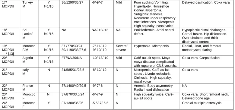
Family Ethnic origin/ Gender CS /Coefficient f Birth : WOG/ Weight(g)/Heigh t(cm)/HC(cm) Postnatal growth : Weight /Height/ HC (SDS) Mental retardati on
Other clinical features Radiological features
1 /SCKL* (case 4[12]) Algeria F Y f=1/64
41/2160/45/32 -4/-4/-4 Mild Café au lait spots. VSD.
Pyelic bifidity. Osteosarcoma
Mild bowing of the femora
2/SCKL NA M Y NA NA NA NA 3/ SCKL* (case 3[12]) Mali M Y f=1/8
FT/2000/40/24 -4/-5 ,5/-10 Severe Hypertonia. Ichtyosis.
Abnormal gyration pattern
Slight platyspondyly 4/SCKL France M Y f=1/16 NA NA NA NA NA 5/SCKL Algeria F/F Y NA NA NA NA NA 6/SCKL Algeria M Y f=1/32
FT/3300/47/30,5 -4/-4/-6 Mild Hip dislocation.
Chromosomal breakage Normal 7/SCKL* (case 2[12]) Morocco F/F Y f=1/16 FT/1400/40/28 FT/NA/40/NA Mild/ N
Delayed puberty. Cataract Catarct Thoracolumbar scoliosis 8/SCKL* (case 5[12]) Algeria M/M/M Y f=1/16 FT/2040/43,5/30 FT/2140//42/NA FT/2300/NA/NA NA/-5/NA NA/-5/NA NA/-4/NA Mild Cryptorchidia.Diabetes mellitus. Craniosynostosis
Scoliosis. Thick long bones.
9/SCKL Lebanon F Y NA NA NA NA NA NA 10/SCKL France F/F N FT/2030/41/NA NA/NA/NA/NA -3.5/-7/-6 -5/-8/-7.5 N/ Mild Pyelic duplicity Scoliosis NA Severe scoliosis 11/SCKL* (case 6[12]) France M/F N NA NA NA NA/-4/-8 NA/-4/-6 NA 12/ SCKL* (case 1[12]) Morocco F/NA (TOP) Y f=1/16 37/1340/38/28 -8/-8/-8 Mild Microdontia. Pyelic ectasia
Thick diaphyseal cortex. Carpal fusion. Gracile long bones. Brachymesophalangia
13/SCKL Morocco F
Y f=1/16
32/950/34/25,5 -5/-8/-6.6 N Café au lait spots, areas of
depigmention Hepatic cytolysis. Subglottic stenosis Recurrent upper respiratory tract infections Coxa vara 14/SCKL Pakistan F Y f=1/16
FT/1650/42/30 -3.5/-7/-5 N Café au lait spots,
area of depigmentation. Polycystic ovaries. Chromosomal breakage
Gracile long bones. Short femoral neck. High vertebral bodies. Carpal fusion. Overtubulated and thick diaphyseal cortex. 15/SCKL France
M
N FT/1720/40/27,3 -2.7/-6/-8 Mild Horseshoe kidney.
Clinodactyly of fifth finger. Rupture of CNS vessels leading to death (20 years)
High vertebral bodies. Thick diaphyseal cortex. Short femoral neck
16/SCKL Lebanon M
Y F=1/16
NA/800/30/NA NA/-9/-13 Y Sparse scalp hair.
Receeding forehead. Prominent curved nose, Micrognatia. Low set ears. Clinodactyly. High-pitched voice. Stridor. Upper respiratory tract infections
Brain MRI : minimal bilateral ill-defined areas of hypersignal intensity in white matter (terminal zone of myelination) Skeletal X rays: NA
Table 1: Clinical and radiological features of the 24 families. Patients 1-11: Patients with Seckel diagnosis - PCNT excluded
Patients 12-16: Patients with Seckel diagnosis - PCNT mutation identified Patients 17-24: Patients with MOPDII diagnosis - PCNT mutation identified
CNS: central nervous system, CS : consanguinity, F: female, FT: full-term pregnancy, HC : head circumference, M: male, MOPDII: Patient with microcephalic osteodysplastic primordial dwarfism type II syndrome N: no, NA: non available, SCKL: Patient with Seckel syndrome, VSD: ventricular septal defect, WOG: week of gestation, Y: presence
17/ MOPDII Turkey M Y f=1/16
36/1290/35/27 -6/-9/-7 Mild Poor sucking.Vomiting.
Hyperlaxity. Horseshoe kidney.Hypertonia. Subglottic stenosis. Recurrent upper respiratory tract infections. Micropenis High squeaky, nasal voice
Delayed ossification. Coxa vara
18/ MOPDII Sri Lanka/ M Y f=1/16
NA NA/-12/-12 NA Poïkilodermia. Atrial septal
defect.
Hypoplastic distal phalanges. Carpal fusion. Hip dislocation. Overtubulated and thick diaphyseal cortex 19/ MOPDII * [13] Morocco F/M Y f=1/16 37 /770/30/24 39/1190/33/27,6 -7/-11/-12 -9/-10/-10 Severe/ severe
Hypertonia. Micropenis. Radial, ulnar, and femoral metaphyseal flaring. 20/ MOPDII Algeria M Y f=1/16
FT/NA/30/NA -10/-13/-10 Mild Café au lait spots.
Moya-moya disease complicated with rupture of CNS vessels.
Coxa vara. Carpal fusion
21/ MOPDII
Italy M
N 31/585/31/23,5 -8/-12/-12 N Micropenis. Café au lait
spots . Livedo reticularis. Cirrhosis. High squeaky, nasal voice Coxa vara 22/ MOPDII France M
N 37/1400/40/29,5 -9/-7/-6 N Anemia. Body asymmetry
Radial head dislocation
NA
23/ MOPDII
Morocco F
N 37/870/33,5/24 -6/-7/-9 N High squeaky voice.
Café-au-lait spots
Coxa vara. Short femoral neck. Delayed bone age.
24/ MOPDII
Morocco F
Table 2: Mutations identified in our series. (#) This mutation was previously identified by Rauch in a MOPDII patient with the same Turkish ethnic background
Family Diagnosis Identified mutation Position Status Protein
12 SCKL c.1753C>T Exon 11 homozygous p.Arg585X
13 SCKL c.3840G>C Exon 19 homozygous p.Gln1280His
Splicesite : Pro1204Glyfs*11
14 SCKL c.6176_6189delGTCA
GCTGCCGAAG
Exon 30 homozygous p.Gln2060Argfs*48
15 SCKL c.3271_3272delTT Exon 16 homozygous p.Leu1091Valfs*101
16 SCKL c.5266dupA Exon 28 homozygous p.Met1756Asnfs*53
17 MOPDII c.3109G>T Exon 15 homozygous p.Glu1037X (#)
18 MOPDII c.9099+2T>C Intron 40 homozygous Splicesite
19 MOPDII c.6316_6325delGTTT
GGAGAGCA
Exon 30 homozygous p.Leu2106Alafs*18
20 MOPDII c.2326_2327delGA Exon 14 homozygous p.Glu776Lysfs*3
21 MOPDII c.3608-2A>G Intron 18 homozygous Splicesite
22 MOPDII c.5578G>T Exon 28 heterozygous p.Glu1860X
23 MOPDII c.7338C>A Exon 34 homozygous p.Cys2446X