HAL Id: hal-01589451
https://hal.archives-ouvertes.fr/hal-01589451
Submitted on 23 Sep 2017
HAL is a multi-disciplinary open access archive for the deposit and dissemination of sci-entific research documents, whether they are pub-lished or not. The documents may come from teaching and research institutions in France or abroad, or from public or private research centers.
L’archive ouverte pluridisciplinaire HAL, est destinée au dépôt et à la diffusion de documents scientifiques de niveau recherche, publiés ou non, émanant des établissements d’enseignement et de recherche français ou étrangers, des laboratoires publics ou privés.
Cytokines in Atherosclerosis: Pathogenic and
Regulatory Pathways
Alain Tedgui, Ziad Mallat
To cite this version:
Alain Tedgui, Ziad Mallat. Cytokines in Atherosclerosis: Pathogenic and Regulatory Pathways. Physiological Reviews, American Physiological Society, 2006, 86 (2), pp.515-581. �10.1152/phys-rev.00024.2005�. �hal-01589451�
Cytokines in Atherosclerosis:
Pathogenic and Regulatory Pathways
ALAIN TEDGUI AND ZIAD MALLAT
Institut National de la Sante´ et de la Recherche Me´dicale U. 689, Cardiovascular Research Center Lariboisiere, and University Paris 7, Paris, France
I. Introduction 516
A. Historical perspective 516
B. Atherosclerosis as an immunoinflammatory disease 517
II. The Atherosclerotic Cytokine Network 518
A. Cytokine families 518
B. Cytokine-associated signaling pathways 519
III. Inducers of Cytokine Production in Atherosclerosis 526
A. Initial trigger(s) 526
B. Secondary triggers 528
IV. Cytokines and Cytokine Receptors in Human Atherosclerotic Plaques 535
A. Cytokine expression in plaques 535
B. Cellular sources of cytokines 535
C. Biological effects of cytokines 538
V. Cytokine and Cytokine Receptor-Associated Modulation of Plaque Development and Stability 541
A. Proinflammatory cytokines 541
B. Anti-inflammatory cytokines 545
C. Chemokines/chemokine receptors 546
D. Hematopoietic factors/M-CSF 547
E. Platelet-derived factors 548
VI. Cytokines and Adaptive Immunity in Atherosclerosis 548
A. Role of T/B cells in atherosclerosis 548
B. Cytokines and pathogenic immune response in atherosclerosis 549
C. Immunological tolerance and regulatory T cells 551
VII. Cytokines and Cardiovascular Risk 555
A. TNF-␣ 555 B. IL-2 556 C. IL-6 556 D. IL-7 556 E. IL-8 556 F. IL-18 556 G. sCD40L 556 H. IL-10 557 I. M-CSF 557
VIII. Therapeutic Potential 557
A. Use of anticytokines 557
B. Targetting downstream inflammasome 557
C. Targetting the JAK/STAT pathway 558
D. Activation of the natural anti-inflammatory intracellular pathway (SOCS) 558
E. Stimulation of Treg cells 558
F. Stimulation of macrophage emigration from atherosclerotic lesions 559
IX. Conclusion 559
Tedgui, Alain, and Ziad Mallat.Cytokines in Atherosclerosis: Pathogenic and Regulatory Pathways. Physiol Rev
86: 515–581, 2006; doi:10.1152/physrev.00024.2005.—Atherosclerosis is a chronic disease of the arterial wall where both innate and adaptive immunoinflammatory mechanisms are involved. Inflammation is central at all stages of
doi:10.1152/physrev.00024.2005.
on April 7, 2006
physrev.physiology.org
atherosclerosis. It is implicated in the formation of early fatty streaks, when the endothelium is activated and expresses chemokines and adhesion molecules leading to monocyte/lymphocyte recruitment and infiltration into the subendothelium. It also acts at the onset of adverse clinical vascular events, when activated cells within the plaque secrete matrix proteases that degrade extracellular matrix proteins and weaken the fibrous cap, leading to rupture and thrombus formation. Cells involved in the atherosclerotic process secrete and are activated by soluble factors, known as cytokines. Important recent advances in the comprehension of the mechanisms of atherosclerosis provided evidence that the immunoinflammatory response in atherosclerosis is modulated by regulatory pathways, in which the two anti-inflammatory cytokines interleukin-10 and transforming growth factor- play a critical role. The purpose of this review is to bring together the current information concerning the role of cytokines in the development, progression, and complications of atherosclerosis. Specific emphasis is placed on the contribution of pro- and anti-inflammatory cytokines to pathogenic (innate and adaptive) and regulatory immunity in the context of atherosclerosis. Based on our current knowledge of the role of cytokines in atherosclerosis, we propose some novel therapeutic strategies to combat this disease. In addition, we discuss the potential of circulating cytokine levels as biomarkers of coronary artery disease.
I. INTRODUCTION
Atherosclerosis is a pathological condition that un-derlies several important adverse vascular events includ-ing coronary artery disease (CAD), stroke, and peripheral arterial disease, responsible for most of the cardiovascu-lar morbidity and mortality in the Western world today. Epidemiological studies indicate that the prevalence of atherosclerosis is increasing all over the world due to the adoption of Western life-style and is likely to reach epi-demic proportions in the coming decades (72, 412).
The earliest visible lesion in the development of atherosclerosis is the fatty streak. This comprises an area of intimal thickening composed of macrophages distended by lipid droplets (known as foam cells), lym-phocytes, and smooth muscle cells. The American Heart Association (AHA) Committee on Vascular Le-sions provided a classification of human atheroscle-rotic lesions which correlate the histological lesion types, from type I to type VI, with corresponding clin-ical syndromes (648, 649). This classification should not be understood as an orderly, linear pattern of plaque progression (704). Plaques develop as a result of the accumulation of low-density lipoproteins (LDL) in the subendothelial space, followed by the diapedesis of leukocytes and formation of foam cells, proliferation of smooth muscle cells, and production of connective tis-sue. The landmark work of Seymour Glagov showed that the arterial wall can remodel itself in response to plaque growth by increasing its external diameter to accommodate the plaque without narrowing of the lu-men (234). Thrombosis is the ultimate stage in the disease process that is responsible for clinically observ-able adverse events implicating coronary, cerebrovas-cular, and peripheral vascular beds (394). Studies indi-cate that in patients with atherothrombotic disease plaque formation is likely to be widespread throughout the vasculature, often affecting more than one vascular bed (93).
A. Historical Perspective
Even though atherosclerosis is reaching epidemic proportions nowadays, it is not in any way a disease specific to the modern times; it was already present in antiquity. Sir Marc Ruffer was able to identify in 1911 degenerative arterial changes suggestive of atherosclero-sis in the left subclavian artery from an Egyptian mummy (583). Later on, paleopathologist A. T. Sandison, using modern technical methods for tissue fixation, confirmed that Egyptian mummies had histological evidence of ath-erosclerosis with lipid deposits, reduplication of the in-ternal elastic lamina, and medial calcification in arteries (593).
Atherosclerosis is nowadays recognized as a chronic inflammatory disease of large arteries (235, 265, 395, 417, 578). Remarkably, the very first description of the cause of angina pectoris referred to inflammation. Yet, the belief in this notion was subjected to peaks and troughs from early dates up to recent times.
According to the historian J. O. Leibowitz (381), the Italian surgeon and anatomist Antonio Scarpa (1752–1832) was the first to present an anatomopathologi-cal description of arterial wall degeneration in full detail. In his 1804 monograph on aneurysms, Scarpa opposed the view that a dilatation of the aorta was the intrinsic cause of an aneurysm leading to rupture. He emphasizes that “. . . especially the internal coat is subject, from slow internal cause, to an ulcerated and steatomatous disorga-nization, as well as to a squamous and earthy rigidity and brittleness,” introducing the concept of an underlying metabolic disorder in the process of atherosclerosis, rather than the theory of inflammation that already pre-vailed at that time, the expression “heart abscess” being frequently used to describe heart pathology (reviewed in Ref. 381).
The term atheroma, derived from Greek and meaning “porridge,” was first proposed by Albrecht von Haller in 1755 to designate the degenerative process observed in the intima of arteries. London surgeon Joseph Hodgson
on April 7, 2006
physrev.physiology.org
(1788 –1869) published in 1815 his Treatise on the Dis-eases of Arteries and Veins in which he claimed that inflammation was the underlying cause of atheromatous arteries. But thereafter, most of pathologists of the 19th century following Carl Rokitanski (1804 –1878) aban-doned the view that inflammation was an etiological fac-tor and considered that atherosclerosis was a degenera-tive process, with intimal proliferation of connecdegenera-tive tis-sue and calcification, best described by the term arteriosclerosisproposed in 1833 by French pathologist Jean Lobstein (1777–1835). However, German pathologist Rudolf Virchow (1821–1902), a leading authority of his day in pathology and the greatest contributor to the no-tion of thrombosis, considered atheroma as a chronic inflammatory disease of the intima, that he called “chronic endarteritis deformans”. In his opinion, the accumulation of lipids was a late manifestation of ath-eroma (701). Finally, the Leipzig pathologist Marchand in 1904 first used the term atherosclerosis, which since has been widely adopted, instead of arteriosclerosis, to designate the degenerative process of the intimal layer of the arteries.
Until the beginning of the 20th century, the theories put forward to explain the pathogenesis of atherosclero-sis remained purely descriptive and were based on the anatomical observation of human atherosclerotic vessels. A first revolution in the mechanistic assessment of ath-erosclerosis was initiated in 1908 when the Russian sci-entist Alexander Ignatowski showed that experimental atherosclerosis could be induced in rabbits by feeding them a diet of milk and egg yolk (301). Soon thereafter, in 1913, N. Anitschkov and S. Chalatov reproduced experi-mental atherosclerosis by adding pure cholesterol to rab-bit food (21). This gave rise to the lipid theory of athero-sclerosis that predominated for most of the 20th century. The next significant leap only came during the 1970s when Brown and Goldstein showed that the LDL receptor that they had discovered, a cell surface protein that binds LDL and removes them from blood (reviewed in Ref. 88), is not involved in macrophage foam-cell formation and pro-posed that a macrophage receptor that recognized acety-lated LDL plays a key role in this process (237). Subse-quently, during the 1980s, the central role of oxidized LDL (oxLDL) in the pathogenesis of atherosclerosis was ex-posed by Daniel Steinberg and his group (650), and a number of scavenger receptors mediating their uptake by macrophages were identified (reviewed in Ref. 387). The model of the Watanabe heritable hyperlipidemic (WHHL) rabbit, introduced in 1980 (726) was particularly useful in establishing the role of oxLDL in atherogenesis. A second revolution occurred at the beginning of the 1990s when mouse models of atherosclerosis, apolipoprotein E (apoE)- and LDL receptor (LDLr)-deficient mice, were derived by homologous recombination techniques (304, 306, 543, 784). In contrast to the previous models, mice
lacking functional apoE or LDLr genes were shown to develop widely distributed arterial lesions that progress from foam cell-rich fatty streaks to fibro-proliferative plaques with lipid/necrotic cores, typical of the spectrum of human lesions (305, 487, 564). The possibility of abol-ishing the expression of a single gene of interest, or of overexpressing it, in these mouse models opened a new era of atherosclerosis research at a mechanistic level.
B. Atherosclerosis as an
Immunoinflammatory Disease
A ripple in the lipid theory appeared in the mid 1970s, when Russel Ross developed his popular “response to injury” hypothesis of atherogenesis, postulating that ath-erosclerotic lesions arise as a result of focal injury to the arterial endothelium, followed by adherence and aggrega-tion of platelets (580). During the resulting release reac-tion, platelet-derived growth factor (PDGF) is secreted from the platelets and promotes the proliferative re-sponse of smooth muscle cells (SMC). Uncontrolled exu-berant SMC proliferation was believed to eventually cause artery occlusion. SMC were considered at that time to be the main promoter of atherosclerotic lesion forma-tion. Instead, it has since been clearly established that SMC proliferation in the plaque is rather modest, and actually tends to be beneficial since it contributes to plaque stabilization (158, 731). In addition, the endothe-lium actually remains morphologically intact during the development of atherosclerosis (197, 578), although it is activated and directly involved in the immunoinflamma-tory response. Poole and Florey (547) were the first to observe that soon after initiation of cholesterol feeding in rabbits, monocytes adhere to the endothelium and mi-grate through the yet intact endothelial monolayer. Mi-chael Gimbrone first proposed the concept of endothelial dysfunction that acknowledged the central role of the normal endothelium in protecting against atherosclerosis while hypothesizing that its cellular functions were al-tered, “activated” in the disease (232). Ross revisited his “response to injury” theory in 1986 (579) considering that “subtle endothelial injury” was the primum movens in atherosclerosis, and published in 1999 in the New En-gland Journal of Medicinea remarkable review entitled: “Atherosclerosis: a chronic inflammatory disease” (578). The view that atherosclerosis is indeed a chronic inflam-matory disease initiated by monocyte/lymphocyte adhe-sion to activated endothelial cells (EC) is now widely accepted and substantiated by experimental and clinical observations. Several excellent reviews have been pub-lished on the theme of atherosclerosis and inflamma-tion since the founding Ross review (52, 235, 265, 395, 417, 578).
Instrumental in the change of opinion regarding the role of inflammation and immunity, rather than SMC
on April 7, 2006
physrev.physiology.org
liferation, in the pathogenesis of atherosclerosis was the precise identification of the cell components of human atherosclerotic plaques using modern immunohistochem-ical techniques by Go¨ran Hansson and colleagues (316). Histologically, the lipid-laden foam cells of the fatty streak, which characterizes the plaque at an early stage, are derived from macrophages. In time, the lipid/necrotic core is covered with fibrous tissue composed mainly of ␣-actin positive SMC, and thus forms the fibrolipid plaque. Rather large amounts of T lymphocytes,⬃20%, are found as well, surrounding the plaque and in the fibrous cap, pointing to a role of immunity in athero-sclerosis (268, 316).
Also determinant in the understanding of the patho-genesis of atherosclerosis were the works by the pathol-ogists Michael Davies (158, 159) and Erling Falk (198), later confirmed and extended by the group of Renu Vir-mani (704), in their quest for the causes of acute coronary syndromes. Their works emphasized that coronary ath-erosclerotic plaques exist under two major phenotypes: 1) stable plaques, characterized for the most part by a thick fibrous cap isolating a relatively small lipid core from the lumen, which are associated with a very low risk of thromboembolic complications; and 2) unstable (or vulnerable) plaques, most of which are characterized by a large lipid core covered by a thin fibrous cap prone to rupture and thrombus formation, and which are thought to be associated with a higher risk for thromboembolic complications (218). Analysis of culprit atherosclerotic lesions in patients with acute myocardial infarction re-vealed that inflammation is crucially determinant in pre-cipitating plaque rupture and some forms of superficial plaque erosion (157, 353, 690).
Virmani uncovered another mechanism of coronary thrombosis occurring in unruptured noninflammatory plaques, described as plaque erosion (199, 703). Eroded plaques differ from ruptured plaques in that they have a base rich in proteoglycans and SMCs. These lesions are more often seen in younger individuals and women, they are associated with less luminal narrowing and less cal-cification, and they are less likely to have foci of macro-phages and T cells compared with ruptured plaque (199). We recently provided experimental evidence that endo-thelial apoptosis might be a major determinant of plaque erosion (182, 679).
Inflammation, which “is a complex set of interactions among soluble factors and cells that can arise in any tissue in response to traumatic, infectious, postischemic, toxic or autoimmune injury” (493) appears to be involved at all stages of atherosclerosis. It is implicated in the formation of early fatty streaks, when the endothelium is activated and expresses chemokines, including monocyte chemotactic protein (MCP)-1 and interleukin (IL)-8, and adhesion molecules, including intercellular adhesion mol-ecule (ICAM)-1, vascular adhesion molmol-ecule (VCAM)-1,
E-and P-selectin, leading to monocyte/lymphocyte recruit-ment and infiltration into the subendothelium (265). It also acts at the onset of adverse clinical vascular events, when activated cells within the plaque secrete matrix proteases that degrade extracellular matrix proteins and fragilize the fibrous cap, leading to rupture and thrombus formation (399). Cells involved in the atherosclerotic pro-cess include vascular (endothelial and smooth muscle) cells, monocytes/macrophages, lymphocytes (T, B, NKT), dendritic cells, and mast cells. They secrete or are stim-ulated by soluble factors including peptides, glycopro-teins, proteases, and a set of cytokines.
The purpose of this review is to bring together the current information concerning the role of cytokines in the development, progression, and complications of ath-erosclerosis. Specific emphasis is placed on the contribu-tion of pro- and anti-inflammatory cytokines, in modulat-ing innate, adaptive, and regulatory immunity in the con-text of atherosclerosis. In addition, we discuss the potential of the circulating cytokine levels as biomarkers of (coronary) artery disease. Finally, we propose some novel therapeutic strategies targeting the cytokine net-work to combat atherosclerosis.
II. THE ATHEROSCLEROTIC CYTOKINE NETWORK
A. Cytokine Families
Stanley Cohen introduced for the first time the word cytokinein 1974 (132, 133). Until then the term lympho-kine, proposed by Dudley Dumonde in 1969, had been used to designate lymphocyte-derived factors and more generally proteins secreted from a variety of cell sources, affecting the growth or function of many types of cells, collectively (181). At the second International Lympho-kine Workshop held in 1979, the name interleukin was proposed to characterize proteins with “the ability to act as communication signals between different populations of leukocytes” (473). Later on in 1989, Balkwill and Burke (33) defined cytokine as “one term for a group of protein cell regulators, variously called lymphokines, monokines, interleukins, interferons (we should add “chemokines”), which are produced by a wide variety of cells in the body, play an important role in many physiological responses, are involved in the pathophysiology of a range of diseases, and have therapeutic potential.”
Nowadays, the cytokines consist of more than 50 secreted factors involved in intercellular communication, which regulate fundamental biological processes includ-ing body growth, lactation, adiposity, and hematopoiesis (77). Cytokines are clustered into several classes: inter-leukins (33 have been identified to date), tumor necrosis factors (TNF), interferons (IFN), colony stimulating
on April 7, 2006
physrev.physiology.org
tors (CSF), transforming growth factors (TGF), and che-mokines. They are especially important for regulating inflammatory and immune responses and have crucial functions in controlling both innate and adaptive immu-nity. The predominant actors in adaptive immunity, helper-T (helper-Th) cells, have been categorized on the basis of the pattern of cytokines that they can secrete, resulting in either a cell-mediated immune response (Th1) associated with IL-2 and IFN-␥ secretion, or a humoral immune response (Th2), associated with IL-4, IL-5, IL-10, and IL-13 secretion.
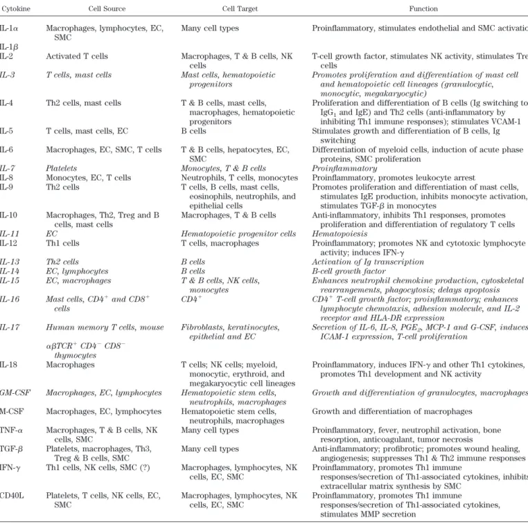
Cytokines are categorized according to the structural homology of their receptors as class I or class II cytokines (77, 369) (Table 1). Most ILs, CSFs, and IFNs belong to one of these two classes of cytokines, which mediate their effects through the Janus kinase-signal transducers and activators of transcription (JAK-STAT) pathway. Three other major cytokine families encompass the IL-1 family (including IL-1␣, IL-1, IL-1ra, and IL-18), TNF family, and TGF- superfamily (Table 1). IL-1 and TNF family mem-bers activate the nuclear factor-B (NF-B) and mitogen-activated protein (MAP) kinase signaling pathways, while TGF- superfamily members activate signaling proteins of the Smad family.
The TGF- superfamily is composed of many multi-functional cytokines including TGF-1–2-3, activins, in-hibins, anti-Mu¨llerian hormone (AMH), bone morphoge-netic proteins (BMPs), and myostatin (540). TGF- family members are secreted as inactive complexes bound to the latency-associated peptide (LAP), a protein derived from the NH2-terminal region of the TGF- gene product. The
LAP forms covalent bonds with the latent TGF- binding proteins (LTBPs), high-molecular-weight proteins of which four different isoforms exist (571). The resulting large latent complexes are sequestered within the extra-cellular matrix. Proteases in the extraextra-cellular matrix can digest LTBP, dissociating LAP from TGF-.
Cytokines share a number of specific features. 1) They show pleiotropic activities: a cytokine can trigger several different cellular responses depending on cell type, timing, and context.
2) They act synergistically: the association of two cytokines (for example, IL-12 and IL-18, TNF-␣ and IFN-␥, MCP-1 and IL-8) markedly amplifies their activity. This also holds true when a cytokine induces the expression of (an)other cytokine receptor(s).
3) They act in an autocrine, paracrine, or juxtacrine manner: cytokines can stimulate on the cells that produce them, or adjacent cells, or they can intervene through direct cell-cell interaction. This local mode of action sets cytokines apart from classical hormones.
4) They commonly share cytokine receptor subunits: for example, several members of the IL-2 family (IL-7, IL-9, IL-15, IL-21) share the IL-2 receptor␥-chain, the IL-6 family cytokines share the gp130 subunit, and the three
IFN- isoforms utilize a heterodimeric receptor com-posed of its specific receptor subunit IFN-R (or IL-28R␣) and the subunit IL-10R2 of the IL-10R, also shared with IL-10 and the IL-10-related cytokines, IL-22 and IL-26.
One must admit that many of these properties are also shared by growth factors. However, one difference is that the production of growth factors, including PDGF, epidermal growth factor (EGF), fibroblast growth factor (FGF), and vascular endothelial growth factor (VEGF), tends to be constitutive and is not as tightly regulated as that of cytokines. Also, the target cells of growth factors are mainly nonimmune.
Cytokines are often classified according to their pro-(TNF, IL-1, IL-12, IL-18, IFN-␥) or anti-inflammatory (IL-4, IL-10, IL-13, TGF-) activities. In light of the data obtained from experimental and clinical studies, described below, regarding the pathophysiological role of cytokines in ath-erosclerosis, we propose to cluster cytokines as pro- or antiatherogenic (Table 2).
B. Cytokine-Associated Signaling Pathways
1. NF-B
The NF-B pathway is one of the main signaling pathways activated in response to proinflammatory cyto-kines, including TNF-␣, IL-1, and IL-18, as well as follow-ing activation of the Toll-like receptors (TLR) by the pattern recognition of pathogen-associated molecular pat-terns (PAMPs). Activation of this pathway plays a central role in inflammation through the regulation of genes en-coding pro-inflammatory cytokines, adhesion molecules, chemokines, growth factors, and inducible enzymes such as cyclooxygenase-2 (COX2) and inducible nitric oxide synthase (iNOS) (166). NF-B is a dimeric transcription factor formed by the hetero- or homodimerization of pro-teins of the Rel family, including p50 and p65. In its inactive form NF-B is bound to inhibitor of B (I-B␣/) in the cytoplasm. Proinflammatory cytokines and patho-gens act through distinct signaling pathways that con-verge on the activation of an IB kinase (IKK) complex containing two kinases IKK1/IKK␣ and IKK2/IKK, and the regulatory protein NEMO (NF-B essential modifier, also named IKK␥) (762); IKK activation initiates IB␣/ phosphorylation at specific NH2-terminal serine residues
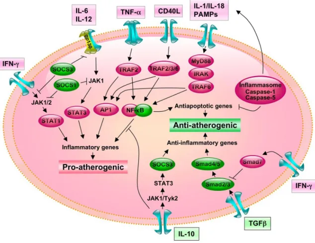
(782). Phosphorylated IB is then ubiquitinated, leading to its degradation by the 26S proteasome. This releases NF-B dimers from the cytoplasmic NF-B-IB complex, allowing them to translocate to the nucleus (Fig. 1). Once in the nucleus, NF-B binds to B enhancer elements on specific genes promoting transcription. Targets genes of NF-B include IB␣, the synthesis of which ensures that NF-B is transiently activated. This negative-feedback regulation gives rise to oscillations in NF-B translocation (496).
on April 7, 2006
physrev.physiology.org
TABLE1. Cytokine classification s y a w h t a P g n i l a n g i S s r o t p e c e R s e n i k o t y C Class I cytokines IL-6/IL-12 family Neuropoietic cytokines CLC R F I L F T N C STAT3 CT-1 gp130 JAK1/JAK2/Tyk2 LIF R M S O M S O STAT3 5 T A T S / 3 T A T S 1 K A J ) L P G ( r o t p e c e r e k i l -0 3 1 p g 1 3 -L I Hematopietic cytokines 3 T A T S 2 k y T / 2 K A J / 1 K A J R s f c g F S C -G R 6 -L I 6 -L I gp130 JAK1/JAK2/Tyk2 STAT3 R 1 1 -L I 1 1 -L I IL-12 IL-12R 2 JAK2/Tyk2 STAT3 IL-23 IL-23R JAK2 STAT3 IL-27 IL-27R ? ?
-Chain users (IL-2 family)
R 2 -L I 2 -L I JAK1/JAK3 STAT5 IL-15 R 7 -L I 7 -L I JAK1/JAK3 STAT5 TSLP R 9 -L I 9 -L I JAK1/JAK3 STAT5 R 1 2 -L I 1 2 -L I JAK1/JAK3 ? IL-4 family R 4 -L I 4 -L I JAK1 STAT5 IL-13 IL-3 R 3 -L I F S C -M G JAK2 STAT5 IL-5 Class II cytokines IFN family Type I IFN- IFNAR1 IFNAR2 JAK1/Tyk2 STAT1/STAT2 IFN-IFN- 1 (IL-28A) IFN- 2(IL-28B) IFN R1(IL-28R ) IL-10R2 JAK1/Tyk2 STAT1/STAT2 IFN- 3 (IL-29) Type II IFN- IFN-GR1 IFNGR2 JAK1/JAK2 STAT1
IL-10-related cytokine family
1 R 0 1 -L I 0 1 -L I IL-10R2 JAK1/Tyk2 STAT1/STAT3/STAT5 1 R 0 2 -L I 9 1 -L I IL-20R2 JAK1/Tyk2 STAT3 1 R 0 2 -L I 0 2 -L I IL-20R2 JAK1/Tyk2 STAT3 1 R 2 2 -L I 2 2 -L I IL-10R2 JAK1/Tyk2 STAT1/STAT3/STAT5 1 R 0 2 L I 4 2 -L I IL-22R1 JAK1/Tyk2 STAT3 1 R 0 2 -L I 6 2 -L I IL-10R2 JAK1/Tyk2 STAT1/STAT3 TNF superfamily TNF- TNFR1(p55)/TNFR2(p75) NF- B/JNK/p38/ERK Lymphotoxin TNFR1(p55)/TNFR2(p75) NF- B/JNK/p38/ERK s a F L s a F NF- B/JNK 0 4 D C ) 4 5 1 D C ( L 0 4 D C NF- B/JNK K N A R L K N A R NF- B/JNK/p38/ERK -F N R -L I A R T L I A R T B/JNK/p38/ERK TGF family TGF- 1/2/3 TGF- -R1/ TGF- -RII Smad2/Smad3 3 d a m S / 2 d a m S I I R t c A / I R t c A A n i v i t c A IL-1 family -F N P c A R 1 -L I / I R 1 -L I 1 -L I B/JNK/p38/ERK R 8 1 -L I 8 1 -L I /IL-18R NF- B/JNK/p38 ? 2 3 -L I NF- B/p38 2 T S 3 3 -L I NF- B/p38/ERK
CLC, CT-1-like factor; LIF, leukemia inhibitory factor; CNTF, ciliary neurotrophic factor; CT-1, cardiotrophin-1; OSM, oncostatin M; G-CSF, granulocyte-colony stimulating factor; TSLP, thymic stromal lymphopoietin; RANK, receptor activator of NF- B; RANKL, RANK ligand; ActRI, activin type I receptor; IL-1RAcP, IL-1 receptor accessory protein.
on April 7, 2006
physrev.physiology.org
NF-B is a redox-sensitive transcription factor, and the intracellular redox status of the cell is extremely important in the regulation of NF-B activity (reviewed in Ref. 311). Antioxidants, such as aspirin, N-acetylcysteine (NAC), and flavonoids can therefore inhibit the activation of NF-B. A number of natural constitutive or inducible pathways inhibiting NF-B activity also exist (see review in Ref. 669). For example, A1 and A20, two cytoprotective genes, are induced in response to inflammatory stimuli to protect EC from exaggerated activation and from under-going apoptosis even when NF-B is blocked (139). A20 terminates NF-B activation by promoting reaccumula-tion of IB through its interaction with proteins involved in TNF-␣ signaling upstream of IB degradation (375). Consequently, A20-deficient mice fail to terminate TNF-induced NF-B activity, having a persistently active IKK complex that prevents reaccumulation of IB protein, are hypersensitive to TNF-␣, and suffer from severe inflam-mation. The inducible form of the heme oxygenase (HO-1) is another example of endogenous anti-inflammatory pathway induced in response to inflammatory stimuli. HO-1 can be upregulated in human EC by TNF and IL-1 (674), and HO-1 possesses potent antiapoptotic and anti-inflammatory properties (742). HO-1 deficiency in humans is associated with the presence of severe and persistent endothelial damage (761). The anti-inflammatory proper-ties of HO-1 seem to be related to an inhibitory action on P- and E-selectin expression on EC (688).
Activated NF-B has been identified in SMC, macro-phages, and EC of human atherosclerotic lesions (78, 82, 480). Enhanced endothelial activation of NF-B has been shown to occur in LDLr-deficient mice very early on fol-lowing a high-fat diet, in regions of the proximal aorta with high probability for atherosclerotic lesion develop-ment (262). Furthermore, supershift analysis in cells iso-lated from human carotid atherosclerotic plaques, com-posed in majority of macrophages and SMC, demonstrate that activated NF-B consists of p65, c-Rel, and p50, but not relB or p52 subunits (480). NF-B activation in these cells controls the expression of proinflammatory cyto-kines TNF-␣; IL-6 and IL-8; matrix metalloproteinases (MMP)-1, -3, and -9; and tissue factor (TF), as shown by their selective inhibition following blockade of the NF-B pathway by overexpression of IB␣ or dominant-negative IKK-2 (480). Interestingly, in this study NF-B inactivation did not affect the expression of the anti-inflammatory cytokine IL-10 or the matrix metalloproteinase inhibitor TIMP-1.
The actual in vivo role of the NF-B pathway has recently been addressed in experimental models of ath-erosclerosis. Kanters et al. (324), using LDLr-deficient mice with a cell-specific deletion of IKK2 preventing NF-B activation in macrophages, unexpectedly found increased atherosclerotic lesion formation and inflamma-tion in these animals. This result was associated with a significant reduction in the inflammatory and anti-atherogenic cytokine IL-10, suggesting that a certain level of NF-B activation is required to modulate the inflam-matory reaction and counteract proatherogenic re-sponses (Fig. 2). This finding is in favor of a central role for NF-B in the induction of “protective” antiapoptotic and anti-inflammatory genes, critical to the resolution of the inflammatory process (374). However, the detrimental effect of NF-B inhibition in atherogenesis is likely to depend on how NF-B activity is inhibited. In a subse-quent study, Kanters et al. (323) examined the effects of hematopoietic NF-B1 (the p50 subunit of NF-B) defi-ciency in the development of atherosclerotic lesions, transplanting bone marrow from mice deficient in NF-B1 into irradiated LDLr⫺/⫺ mice. Instead of promoting the formation of larger inflammatory lesions, as was the case with specific IKK2 deficiency in macrophages, hemato-poietic NF-B1 deficiency was associated with a signif-icant decrease in lesion size, despite enhanced accu-mulation of T and B lymphocytes within the lesions. This could be explained, at least in part, by the obser-vation that in contrast to IKK2 deficiency, NF-B1 de-ficiency did not alter the inflammatory balance in favor of a proatherogenic phenotype. Despite increased TNF-␣ expression by NF-B1-deficient macrophages, other major proatherogenic molecules such as MCP-1 were downregulated, whereas critical antiatherogenic factors such as IL-10 were significantly upregulated.
TABLE2. Pro- and antiatherogenic cytokines
Proatherogenic Cytokines Antiatherogenic Cytokines
TNFR family TNF-␣ Lymphotoxin Osteoprotegerin CD40L IL-1 family IL-1 IL-1ra IL-18 IL-18BP Class I cytokines IL-2 IL-4 IL-6 IL-6 IL-12 IL-9 Class II cytokines IFN-␥ IL-10 Hematopoietic factors M-CSF Chemokines/chemokine receptors IL-8/CXCR2 MCP-1/CCR2 Fractalkine/CX3CR1 RANTES MIF Bone-associated cytokines Osteopontin TGF- family TGF-
TNF, tumor necrosis factor; IFN, interferon; TGF, transforming growth factor.
on April 7, 2006
physrev.physiology.org
Decreased MCP-1 production and increased IL-10 ex-pression may have contributed to the limitation of plaque size despite enhanced accumulation of T cells. Another plausible explanation for reduced lesion devel-opment in NF-B1-deficient animals could be a defect in the uptake of oxLDL by macrophages, as character-istic foam cells were absent in NF-B1-deficient le-sions. Moreover, both scavenger receptor class A (SR-A) expression and uptake of oxLDL were signifi-cantly reduced in NF-B1-deficient macrophages stim-ulated ex vivo with lipopolysaccharide (LPS), although in vivo relevance of this in vitro effect remains to be determined. In summary, NF-B appears to be at the
crossroads of the inflammatory response in atheroscle-rosis, fine-tuning the response of the vessel wall to injury (Fig. 2).
2. JNK/AP-1
AP-1 (activator protein-1) is a transcription factor consisting of homodimers or heterodimers of Fos (c-Fos, FosB, Fra-1 and Fra2), Jun (c-Jun, JunB, JunD), or ATF subunits which recognize either 12-O-tetradecanoylphor-bol-13-acetate (TPA) response elements or cAMP re-sponse elements (CRE) (626). Jun proteins can ho-modimerize, but Fos proteins can only form stable dimers
FIG. 1. Principal signaling pathways involved in atherogenesis. Proinflammatory cytokines (IL-1, IL-18) and pathogens (represented as pathogen-associated molecular patterns, PAMP), as well as nonpathogen activators of TLR, act through distinct signaling pathways that converge on the activation of NF-B. MyD88 functions as an adaptor between receptors of the TLR or IL-1R families and downstream signaling kinases. Following association of MyD88 with IL-1-associated kinase IRAK-4, IRAK-4 is autophosphorylated, dissociates from the receptor complex, and interacts with TNF-receptor-associated factor-6 (TRAF-6), which also mediates CD40 signaling. Once activated, TRAF6 associates with the MAP3 kinase TAK1 (716). From TAK1, two signaling pathways diverge; one ultimately leads to NF-B activation and the other to MAP kinase activation. In its inactive form, NF-B is bound to inhibitor of B (IK-B␣/) in the cytoplasm and consists of an IB kinase (IKK) complex containing two kinases IKK␣ and IKK, and the regulatory protein IKK␥ (also named NEMO); IKK activation initiates IB␣/ phosphorylation. Phosphorylated IB is then ubiquitinated, leading to its degradation by the 26S proteasome. This releases NF-B dimers from the cytoplasmic NF-B-IB complex, allowing them to translocate to the nucleus. JNK phosphorylation is mediated by two MAPK kinases (MAPKKs), MKK4 and MKK7, that they can cooperatively activate JNK. Both kinases are required for full activation of JNK by environmental stressors, and MKK7 is essential for JNK activation by TNR engagement. Tyrosine phosphorylation activates the cytosolic inactive STATs, resulting in their nuclear translocation and gene activation. This pathway was originally found to be activated by IFNs, but a number of cytokines, growth factors, and hormonal factors also activate JAK and/or STAT proteins. IFN-␥ utilizes JAK1 and JAK2, and usually activates STAT1. TGF--triggered signals are transduced by proteins belonging to the Smad (for vertebrate homologs of Sma and Mad) family. The type I receptor recognizes and phosphorylates Smad2 and Smad3, which associates with Smad4, forming complexes that participate in DNA binding and recruitment of transcription factors. Smad3 may also have antagonistic properties, as it plays a critical role in TGF--dependent repression of vascular inflammation by inhibiting AP-1 activity. Smad7 inhibits Smad2 and Smad3 phosphorylation.
on April 7, 2006
physrev.physiology.org
with Jun. Phosphorylation of c-Jun by c-Jun NH2-terminal
kinases (JNKs) results in enhanced transcriptional activ-ity of complexes containing AP-1 dimers (734).
JNK belongs to the family of stress-activated pro-tein kinases that also includes the p38 propro-tein kinases. Three highly related but distinct gene products, JNK1, JNK2, and JNK3, can be expressed as a total of 10 isoforms as a result of variable mRNA splicing (259). JNK1 and JNK2 show a broad tissue distribution, whereas JNK3 is expressed predominantly in neurons but also in cardiac smooth muscle and the testes (770). Targeted deletion of the genes coding for JNK1 or JNK2 results in abnormal thymocyte selection (588) and loss of T-lymphocyte differentiation and effector function (179). JNK3 knockout mice show resistance to neuro-nal apoptosis, directly implicating JNK in at least some specific instances of programmed cell death (678, 768).
JNK phosphorylation is mediated by two MAPK ki-nases (MAPKKs), MAP2K4 (or MKK4) and MAP2K7 (or MKK7), that they can cooperatively activate JNK (Fig. 1).
Gene disruption studies in mice demonstrate that both MAP2K4 and MAP2K7 are required for full activation of JNK by environmental stressors and that MKK7 is essen-tial for JNK activation by TNF (677).
Many proinflammatory genes, including those en-coding TNF-␣, IL-2, IL-6, E-selectin, ICAM-1, VCAM-1, MCP-1, COX2, and MMPs-1, -9, -12, and -13 (500), are regulated by the JNK pathway, through interaction of AP-1 with other cis-acting sequences in their promoters and with certain transcription factors that bind to these sequences (Fig. 2).
A recent study showed that atherosclerotic lesions were significantly reduced in JNK2-deficient apoE⫺/⫺ mice, but not in JNK1-deficient apoE⫺/⫺mice, compared with apoE⫺/⫺mice (568). JNK2 expression in leukocytes, rather than in vascular cells, appeared to be responsible for this effect. Indeed, transplantation of apoE⫺/⫺ JNK2⫺/⫺ bone marrow into apoE⫺/⫺ mice reduced ath-erosclerosis to an extent similar to that of apoE⫺/⫺ JNK2⫺/⫺mice transplanted with apoE⫺/⫺JNK2⫺/⫺bone marrow, whereas apoE⫺/⫺JNK2⫺/⫺ mice transplanted
FIG. 2. Cross-talks between proinflammatory/proatherogenic and anti-inflammatory/antiatherogenic signal transduction pathways. Inhibitory Smads such as Smad7 downstream of IFN-␥ signaling associate with activated receptors and interfere with Smad2 and Smad3 binding. It is noteworthy that like IFN-␥, the anti-inflammatory cytokine IL-10 also activates JAK and/or STAT proteins. However, the IL-10/IL-10R interaction activates JAK1 and Tyk2, leading to STAT3 and SOCS3 activation, which is central for the anti-inflammatory responses of IL-10 in macrophages. The inflammasome may be a central link between apoptosis and inflammation in pathological conditions. NF-B may have a dual role in atherosclerosis, being proatherogenic through its proinflammatory properties, and antiatherogenic through its antiapoptotic activities.
on April 7, 2006
physrev.physiology.org
with apoE⫺/⫺ bone marrow showed atherosclerotic le-sions equivalent to those of apoE⫺/⫺ mice transplanted with apoE⫺/⫺bone marrow (568).
3. JAK/STAT
The class I and II cytokines induce homodimerization and activation of their cognate receptors, resulting in the activation of associated JAK kinases (JAK1, JAK2, JAK3, and Tyk2) (Table 1) (520a). The activated JAKs phosphor-ylate the receptor cytoplasmic domains, which creates docking sites for SH2-containing signaling proteins. Among the tyrosine phosphorylated substrates are mem-bers of the STAT family of proteins (Table 1) (520a). Receptor engagement and tyrosine phosphorylation acti-vate the cytosolic inactive STATs, resulting in their nu-clear translocation and gene activation. This pathway was originally found to be activated by IFNs, but a number of cytokines, growth factors, and hormonal factors also ac-tivate JAK and/or STAT proteins (Fig. 1). In particular, IL-6 binds to the IL-6 receptor␣-chain and gp130, which activate JAK1 and STAT3. IFN-␥ utilizes JAK1 and JAK2, and usually activates STAT1. It is noteworthy that the anti-inflammatory cytokine IL-10 also activates JAK and/or STAT proteins (reviewed in Ref. 481). The IL-10/ IL-10R interaction activates JAK1 and Tyk2, which are associated with the IL-10R1 and IL-10R2, respectively.
STAT3 can be activated by a number of cytokines, especially those of the IL-6 family, mediating the expres-sion of several acute-phase response genes. Yet, STAT3 appears to play a critical negative role in controlling inflammation, as shown in mice with STAT3 deletion in specific cell types, including keratinocytes (594), T cells (666), macrophages/neutrophils (664), cardiomyocytes (309), or endothelial cells (322), STAT3 deficiency being embryonically lethal. STAT3-deficient T cells show se-verely impaired IL-6-induced cell proliferation, due to the lack of IL-6-mediated prevention of T-cell apoptosis (666). STAT3 deletion in mice within the macrophage/neutrophil lineage results in chronic inflammation and pathological colitis with age, due to the enhancement of the Th1 re-sponse by blockade of IL-10 signaling (664). Removal of STAT3 from hematopoietic progenitors also results in increased proinflammatory cytokine production, inflam-matory bowel disease, and an expanded macrophage pop-ulation (732). Interestingly, STAT3-deficient macrophages and neutrophils show increased production of inflamma-tory cytokines in response to LPS, which cannot be re-duced by IL-10 (664). STAT3 activation by IL-10 is there-fore central for anti-inflammatory responses in macro-phages and neutrophils (Fig. 2). It is noteworthy that mice with conditional STAT3 deletion in endothelium also show exaggerated inflammation and leukocyte infiltration in multiple organs upon LPS challenge (322). An endothe-lium-derived soluble factor that is dependent on STAT3 is
likely to control IFN-␥ production during LPS-induced inflammation (322).
In terms of immunoregulation, STAT4 and STAT6 are crucially important for the differentiation of Th cells. IL-4 activates STAT6 and promotes the differentiation of Th2 cells (634). Conversely, IL-12 activates STAT4 and drives the differentiation of naive T cells into Th1 cells that produce IFN-␥ (325). In atherosclerosis, the Th cell re-sponse is of the Th1 type, characterized by abundant secretion of IFN-␥ (264). Yet, Th2 profile does not neces-sarily offer protection against atherosclerosis and might even be proatherogenic (see sect. VIB3). Therefore,
tar-geting STAT4 and STAT6 could be of use in the treatment of atherosclerosis. Interestingly, statins, which are be-lieved to exert beneficial effects in cardiovascular disease beyond cholesterol lowering (350), have been reported to inhibit Th1-mediated disease and to block activation of STAT4 (386, 492, 778) and induction of major histocom-patibilty complex (MHC)-II expression by IFN-␥ (366). Other drugs, including rapamycin and lisofylline, have also been reported to block STAT4 activation (127, 771). Interestingly, a recent study showed that rapamycin re-duces atherosclerosis in apoE⫺/⫺mice, with concomitant decreased expression of IL-12p40, IFN-␥ and IL-10 mRNA, and enhanced expression of TGF-1 (190). Pentoxifylline, a methylxanthine derivative of lisofylline, has been re-ported to have protective effects against atherosclerosis in apoE⫺/⫺mice, associated with a reduced Th1 polariza-tion of Th lymphocytes (373).
Cytokine signaling by the JAK/STAT pathway is reg-ulated, in part, by a family of endogenous JAK kinase inhibitor proteins termed suppressors of cytokine signal-ing (SOCS) (748). The SOCS family consists of eight mem-bers [SOCS-1 to SOCS-7 and cytokine-inducible SH2 pro-teins (CIS)] all sharing a central SH2 domain and a COOH-terminal SOCS box. Both SOCS1 and SOCS3 inhibit JAK tyrosine kinase activity; SOCS1 directly binds to the acti-vation loop of JAKs through the SH2 domain, while SOCS3 binds to cytokine receptors (Fig. 2). SOCS1 regu-lates INF␥ signaling, and deficiency leads to lethal dis-ease, which is characterized by exaggerated effects of IFN-␥. Interestingly, mice lacking both SOCS-1 and IFN-␥, though saved from the lethal perinatal syndrome ob-served in SOCS-1-deficient mice, develop a variety of chronic infections or inflammatory lesions as adults (466). In contrast, SOCS2 regulates growth hormone, and SOCS-knockout mice show gigantism. SOCS3 is preferentially expressed in Th2 cells and plays an important role in regulating the onset and maintenance of Th2-mediated allergic immune disease (619).
Very little is known regarding the role of SOCS in atherosclerosis. It has been reported that SOCS-1 inhibits IFN-␥-induced CD40 expression in macrophages by blocking IFN-␥-mediated STAT-1 activation, and in so doing suppressing IFN-␥-induced TNF-␣ secretion and
on April 7, 2006
physrev.physiology.org
subsequent NF-B activation (733). Inasmuch as the CD40/CD40L pathway actively participates in plaque de-velopment and progression (reviewed in Ref. 605), mim-ics or inducers of SOCS1 might be useful to attenuate the effects of IFN-␥ in the context of atherosclerosis. 4. Smads
TGF--triggered signals are transduced by proteins belonging to the Smad (for vertebrate homologs of Sma and Mad) family. Smads serve as substrates for TGF- receptors type I and II, in which the cytoplasmic domain possesses serine/threonine kinase activity (453). The type I receptor recognizes and phosphorylates Smad2 and Smad3, which associates with Smad4, forming complexes that participate in DNA binding and recruitment of tran-scription factors (Fig. 1). Smad3 may also have antago-nistic properties, as it plays a critical role in TGF- -dependent repression of vascular inflammation by inhib-iting AP-1 activity (Fig. 2) (200, 201). In addition to these agonistic Smads, inhibitory Smads (I-Smad) such as Smad6 and Smad7, which associate with activated recep-tors and interfere with Smad2 and Smad3 binding, are present. Expression of Smad7 is induced by IFN-␥ as a negative regulator of the TGF-/Smad pathway (684). Re-cent advances in the study of atherosclerosis point to an important role of TGF- signaling in the protection against excessive plaque inflammation, loss of collagen content, and induction of regulatory immunity (see below and reviews in Refs. 243, 444, 445). Immunohistochemis-try and RT-PCR anlaysis of human plaques reveal Smad2, Smad3, and Smad4 expression in macrophages of fibro-fatty lesions and in SMC of fibrous caps (320). We also detected phosphorylated Smad2 in the aortic sinus of apoE⫺/⫺mice, indicative of TGF- activity in atheroscle-rotic lesions (438).
5. TLR/Myd88 signaling pathways
At least 10 TLRs (TLR1–10) recognize different PAMPs associated with different classes of pathogens (review in Refs. 303, 665). For example, TLR4 recognizes LPS, which is unique to Gram-negative bacteria, and TLR2 recognizes peptidoglycan found in Gram-positive bacte-ria. TLR9 recognizes unmethylated CpG motifs, which are abundant in prokaryotic genomes and virus DNA. TLR3 recognizes double-stranded RNA (dsRNA) produced dur-ing viral infections. TLRs are characterized by a 150-amino acid intracytoplasmic domain named TIR (Toll/IL-1R/R), which they share with members of the IL-1 recep-tor (IL-1R) family and plant disease resistance (R) genes, and by an extracellular domain composed of NH2
-termi-nal leucine-rich repeats (LRRs) flanked by characteristic cysteine clusters on the COOH-terminal (CF motif) or NH2-terminal (NF motif) side of the LRRs. Upon
stimula-tion, TLRs and the IL-1R family activate the transcription
factors NF-B and AP-1, leading to production of proin-flammatory cytokines. TIR domains play a critical role in TLR signaling. They allow homophilic interactions with the cytoplasmic factor MyD88 that also contains a TIR domain (Fig. 1). MyD88, which is recruited to the recep-tors after stimulation, contains an NH2-terminal death
domain that enables it to bind the death domain-contain-ing serine-threonine kinases of the IL-1R-associated ki-nases (IRAK) family (reviewed in Ref. 312). As a result, MyD88 functions as an adaptor between receptors of the TLR or IL-1R families and downstream signaling kinases. Following association of MyD88 with IRAK-4, IRAK-4 is autophosphorylated, dissociates from the receptor com-plex, and interacts with TNF-receptor-associated factor (TRAF)-6. Once activated, TRAF6 activates a heterodimer composed of two ubiquitination proteins called Uev1A and Ubc13, which triggers its association with the MAP3 kinase TAK1 (716). From TAK1, two signaling pathways diverge; one ultimately leads to NF-B activation and the other to MAP kinase activation. Studies using MyD88-deficient mice showed that this factor is essential for the NF-B-dependent induction of TNF-␣ and IL-6 in re-sponse to TLR agonists (331). Interestingly, analysis of MyD88 mutant mice unexpectedly pointed to the exis-tence of a MyD88-independent pathway downstream of some TLRs. Indeed, TLR4- or TLR3-mediated activation of NF-B and AP-1 by LPS and dsRNA, respectively, was not abolished but only delayed in MyD88-deficient mice (13, 331).
Recently, enhanced expression of TLR4 was detected in murine (apoE⫺/⫺ mice) and human carotid and coro-nary atherosclerotic plaques (186, 760). TLR1 and TLR2 expression has also been found in human carotid (186) but not in coronary plaques (760). Human epidemiological data demonstrate that an Asp299Gly TLR4 polymorphism, which attenuates receptor signaling, is associated with a decreased risk of atherosclerosis and acute coronary events (18, 64, 335). Functional TLR4 expression has also been correlated with the development of aortic intimal hyperplasia in a mouse model of artery injury (699), and TLR4 activation by LPS increases atherosclerotic plaque formation in the apoE3*Leiden atherosclerotic mouse model (287).
Interestingly, TLR4 appears to be involved in several aspects of the inflammatory response even in the absence of infection, by recognizing endogenous ligands produced during inflammation. Extracellular matrix components, including the type III repeat extra domain A of fibronec-tin, low-molecular-weight oligosaccharides of hyaluronic acid, and polysaccharide fragments of heparan sulfate, provoke immunostimulatory responses similar to those induced by LPS, via TLR4 (315, 517, 673). Moreover, fi-brinogen (642) and minimally modified LDL (mmLDL) (471) are able to induce the production of chemokines and cytokines from macrophages through recognition by
on April 7, 2006
physrev.physiology.org
TLR4. Together, these recent findings indicate that TLR4 may exert LPS-independent atherogenic activities (468, 469). Two lines of evidence support this hypothesis: 1) oxLDL enhances TLR4 expression in macrophages (760), and 2) TLR4 or its intracellular adaptor protein, MyD88, reduces atherosclerosis in uninfected apoE-deficient mice, concomitant with a marked reduction in macro-phage infiltration and MCP-1 expression in the atheroscle-rotic lesions (54), and decreased circulating levels of IL-12 and MCP-1 (470).
Remarkably, CD4⫹CD25⫹ Treg cells selectively ex-press TLR4 –5-7– 8 (110). This is of particular importance given the role that Treg cells play in atherosclerosis, as we recently reported (8, 437; see sect.VIC).
III. INDUCERS OF CYTOKINE PRODUCTION IN ATHEROSCLEROSIS
A. Initial Trigger(s)
According to the classical view of inflammation, cy-tokines are produced by cells of the innate immune sys-tem (monocytes, neutrophils, NKT cells) in response to microbial infection, toxic reagents, trauma, antibodies, or immune complexes (493). In the host, TLRs and intracel-lular proteins (NOD1 and NOD2, for “nucleotide-binding oligomerization domain”) act as sensors of the conserved molecular motifs present on a wide range of different microbes, the PAMPs. Hence, cytokines are secondary mediators of inflammation and not the primary triggers. An etiologic role for infectious agents in atherosclerosis, especially Chlamydia pneumoniae and cytomegalovirus (CMV), has been repeatedly evoked (396) since the first seroepidemiologic evidence of an association of the chla-mydia TWAR strain with acute myocardial infarction and chronic coronary disease was reported in 1988 (589). However, the most recent clinical trials, including Weekly Intervention with Zithromax for Atherosclerosis and its Related Disorders (WIZARD) (511), Azithromycin in Acute Coronary Syndrome (AZACS) (114), Antibiotic Therapy After Acute Myocardial Infarction (ANTIBIO) (781), Pravastatin or Atorvastatin Evaluation and Infec-tion Therapy (PROVE-IT) (108), and Azithromycin and Coronary Events Study (ACES) (250), assessing the po-tential benefits of antibiotic therapy with the goal of tar-geting Chlamydia pneumoniae showed no effect of treat-ment in patients with CAD. Moreover, experitreat-mental stud-ies showed that infection is not necessary for initiation or progression of atherosclerosis in apoE-deficient mice. Atherosclerosis develops identically in germ-free animals and in animals raised with ambient levels of microbial challenge (749). One must therefore conclude that patho-gens do not serve as etiologic agents for atherosclerosis, even though one cannot rule out a role in disease
exac-erbation. Several reports indicate that inoculation of ath-erosclerosis-prone mice with high doses of C. pneu-moniaefosters atherosclerosis (292, 475). Yet, the athero-genic effect of C. pneumoniae requires elevated serum cholesterol levels (292).
Atherosclerosis clearly does not develop in any ani-mal model without a significant level of plasma choles-terol, and the dominant role of cholesterol is also well established in humans. While hypertension, diabetes, and smoking are factors that dramatically increase the risk of atherosclerosis, it is not rare to have clinically significant atherosclerosis in the absence of these risk factors. In contrast, below a certain level of cholesterol (150 mg/dl), atherosclerosis is practically absent in human popula-tions (106), and risk gradually increases with increased plasma cholesterol levels (647). Moreover, primary and secondary clinical trials have established the efficacy of lowering cholesterol with statins for prevention of cardio-vascular disease (256, 694). It is therefore tempting to hypothesize that the primary trigger of cytokine release in atherosclerosis has a link with cholesterol. Atherogenic cholesterol exists mainly in the form of LDL, which are the main culprit in CAD. In fact, several lines of evidence support the hypothesis that oxidized lipids, including ox-LDL, are the most likely triggering factors for cytokine production.
Quantitative analysis of atherosclerosis in fetal aorta showed that fatty streaks are already present at this early stage of life, lesions being more abundant in fetus from hypercholesterolemic mothers than from normocholes-terolemic mothers (489). Interestingly, qualitative analy-sis of lesions depicted similar distribution of native LDL, oxLDL, and macrophages in lesions of offspring from both hypercholesterolemic and normocholesterolemic mothers. The presence of macrophages alone, without native LDL or oxLDL, or their association with native LDL, was almost never observed, and most of the lesions contained both oxLDL and macrophages. A few lesions with native LDL or oxLDL without macrophages were also present. This seminal study allows us to describe the exact chronology of events leading to fatty streak for-mation in humans, starting with native LDL uptake by the arterial intima, followed by LDL oxidation and, finally, monocyte recruitment after endothelial activa-tion by oxLDL.
C3H mice, which do not develop atherosclerotic le-sions either when fed an atherogenic diet or when crossed with the atherosclerosis prone apoE⫺/⫺mice, do not re-spond to in vivo administration of oxLDL, in contrast to C57BL/6 mice (391). Their EC are not activated in the presence of oxLDL, whereas cells from C57BL/6 mice express M-CSF, MCP-1, and VCAM-1 in the same condi-tions. Yet, C3H EC respond perfectly well to activation by the proinflammatory cytokines IL-1 and TNF-␣ (628, 629).
on April 7, 2006
physrev.physiology.org
oxLDL behaves as a potent inflammatory agent. In vivo administration of oxidized LDL to C57BL/6 mice causes rapid induction of circulating M-CSF and upregu-lation of genes encoding JE (the murine analog of MCP-1) as well as other inflammatory proteins in various tissues (392). OxLDL stimulates the expression of adhesion mol-ecules on EC (337). OxLDL has chemoattractant activity on monocytes, promotes their differentiation into macro-phages, but inhibits their mobility (555, 556). Binding of oxLDL to CD36 triggers the release of proinflammatory cytokines in macrophages (310). In addition, incubating human blood mononuclear cells with oxLDL results in T-lymphocyte activation, as assessed by increased expres-sion of IL-2 receptors and HLA-DR antigens on T lympho-cytes (215).
Oxidation of LDL generates many “neo-self determi-nants” that induce an active immune response (288) and may challenge the regulatory pathways responsible for immune homeostasis. Both humoral and cellular immune responses can profoundly affect atherosclerotic develop-ment and progression (268).
The amount of lipid retained in macrophages de-pends on unregulated uptake of oxidized lipoproteins by scavenger receptors, as first identified by Brown and Goldstein (87), counterbalanced by degradation and efflux.
Altogether these findings point to a role of oxLDL as a very early trigger of vascular inflammation. LDL accu-mulation and modification in the subendothelium trigger monocyte and lymphocyte recruitment. Thereafter, acti-vated macrophages and lymphocytes secrete abundant amounts of cytokines that in turn can activate EC, SMC, and macrophages/lymphocytes to foster cytokine produc-tion, leading to a self-perpetuating inflammatory process that becomes less dependent on the presence of oxLDL. This might explain why oxLDL, while instrumental in triggering the early atherosclerotic events, are less critical in upholding the inflammatory environment. This might also explain in part the efficiency of antioxidant therapies in the prevention of atherosclerosis when these therapies are administered at the very beginning of the atheroscle-rotic process in animal models, but their failure to do so in most secondary or primary prevention clinical trials in humans (reviewed in Ref. 743), where treatment is admin-istered at later stages of the disease when secondary inflammatory mediators become as important as the ini-tial oxidative-related stimulus. It is noteworthy that ath-erosclerotic plaques do not regress, or regress very slowly, in cholesterol-fed rabbits following short-term withdrawal of cholesterol feeding and normalization of cholesterol plasma levels (2, 155). It is only after a pro-longed cholesterol withdrawal period that decrease in plaque size, together with reduced vascular inflammation and plaque stabilization, is observed (7, 347, 697). In humans, aggressive lipid lowering treatment using statins
has been shown to be very effective in limiting plaque development and reducing plaque progression (142, 143, 505, 506, 777). The cytokine network may thus serve as a final common proinflammatory pathway regardless of the initiating event and provides a supplemental therapeutic target, especially in late stages of the disease.
Several oxidized lipids and/or phospholipids are lipid bioactive mediators and may serve as primary triggers of the atherosclerotic process. Bioactive lipids have been identified in the atherosclerotic plaque, including the po-tent inflammatory mediator platelet activating factor (PAF), PAF-like lipids, oxidized phospholipids (oxPL), and lysophosphatidylcholine (lysoPC) (494). Like oxLDL, PAF induces TNF-␣ production by monocytes (71, 213) and MHC class II dependent IFN-␥ secretion by T lym-phocytes (213). Oxidized phospholipids upregulate tissue factor expression in EC (62), as well as in SMC (146). Similarly, lysoPC can enhance IFN-␥ secretion and gene expression in human T lymphocytes (503) and stimulate the production of IL-1 in macrophages (407). It also stimulates ICAM-1 and VCAM-1 expression (362, 793) and induces the release of IL-6 and IL-8 (662) in EC and MCP-1 in EC (663) and SMC (575).
Lipid oxidation products such as lysoPC, 4-hydroxy-2-nonenal (4HNE), and oxysterols are contained in oxLDL (662). Oxidized 1-palmitoyl-2-arachidonyl-sn-glycero-3-phosphorylcholine (OxPAPC), which is present in mini-mally modified LDL, is a PAF-like lipid that is found in atherosclerotic plaques. OxPAPC, but not native PAPC, is able to stimulate EC to bind monocytes and to secrete MCP-1, IL-8, and growth-related oncogene (GRO)-␣ (376, 565, 773; see review in Ref. 382). Individual lipids identi-fied in OxPAPC include (5-oxovaleroyl)-sn-glycero-3-phosphorylcholine (POVPC), 1-palmitoyl-2-glutaroyl-sn-glycero-3-phosphorylcholine (PGPC), and epoxy-isoprostane-PC (727, 728). Oxidized 1-palmitoyl-2-linoleoyl-sn-glycero-3-phosphorylcholine (PLPC) also promotes monocyte-endothelial interactions (4). More-over, epoxyisoprostane and epoxycyclopentenone phos-pholipids have been identified in OxPAPC that induce MCP-1 and IL-8 in EC (653). Oxidized phospholipids can upregulate tissue factor both in EC (62) and in SMC (146). Interestingly, it has been shown that OxPAPC inhib-its the binding of LPS to LPS-binding protein and CD14, which are required for presenting LPS to TLR-4 (61). It is therefore likely that upon acute bacterial inflammation oxidized phospholipids exert anti-inflammatory proper-ties by inhibiting NF-B pathway, while under conditions of chronic inflammation, the pro-inflammatory activity of lipid oxidation products becomes more pathologically rel-evant (382).
Eicosanoids are well-known lipid mediators of in-flammation. They comprise a variety of compounds [pros-taglandins, thromboxanes, leukotrienes (LT),
on April 7, 2006
physrev.physiology.org
and epoxy-fatty acids, lipoxins, and isoprostanes] that are derived from arachidonic acid. Leukotrienes are a class of eicosanoids that are derived through the action of the 5-lipoxygenase (5-LO). 5-LO is pivotal for the generation of both proinflammatory (LTB4and LTC4) and
anti-inflam-matory (lipoxins) mediators. However, in contrast to their inhibitory effects on PMN and eosinophils, lipoxins are potent stimuli for peripheral blood monocytes, stimulat-ing monocyte chemotaxis and adherence (428). Recent biologic and genetic findings implicate the 5-LO pathway in atherosclerosis (162, 279, 461, 552). Mehrabian et al. (461) reported that heterozygotes for the 5-LO gene on the LDLr⫺/⫺background had considerably reduced aortic le-sions, compared with the advanced lesions observed in 5-LO⫹/⫹LDLr⫺/⫺mice, despite equivalent hypercholester-olemia. 5-LO pathway also promotes pathogenesis of hy-perlipidemia-dependent aortic aneurysm (787). Further-more, clinical findings showed that variant alleles of 5-LO genes were associated with a significant increase of ca-rotid intima thickness (184). Most recently, a significant association was drawn between the gene encoding 5-LO activating protein (FLAP) and myocardial infarction by analysis of single-nucleotide polymorphism haplotype in humans (279).
Another significant recent discovery is the chemotac-tic activity of LTB4on activated CD4⫹ and CD8⫹ T cells
expressing the LTB4receptor, BLT1 (239, 522, 661). It was
found that both Th1- and Th2-polarized CD4⫹ T cells and antigen-specific CD4⫹ T cells, but not naive T cells, ex-press BLT1 (661). Therefore, the antiatherogenic effects of the blockade of LTB4/BLT1 pathway (6, 280) might
result in part from decreased Th1/Th2 cells recruitment in the plaque.
B. Secondary Triggers
Once inflammation has been triggered and cytokine release is initiated at the onset of atherosclerotic lesion development, a number of factors that are found in the atherosclerotic plaque can participate in maintaining and amplifying cytokine production (Table 3).
1. HSP
Evidence suggests that the inflammatory component of atherosclerosis might, at least in part, involve immune reactivity to heat shock proteins (HSPs) (446, 544, 737). Animal models of atherosclerosis have shown a very early role for HSP60 in the development of the disease (see review in Ref. 738). HSP60 might be an important autoan-tigen in atherosclerosis and might play a role similar to that of oxLDL in triggering an autoreactive T-cell re-sponse. Rabbits (757, 758) or mice (226) immunized with mycobacterial HSP65, which has a high degree of se-quence homology with mammalian HSP60, develop
en-hanced early atherosclerotic lesions. High levels of auto-antibodies specific for human HSP60 have been reported to be associated with CAD (792).
Besides their role as autoantigens, HSPs can act as an amplifier of the cytokine production. Although HSPs are typically regarded as intracellular proteins, HSP60 and HSP70 are present in the sera of clinically normal individ-uals (546, 759), and enhanced levels of circulating HSP60 are associated with early atherosclerosis in clinically nor-mal subjects (546, 759), as well as with peripheral vascu-lar disease (749). Elevated HSP60 levels also predict the progression of atherosclerosis in hypertensive patients according to the European Lacidipine Study on Athero-sclerosis (ELSA) (545). HSP60, HSP70, HSP90, and gp96 are capable of inducing the production of proinflamma-tory cytokines by macrophages, and they can stimulate the activation and maturation of dendritic cells as well, via CD14/TLR2 and CD14/TLR4 receptor complex-medi-ated signal transduction pathways, in a manner similar to the effects of LPS and bacterial lipoprotein (714). In par-ticular, chlamydial and human HSP60 induce TNF-␣ and MMP production by macrophages (351) and stimulate E-selectin, ICAM-1, and VCAM-1 expression on EC (349). HSP60 also markedly enhances IL-6 production by EC, SMC, and macrophages (349). However, recent evidence suggests that the reported cytokine-inducing effects of HSPs may in part be due to contaminating LPS and LPS-associated molecules (see review in Ref. 680).
TABLE3. Primary and secondary triggers of cytokine release in atherosclerosis
Primary: bioactive lipid mediators Oxidized low-density lipoprotein 4-Hydroxy-2-nonenal (4HNE) Oxysterols
Oxidized phospholipids (oxPL) Lysophosphatidylcholine (lysoPC)
Oxidized 1-palmitoyl-2-arachidonyl-sn-glycero-3-phosphorylcholine (oxPAPC)
Platelet activating factor (PAF) Secondary
Heat shock proteins Immune complexes Infectious agents
Defective clearance of apoptotic cells Matrix metalloproteinases
Inflammasome Oxygen radicals Angiotensin II
Advanced glycated end products Proinflammatory cytokines
Toll-like receptor endogenous ligands Mechanical factors Hypertension Disturbed flow Adipokines Leptin Resistin Platelet products on April 7, 2006 physrev.physiology.org Downloaded from
2. Immune complexes
OxLDL is a major autoantigen involved in atheroscle-rosis (reviewed in Ref. 52), and both oxLDL and anti-oxLDL antibodies are present in atherosclerotic lesions (526, 775). Immune complexes consisting of oxLDL and anti-oxLDL may be ingested by macrophages via Fc-␥ receptors, leading to their activation and subsequent re-lease of inflammatory cytokines, oxygen-activated radi-cals, and MMPs (702). Immune complexes may also in-duce dendritic cell maturation and the production of im-munostimulatory cytokine via ligation of the Fc-␥ receptors.
3. Infectious agents
We have previously indicated why infectious agents are unlikely to be etiological factors in atherogenesis. However, they may participate in exacerbating the inflam-matory process associated with atherosclerosis. Chla-mydia pneumoniae can infect EC, SMC, and macro-phages, resulting in the production of large amounts of chlamydial HSP60 during chronic, persistent chlamydial infections. Chlamydia pneumoniae induces the expres-sion of the adheexpres-sion molecules E-selectin, ICAM-1, and VCAM-1 in EC (329, 349) and stimulates the production of TNF-␣, IL-1, IL-6, MMP-2, and MMP-9 in macrophages (330, 349, 351, 499). In addition, Chlamydia pneumoniae is a potent inducer of IL-18 and IFN-␥ production in peripheral blood mononuclear cells, the latter depending on the release of endogenous IL-18, IL-12, and IL-1 but not on TNF-␣ (499). Chlamydia pneumoniae-induced synthesis of TNF-␣ and IL-1 involves TLR2-mediated signals, whereas stimulation of IL-18 production is medi-ated through MyD88-dependent pathways independent of TLR2 or TLR4 (498, 499).
A role for viruses in atherosclerosis was proposed in the 1970s by Catherine Fabricant showing that Marek’s disease virus induces atherosclerosis in hypercholester-olemic chickens (196). Other herpes viruses such as her-pes simplex virus (HSV) and CMV can contribute to ath-erosclerosis (90). In the human aorta, EC and SMC appear to be a primary site of infection with CMV, suggesting that the vasculature may serve as a reservoir for CMV (527). Interestingly, the production of IL-6 and IL-8 has been shown to be enhanced in CMV-infected SMC and EC (14, 172). Likewise, infection of SMC with human CMV in-duces strong production of chemokines, RANTES (regu-lated upon activation, normal T expressed and presum-ably secreted), and IP-10 (IFN-inducible protein 10) (249). One of the most relevant contributions of herpes viruses to atherosclerosis could be through their potential to initiate the generation of thrombin by having essential phospholipids and TF activities on their surface (656).
4. Defective clearance of apoptotic cells
A variety of mechanisms are involved in apoptotic cell recognition by phagocytes. Innate recognition of non-self involves CD91-calreticulin complex (binding to C1q or to mannose binding lectin which recognizes apoptotic cell-associated molecular patterns by phagocytes), CD14 and2-integrins (which bind the inactivated complement
fragment iC3b) (reviewed in Ref. 596). Recognition of altered self (oxidized epitopes) is achieved through scav-enger receptors, or ligation to bridging proteins, like Gas6 and milk fat globule epidermal growth factor 8 (MFG-E8), which bind Mer kinase receptor and␣v3-integrin,
respec-tively, on phagocytes (596). Apoptosis is a mechanism of cell death that does not generate an inflammatory re-sponse, since appropriate clearance of apoptotic bodies by professional phagocytes induces the release of the anti-inflammatory cytokines IL-10 and TGF- (596). How-ever, intrinsic defects in the clearance of apoptotic cells are associated with spontaneous and persistent tissue inflammation and autoimmunity. This may be due to re-duced production of immunoregulatory cytokines due to defective phagocytosis and/or to the immunogenic and proinflammatory potential of the unremoved apoptotic cells (53). For instance, impaired clearance of apoptotic cells has been described in patients with cystic fibrosis and bronchiectasis (691) and has also been linked to the pathogenesis of systemic lupus erythematosus (SLE) (668). Interestingly, a recent study by Grainger et al. (246) provided evidence that apoE deficiency results in im-paired clearance of apoptotic cell debris. This in turn was associated with a systemic increase in proinflammatory markers in apoE⫺/⫺ mice, including TNF-␣ and fibrino-gen, which was independent of lipoprotein metabolism (246). With regard to atherosclerotic plaques, we have shown that apoptotic microparticles accumulate in the lipid core (442), most likely as a result of reduced capacities of clearance of apoptotic cells by foam mac-rophages that are in an oxidant-rich environment (443, 592). Defect in the clearance of apoptotic cells/micro-particles may promote and perpetuate proinflammatory cytokine production.
5. Cellular microparticles
Microparticles (MPs) are plasma membrane-derived vesicles shed from the plasma membrane of stimulated or apoptotic cells. They are now acknowledged as cellular effectors involved in fundamental physiological processes including intercellular communication, hemostasis, and immunity (reviewed in Refs. 296, 482). MPs are ideal links between inflammation, thrombosis, and atherosclerosis. MPs express a number of proinflammatory and prothrom-bogenic molecules and could play an important role in the dissemination of these factors to sites remote from the site of their production. MPs are a source for IL-
on April 7, 2006
physrev.physiology.org