HAL Id: hal-01130628
https://hal.sorbonne-universite.fr/hal-01130628
Submitted on 12 Mar 2015
HAL is a multi-disciplinary open access
archive for the deposit and dissemination of
sci-entific research documents, whether they are
pub-lished or not. The documents may come from
teaching and research institutions in France or
abroad, or from public or private research centers.
L’archive ouverte pluridisciplinaire HAL, est
destinée au dépôt et à la diffusion de documents
scientifiques de niveau recherche, publiés ou non,
émanant des établissements d’enseignement et de
recherche français ou étrangers, des laboratoires
publics ou privés.
Delving into the complexity of hereditary spastic
paraplegias: how unexpected phenotypes and
inheritance modes are revolutionizing their nosology
Christelle Tesson, Jeanette Koht, Giovanni Stevanin
To cite this version:
Christelle Tesson, Jeanette Koht, Giovanni Stevanin. Delving into the complexity of hereditary spastic
paraplegias: how unexpected phenotypes and inheritance modes are revolutionizing their nosology.
Human Genetics, Springer Verlag, 2015, pp.1-28. �10.1007/s00439-015-1536-7�. �hal-01130628�
DOI 10.1007/s00439-015-1536-7
REVIEW PAPER
Delving into the complexity of hereditary spastic paraplegias:
how unexpected phenotypes and inheritance modes
are revolutionizing their nosology
Christelle Tesson · Jeanette Koht · Giovanni Stevanin
Received: 31 December 2014 / Accepted: 23 February 2015
© The Author(s) 2015. This article is published with open access at Springerlink.com
metabolism and organelle structures, which represent in
fact a relatively small number of cellular processes that
could help to find common curative approaches, which are
still lacking.
Introduction
Hereditary spastic paraplegia (HSP) refers to a group of
neurological diseases caused by corticospinal tract
degen-eration (Tallaksen et al.
2001
; Fink
2003
,
2013
).
Approxi-mately, 1 to 10/100,000 people are affected by HSP,
depending on the geographical area (Ruano et al.
2014
).
Patients suffer from the presence of pyramidal signs
pre-dominating in lower limbs (LL), which include spasticity
(stiff legs) and exaggerated reflexes, associated to muscular
weakness that can progress to spastic paralysis of the legs
(paraplegia) (Harding
1983
; Fink
2003
). Pyramidal signs in
the upper limbs (UL), as well as distal LL muscle wasting,
may appear after long disease durations. Spasticity is
usu-ally more severe during gait than at rest. Patients present a
swaying, scissor-like, shuffling gait. Age at onset is widely
variable, from early childhood to late adulthood. An early
sign of spastic paraplegia is the wearing down of the soles
of the shoes at the toes and on the inner sides, because of
the typical spasticity of adductor muscles and tiptoe gait.
Historically, cases are distinguished as pure or
compli-cated on clinical grounds (Harding
1983
), even if recent
knowledge of these diseases has demonstrated that this
is not always correlated with their genetic bases and can
vary between patients in the same family. Pure forms are
characterized by pyramidal signs, associated with
mus-cle weakness and bladder dysfunction, but patients may
also have decreased vibration sense at ankles or pes cavus.
Patients rarely need a wheelchair but may use canes during
Abstract Hereditary spastic paraplegias (HSP) are rare
neurodegenerative diseases sharing the degeneration of
the corticospinal tracts as the main pathological
character-istic. They are considered one of the most heterogeneous
neurological disorders. All modes of inheritance have been
described for the 84 different loci and 67 known causative
genes implicated up to now. Recent advances in
molecu-lar genetics have revealed clinico-genetic heterogeneity of
these disorders including their clinical and genetic overlap
with other diseases of the nervous system. The systematic
analysis of a large set of genes, including exome
sequenc-ing, is unmasking unusual phenotypes or inheritance modes
associated with mutations in HSP genes and related genes
involved in various neurological diseases. A new nosology
may emerge after integration and understanding of these
new data to replace the current classification. Collectively,
functions of the known genes implicate the disturbance of
intracellular membrane dynamics and trafficking as the
consequence of alterations of cytoskeletal dynamics, lipid
Electronic supplementary material The online version of this
article (doi:10.1007/s00439-015-1536-7) contains supplementary
material, which is available to authorized users. C. Tesson · G. Stevanin (*)
INSERM U1127, CNRS UMR7225, Sorbonne Universités, UPMC Univ Paris 06 UMR_S1127, EPHE, Institut du Cerveau et de la Moelle épinière, CHU Pitié-Salpêtrière,
47 bd de l’Hôpital, 75013 Paris, France e-mail: giovanni.stevanin@upmc.fr J. Koht
Department of Neurology, Drammen Hospital, Vestre Viken Health Trust, Drammen, Norway G. Stevanin
Département de Génétique et Cytogénétique, APHP, Hôpital de la Pitié-Salpêtrière, 75013 Paris, France
the disease course, and they have usually, except in some
clinico-genetic entities, a normal lifespan. In complicated
forms, additional neurological signs are observed, such as
cerebellar signs, neuropathy, mental/cognitive impairment,
epilepsy, extrapyramidal and retinal signs, as well as
extra-neurological signs such as gastroesophageal reflux, cataract
and abnormal skin pigmentation. In complex forms, the
functional handicap and lifespan will depend on the full
clinical picture.
At present, therapeutic options are very limited. For all
patients except those with inborn errors of metabolism,
rehabilitation therapies with an interdisciplinary approach
to maintain autonomy as much as possible, physiotherapy
and training are the best treatment options. Regular
physi-cal therapy is important to maintain muscle strength and
to preserve range of motion and, based on passive tendon
stretching, gait and equilibrium rehabilitation. According to
the functional repercussion of spasticity, medications such
as oral baclofen, intramuscular botulinum toxin or
intrathe-cal baclofen can be of some benefit to patients. Orthopedic
options such as special shoes for pes cavus or achilles
ten-dotomy for equinovarus are also proposed to allow a longer
autonomous gait. Sphincter disturbances should be
investi-gated by specialists and with a view to possible treatment
with anticholinergics, antimuscarinic agents or botulinum
toxin injections into the bladder (Fink
2013
; Ginsberg et al.
2013
). Additional symptoms of complex forms can also be
treated, such as parkinsonism with levodopa (Anheim et al.
2009
).
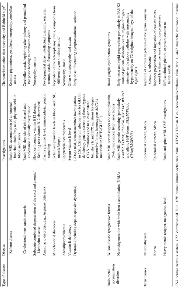
Exclusion diagnosis
There are various acquired and genetic causes that should
be ruled out in patients with the symptom of spastic
para-plegia without a family history (Table
1
). Cerebral and
spi-nal magnetic resonance imaging (MRI) investigations are
important to rule out common neurological conditions and
structural anomalies (e.g., spinal cord compression). For
example, a frontal interhemispheric tumor may manifest as
progressive spastic paraplegia with sphincter disturbances
before other signs such as cognitive deterioration, headache
or visual troubles appear. Disease progression, age at onset,
additional symptoms and results from other supplementary
investigations such as cerebrospinal fluid (CSF) analyses,
blood biochemistry and serology, electroneuromyography
and ophthalmological examination can give important clues
to the diagnosis (Table
1
). All these investigations will first
exclude acquired causes of spastic paraplegia but will
sub-sequently help with the diagnostic workflow to find the
correct genetic diagnosis. Some apparently sporadic cases
are in fact masked familial diseases. The absence of a
fam-ily history in neurogenetic disorders is frequent in clinical
practice and several explanations for apparent isolation are
reduced penetrance, age-dependent penetrance, variable
expressivity, de novo mutation, early death of the
transmit-ting parent or underdiagnosis in pure dominant forms with
mild symptoms, autosomal recessive inheritance in small
kindreds or, more rarely, X-linked inheritance in affected
men. Among other inherited neurogenetic conditions that
must be ruled out are leukodystrophies, in the absence of
inflammation but in the presence of MRI abnormalities.
Biochemical analyses in serum and/or CSF can suggest
neurometabolic diseases. Finally, dopa-responsive
dysto-nias (DRD) are a group of autosomal dominant or recessive
diseases, which may present with spasticity and can mimic
HSP. The dystonic toe is well known and can be
misdiag-nosed as extensor plantar reflex (Furukawa et al.
2001
).
Diurnal fluctuations and high and sustained sensibility to
levodopa are characteristic of DRD.
The exploration of rare genetic disorders is an
impor-tant issue since some diseases associated with spasticity
are treatable. In particular, spastic paraparesis can be one of
the multiple presentations of inborn errors of metabolism in
children and adults and in some cases the symptom spastic
paraparesis remains the only symptom for years; therefore,
these metabolic causes should be included in the general
diagnostic approach to sporadic spastic paraparesis due to
treatment options (e.g., diet for argininemia, biotin in
bioti-nidase deficiency) (Tanyel and Mancano
1997
; Sedel et al.
2007
) (
www.treatable-id.org
).
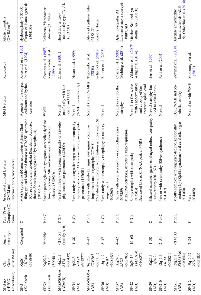
Genetic aspects of HSP
Genetic analysis of HSP genes can be performed when,
according to the clinical symptoms and signs, other
impor-tant causes have been excluded. HSP genes are denoted
Spastic Paraplegia Gene followed by a number according to
their order of discovery (SPGn). Up to now, the clinical
phe-notype and age at onset were critical to prioritize molecular
testing because of the heterogeneity of these diseases at the
clinical and genetic levels (Supplementary Fig. S1). More
than 25 novel causative genes have been reported in 2013–
2014 due to next-generation sequencing methods, making
this genetic workflow time-consuming (Martin et al.
2013
;
Oates et al.
2013
; Boukhris et al.
2013
; Landouré et al.
2013
; Novarino et al.
2014
; Dor et al.
2014
; Esteves et al.
2014
; Crow et al.
2014
), even if there are some genes that
are still more frequent than others and may be analyzed first,
such as SPAST (SPG4) and KIAA1840 (SPG11) (see below).
All classical modes of transmission can be found and there
are at least 67 genes that, when mutated, can account for
these diseases (Table
2
) to which can be added additional
genes for which spasticity can be present as part of the
clini-cal presentation (Supplementary Table 1).
Table
1
List of the most important dif
ferential diagnoses to hereditary spastic paraple
gia with suggested supplementary in
vestig ations Type of disease Disease In vestig ations
Characteristics other than spasticity and Babinski sign
b
Structural anoma
-lies and trauma
Arnold–Chiari malformation
Brain and spine MRI
Ataxia, dizziness, unsteadiness
T
umor
Brain and spine MRI
Headache if brain tumor
, other focal symptoms
Spinal cord v
ascular malformation
MRI/spinal angiograph
y
Fluctuating symptoms/sudden onset
V
ertebral disorders with myelopath
y
Spine MRI
Sensory symptoms, pain
Spinal cord injury
Spine MRI
Sudden onset, trauma
Inflammatory
Primary progressi
ve or relapsing-remitting multiple sclerosis
Brain and spine MRI, CSF in
vestig ations includ -ing immunoelectrophoresis (e vok ed responses)
Symptoms from dif
ferent topographic re gions Neurode generati ve Spinocerebellar ataxias
Genetic screening, brain MRI
Ataxia
Amyotrophic lateral sclerosis (ALS) and primary lateral sclerosis (PLS)
Spine and brain MRI, neurograph
y, electromyo -graph y, CSF in vestig ations Often b
ulbar signs and rapid progression, weakness,
increased refle
xes. In
ALS; upper and lo
wer motor
neuron signs
Acquired
Diple
gic cerebral palsy (Little disease)
Brain MRI, antenatal, birth or postnatal history
Non-progressi ve Infectious Neurosyphilis Syphilis serology/CSF in vestig ations
Acute/subacute, and chronic, laboratory findings, often peripheral nerv
ous system findings
HTL
V
-1 infection (tropical spastic paraparesis)
Serum/CSF HTL
V
-1 antibodies
Subacute onset, laboratory findings
Acquired immune deficienc
y syndrome (AIDS)
HIV test
Subacute onset, laboratory findings
Neuroborreliosis
Serology/CSF in
vestig
ations
Subacute onset, laboratory findings and/or symptoms from other topographic re
gions other than upper motor
neuron Metabolic a Leuk odystrophies X-link ed adrenoleuk odystrophies
Brain MRI, measurement of v
ery long-chain
fatty acids in plasma
Neuropath
y, cogniti
ve decline, white matter changes
Metachromatic leuk
odystroph
y (late-onset forms)
Brain MRI, arylsulphatase
A dosage
Neuropath
y, beha
vioral signs and re
gression
Hereditary CNS demyelinating disease Krabbe leuk
odystroph y (late-onset forms) Brain MRI, g alactocerebrosidase deficienc y Neuropath y, re gression Pelizaeus–Merzbacher disease Brain MRI Nystagmus, ataxia, de velopmental delay Cana van disease Brain MRI, e xcessi ve urinary N AA e xcretion Blindness, se
vere mental defect, me
galocephaly
Leuk
oencephalopath
y with v
anishing white matter
Brain MRI
Also kno
wn as childhood ataxia with central nerv
ous system h ypomyelination (CA CH) or v anishing white matter disease Ale xander disease Brain MRI Seizures, me galencephaly , de
velopmental delay; In older
patients, b
ulbar or pseudob
ulbar signs
Sjögren–Larsson syndrome (progressi
ve forms) Brain MRI, lo w f atty aldeh yde deh ydrogenase acti vity Ichth
yosis, mental retardation, macular dystroph
y and
leuk
oencephalopath
Table 1 continued Type of disease Disease In vestig ations
Characteristics other than spasticity and Babinski sign
b
Refsum disease
Brain MRI, accumulation of an unusual branched-chain f
atty acid, ph
ytanic acid, in
blood and tissues
Retinitis pigmentosa, peripheral neuropath
y, cerebellar
ataxia
Cerebrotendinous xanthomatosis
Brain MRI, deposits of cholesterol and cholestanol in virtually e
very tissue
Cerebellar ataxia be
ginning after puberty and pseudob
ul
-bar phase leading to premature death
Subacute combined de
generation of the cord and anemia/
Lichtheim disease
Blood cell counts, vitamin B12 dosage, Schilling test (vitamin B12 absorption)
Neuropath
y, anemia
Amino acid disorders, e.g.,
Ar
ginase deficienc
y
Plasma ar
ginine le
vel, aminoaciduria, genetic
screening De velopmental delay , intellectual disability , seizures, tremor
, ataxia, fluctuating symptoms
Mitochondrial disorders
Lactate and p
yruv
ate le
vels in blood and CSF
,
muscle biopsy
Dependent of the heteroplasmy le
vels, symptoms from
dif ferent or gans (multisystemic) Abetalipoproteinemia Lipoprotein electrophoresis Neuropath y, ataxia V itamin E deficienc y Serum vitamin E le vel
Often with neuropath
y and ataxia
Dystonia (including dopa-responsi
ve dystonias)
l
-Dopa trial, neurotransmitter in
vestig
ations
in CSF
, CSF/serum glucose ratio for GLUT1
deficienc
y, genetic screening (heterozygous
GCH1
mutations and to a lesser e
xtend
biallelic
TH
and
SPR
mutations for
dopa-responsi ve forms, heterozygous SLC2A1 mutations in D YT9/GLUT1)
Early-onset, fluctuating symptoms/diurnal v
ariation
Brain metal accumulation disorders
W
ilson disease (progressi
ve forms)
Brain MRI, serum copper and ceruloplasmin, 24-h urine copper; li
ver tissue biopsy
Basal g
anglia dysfunction symptoms
Neurode
generation with brain iron accumulation (NBIA)
Brain MRI, genetic screening (particularly PANK2, CO
ASY
, PLA2G6,
A
TP13A2,
WDR45
and allelic HSP forms;
FA2H/SPG35,
C19orf12/SPG43
)
Early onset and rapid progression particularly in P
ANK2
mutated patients, dystonia, central re
gion of h
yper
-intensity in the glob
us pallidus with surrounding
hypointensity on
T2-weighted images (“e
ye-of-the-tiger sign”) Toxic causes Neurolath yrism Epidemical conte xt, Africa Ingestion of certain v
egetables of the genus
Lathyrus (peas…), subacute K onzo Epidemical conte xt, Africa
Improper preparation and ingestion of cassa
va roots,
onset less than one week later
, non-progressi ve Hea vy metals (copper , mang anese, lead)
Brain and spine MRI, CSF in
vestig
ations
Dif
fuse clinical picture, e
xposure conte xt to hea vy metals CNS central nerv ous system, CSF cerebrospinal fluid, HIV human immunodeficienc y virus, HTL V -1 Human T cell leuk
emia/lymphoma virus type 1,
MRI
magnetic resonance imaging,
NA
A
N
-acetylaspartic acid
a The list is not complete, b
ut the main groups with the most important subgroups are mentioned
b Extensor response of the cutaneous plantar refle
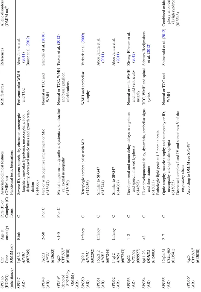
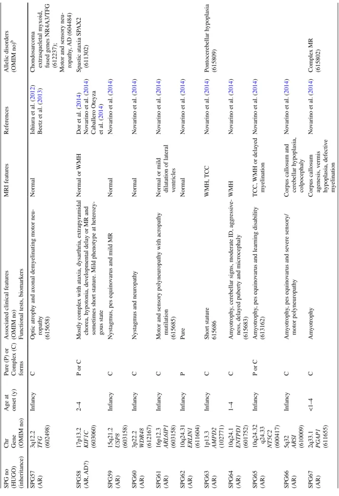
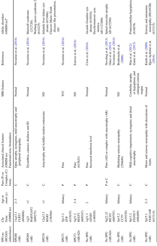
Table
2
HSP genes and their associated phenotypes
SPG no (HUGO) (inheritance) Chr Gene (OMIM no) Age at onset (y) Pure (P) or Comple
x (C)
forms
Associated clinical features (OMIM no) Functional tests, biomark
ers
MRI features
References
Allelic disorders (OMIM no)
b SPG1 (X-link ed) Xq28 L1CAM (308840) Congenital C
MASA syndrome (Mental retardation
Aphasia Shuf
-fling g
ait and
Adducted thumb) or CRASH syndrome
(Corpus callosum h
ypoplasia Retardation
Adducted
thumb Spastic paraple
gia and Hydrocephalus)
(303350)
Agenesis of the corpus callosum and h
ydro -cephalus Rosenthal et al. ( 1992 ) Jouet et al. ( 1994 )
Hydrocephaly (307000); Corpus callosum agenesis (304100)
SPG2 (X-link ed) Xq22.2 PLP1 (300401) V ariable P or C Spastic paraple gia with n
ystagmus, cerebellar dysfunc
-tion, h
ypotonia, MR and sometimes dementia or
seizures (312920) WMH Cremers et al. ( 1987 ) Saugier -V eber et al. ( 1994 ) Pelizaeus–Merzbacher disease (312080) SPG3/SPG3A (AD/AR) 14q22.1 ATL1 (606439) <1 to 51 (mainly <10) P (C)
Pure form, rarely with axonal neuropath
y or amyotro
-ph
y, incomplete penetrance (182600)
Normal (one f
amily with late
onset and TCC) Zhao et al. ( 2001 )
Hereditary sensory neuropath
y type ID, AD (613708) SPG4 (AD) 2p22.3 SPAST (604277) 1–80 P (C)
Pure, rarely with cogniti
ve impairment or neuropath y; epilepsy , ataxia and ALS in one f amily , incomplete penetrance (182601) Normal (WMH in one f amily) Hazan et al. ( 1999 ) SPG5/SPG5A (AR) 8q12.3 CYP7B1 (603711) 4–47 P or C
Pure or with cerebellar signs, n
ystagmus, cogniti
ve
impairment and amyotroph
y
(270800)
27-h
ydroxy-cholesterol accumulation in blood and CSF
Normal (rarely WMH) Tsaousidou et al. ( 2008 ) Schüle et al. ( 2010 )
Bile acid synthesis defect (613812); Sensory ataxia
SPG6 (AD) 15q11.2 NIP A1 (608145) 8–37 P (C)
Pure, rarely with neuropath
y or epilepsy or memory impairment (600363) Normal Rainier et al. ( 2003 ) SPG7 (AR) 16q24.3 SPG7 (602783) 4–42 P or C
Pure or with optic neuropath
y or cerebellar ataxia
(607259) Mito DN
A deletions, defects in Mito respiration
Normal or cerebellar atroph
y Casari et al. ( 1998 ) W edding et al. ( 2014 ) Optic neuropath y, AD;
Late-onset ataxia suscepti
-bility , AD SPG8 (AD) 8q24.13 KIAA0196 (610657) 10–60 P (C) Rarely comple x with neuropath y
(603563) Decreased Cho & Cr/N
AA peak at PMRS
Normal, or fe
w white
matter abnormalities and atroph
y of the
thoracic spinal cord
V aldmanis et al. ( 2007 ) W ang et al. ( 2014 ) Ritscher–Schinzel syn -drome, AR (220210) SPG9 (AD) 10q23.3- q24.2-1–30 C Bilateral cataracts, g
astroesophageal reflux, neuropath
y, amyotroph y (601162) Normal (atroph y lim
-ited to spinal cord)
Seri et al. ( 1999 ) SPG10 (AD) 12q13.3 KIF5A (602821) 2–51 P or C
Pure or with neuropath
y (Silv er syndrome) (604187) Normal Reid et al. ( 2002 ) SPG11 (AR) 15q21.1 KIAA1840 (610844) <1 to 33 P or C Mostly comple x with cogniti ve decline, neuropath y, retinopath
y (Kjellin syndrome) and cerebellar signs
(604360) TCC, WMH and cer -ebellar atroph y Ste vanin et al. ( 2007b ) Juv enile amyotrophic
lateral sclerosis (ALS- 5), Orlacchio et
al. ( 2010 ) SPG12 (AD) 19q13.32 RTN2 (603183) 7–24 P Pure (604805) Normal or with WMH Montene gro et al. ( 2012 )
Table
2
continued
SPG no (HUGO) (inheritance) Chr Gene (OMIM no) Age at onset (y) Pure (P) or Comple
x (C)
forms
Associated clinical features (OMIM no) Functional tests, biomark
ers
MRI features
References
Allelic disorders (OMIM no)
b SPG13 (AD) 2q33.1 HSPD1 (118190) 17–68 P Pure (605280) Normal Hansen et al. ( 2002 ) Hypomyelinating leu -kodystroph y type 4, AR (612233) SPG14 (AR) 3q27-q28 ~30 C
Distal motor neuropath
y, mild MR, visual agnosia, and
memory deficienc y (605229) Normal V azza et al. ( 2000 ) SPG15 (AR) 14q24.1 ZFYVE26 (612012) 4–19 P or C Mostly comple x with cogniti ve decline, neuropath y, retinopath
y (Kjellin syndrome) and cerebellar signs
(270700) TCC, WMH and cer -ebellar atroph y Hanein et al. ( 2008 ) SPG16 (X-link ed) Xq11.2 Early inf anc y P or C Pure or comple x with quadriple
gia, motor aphasia, mild
MR, and bo
wel and bladder dysfunction
(300266) Delayed myelination Steinmüller et al. ( 1997 ) Tamag aki et al. ( 2000 ) SPG17 (AD) 11q12.3 BSCL2 (606158) 2–60 C Silv er syndrome: neuropath y, amyotroph y (270685) Normal Magré et al. ( 2001 ); W indpassinger et al. ( 2004 ) Congenital lipodystroph y type 2, AR (260700);
Hereditary motor neu
-ropath y type V A, AD (600794); Progressi ve encephalopa -th y, AR (615924) SPG18 (AR) 8p11.23 ERLIN2 (611605) <2 C ID and contractures (611225) Normal Yıldırım et al. ( 2011 ) Juv
enile primary lateral
sclerosis, AR SPG19 (AD) 9q33-q34 36–55 P Pure (607152) Normal V alente et al. ( 2002 ) SPG20 (AR) 13q12.3 SPG20/ KIAA0610 (607111) Inf anc y C T ro
yer Syndrome: dysarthria, distal amyotroph
y in
hands and feet, cerebellar signs, mild ID and sk
eletal
abnormalities (short stature)
(275900) WMH Patel et al. ( 2002 ) SPG21 (AR) 15q22.31 SPG21/ A CP33 (608181) Adulthood C
Mast syndrome: speech decline leading to akinetic mutism, personality disturbances, psychotic episodes, cogniti
ve decline and cerebellar dysfunction (inco
-ordination and dysdiadochokinesia). F
or a Japanese
family: cogniti
ve decline and apraxia (248900)
TCC, WMH and cer -ebellar atroph y Simpson et al. ( 2003 ) SPG22 (X-link ed) Xq13.2 SLC16A2 (300095) Early inf anc y C Allan–Herndon–Dudle
y syndrome: spastic quadriple
-gia, se vere MR, central h ypotonia, muscle h ypoplasia, dystonia, ataxia (300523) Abnormal relati
ve concentrations of circulating iodo
-th
yronines
Normal or most often delayed myelination with
sometimes TCC
and mild cortical atroph
y Dumitrescu et al. ( 2004 ) Schw artz et al. ( 2005 )
Table
2
continued
SPG no (HUGO) (inheritance) Chr Gene (OMIM no) Age at onset (y) Pure (P) or Comple
x (C)
forms
Associated clinical features (OMIM no) Functional tests, biomark
ers
MRI features
References
Allelic disorders (OMIM no)
b SPG23 (AR) 1q24-q32 Inf anc y C
Lison syndrome: abnormal skin and hair pigmenta
-tion, ± dysmorphisms, sk eletal deformities, MR or sensorimotor neuropath y (270750)
Normal or slight enlar
gement of the v entricles with ± microcephaly Blumen et al. ( 2003 ) SPG24 (AR) 13q14 Inf anc y P Pure (607584) Normal Hodgkinson et al. ( 2002 ) SPG25 (AR) 6q23-24.1 30–46 C
Mild sensorimotor neuropath
y
(608220)
Spinal disc herniation with minor spondy
-losis Zortea et al. ( 2002 ) SPG26 (AR) 12q13.3 B4GALNT1 (601873) 2–19 C
ID, cerebellar ataxia, peripheral neuropath
y, and one
family presents beha
vioral problems
(609195) Decreased GM2 and increased GM3 in fibroblasts. Lo
w
testosterone le
vel in men
Normal or after long disease duration corti
-cal and subcorti-cal atroph
y and/or WMH Boukhris et al. ( 2013 ) Harlalka et al. ( 2013 ) SPG27 (AR) 10q22.1- q24.1 P: 25–45 C: 2–7 P or C
Pure or with sensorimotor polyneuropath
y and
sometimes with MR, cerebellar signs and sk
eletal
abnormalities
(609041)
Normal or mild cortical and cerebellar atroph
y Meijer et al. ( 2004 ) Ribai et al. ( 2006 ) SPG28 (AR) 14q22.1 DDHD1 (614603) 7–15 P or C
Pure or with cerebellar oculomotor disturbances or axonal neuropath
y
(609340) Ventricular lactate accumulation and reduction of PCr/ Pi ratio in muscles
Normal Tesson et al. ( 2012 ) Liguori et al. ( 2014 ) SPG29 (AD) 1p31.1-21.1 Inf anc y C Neonatal h
yperbilirubinemia, hearing impairment due
to auditory neuropath y and persistent v omiting due to hiatal hernia (609727) Normal Orlacchio et al. ( 2005 ) SPG30 (AR) 2q37.3 KIF1A (601255) 10–39 P or C
Pure or with sensory neuropath
y and cerebellar ataxia
(610357)
Normal or mild cer
-ebellar atroph y Erlich et al. ( 2011 ); Klebe et al. ( 2012b ) Comple x MR with axial
hypotonia, spasticity and cerebellar
atroph
y, AD
(614255);
Sensory and autonomic neuropath
y, AR (614213) SPG31 (AD) 2p11.2 REEP1 (609139) V ariable P or C
Pure or sometimes comple
x with neuropath y (610250) Normal Züchner et al. ( 2006 )
Distal hereditary motor neuropath
y type VB, AD (614751) SPG32 (AR) 14q12-q21 6–7 C Mild MR (611252) Cerebellar atroph y and
pontine dysraphia, moderate TCC
Ste
vanin et
al. (
2007a
Table
2
continued
SPG no (HUGO) (inheritance) Chr Gene (OMIM no) Age at onset (y) Pure (P) or Comple
x (C)
forms
Associated clinical features (OMIM no) Functional tests, biomark
ers
MRI features
References
Allelic disorders (OMIM no)
b SPG33 (AD) 10q24.2 ZFYVE27 (610244) 42–50 P Pure (610248) ND Mannan et al. ( 2006 ) SPG34 (X-link ed) Xq24-q25 16–25 P Pure (300750) ND Macedo-Souza et al. ( 2008 ) SPG35 (AR) 16q23.1 FA2H (611026) 2–17 one family with late onset
C
Dystonia, LL amyotroph
y, seizures, cerebellar signs,
cogniti
ve decline and optic atroph
y
(612319) Reduced h
ydroxylated f
atty acid sphingomyelin in
fibroblasts and erythroc
ytes Leuk odystroph y, hypointensities of glob us pallidus, TCC
and cerebellar atroph
y Edv ardson et al. ( 2008 ) Dan et al. ( 2011 ) Leuk odystroph y/NBIA, AR SPG36 (AD) 12q23-24 14–33 C
Peripheral sensorimotor neuropath
y (613096) Normal Schüle et al. ( 2009a ) SPG37 (AD) 8p21.1-q13.3 8–60 P Pure (611945) Normal Hanein et al. ( 2007 ) SPG38 (AD) 4p16-p15 16–19 P
Clinical features similar to SPG4 (612335)
ND Orlacchio et al. ( 2008 ) SPG39 (AR) 19p13.2 PNPLA6 (603197) Inf anc y, ado -lescence C Muscle w
asting and motor axonopath
y of the LL and UL (612020) Normal Rainier et al. ( 2008 ); Synofzik et al. ( 2014 ) Boucher -Neuhauser syn -drome (215470);
Gordon Holmes syndrome; Spastic ataxia
SPG41 (AD) 11p14.1- 11p.2 Mean 17 ± 3 P Pure (613364) Normal Zhao et al. ( 2008 ) SPG42 (AD) 3q25.31 SLC33A1 (603690) 4–42 P Pure (612539) Normal Lin et al. ( 2008 )
Congenital cataracts, hear
-ing loss and neurode
gen -eration, AR (614482) SPG43 (AR) 19p13.11- q12 C19orf12 (614297) 7–12 C Neuropath y and se vere atroph
y and decreased sensation
in the arms and le
gs (615043) Normal Landouré et al. ( 2013 ) NBIA4 (614298); Pallido-p yramidal syn -drome SPG44 (AR) 1q42.13 GJC2 (608803) 1st or 2nd decade C
Dysarthria, cerebellar ataxia, mental impairment (613206) Reduced Cho/N
AA and Cho/Cr ratios
WMH Uhlenber g et al. ( 2004 ) Orthmann-Murph y et al. ( 2009 ) Pelizaeus–Merzbacher -lik e hypomyelinating leuk od -ystroph y (608804); Hereditary lymphedema, AD (613480) SPG45 (AR) 10q24.3- q25.1 Inf anc y C
MR and ocular signs (613162)
ND Dursun et al. ( 2009 ) SPG46 (AR) 9p13.3 GB A2 (609471) 1–16 C
Cerebellar ataxia, cataract and mental impairment, infertility in males (614409) GB
A2 acti
vity abolished in lymphoblasts and leuk
oc
ytes
TCC, cerebral and cerebellar atroph
y Martin et al. ( 2013 ) Spastic ataxia
Table
2
continued
SPG no (HUGO) (inheritance) Chr Gene (OMIM no) Age at onset (y) Pure (P) or Comple
x (C)
forms
Associated clinical features (OMIM no) Functional tests, biomark
ers
MRI features
References
Allelic disorders (OMIM no)
b SPG47 (AR) 1p13.2 AP4B1 (607245) Birth C Se
vere ID, absent speech, sh
y character , stereotypic laughter , muscular h ypotonia, microcephaly , foot deformity
, decreased muscle mass and gro
wth retar -dation (614066) Peri ventricular WMH and TCC Abou Jamra et al. ( 2011 ) Bauer et al. ( 2012 ) SPG48 (AR) 7p22.1 AP5Z1 (613653) 2–50 P or C
Pure or with cogniti
ve impairment or MR (613647) Normal or TCC and WMH Słabicki et al. ( 2010 ) SPG49 a
(denoted SPG56 by OMIM) (AR) 4q25 CYP2U1
a
(615030)
<1–8
P or C
Mental impairment, dysarthria, dystonia and infraclini
-cal axonal neuropath
y (615030) Normal or TCC, WMH and basal g anglion calcifications Tesson et al. ( 2012 ) SPG50 (AR) 7q22.1 AP4M1 (602292) Inf anc y C Tetraple
gic cerebral palsy with MR
(612936)
WMH and cerebellar atroph
y V erk erk et al. ( 2009 ) SPG51 (AR) 15q21.2 AP4E1 (607244) Inf anc y C Similar to SPG47 (613744) Abou Jamra et al. ( 2011 ) SPG52 (AR) 14q12 AP4S1 (607243) Inf anc y C Similar to SPG47 (614067) Abou Jamra et al. ( 2011 ) SPG53 (AR) 8p22 VPS37A (609927) 1–2 C De
velopmental and motor delay
, delays in cognition
and speech, mark
ed k yphosis (614898) Normal or mild WMH and mild v entriculo -me galy Zi von y-Elboum et al. ( 2012 ) SPG54 (AR) 8p11.23 DDHD2 (615003) <2 C ID or de velopmental delay
, dysarthria, cerebellar signs
and short stature
(615033) Pathologic lipid peak at 1.3
ppm in brain TCC, WMH and spinal syrinx Schuurs-Hoeijmak ers et al. ( 2012 ) SPG55 (AR) 12q24.31 C12orf65 (613541) 2–7 C Optic atroph y, muscle atroph y and neuropath y or ID, neuropath y and ophthalmople gia (615035) Decreased comple
x I and IV and sometimes
V of the respiratory chain Normal or TCC and WMH Shimazaki et al. ( 2012 ) Combined oxidati ve phos -phorylation deficienc y 7 (Leigh syndrome) (613543) SPG56 a (AR) 4q25 CYP2U1 a (615030)
According to OMIM see SPG49
Table
2
continued
SPG no (HUGO) (inheritance) Chr Gene (OMIM no) Age at onset (y) Pure (P) or Comple
x (C)
forms
Associated clinical features (OMIM no) Functional tests, biomark
ers
MRI features
References
Allelic disorders (OMIM no)
b SPG57 (AR) 3q12.2 TFG (602498) Inf anc y C Optic atroph
y and axonal demyelinating motor neu
-ropath y (615658) Normal Ishiura et al. ( 2012 ) Beetz et al. ( 2013 )
Chondosarcoma extrasqueletal myxoid, fused genes NR4A3/TFG (612237); Motor and sensory neu
-ropath y, AD (604484) SPG58 (AR, AD?) 17p13.2 KIF1C (603060) 2–4 P or C Mostly comple
x with ataxia, dysarthria, e
xtrap
yramidal
chorea, h
ypotonia, de
velopmental delay or MR and
sometimes short stature. Mild phenotype at heterozy
-gous state Normal or WMH Dor et al. ( 2014 ) No varino et al. ( 2014 ) Caballero Ote yza et al. ( 2014 ) Spastic ataxia SP AX2 (611302) SPG59 (AR) 15q21.2 USP8 (603158) Inf anc y C
Nystagmus, pes equino
varus and mild MR
Normal No varino et al. ( 2014 ) SPG60 (AR) 3p22.2 WDR48 (612167) Inf anc y C
Nystagmus and neuropath
y Normal No varino et al. ( 2014 ) SPG61 (AR) 16p12.3 ARL6IP1 (603158) Inf anc y C
Motor and sensory polyneuropath
y with acropath
y
mutilation
(615685)
Normal or mild dilatation of lateral ventricles
No varino et al. ( 2014 ) SPG62 (AR) 10q24.31 ERLIN1 (611604) Inf anc y P Pure Normal No varino et al. ( 2014 ) SPG63 (AR) 1p13.3 AMPD2 (102771) Inf anc y C Short stature 615686 WMH, TCC No varino et al. ( 2014 ) Pontocerebellar h ypoplasia (615809) SPG64 (AR) 10q24.1 ENTPD1 (601752) 1–4 C Amyotroph
y, cerebellar signs, moderate ID, aggressi
ve
-ness, delayed puberty and microcephaly
(615683) WMH No varino et al. ( 2014 ) SPG65 (AR) 10q24.32 q24.33 NT5C2 (600417) Inf anc y P or C Amyotroph y, pes equino
varus and learning disability
(613162) TCC, WMH or delayed myelination No varino et al. ( 2014 ) SPG66 (AR) 5q32 ARSI (610009) Inf anc y C Amyotroph y, pes equino varus and se vere sensory/ motor polyneuropath y
Corpus callosum and cerebellar h
ypoplasia, colpocephaly No varino et al. ( 2014 ) SPG67 (AR) 2q33.1 PGAP1 (611655) <1–4 C Amyotroph y
Corpus callosum agenesis, v
ermis hypoplasia, defecti ve myelination No varino et al. ( 2014 ) Comple x MR (615802)
Table
2
continued
SPG no (HUGO) (inheritance) Chr Gene (OMIM no) Age at onset (y) Pure (P) or Comple
x (C)
forms
Associated clinical features (OMIM no) Functional tests, biomark
ers
MRI features
References
Allelic disorders (OMIM no)
b SPG68 (AR) 11q13.1 FLRT1 (604806) 2–3 C Optic atroph y, n
ystagmus, mild amyotroph
y and peripheral neuropath y Normal No varino et al. ( 2014 ) SPG69 (AR) 1q31 RAB3GAP2 (609275) <1 C
Dysarthria, cataract, deafness and ID
Normal No varino et al. ( 2014 )
Martsolf syndrome: (212720); Warb
ur g micro syndrome 2 (614225) SPG70 (AR) 12q13.3 MARS (156560) <1 C Amyotroph y and
Achilles tendon contracture
ND No varino et al. ( 2014 ) Inf antile li ver f ailure syn -drome (615486); Charcot–Marie–T ooth disease lik e presentation, AD SPG71 (AR) 5p13.3 ZFR (615635) Inf anc y P Pure TCC No varino et al. ( 2014 ) SPG72 (AR/AD) 5q31.2 REEP2 (609347) 3–4 P Pure (615625) ND Este ves et al. ( 2014 ) No SPG (AR) 1q21.3 AD AR1 (146920) 2 P
Pure Increased interferon le
vel Normal Cro w et al. ( 2014 ) Aicardi–Goutière syn -drome (615010); Dyschromatosis sym -metrica AD (127400) No SPG (AR/AD) 9q22.32 BICD2 (609797) Inf anc y P or C
Pure (AD) or comple
x with amyotroph y (AR) Normal Ne veling et al. ( 2013 ) Oates et al. ( 2013 ) No varino et al. ( 2014 )
Spinal muscular atroph
y AD (615290) No SPG (AR) 5p15.2 CCT5 (610150) Inf anc y C
Mutilating sensory neuropath
y (256840) ND Bouhouche et al. ( 2006 ) No SPG (AR) 9p13.2 EXOSC3 (606489) Inf anc y C Mild cogniti ve impairment, n
ystagmus and distal
amyotroph y Cerebellar atroph y or h ypoplasia, and enlar ged cisterna magna W an et al. ( 2012 ) Zanni et al. ( 2013 ) Pontocerebellar h ypoplasia (614678) No SPG (AR) 5p15.1 FAM134B (613114) 2–3 C
Motor and sensory neuropath
y with ulcerations of limbs Normal K urth et al. ( 2009 ) Ilg az-A ydinlar et al. ( 2014 )
Sensory and autonomic neuropath
y (HSAN2B)
Table
2
continued
SPG no (HUGO) (inheritance) Chr Gene (OMIM no) Age at onset (y) Pure (P) or Comple
x (C)
forms
Associated clinical features (OMIM no) Functional tests, biomark
ers
MRI features
References
Allelic disorders (OMIM no)
b No SPG (AR) 1q42.13 IBA57 (615316) 3–12 C Distal amyotroph y, peripheral neuropath y optic nerv e atroph
y and reduced visual acuity (SPO
AN-lik
e
phenotype)
Normal or
WMH foci
sometimes with TCC and cerebellar atroph
y.
Lossos et
al. (
2015
)
Multiple mitochondrial dysfunctions syndrome (615330),
Ajit Bolar et al. ( 2013 ) No SPG (AR) 2q24.2 IFIH1 (606951) 2 P
Pure Increased Interferon le
vel Normal Cro w et al. ( 2014 ) Aicardi–Goutière syn -drome (615846) No SPG (AR) 1q42.3 LYST (606897) Late (48–58) C
Cerebellar ataxia, peripheral neuropath
y and lar
ge
peroxidase-positi
ve granules in granuloc
ytes
Mild cerebellar atroph
y Shimazaki et al. ( 2014 ) Chediak–Hig ashi syndrome (214500) No SPG (AR) 19q13.1 MAG (159460) Inf anc y C Cerebellar signs, n
ystagmus, and amyotroph
y Normal No varino et al. ( 2014 ) No SPG (Mito) MT -A TP6 (516060) 30–50 P or C
Pure or with neuropath
y, cerebellar signs and cardio
-myopath y ND V ern y et al. ( 2011 )
Leigh syndrome (551500); Leber optic atroph
y
(535000);
Inf
antile bilateral striatal necrosis (500003); Epilepsy and lactic acidosis Inf
antile cardiomyopath y No SPG (Mito) MT -CO3 (516050) Inf anc y C
Spastic paraparesis, ophthalmoparesis and lactic acidosis
Basal g
anglia h
yper
-intensities (Leigh syndrome-lik
e) and
mild cerebral and cerebellar atroph
y T iranti et al. ( 2000 ) No SPG (Mito) MT -TI (590045) Adulthood P or C Pure with lo w heteroplasmy le vels. Comple x with high heteroplasmy le
vels, with ataxia, deafness, epilepsy
, cardiomyopath y and h ypogonadism ND Corona et al. ( 2002 ) No SPG (AR) 13q14.3 RN ASEH2B (610326) 18– 21 months P Pure Normal Cro w et al. ( 2014 ) Aicardi–Goutière syn -drome (610181) No SPG (AR) 13q11 SACS (604490) Inf anc y C
Spastic ataxia of Charle
voix Saguenay: early child
-hood onset of cerebellar ataxia, p
yramidal tract signs
and peripheral neuropath
y,
±
retinal striations on
fundoscop
y and thick
ening of the retinal nerv
e fiber layer on OCT Atroph y of the superior cerebellar v ermis,
hyperintensity of corticospinal tracts
Engert et al. ( 2000 ) No SPG a (denoted SPG49 a by OMIM) (AR) 14q32.31 TECPR2 a (615000) Inf anc y C Se
vere ID, rigid ataxic g
ait, brach ycephalic micro -cephaly , fluctuating central h ypo ventilation, g astroe
-sophageal reflux disease, w
ak e apnea, arefle xia and dysmorphic features (615031) V entriculome galy ,
TCC, cerebral and cerebellar atroph
y Oz-Le vi et al. ( 2012 )
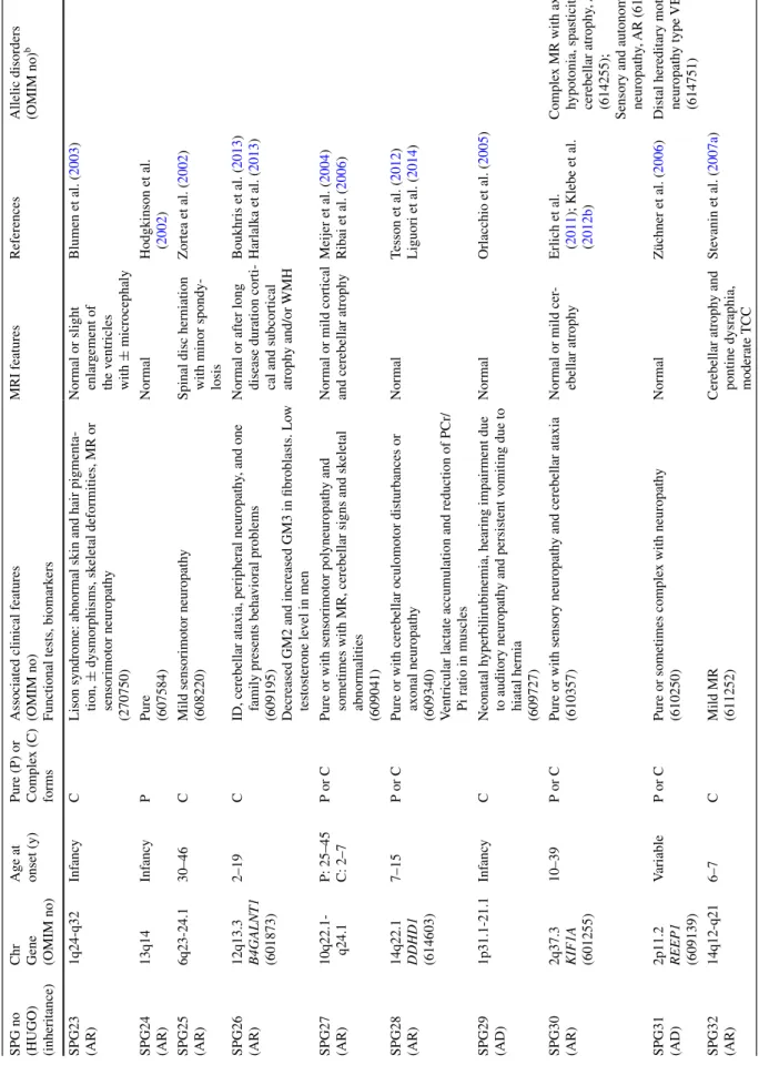
Autosomal dominant (AD) forms of HSP are mainly
pure forms with ages at onset that can range from
infancy to late adulthood. Mutations in SPAST (SPG4),
ATL1
(SPG3), KIF5A (SPG10) and REEP1 (SPG31) are
described as being responsible for around 50 % of all
cases (Fig.
1
) (Finsterer et al.
2012
). SPAST point
muta-tions and exonic rearrangements have been implicated in
10–40 % of the HSP patients (Hazan et al.
1999
; Meijer
et al.
2002
; Beetz et al.
2006
; Loureiro et al.
2013
) and in
up to 12 % of sporadic forms (Depienne et al.
2006
).
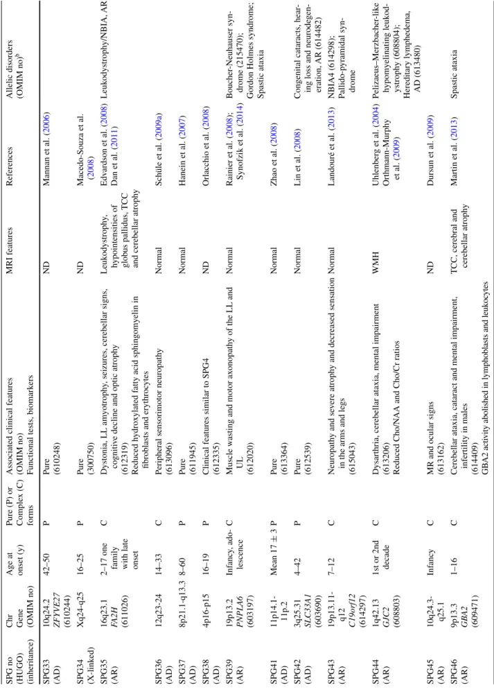
The autosomal recessive (AR) forms appear to be
particularly prevalent where consanguinity is common
such as in the Middle East or Mediterranean countries
(Coutinho et al.
1999
; Boukhris et al.
2009
; Ruano et al.
2014
), and lesser frequent in central Europe, Japan
(Tak-iyama
2014
) and USA (with the exception of
communi-ties such as the Amish). They are also more complex in
clinical terms, associated with greater genetic
heteroge-neity (Table
2
) with an onset of symptoms that is
gen-erally early. Only two forms are associated with pure
HSP, but this likely results from the assignment of few
families each: SPG71 and SPG72. In complex forms, the
associated signs may be subtle but important indicators
of the mutated gene, such as cerebellar atrophy or
cer-ebellar ataxia with optic atrophy in SPG7,
developmen-tal delay and short stature in SPG20 (Troyer syndrome),
dysarthria, distal amyotrophy, premature aging and
cog-nitive decline in SPG21 (Mast syndrome), peripheral
neuropathy and abnormal skin and hair pigmentation in
SPG23 (Lison syndrome) (Table
2
). Mental retardation
or intellectual deterioration, thin corpus callosum (TCC)
and axonal neuropathy are highly suggestive of SPG11
(Stevanin et al.
2008a
). Finally, spastic ataxia with
dys-arthria, nystagmus and retinal striations is suggestive
of autosomal recessive spastic ataxia of
Charlevoix-Saguenay (ARSACS). Mutations in the CYP7B1 (SPG5),
SPG7
, KIAA1840 (SPG11) and ZFYVE26 (SPG15)
genes are among the most frequently found but their
relative frequencies vary according to the geographical
origin (Stevanin et al.
2008a
; Paisan-Ruiz et al.
2008
;
Erichsen et al.
2009
; Goizet et al.
2009a
; Schüle et al.
2009b
; Arnoldi et al.
2012
; Klebe et al.
2012a
; Pfeffer
et al.
2014
) (Fig.
1
). Point mutations or rearrangements
in KIAA1840 (SPG11) have been shown to account
for approximately 20 % of AR-HSP (Stevanin et al.
2008a
).
X-linked forms are rare and include two clinical entities
well recognized by pediatricians (Table
2
): SPG1, caused
by mutations in the neural cell adhesion molecule L1CAM
gene, and SPG2, which results from mutations in the gene
encoding the proteolipid protein (PLP1), a myelin
compo-nent. SPG2 can also account for late-onset cases in women
(Sivakumar et al.
1999
).
Table 2 continued AD autosomal dominant, ALS amyotrophic lateral sclerosis, AR autosomal recessi ve, Chr chromosome, Cho/Cr and Cho/N AA ratio choline to creatine or to N AA, CSF cerebrospinal fluid, GM2/3 gangliosides monosialic 2 and 3,
ID intellectual disability , LL lo wer limb, Mito mitochondrial, MR mental retardation, MRI
magnetic resonance imaging,
NA A N -acetyl aspartate, nb number , NBIA
neuronal brain iron accumulation disorders,
ND
not described,
OCT
ocular coherence tomograph
y,
PCr/Pi
ratio of phosphocreatine to inor
ganic phosphate,
PPM
parts per mil
-lion, PMRS proton magnetic resonance spectrometry , SPO AN spastic paraple gia, optic atroph y and neuropath y, TCC thin corpus callosum, UL upper limb, WMH white matter hyperintensity , y
years a According to the HUGO
nomenclature, SPG49 has been associated with
CYP2U1
mutations and SPG56 has not been associated to a specific gene.
According to
the OMIM numbering,
SPG49 has been associated to
TECPR2
mutations and SPG56 to
CYP2U1
mutations
b Inheritance mode is indicated when it dif
fers from the one described in f
amilies with spasticity
SPG no (HUGO) (inheritance) Chr Gene (OMIM no) Age at onset (y) Pure (P) or Comple
x (C)
forms
Associated clinical features (OMIM no) Functional tests, biomark
ers
MRI features
References
Allelic disorders (OMIM no)
b
No SPG (AD) 9p13 VCP (601023)
54–57
C
Case report: hereditary spastic paraple
gia with P aget’ s disease of bone. Normal W atts et al. ( 2004 ) De bot et al. ( 2012 )
Inclusion body myopath
y
(167320);
Amyotrophic lateral sclero
Phenotype–genotype correlations in HSP
Many studies have failed in the past to determine reliable
phenotype–genotype correlations. However, the systematic
analysis of a large set of genes, including exome
sequenc-ing, is regularly unmasking unusual phenotypes and
inher-itance modes associated with mutations in HSP genes and
the nature of the mutation in some of them can now be
cor-related to a specific phenotype.
Instances where similar mutations are associated with a
wide spectrum of HSP phenotypes; extension of the
clinical picture previously observed
There are good examples of variable phenotypes among HSP
subtypes, as SPG4 in which age at onset can vary from early
childhood to asymptomatic status at old ages. As more
fami-lies are reported with a mutation in a specific gene, the full
spectrum of each genetic entity extends and there are now
fewer than ten HSP loci/genes associated exclusively with
pure forms of the disease, most of them accounting for
sin-gle or only a few families so far (Table
2
; Supplementary
Table 1). This was the case, for example, with SPG10, which
was initially thought to be a pure form but now also accounts
for 10 % of the complex AD families (Goizet et al.
2009b
).
In SPG7, the occurrence of cerebellar ataxia and/or atrophy
(Klebe et al.
2012a
) or progressive external ophthalmoplegia
(Wedding et al.
2014
; Pfeffer et al.
2014
) suggests that the
analysis of this gene should be extended to other phenotypes.
Patients with isolated optic neuropathy should also be tested
for mutations on SPG7 (Klebe et al.
2012a
).
Instances where the nature of the mutations of a specific
HSP gene can determine the inheritance model and/or
associated phenotype
One of the recent advances in HSP genetics is the
identi-fication of various modes of inheritance of the mutations
in single HSP genes. This is what occurs for REEP2
muta-tions that have recently been implicated in three families
with recessive or dominant transmission of a pure HSP,
namely SPG72 (Novarino et al.
2014
; Esteves et al.
2014
).
In one Portuguese family with AR inheritance, two
muta-tions segregated in trans including a splice site mutation
leading to a loss of function of the corresponding allele
and a missense mutation responsible for reduced binding
capacities to membranes of the protein formed from the
second allele. In a French autosomal dominant family, the
disease segregated with a heterozygous missense
muta-tion that had a dominant negative effect on the capacity of
the wild-type protein to bind membranes. In both cases,
AD and AR mutations led to a complete loss of membrane
binding capacities of the REEP2 protein with consequences
for the tubular structure of the endoplasmic reticulum (ER)
(Esteves et al.
2014
). Recently, position p.Arg415 of
Atlas-tin-1 (SPG3A) was shown to be a hotspot for missense
mutations, first associated with incomplete penetrance
with an AD inheritance pattern (D’Amico et al.
2004
), and
then with AR transmission (Varga et al.
2013
) (Khan et al.
2014
). Similarly, an unusual recessive or dominant
inher-itance has been suggested in SPAST (SPG4, Lindsey et al.
2000
) and SPG7 (McDermott et al.
2001
; Sánchez-Ferrero
et al.
2013
), respectively.
The nature of the mutation and/or its localization in the
protein can sometimes impact both the inheritance model
and the phenotype at the same time, so that the nature of the
mutation can predict the phenotype. This was observed with
KIF1A
(SPG30), in which missense homozygous mutations
located in the kinesin motor domain account for a relatively
pure HSP (Erlich et al.
2011
; Klebe et al.
2012b
), whereas
heterozygous mutations located in the ATP binding site of
KIF1A were found in patients with severe mental
retarda-tion with axial hypotonia, peripheral spasticity and mild
atrophy of cerebellum and or corpus callosum, a
pheno-type reminiscent of SPG11 (Hamdan et al.
2011
; Chang
et al.
2014
). Homozygous KIF1A frameshift mutations lead
Fig. 1 Relative frequencies of
the main autosomal dominant (a) and recessive (b) mutations
in the SPATAX (http://spatax.
wordpress.com/) cohort (Goizet
et al. 2009a, b, 2011;
Steva-nin et al. 2008a; Tesson et al.
to hereditary sensory neuropathy type IIC (Rivière et al.
2011
). Similarly, heterozygous mutations in HSPD1 lead
to SPG13 (Hansen et al.
2002
), but homozygous missense
mutations of the same gene are implicated in
hypomyelinat-ing leukodystrophy type 4 (Magen et al.
2008
). Mutations
in TFG are responsible for SPG57, an AR-HSP associated
with optic atrophy and neuropathy (Beetz et al.
2013
) but
can also be responsible for AD motor and sensory
neuropa-thy (Ishiura et al.
2012
). Interestingly, the TFG mutations
affect different domains of the protein: the coil–coil domain
in the HSP family, the P/Q rich domain in the family with
neuropathy, suggesting different pathological mechanisms.
In addition, one patient with neuropathy had ubiquitin- and
TDP43-positive cytoplasmic neuronal inclusions
reminis-cent of amyotrophic lateral sclerosis (ALS) (Supplementary
Table 2), suggesting a toxic gain of function effect
result-ing in a dominant inheritance pattern. In contrast, biallelic
mutations affect the capacity of TFG to self-assemble and
then probably lead to a loss of function (Beetz et al.
2013
).
This is also the case with REEP1, in which frameshift
muta-tions or missense mutamuta-tions that abolish ER targeting and
affect the capacity of the protein to bind ATL1 (Falk et al.
2002
; Beetz et al.
2012
) lead to HSP (Züchner et al.
2006
;
Beetz et al.
2008
; Hewamadduma et al.
2009
; Goizet et al.
2011
), whereas in-frame deletions do not impact the
capac-ity of the protein to bind ATL1 and lead to hereditary motor
neuropathy type V (Beetz et al.
2012
).
Instances where the mutations of a specific HSP gene lead
to overlapping diseases
During the past few years, it has appeared that HSP and
other neurological conditions are at opposite ends of a
continuum of overlapping diseases. The clinical overlap
of HSP with peripheral neuropathies, cerebellar ataxias or
mental disabilities is not new since mutated HSP patients
can have a clinical presentation associating symptoms
spe-cific to these groups of disorders. ARSACS is a good
illus-tration of this clinical overlap between ataxias and spastic
paraplegias (Bouhlal et al.
2011
). It is sometimes difficult
to decide which symptom is most prominent in the clinical
presentation and this may also depend on the physician’s
expertise. The overlap between HSP and ataxias was again
recently highlighted by mutations in GBA2 (SPG46), which
have been found in patients with spastic ataxia associated
with cataract, having ataxia (Hammer et al.
2013
; Votsi
et al.
2014
) or spasticity (Martin et al.
2013
; Citterio et al.
2014
) as the prominent clinical feature. Point mutations
and exonic deletions in KIF1C have both been reported in
HSP (Novarino et al.
2014
) and spastic ataxia (Dor et al.
2014
), illustrating the fact that both diseases are part of
the same clinical spectrum. PNPLA6 mutations are found
in patients with a wide range of phenotypes: Gordon
Hol-mes spinocerebellar syndrome (ataxia with brisk reflexes
and hypogonadism), Boucher-Neuhäuser syndrome (ataxia
with chorioretinal dystrophy and hypogonadism), isolated
cerebellar ataxia and isolated spastic paraplegia (SPG39)
(Rainier et al.
2008
; Synofzik et al.
2014
).
Regarding motor neuron diseases and polyneuropathies
(Fig.
2
), HSP genes have been found mutated in patients with
(i) peripheral nerve affections such as Charcot–Marie–Tooth
(CMT) neuropathies, (ii) first and secondary motor neuron
degeneration, such as ALS, and (iii) lower motor neuron
dis-orders such as spinal muscular atrophy (SMA). For
exam-ple, KIAA1840 (SPG11) mutations may mimic ALS5 when
muscle wasting is marked in absence of other complicated
Fig. 2 Clinico-genetic entities
associated with hereditary spas-tic paraplegia (HSP) according to the motor neuron phenotypic presentation. When mutated, HSP genes can be associated with various phenotypes that overlap with upper and lower motor neuron diseases