HAL Id: hal-02364352
https://hal.archives-ouvertes.fr/hal-02364352
Submitted on 14 Nov 2019HAL is a multi-disciplinary open access
archive for the deposit and dissemination of sci-entific research documents, whether they are pub-lished or not. The documents may come from teaching and research institutions in France or abroad, or from public or private research centers.
L’archive ouverte pluridisciplinaire HAL, est destinée au dépôt et à la diffusion de documents scientifiques de niveau recherche, publiés ou non, émanant des établissements d’enseignement et de recherche français ou étrangers, des laboratoires publics ou privés.
Non-canonical mTOR-Independent Role of DEPDC5 in
Regulating GABAergic Network Development
Amrutha Swaminathan, Rahma Hassan-Abdi, Solène Renault, Aleksandra
Siekierska, Raphaëlle Riché, Meijiang Liao, Peter de Witte, Constantin
Yanicostas, Nadia Soussi-Yanicostas, Pierre Drapeau, et al.
To cite this version:
Amrutha Swaminathan, Rahma Hassan-Abdi, Solène Renault, Aleksandra Siekierska, Raphaëlle Riché, et al.. Non-canonical mTOR-Independent Role of DEPDC5 in Regulating GABAer-gic Network Development. Current Biology - CB, Elsevier, 2018, 28 (12), pp.1924-1937.e5. �10.1016/j.cub.2018.04.061�. �hal-02364352�
Current Biology
Non-canonical mTOR-independent role of DEPDC5 in regulating GABAergic network
development
--Manuscript
Draft--Manuscript Number: CURRENT-BIOLOGY-D-17-01554R3
Full Title: Non-canonical mTOR-independent role of DEPDC5 in regulating GABAergic network development
Article Type: Research Article Corresponding Author: Eric Samarut, Ph.D
CRCHUM
Montreal, CANADA
First Author: Amrutha Swaminathan
Order of Authors: Amrutha Swaminathan Rahma Hassan-Abdi Solene Renault Aleksandra Siekierska Raphaelle Riche Meijiang Liao Peter A.M de Witte Constantin Yanicostas Nadia Soussi-Yanicostas Pierre Drapeau
Eric Samarut, Ph.D
Abstract: Mutations in DEPDC5 are causal factors for a broad spectrum of focal epilepsies, but the underlying pathogenic mechanisms are still largely unknown. To address this question, a zebrafish depdc5 knockout model showing spontaneous epileptiform events in the brain, increased drug-induced seizure susceptibility, general hypoactivity and premature death at 2-3 weeks post fertilization as well as the expected
hyperactivation of mTOR signaling was developed. Using this model, the role of DEPDC5 in brain development was investigated using an unbiased whole
transcriptomic approach. Surprisingly, in addition to mTOR-associated genes, many genes involved in synaptic function, neurogenesis, axonogenesis and GABA network activity were found to be dysregulated in larval brains. Although no gross defects in brain morphology/neuron loss were observed, immunostaining of depdc5-/- brains for several GABAergic markers revealed specific defects in the fine branching of
GABAergic network. Consistently, some defects in depdc5-/- could be compensated by treatment with GABA, corroborating that GABA signaling is indeed involved in
DEPDC5 pathogenicity. Further, the mTOR-independent nature of the
neurodevelopmental defects was demonstrated by the inability of rapamycin to rescue the defects of GABAergic networks observed in depdc5-/- brains and conversely, the inability of GABA to rescue the hypoactivity in another genetic model showing mTOR hyperactivation. This study hence provides the first in vivo evidence that DEPDC5 plays previously unknown roles apart from its canonical function as an mTOR inhibitor. Moreover, these results propose that defective neurodevelopment of GABAergic network could be a key factor in epileptogenesis when DEPDC5 is mutated.
Non-canonical mTOR-independent role of DEPDC5 in regulating GABAergic
1
network development.
2 3
Amrutha Swaminathan1, Rahma Hassan-Abdi2,3, Solène Renault2,3, Aleksandra Siekierska4,
4
Raphaëlle Riché1, Meijiang Liao1, Peter A.M. de Witte4, Constantin Yanicostas2,3, Nadia Soussi-5
Yanicostas2,3, Pierre Drapeau1,5# and Éric Samarut1,5,# 6
7
1 Department of Neurosciences, Research Center of the University of Montréal Hospital Center
8
(CRCHUM), Université de Montréal, Montréal, Canada H2X0A9. 9
2 Inserm, U1141, F-75019 Paris, France
10
3 Université Paris Diderot, Sorbonne Paris Cité, UMRS 1141, F-75019 Paris, France
11
4 Laboratory for Molecular Biodiscovery, Department of Pharmaceutical and Pharmacological
12
Sciences, University of Leuven, 3000 Leuven, Belgium 13
5 DanioDesign Inc., Montréal, Canada.
14
# Co-corresponding authors: p.drapeau@umontreal.ca and eric.samarut@umontreal.ca
15
Lead contact: Éric Samarut (eric.samarut@umontreal.ca)
16 17 18
Running title: DEPDC5 regulates GABA network development
19 20 21 Manuscript
Summary
22
Mutations in DEPDC5 are causal factors for a broad spectrum of focal epilepsies, but the 23
underlying pathogenic mechanisms are still largely unknown. To address this question, a 24
zebrafish depdc5 knockout model showing spontaneous epileptiform events in the brain, 25
increased drug-induced seizure susceptibility, general hypoactivity, premature death at 2-3 weeks 26
post fertilization as well as the expected hyperactivation of mTOR signaling was developed. 27
Using this model, the role of DEPDC5 in brain development was investigated using an unbiased 28
whole transcriptomic approach. Surprisingly, in addition to mTOR-associated genes, many genes 29
involved in synaptic function, neurogenesis, axonogenesis and GABA network activity were 30
found to be dysregulated in larval brains. Although no gross defects in brain morphology/neuron 31
loss were observed, immunostaining of depdc5-/- brains for several GABAergic markers 32
revealed specific defects in the fine branching of GABAergic network. Consistently, some 33
defects in depdc5-/- could be compensated for by treatment with GABA, corroborating that 34
GABA signaling is indeed involved in DEPDC5 pathogenicity. Further, the mTOR-independent 35
nature of these neurodevelopmental defects was demonstrated by the inability of rapamycin to 36
rescue the GABAergic network defects observed in depdc5-/- brains and conversely, the inability 37
of GABA to rescue the hypoactivity in another genetic model showing mTOR hyperactivation. 38
This study hence provides the first in vivo evidence that DEPDC5 plays previously unknown 39
roles apart from its canonical function as an mTOR inhibitor. Moreover, these results propose 40
that defective neurodevelopment of GABAergic networks could be a key factor in 41
epileptogenesis when DEPDC5 is mutated. 42
43
Keywords: Epilepsy/DEPDC5/GABA/mTOR/Neurodevelopment/zebrafish
Introduction:
45
Focal seizures, which originate in one part of the brain, are the most prevalent type of 46
epileptic seizures and mutations in DEPDC5 (Dishevelled, Egl-10 and Pleckstrin (DEP)-domain 47
containing 5) account for 12-37% of the cases [1]. Though numerous ion channel mutations have 48
been traditionally associated with epilepsy, recent work using whole genome/exome sequencing 49
has identified other pathways in epilepsy, like mTOR (mechanistic/mammalian target of 50
rapamycin) signaling. The causative gene for familial focal epilepsy with variable foci (FFEVF) 51
remained unknown until the identification of DEPDC5 mutations in a large number of families 52
with FFEVF [2-4]. Mutations in DEPDC5 leading to haploinsufficiency have been associated 53
also to other focal epilepsies including autosomal dominant nocturnal frontal lobe epilepsy and 54
familial temporal lobe epilepsy [3, 5]. 55
DEPDC5, along with NRPL2 and NRPL3 forms the mTOR inhibitory GATOR1 56
complex, which blocks the activation of the classical mTOR complex, mTORC1, when amino 57
acid concentration is low [6]. Mutations in all three components have been linked to focal 58
epilepsy with/without cortical malformations [7-10]. The mTOR pathway plays a significant role 59
in the regulation of protein synthesis and in critical cell fate decisions like autophagy. Absence of 60
regulation of this pathway results in constitutively high mTORC1 activity, and mTOR 61
hyperactivation has been implicated in various neurological conditions associated with 62
intractable seizures [11]. Thus, mTOR inhibitors like rapamycin and its analogs hold significant 63
potential in anti-epileptogenic therapies [12, 13]. 64
Since the components of the GATOR1 complex have only been recently associated with 65
epilepsy, their pathophysiological roles related to focal epilepsy are yet to be completely 66
understood. Moreover, some initial discoveries found that multiple DEPDC5 mutations in 67
patients are located in a domain of unknown function (DUF3608) [3], thus suggesting that 68
unexpected roles of DEPDC5 may be associated with its pathogenicity. There are three rodent 69
models of DEPDC5 knockout, where homozygotes show mTOR hyperactivation, severe 70
morphological defects and premature death [14-16]. The conditional knockout mouse model 71
reported recently describes cortical defects and increased seizure susceptibility in adult mice 72
recapitulating many features of mTORopathies [16]. However, in order to investigate the 73
function of DEPDC5 during neurodevelopment, new animal models more conducive to the study 74
of early development are required, and hence, we used zebrafish as a handy neurodevelopmental 75
model. 76
In this study, we report the generation and use of a zebrafish model of DEPDC5 loss-of-77
function to understand the underlying pathological mechanisms. This depdc5 knockout model 78
showed mTOR hyperactivation, behavioral hypoactivity, spontaneous epileptiform brain events, 79
increased drug-induced seizure susceptibility and premature death. An unbiased transcriptomic 80
analysis of the brains of depdc5-/- larvae was performed which showed that, apart from the 81
metabolic perturbation occurring due to mTOR hyperactivation, the regulation of 82
neurodevelopmental pathways was also affected. In particular, fine branching of the GABA 83
network was specifically affected by DEPDC5 knockout. Consistently, treatment with GABA 84
could compensate for some of the defects. Further, these neurodevelopmental changes as well as 85
the transcriptomic changes induced by DEPDC5 knockout occur independently of mTOR 86
signaling. As a result, this study presents new evidence that DEPDC5 is not only involved in 87
mTOR-dependent pathways but is also involved in previously unknown mTOR–independent 88
functions especially during neurodevelopment, a combination of which result in focal epilepsies. 89
These novel insights into the function of DEPDC5 in brain development bring the first evidence 90
of how DEPDC5 loss-of-function could lead to epilepsy. 91
92
Results:
93 94
Depdc5 is essential for vertebrate survival
95
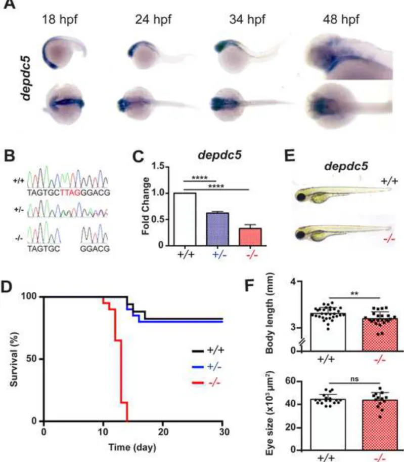
Though DEPDC5 mutations were associated with epilepsy, the underlying pathology of 96
this class of focal epilepsies remains unstudied. Zebrafish has only one ortholog of DEPDC5, 97
and sequence alignment of the zebrafish and human proteins showed a high degree of 98
conservation with 75% identity and 84% similarity. Whole mount RNA in situ hybridization 99
showed that depdc5 is expressed in the forebrain, midbrain, hindbrain and also in the neural tube 100
and notochord during zebrafish development (Figure 1A). 101
Using CRISPR/Cas9 genome editing, the 14th exon of depdc5 encoding the DUF3608, 102
which was initially found to be mutated in many patients [2, 17], was targeted for disruption. A 103
positive founder transmitting a 4-nucleotide frame-shifting deletion was selected, which would 104
lead to the expression a truncated protein (352 amino acids instead of the 1568 residue wild-type 105
protein, Figure 1B). Quantification of mRNA expression showed significant reduction in 106
heterozygous and homozygous larvae (Figure 1C). Since crosses of heterozygotes never resulted 107
in viable juvenile/adult homozygotes, according to the 1:2:1 Mendelian ratio, the survival of all 108
the three genotypes: depdc5+/+ (wild type), depdc5+/- and depdc5-/- was monitored for a 109
month from 0 day post fertilization (dpf). While most of the depdc5+/+ and +/- larvae survived 110
for this period, all the depdc5-/- larvae died by two weeks of age (Figure 1D). Notably, the 111
survival of depdc5-/- can be extended by few days if they are separated early and raised with 112
extra care, although they never survive after 20 dpf. However, no obvious morphological defects 113
or changes in eye size were observed, though a small reduction in body length was seen 114
following quantification (Figures 1E and 1F). While this observation is consistent with the 115
premature death of rodent Depdc5-/- models [14-16], the growth delay observed in the complete 116
knockout rodent embryos was not evident in case of depdc5-/- zebrafish larvae. Hence, though 117
vertebrates can survive haploinsufficiency of DEPDC5, total absence leads to death during 118
development. 119
120
depdc5-/- larvae show mTOR hyperactivation, brain epileptic discharges, hypoactivity and
121
increased seizure susceptibility
122
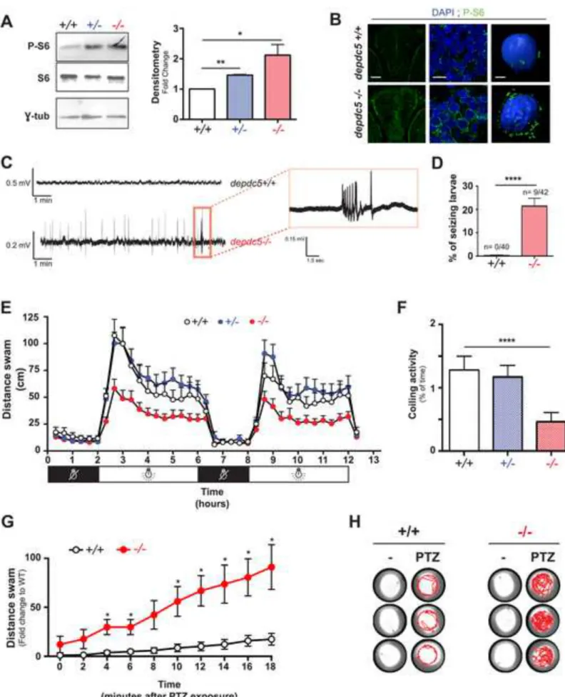
Since DEPDC5 is known to negatively regulate mTOR, mTORC1 activity was assessed 123
through the phosphorylation state of the ribosomal protein S6, which is a downstream effector of 124
mTORC1 [18]. Lysates of 8 dpf depdc5+/+, depdc5+/- and depdc5-/- larvae were analyzed by 125
western blotting using antibodies against the S6 Ser240/244 phospho-epitope, and a gradual 126
increase in the phosphorylation was observed (Figure 2A), indicating high mTORC1 activity in 127
the absence of DEPDC5. This was confirmed by performing immunohistological analysis where 128
depdc5-/- larval brains showed greater levels of phospho-S6 (Figure 2B). However, no abnormal
129
or dysmorphic cells were visible. In order to monitor epileptic activity, brain activity was 130
monitored using non-invasive local field potential (LFP) recordings from the optic tectum [19]. 131
Recurrent spontaneous epileptiform events were observed in depdc5-/- larval brains at 9 dpf 132
(Figures 2C and 2D) at a mean frequency of 9.1 + 5.0 events/10 minutes and mean duration of 133
309.7 + 152.3 msec, confirming the pathogenicity of DEPDC5 loss-of-function. However, since 134
the LFPs were monitored only for 10 minutes, this epileptic brain activity was observed in only 135
9/42 larvae, and it is possible that many larvae show epileptiform discharges outside this period. 136
larvae). It is also possible that these epileptiform discharges manifest earlier but were only 138
monitored at 9 dpf in order to increase the chances of detecting them. The absence of 139
epileptiform discharges in 100% of the animals is consistent with similar observations in Depdc5 140
conditional knockout mice and patients with focal epilepsy [16]. In order to have a more accurate 141
readout of the abnormalities in depdc5-/- larvae, their locomotor activity was quantified in light-142
dark cycles. depdc5-/- larvae were significantly hypoactive (Figure 2E), and this was observed in 143
100% of the larvae across generations from 5 dpf onward (N>5; n>300), till before death. These 144
results are also consistent with other mTORopathy zebrafish models which show epileptic brain 145
activity and are hypoactive [20]. Interestingly, when coiling (rotating movement of embryo 146
inside the chorion) was monitored at 20 hpf, this earliest motor behavior was already lower in 147
depdc5-/- embryos (Figure 2F), indicating that the hypoactivity begins very early during
148
development, suggesting that DEPDC5 loss-of-function may have consequences on early 149
neurodevelopment. Lastly, the susceptibility of depdc5-/- larvae to chemically-induced seizures 150
by pentylenetetrazol (PTZ) was tested. Consistently with what have been reported in mice [16], 151
depdc5-/- larvae show a significantly increased response to PTZ (shorter delay of response to
152
PTZ exposure and higher swimming activity) compared to their siblings (Figure 2G and 2H). 153
Altogether, the phenotypes observed upon knocking out depdc5 in zebrafish are 154
reminiscent of what is observed in the existing rodent models of Depdc5 knockout and also to an 155
extent, in patients. Altogether, these results validate the pathogenicity of depdc5 loss-of-function 156
in zebrafish. 157
158
Alteration of brain gene expression profile in depdc5-/-
In order to obtain new unbiased insights into the molecular perturbations occurring 160
during neurodevelopment in the absence of DEPDC5, brains of 10 dpf depdc5+/+ and -/- larvae 161
were dissected out and total RNA was extracted for deep sequencing (Figure 3A). This age was 162
chosen as an intermediate stage to perform exhaustive analysis of the molecular phenotype at a 163
stage when the swimming phenotype is distinct, while preventing the perturbation of gene 164
expression by the premature death that occurs 5 days later. From the differential gene expression 165
analysis, 1191 genes out of approximately 12,000 genes expressed in the larval brain were found 166
to be significantly up- (532 genes) or downregulated (659 genes) (p<0.05) (full list can be found 167
in Table S1, description provided for top dysregulated genes). Notably, 16 out of the 20 168
aminoacyl-tRNA synthetases in zebrafish were upregulated in the depdc5-/- brains, presumably 169
due to improper mTOR regulation. Functional annotation and clustering of the significantly 170
dysregulated genes showed that apart from aminoacyl-tRNA biosynthesis and electron transport 171
chain that can be attributed to mTOR pathway dysfunction, the expression of genes involved in 172
other pathways including chordate embryonic pattern specification, regulation of neuron 173
projection morphogenesis/axonogenesis, synapse formation, zinc finger/transcription factors and 174
ion channels were also perturbed (Figure 3B). This suggested that DEPDC5 may also play a role 175
in embryonic neurodevelopment. 176
Since genes related to neurodevelopment were altered, the number of neurons during 177
developmental stages was analyzed. Measurement of the head size and staining with cresyl violet 178
showed that the size and morphology of depdc5-/- larval brains were similar to that of the control 179
(Figures 3D and 3F). In addition, using immunostaining against acetylated tubulin and 180
phosphorylated histone H3 (which label neuronal fibres and mitotic cells respectively), the main 181
brain networks and neurogenesis itself did not appear to be affected (Figures 3C and 3E). 182
However, many genes related to GABAergic networks which have conventionally been 183
correlated with other kinds of epilepsy, were downregulated. Indeed, 3 GABA receptors 184
(GABRA1, GABBR1b and GABRG2) and other associated factors required for synthesis and 185
transport of GABA, GABAergic synapse formation, maturation and transmission (like Gat3, 186
pyruvate carboxylase, Kcc2 and neuroligin 2) were all downregulated (Table S3). 187
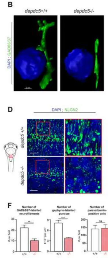
Since the expression of multiple factors important for GABAergic network formation and 188
synaptic function was perturbed, the expression of three markers of inhibitory synapses, 189
Gad65/67, gephyrin and neurogilin-2 (Nlgn-2), was examined by immunostaining of transverse 190
sections from 8 dpf depdc5+/+ and depdc5-/- larval brains. Interestingly, close examination of 191
the sections immunostained with Gad65/67 showed that finer branched structures were not 192
present as in wild type siblings (Figure 4A). 3D reconstruction of Gad65/67 staining confirmed 193
that the Gad65/67 projections (in blue in Figure 4B) are less arborized in the mutant brains. 194
Since GABA synthetizing enzymes are expressed in clusters at the pre-synaptic GABA synapses, 195
these results confirm defects in the GABAergic network connectivity. Immunolabeling against 196
gephyrin and Nlgn2 also showed a significant decrease of the complexity in depdc5-/- brains 197
(Figures 4C and 4D). Upon quantifying the GAD65/67 and gephyrin positive neurofilaments and 198
puncta from at least three biological replicates, both were decreased by >50% in depdc5-/- 199
(Figure 4F). Lastly, immunolabelling and quantification of the interneuron population using 200
parvalbumin 7 antibody did not show any difference in the number of positive cells (Figure 4E 201
and 4F). These results demonstrate that DEPDC5 loss-of-function significantly impacts fine 202
branching of GABAergic network during neurodevelopment without causing major loss of 203
interneurons. 204
GABA exposure rescues hypoactivity and seizure susceptibility in depdc5-/- larvae through
206
paracrine effects
207
Since the expression of GABA synthetizing enzymes seemed to be affected, we 208
hypothesized that exposure to GABA may compensate for this decrease and could therefore 209
rescue the consistent hypoactivity and seizure susceptibility phenotypes of depdc5-/- larvae. 210
Concurrently with this hypothesis, GABA treatment could completely rescue the hypoactivity of 211
depdc5-/- larvae (swimming at 7 dpf and coiling at 24 hpf) (Figures 5A and 5B). Rapamycin,
212
which has a positive effect on other mTORopathy models, was used as a positive control, and 213
could also completely rescue the hypoactivity of depdc5-/- embryos and the increased phospho-214
S6 levels (Figures 5A-C). Moreover, GABA treatment, as well as rapamycin could rescue the 215
seizure susceptibility of depdc5-/- larvae exposed to PTZ, to the level of their siblings (Figures 216
5D and 5E). Hence, these results confirm earlier observations from transcriptomic and 217
immunolabelling experiments that GABA network/signalling is indeed altered in depdc5-/- 218
larvae. Moreover, it shows that both GABA and mTOR signalling are involved in the 219
pathogenicity of depdc5 loss-of-function. 220
In order to assess if this rescuing effect could be a result of a rescue of the 221
neurodevelopmental defects themselves, neuronal branching of GABAergic neurons was 222
monitored following GABA treatment. However, supplementation with GABA could not rescue 223
the fine neuronal branching defects observed in depdc5-/- larvae (Figures 5F and 5G) thus 224
suggesting that GABA can only compensate for, but not rescue the neurodevelopmental defects. 225
Further, when the effect of GABA and rapamycin treatment on larval survival was tested, GABA 226
exposure could not rescue the premature death at 2-3 weeks post fertilization (wpf), while 227
rapamycin treatment extended the life span as long as it was supplemented (Figure 5H). This 228
supports our conclusion that GABA exposure can only partially compensate for the 229
neurodevelopmental defects. 230
Hence, these results show that while increasing GABA levels could compensate for 231
defective GABAergic branching, it could not rescue the neurodevelopmental defect itself. This 232
also indicates that the premature death is essentially due to perturbation of mTOR signaling 233
(which could be rescued by rapamycin), rather than the GABA network defects. Indeed, 234
rapamycin needed to be continually supplemented, since withdrawal of rapamycin led to gradual 235
death (Figure 5H), reiterating the contribution of mTOR deregulation to premature death. 236
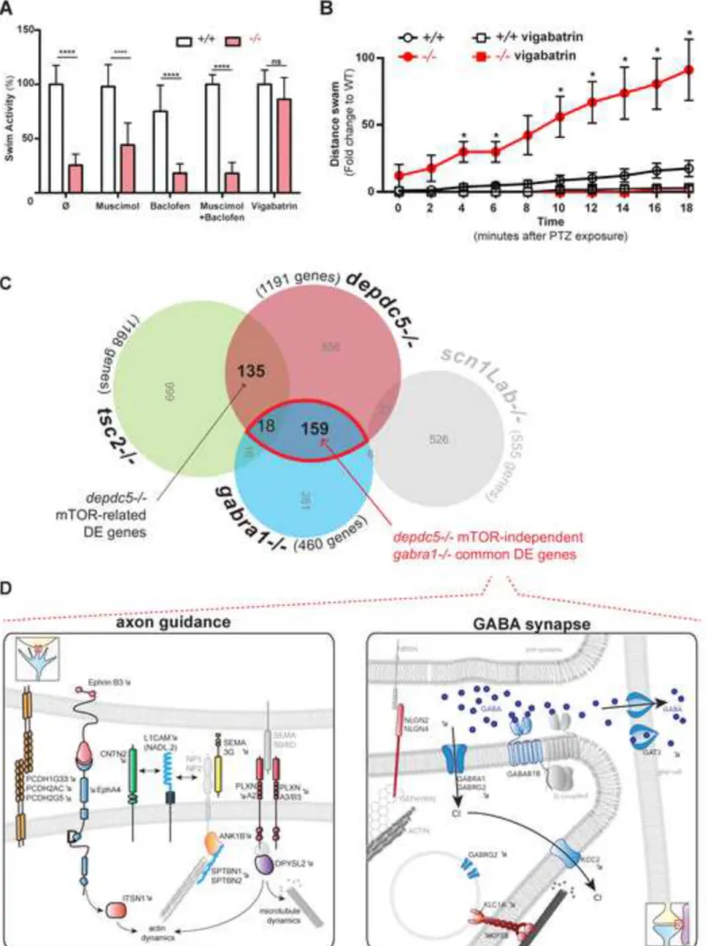
In order to understand if the compensating effect of GABA was mediated through GABA 237
receptors or independently of them, muscimol (a GABAA receptor agonist), baclofen (a GABAB
238
receptor agonist) and vigabatrin (an inhibitor of GABA transaminase which increases local 239
GABA concentration) were tested for their effect on depdc5-/- larvae (Figure 6A). Interestingly, 240
treatment with the GABA receptor agonists, muscimol, baclofen, and even both in combination 241
could not rescue the hypoactivity. However, exposure to vigabatrin, which increases endogenous 242
GABA level by inhibiting its metabolism, completely rescued the hypoactivity of depdc5-/- 243
larvae as well as PTZ hypersensitivity (Figure 6B). This proves that GABA acts through 244
receptor-independent mechanisms in a paracrine fashion. Moreover, the fact that inhibiting 245
GABA metabolism using vigabatrin is sufficient to rescue the phenotype suggests that GABA 246
itself mediates the compensating effects rather than metabolites of GABA. 247
Altogether these results support the idea that DEPDC5 loss-of-function impairs early 248
neurodevelopment especially GABA network fine branching which is likely to contribute to 249
epileptogenesis, and that mTOR-dependent metabolic changes are more generalized and severe, 250
eventually resulting in premature death. 251
252
mTOR-dependent and independent pathways are perturbed in depdc5-/- larvae
253
In order to further understand whether these two phenomena: upregulation of mTOR 254
signaling, and defective development of GABAergic networks are related and act together in the 255
same pathway, or are controlled independently by DEPDC5, the depdc5-/- differentially 256
expressed gene dataset was compared to datasets from three other zebrafish models of epilepsy. 257
These included: (1) a gabra1-/- zebrafish line recapitulating idiopathic generalized epilepsy, in 258
which GABAergic network is defective (Samarut et al., under review) (2) an mTORopathy 259
epilepsy (tsc2-/-) zebrafish model in which mTORC1 activity is increased [20], and (3) a Dravet 260
syndrome zebrafish model (scn1Lab mutant carrying a mutation in Nav1.1 [21]), as a neutral
261
mTOR- and GABA-non-related epilepsy model (Figure 6C). 262
177 genes were found to overlap between the depdc5 and gabra1 datasets (15% and 40% 263
of the total number of genes in the depdc5 and gabra1 datasets respectively), and remarkably, 264
99% of them (175/177) showed the same trend of up- or downregulation in both datasets 265
suggesting specific common mechanisms. When functional clustering and annotation of the 266
common genes was performed, these genes were found to be involved in electron transport, 267
embryonic morphogenesis, GABA receptor activity, synapse formation, general synaptic 268
activity, axonogenesis, nucleotide binding and transcription (Figure 6D and Table S3). 269
Interestingly, many factors which have been associated with epilepsy earlier were also 270
dysregulated. This significant overlap between depdc5 and gabra1 datasets provides further 271
evidence that DEPDC5 loss-of-function induces broad defects in GABA network. 272
Since both DEPDC5 and TSC2 are involved in the negative regulation of mTOR, 273
common genes between these two datasets showed mTOR-related pathways as the largest group 274
of genes, upon functional clustering and annotation (Table S4). About 15% genes of the two 275
datasets were found to be genes encoding for mTOR related members like aminoacyl-tRNA 276
synthetases, stress response and protein phosphorylation. The overlap of the depdc5 dataset with 277
data from other models of epilepsy (Scn1Lab) and autism spectrum disorder (unpublished, data 278
not shown) did not show a significant overlap (only 23 genes in common between depdc5-/- and 279
Scn1Lab-/- datasets) thus confirming the specificity of the overlaps described above.
280
Surprisingly, among the 177 common genes between the gabra1 and depdc5 datasets 281
which were involved in neurogenesis and GABA networks, only 18 genes were found to be 282
common with the tsc2 dataset. The non-overlapping nature of 90% (159 of 177) of the genes 283
indicates that these neurodevelopment-associated genes are more likely to be unrelated to 284
mTOR, but rather controlled by DEPDC5 independently of its canonical mTOR-inhibitory 285
function. These observations show that in the absence of DEPDC5, there are mTOR-dependent 286
metabolic changes (which overlap with the tsc2 model) but also mTOR-independent 287
neurodevelopment defects especially in the GABAergic network (which overlap with the gabra1 288
model), a combination of which presumably leads to epilepsy. 289
290
Neurodevelopmental defects in depdc5-/- larvae are independent of mTOR signaling
291
In order to confirm the independent nature of the mTOR and GABA pathways, mTOR 292
signaling was assessed following treatment with GABA in depdc5+/+ and -/- larvae (Figure 293
7A). Consistent with the hypothesis, GABA treatment could not rescue the increase in mTOR 294
activity in depdc5-/- larvae (Figure 7A, upper panel). Moreover, the levels of phospho-S6 were 295
also checked in gabra1-/- larvae, which have defective GABA signaling and it did not show any 296
change (Figure 7A lower panel). These results indicate that GABA and its related metabolism do 297
not control mTOR signaling. 298
Further, in order to confirm that the neurodevelopmental defects induced by depdc5 loss-299
of-function are indeed mTOR-independent, the ability of rapamycin to rescue the fine branching 300
of the GABAergic network in depdc5-/- larval brains was assessed. Brain slices from rapamycin 301
treated depdc5+/+ and -/- larvae were immunostained using -GAD65/67. Rapamycin treatment 302
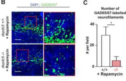
failed to rescue the reduced fine branching of the GABAergic network during development in 303
depdc5-/-. Indeed, similar to what was observed in untreated depdc5-/- brains (Figure 4), the
304
GAD65/67-positive neurofilaments were less arborized even after rapamycin treatment (Figure 305
7B and 7C), proving that the GABAergic branching defects resulting from the absence of 306
DEPDC5 are indeed mTOR-independent. Of note, phospho-S6 levels are significantly reduced 307
when rapamycin was applied at this concentration (Figure 7D). 308
To confirm the mTOR-independent effects at the molecular level, the ability of 309
rapamycin to restore the expression of critical up- and downregulated genes back to normal was 310
tested in order to assess if the transcriptional changes were also mTOR independent. The genes 311
that were chosen for testing by RT-qPCR were related to axon guidance (plxn2a, sema3b, 312
efnb3b) and GABA synapse (gabrg2, gat3, kcc2), which were in common with the gabra1
313
dataset, and also some mitochondrial factors (mt-cyb, mt-co2). Consistently with their 314
involvement in metabolic processes, the expression of mitochondrial cytochrome B and 315
cytochrome C oxidase II genes in depdc5-/- brains was decreased to wild-type level upon 316
rapamycin treatment (Figure 7E). However, for the genes involved in axon guidance and GABA 317
synapse activity, rapamycin treatment could not rescue the reduced expression observed in 318
depdc5-/- larval brains (Figure 7E). This incapability of rapamycin to rescue both, the
neurodevelopmental defects and the transcriptional changes proves that the molecular defects are 320
indeed mTOR-independent. 321
Lastly, if GABAergic network defects induced by DEPDC5 knockout are mTOR 322
independent, GABA would not have any effect on another genetic model of mTOR 323
hyperactivation such as tsc2-/- [20]. Consistently, when tested on tsc2-/- larvae, GABA was 324
unable to rescue the hypoactivity, again validating the mTOR-independent nature of the defects 325
observed in depdc5-/- larvae (Figure 7F). Of note, rapamycin, which was used as a positive 326
control could rescue the hypoactivity observed in tsc2-/- larvae. 327
Hence, these results show that at the phenotypic, cellular and molecular levels, alongside 328
its canonical mTOR inhibitory function, DEPDC5 does control neurodevelopmental aspects of 329
GABAergic networks in an mTOR independent fashion. 330
331
Discussion:
332
Mutations in the members of the mTOR-inhibitory GATOR1 complex have recently been 333
shown to be associated with focal epilepsy [7-10]. However, multiple aspects of this complex 334
remain poorly understood, including the molecular pathways through which it plays a role in 335
normal brain activity and/or development. 336
Depdc5 is strongly expressed in the developing vertebrate nervous system as shown by in
337
situ hybridization of early zebrafish embryos, indicating a role for this protein in neuronal
338
development. DEPDC5 is expressed specifically in neurons in the human brain (Human protein 339
atlas database and Human brain transcriptome project) where the transcript was first identified 340
[2, 22], but this is the first report showing its strong expression in the developing vertebrate 341
neural system. 342
In order to understand the role of DEPDC5 in neurodevelopment and brain activity, the 343
first zebrafish knockout model of depdc5 was generated and studied. Although rodent models 344
have been recently generated [14-16], the zebrafish model proffers specific technical advantages 345
for studying early neurodevelopment. Though depdc5-/- zebrafish larvae do not show obvious 346
phenotypic changes, they exhibit transient spontaneous epileptiform discharges in their brain and 347
increased seizure susceptibility before they die prematurely at about 2 wpf. The zebrafish model 348
also proffers a platform for drug screening purposes. Our zebrafish model would be convenient 349
for initial high-throughput drug screening assays that could identify candidate molecules 350
restoring depdc5-/- larval phenotype (i.e. hypoactivity and seizure susceptibility) and later be 351
confirmed in mammalian models. Such an approach using complementary vertebrate models has 352
proven to be effective and time-saving in identifying new therapeutics for other genetic diseases 353
[23]. 354
Hypoactivity and seizure susceptibility of the larvae was consistently observed and could 355
be used as an accurate readout for further studies. It is noteworthy that another zebrafish model 356
with hyperactive mTOR owing to mutated tsc2 also shows hypoactivity and premature death 357
along with epileptic seizures [20]. Also, since the hypoactivity is observed from very early on, 358
coiling could also be used as a readout in drug screening to economize on time. Notably, no 359
enlarged or abnormal cells were detected in the developing zebrafish brains in this model. This 360
could be explained by the “two-hit” hypothesis where an additional hit along with DEPDC5 361
mutations occurs to cause FCD in patients [9]. Also, though depdc5-/- larvae show considerably 362
less mRNA levels, it was not completely absent, probably due to incomplete degradation of 363
depdc5 mRNA by nonsense-mediated decay. However, the significant reduction of mRNA levels
364
show that the phenotypes observed were a result of haploinsufficiency. 365
With the aim to get further insights into the molecular perturbations in the epileptic 366
depdc5-/- brains, transcriptomic analysis was performed in this study, which showed interesting
367
candidates that were up- or downregulated. This is the first report of an in vivo transcriptomic 368
profile from a model of dysfunctional GATOR1 complex. As expected, various mTOR-regulated 369
pathways like amino acid metabolism were upregulated owing to mTOR deregulation, including 370
most amino acyl tRNA synthetases. While loss-of-function mutations in some aminoacyl-tRNA 371
synthetases have been shown to be associated with epilepsy [24-27], the contribution of such a 372
broad upregulation in expression levels of these factors to epilepsy remains to be understood. 373
Interestingly, though factors associated with neurogenesis, axonogenesis and synaptic 374
functions were downregulated, rather than gross morphological defect or neuron loss, specific 375
downregulation of GABA receptors, transporters and factors involved in inhibitory network 376
development, synapse formation and function was observed, revealing for the first time the 377
possible role of DEPDC5 in the formation, pruning and maintenance of the GABAergic system. 378
GABA exposure could partially compensate the phenotype observed in depdc5-/- larvae. 379
The use of GABA receptor agonists in mTORopathies has been explored only for TSC [28], but 380
muscimol and baclofen were ineffective in depdc5-/- larvae. These observations suggest that the 381
compensating effect of GABA is more likely to be a paracrine effect of GABA rather than 382
through GABA receptors. In this context, the ability of vigabatrin to rescue the phenotype also 383
confirms the direct effect of GABA rather than of its metabolites. Notably, GABA can only 384
partially ameliorate the depdc5-/- phenotype and therefore cannot be considered as a candidate 385
drug against refractory epilepsies. However, the observation that GABAergic networks are 386
affected early during development in a vertebrate DEPDC5 knockout model is an important 387
consideration for treatment strategies. Indeed, it might be useful to design therapeutic strategies 388
that can restore or at least compensate stably, the neurodevelopmental defects, rather than solely 389
trying to treat symptomatically, i.e., alleviate the seizures. Notably, the paracrine effect of 390
GABA observed here is also significant since many patients with mutations in DEPDC5 are 391
refractory to conventional anti-epileptic drugs, which generally act through GABA receptors 392
such as benzodiazepines. Thus, the application of molecules that would increase GABA 393
signaling independently of GABA receptors (such as vigabatrin) could be interesting in the 394
context of these refractory epilepsies. 395
Predictably, rapamycin could rescue the phenotype, and also prevent premature death, 396
suggesting that mTOR deregulation affects multiple organismal functions. Rapamycin and 397
rapalogs have proved to be extremely effective against many mTOR associated diseases 398
including cancer, lymphangioleiomyomatosis (LAM) and tuberous sclerosis [29-31]. Rapamycin 399
has also been shown to extend longevity and have neuroprotective effects [32, 33]. Like in 400
earlier studies with other diseases, though very effective, cessation of rapamycin treatment led to 401
reappearance of the symptoms and death [30, 34]. Due to various reasons including activation of 402
alternate or compensatory pathways, rapamycin treatment has had modest outcomes in clinical 403
trials despite very promising pre-clinical animal model studies [35]. Hence, rapamycin and 404
rapalogs are now being tested for combination therapy as chemotherapeutic agents, and these 405
could also be applied to treat focal epilepsies, since there is a need to identify new targetable 406
mechanisms of focal epilepsy in order to design new drugs against refractory seizures. 407
Through multiple comparison of transcriptomic datasets, a large set of genes from the 408
depdc5 dataset was found to overlap with the gabra1 dataset independently of the tsc2 dataset,
409
suggesting that DEPDC5 has unexpected mTOR-independent activity, especially in regulating 410
GABA network development, as proven experimentally (Figure 7). Hence, it is possible that the 411
two inhibitory complexes, GATOR1 (via DEPDC5) and TSC play distinct roles in different 412
pathways and cell types in addition to their overlapping mTOR-related functions. DEPDC5 413
function is still incompletely understood and many epileptic mutations have been identified in 414
different domains of DEPDC5 including DUF3608 [2, 3], which also supports the idea that there 415
could be other yet undiscovered functions of DEPDC5. Though mTOR activity has been 416
associated with proper neuronal development [36-38], this study shows that the 417
neurodevelopmental role of DEPDC5 is mTOR-independent. Hence, in addition to controlling 418
neuronal development through mTOR, the components of the pathway might also have mTOR-419
independent roles. 420
Altogether, these results show a novel mTOR-independent effect of DEPDC5 on the 421
GABAergic network that is very likely to be a key component of DEPDC5-mediated focal 422
epilepsy substratum. Indeed, abnormal wiring and functioning of the GABAergic system have 423
been observed in epilepsy [39] and GABA agonists have been used for decades alongside other 424
drugs like sodium and calcium channel blockers, glutamate blockers, carbonic anhydrase 425
inhibitors, and other drugs as anti-epileptics [40, 41]. Since GABA is the primary inhibitory 426
neurotransmitter in the cortex, GABA agonists have long been considered the logical choice as 427
anti-epileptic agents. However, in the emerging field of epilepsies caused by mutations in non-428
ion channel genes, the contribution of the GABAergic network to epilepsy has not been studied 429
and this study shows that modulation of the GABAergic network together with targeting mTOR 430
could be very effective against this class of non-ion channelopathies. 431
432
Acknowledgements:
We thank Dr. Louis-Charles Levros and Prof. Patrick Cossette for useful discussions regarding 434
the GABA agonists, Florent Guilloteau and Patrick Gendron from the IRIC genomic platform for 435
discussions regarding the RNA-sequencing experiment, Marina Drits for assistance with fish 436
maintenance. We thank Dr. Kessen Patten and Prof. Jacques Samarut for critical reading and 437
valuable comments on the manuscript. This work was funded by The Savoy Foundation, the 438
Fonds de Recherche Québec Santé (FRQS), the Rare Disease Model and Mechanism network, 439
Dravet Canada, FRQS-affiliated GRSNC (Groupe de Recherche sur le Système Nerveux 440
Central), the Québec MEESR (Ministère de l’Éducation, de l’Enseignement Supérieur et de 441
Recherche), the CRCHUM and the Agence Nationale pour la Recherche (ANR) (grant ANR-16-442 CE18-0010 to N.S.Y.). 443 444 Author contributions: 445
The project was conceptualized by ES and PD. ES generated and characterized the CRISPR line 446
with assistance from ML and RR. AS and ES performed most of the experiments. AlS and 447
PAMW performed the analyzed the brain epileptiform activity. NSY designed and analyzed the 448
immunolabeling experiment, which RHA and SR performed. AS and ES wrote the manuscript, 449
which was edited by PD. 450
451
Declaration of interests: The authors declare no competing interests. 452
453
References:
454
1. Baulac, S. (2014). Genetics advances in autosomal dominant focal epilepsies: focus on 455
DEPDC5. Prog Brain Res 213, 123-139. 456
2. Dibbens, L.M., de Vries, B., Donatello, S., Heron, S.E., Hodgson, B.L., Chintawar, S., 457
Crompton, D.E., Hughes, J.N., Bellows, S.T., Klein, K.M., et al. (2013). Mutations in 458
DEPDC5 cause familial focal epilepsy with variable foci. Nat Genet 45, 546-551. 459
3. Ishida, S., Picard, F., Rudolf, G., Noe, E., Achaz, G., Thomas, P., Genton, P., Mundwiller, 460
E., Wolff, M., Marescaux, C., et al. (2013). Mutations of DEPDC5 cause autosomal 461
dominant focal epilepsies. Nat Genet 45, 552-555. 462
4. Xiong, L., Labuda, M., Li, D.S., Hudson, T.J., Desbiens, R., Patry, G., Verret, S., Langevin, 463
P., Mercho, S., Seni, M.H., et al. (1999). Mapping of a gene determining familial partial 464
epilepsy with variable foci to chromosome 22q11-q12. Am J Hum Genet 65, 1698-1710. 465
5. Picard, F., Makrythanasis, P., Navarro, V., Ishida, S., de Bellescize, J., Ville, D., 466
Weckhuysen, S., Fosselle, E., Suls, A., De Jonghe, P., et al. (2014). DEPDC5 mutations in 467
families presenting as autosomal dominant nocturnal frontal lobe epilepsy. Neurology 468
82, 2101-2106.
469
6. Bar-Peled, L., Chantranupong, L., Cherniack, A.D., Chen, W.W., Ottina, K.A., Grabiner, 470
B.C., Spear, E.D., Carter, S.L., Meyerson, M., and Sabatini, D.M. (2013). A Tumor 471
suppressor complex with GAP activity for the Rag GTPases that signal amino acid 472
sufficiency to mTORC1. Science 340, 1100-1106. 473
7. Ricos, M.G., Hodgson, B.L., Pippucci, T., Saidin, A., Ong, Y.S., Heron, S.E., Licchetta, L., 474
Bisulli, F., Bayly, M.A., Hughes, J., et al. (2016). Mutations in the mammalian target of 475
rapamycin pathway regulators NPRL2 and NPRL3 cause focal epilepsy. Ann Neurol 79, 476
120-131. 477
8. Scheffer, I.E., Heron, S.E., Regan, B.M., Mandelstam, S., Crompton, D.E., Hodgson, B.L., 478
Licchetta, L., Provini, F., Bisulli, F., Vadlamudi, L., et al. (2014). Mutations in mammalian 479
target of rapamycin regulator DEPDC5 cause focal epilepsy with brain malformations. 480
Ann Neurol 75, 782-787. 481
9. Baulac, S., Ishida, S., Marsan, E., Miquel, C., Biraben, A., Nguyen, D.K., Nordli, D., 482
Cossette, P., Nguyen, S., Lambrecq, V., et al. (2015). Familial focal epilepsy with focal 483
cortical dysplasia due to DEPDC5 mutations. Ann Neurol 77, 675-683. 484
10. D'Gama, A.M., Geng, Y., Couto, J.A., Martin, B., Boyle, E.A., LaCoursiere, C.M., Hossain, 485
A., Hatem, N.E., Barry, B.J., Kwiatkowski, D.J., et al. (2015). Mammalian target of 486
rapamycin pathway mutations cause hemimegalencephaly and focal cortical dysplasia. 487
Ann Neurol 77, 720-725. 488
11. Crino, P.B. (2016). The mTOR signalling cascade: paving new roads to cure neurological 489
disease. Nat Rev Neurol 12, 379-392. 490
12. Ostendorf, A.P., and Wong, M. (2015). mTOR inhibition in epilepsy: rationale and clinical 491
perspectives. CNS Drugs 29, 91-99. 492
13. Palavra, F., Robalo, C., and Reis, F. (2017). Recent Advances and Challenges of mTOR 493
Inhibitors Use in the Treatment of Patients with Tuberous Sclerosis Complex. Oxid Med 494
Cell Longev 2017, 9820181. 495
14. Marsan, E., Ishida, S., Schramm, A., Weckhuysen, S., Muraca, G., Lecas, S., Liang, N., 496
Treins, C., Pende, M., Roussel, D., et al. (2016). Depdc5 knockout rat: A novel model of 497
mTORopathy. Neurobiol Dis 89, 180-189. 498
15. Hughes, J., Dawson, R., Tea, M., McAninch, D., Piltz, S., Jackson, D., Stewart, L., Ricos, 499
mice causes severe embryonic dysmorphology with hyperactivity of mTORC1 signalling. 501
Sci Rep 7, 12618. 502
16. Yuskaitis, C.J., Jones, B.M., Wolfson, R.L., Super, C.E., Dhamne, S.C., Rotenberg, A., 503
Sabatini, D.M., Sahin, M., and Poduri, A. (2017). A mouse model of DEPDC5-related 504
epilepsy: Neuronal loss of Depdc5 causes dysplastic and ectopic neurons, increased 505
mTOR signaling, and seizure susceptibility. Neurobiol Dis 111, 91-101. 506
17. Martin, C., Meloche, C., Rioux, M.F., Nguyen, D.K., Carmant, L., Andermann, E., Gravel, 507
M., and Cossette, P. (2014). A recurrent mutation in DEPDC5 predisposes to focal 508
epilepsies in the French-Canadian population. Clin Genet 86, 570-574. 509
18. Hay, N., and Sonenberg, N. (2004). Upstream and downstream of mTOR. Genes Dev 18, 510
1926-1945. 511
19. Zdebik, A.A., Mahmood, F., Stanescu, H.C., Kleta, R., Bockenhauer, D., and Russell, C. 512
(2013). Epilepsy in kcnj10 morphant zebrafish assessed with a novel method for long-513
term EEG recordings. PLoS One 8, e79765. 514
20. Scheldeman, C., Mills, J.D., Siekierska, A., Serra, I., Copmans, D., Iyer, A.M., Whalley, B.J., 515
Maes, J., Jansen, A.C., Lagae, L., et al. (2017). mTOR-related neuropathology in mutant 516
tsc2 zebrafish: Phenotypic, transcriptomic and pharmacological analysis. Neurobiol Dis 517
108, 225-237.
518
21. Baraban, S.C., Dinday, M.T., and Hortopan, G.A. (2013). Drug screening in Scn1a 519
zebrafish mutant identifies clemizole as a potential Dravet syndrome treatment. Nat 520
Commun 4, 2410. 521
22. Ishikawa, K., Nagase, T., Suyama, M., Miyajima, N., Tanaka, A., Kotani, H., Nomura, N., 522
and Ohara, O. (1998). Prediction of the coding sequences of unidentified human genes. 523
X. The complete sequences of 100 new cDNA clones from brain which can code for large 524
proteins in vitro. DNA Res 5, 169-176. 525
23. Patten, S.A., Aggad, D., Martinez, J., Tremblay, E., Petrillo, J., Armstrong, G.A., La 526
Fontaine, A., Maios, C., Liao, M., Ciura, S., et al. (2017). Neuroleptics as therapeutic 527
compounds stabilizing neuromuscular transmission in amyotrophic lateral sclerosis. JCI 528
Insight 2. 529
24. Almalki, A., Alston, C.L., Parker, A., Simonic, I., Mehta, S.G., He, L., Reza, M., Oliveira, 530
J.M., Lightowlers, R.N., McFarland, R., et al. (2014). Mutation of the human 531
mitochondrial phenylalanine-tRNA synthetase causes infantile-onset epilepsy and 532
cytochrome c oxidase deficiency. Biochim Biophys Acta 1842, 56-64. 533
25. Zhang, X., Ling, J., Barcia, G., Jing, L., Wu, J., Barry, B.J., Mochida, G.H., Hill, R.S., Weimer, 534
J.M., Stein, Q., et al. (2014). Mutations in QARS, encoding glutaminyl-tRNA synthetase, 535
cause progressive microcephaly, cerebral-cerebellar atrophy, and intractable seizures. 536
Am J Hum Genet 94, 547-558. 537
26. Hallmann, K., Zsurka, G., Moskau-Hartmann, S., Kirschner, J., Korinthenberg, R., Ruppert, 538
A.K., Ozdemir, O., Weber, Y., Becker, F., Lerche, H., et al. (2014). A homozygous splice-539
site mutation in CARS2 is associated with progressive myoclonic epilepsy. Neurology 83, 540
2183-2187. 541
27. Nishri, D., Goldberg-Stern, H., Noyman, I., Blumkin, L., Kivity, S., Saitsu, H., Nakashima, 542
M., Matsumoto, N., Leshinsky-Silver, E., Lerman-Sagie, T., et al. (2016). RARS2 mutations 543
cause early onset epileptic encephalopathy without ponto-cerebellar hypoplasia. Eur J 544
Paediatr Neurol 20, 412-417. 545
28. Curatolo, P., Verdecchia, M., and Bombardieri, R. (2001). Vigabatrin for tuberous 546
sclerosis complex. Brain Dev 23, 649-653. 547
29. Wander, S.A., Hennessy, B.T., and Slingerland, J.M. (2011). Next-generation mTOR 548
inhibitors in clinical oncology: how pathway complexity informs therapeutic strategy. J 549
Clin Invest 121, 1231-1241. 550
30. Bissler, J.J., McCormack, F.X., Young, L.R., Elwing, J.M., Chuck, G., Leonard, J.M., 551
Schmithorst, V.J., Laor, T., Brody, A.S., Bean, J., et al. (2008). Sirolimus for 552
angiomyolipoma in tuberous sclerosis complex or lymphangioleiomyomatosis. N Engl J 553
Med 358, 140-151. 554
31. Muncy, J., Butler, I.J., and Koenig, M.K. (2009). Rapamycin reduces seizure frequency in 555
tuberous sclerosis complex. J Child Neurol 24, 477. 556
32. Bové, J., Martínez-Vicente, M., and Vila, M. (2011). Fighting neurodegeneration with 557
rapamycin: mechanistic insights. Nat Rev Neurosci 12, 437-452. 558
33. Harrison, D.E., Strong, R., Sharp, Z.D., Nelson, J.F., Astle, C.M., Flurkey, K., Nadon, N.L., 559
Wilkinson, J.E., Frenkel, K., Carter, C.S., et al. (2009). Rapamycin fed late in life extends 560
lifespan in genetically heterogeneous mice. Nature 460, 392-395. 561
34. McCormack, F.X., Inoue, Y., Moss, J., Singer, L.G., Strange, C., Nakata, K., Barker, A.F., 562
Chapman, J.T., Brantly, M.L., Stocks, J.M., et al. (2011). Efficacy and safety of sirolimus in 563
lymphangioleiomyomatosis. N Engl J Med 364, 1595-1606. 564
35. Li, J., Kim, S.G., and Blenis, J. (2014). Rapamycin: one drug, many effects. Cell Metab 19, 565
373-379. 566
36. Fu, C., Cawthon, B., Clinkscales, W., Bruce, A., Winzenburger, P., and Ess, K.C. (2012). 567
GABAergic interneuron development and function is modulated by the Tsc1 gene. Cereb 568
Cortex 22, 2111-2119. 569
37. Westerholz, S., de Lima, A.D., and Voigt, T. (2013). Thyroid hormone-dependent 570
development of early cortical networks: temporal specificity and the contribution of 571
trkB and mTOR pathways. Front Cell Neurosci 7, 121. 572
38. Garza-Lombó, C., and Gonsebatt, M.E. (2016). Mammalian Target of Rapamycin: Its Role 573
in Early Neural Development and in Adult and Aged Brain Function. Front Cell Neurosci 574
10, 157.
575
39. Treiman, D.M. (2001). GABAergic mechanisms in epilepsy. Epilepsia 42 Suppl 3, 8-12. 576
40. Ashton, H., and Young, A.H. (2003). GABA-ergic drugs: exit stage left, enter stage right. J 577
Psychopharmacol 17, 174-178. 578
41. Jembrek, M.J., and Vlainic, J. (2015). GABA Receptors: Pharmacological Potential and 579
Pitfalls. Curr Pharm Des 21, 4943-4959. 580
42. Moreno-Mateos, M.A., Vejnar, C.E., Beaudoin, J.D., Fernandez, J.P., Mis, E.K., Khokha, 581
M.K., and Giraldez, A.J. (2015). CRISPRscan: designing highly efficient sgRNAs for CRISPR-582
Cas9 targeting in vivo. Nat Methods 12, 982-988. 583
43. Thisse, C., and Thisse, B. (2008). High-resolution in situ hybridization to whole-mount 584
zebrafish embryos. Nat Protoc 3, 59-69. 585
44. Huang da, W., Sherman, B.T., and Lempicki, R.A. (2009). Systematic and integrative 586
analysis of large gene lists using DAVID bioinformatics resources. Nature Protocols 4, 44-587
57. 588
45. Puverel, S., Nakatani, H., Parras, C., and Soussi-Yanicostas, N. (2009). Prokineticin 589
receptor 2 expression identifies migrating neuroblasts and their subventricular zone 590
transient-amplifying progenitors in adult mice. J Comp Neurol 512, 232-242. 591
592 593
Figure legends:
594
Figure 1: depdc5 is essential for survival. (A) Whole mount in situ hybridization showing
595
expression of depdc5 at different stages of development. (B) Electropherograms showing 596
confirmation of the 4 bp insertion by Sanger sequencing. (C) Expression of depdc5 transcript in 597
7 dpf depdc5+/+, +/- and -/- larvae (N=3, Student’s t test: p (WT vs HT)=0.0026; p (WT vs 598
HM)=0.001). (D) Survival curve showing that depdc5-/- embryos have reduced survival with 599
most larvae dying around 14 dpf (N=2, n=15 per genotype). (E) Images showing that depdc5-/- 600
larvae don’t show gross morphological changes at 3 dpf. (F) Measurement of body length (upper 601
panel; n=20/genotype; p=0.0045 by unpaired Student’s t-test) and eye size (lower panel; 602
n=12/genotype; p=0.7219 by unpaired Student’s t-test) showed no significant change in either 603
parameter. 604
605
Figure 2: depdc5 knockout results in increased mTOR activity, hypoactivity, epileptiform
606
discharges and seizure susceptibility. (A) Western blotting of 7 dpf larval lysates to examine
607
phospho-S6 levels in depdc5+/+, depdc5+/- and depdc5-/- larvae showing hyperactivation of 608
mTOR signalling. (n=3; p(WTvsHT) = 0.0012; p(WTvsHM) = 0.0455). (B) Images of 6 dpf 609
depdc5+/+ and depdc5-/- larval brains (transversal sections across the optic tectum)
610
immunostained with phospho-S6 antibodies and counterstained with DAPI. The panels on the 611
from left to right panels: 20µm, 2.5µm, 1.5µm). (C) Spontaneous extracellular electrographic 613
activity recorded from the optic tectum of depdc5+/+ or depdc5-/- larvae at 9 dpf showing 614
representative epileptiform activity of depdc5-/- larvae displaying polyspiking discharges. (D) 615
Percentage of depdc5+/+ and depdc5-/- larvae showing epileptiform activity in the brain (N=4; 616
n=10/genotype, Student’s t-test p(WTvs HM)<0.0001). (E) Distance swam by 8 dpf larvae 617
across two 2 hour dark-4 hour light cycles. (F) Spontaneous coiling activity in 20 hpf embryos 618
monitored over 20 minutes represented as the percentage of total time during which the embryos 619
were active (N=3, n=20, Student’s t-test p (WT vs HM)<0.0001). (G) Distance swam by 2 dpf 620
embryos upon exposure to 3mM PTZ represented as fold change compared to basal distance 621
swam by wildtype larvae at t0. (N=3, n>10, student t-test, *: p<0.05) (H) Representative
622
swimming tracks of 2 dpf embryos during a 10-minute period following a 3mM PTZ exposure 623
(tracks of 3 replicates are shown in rows for each genotype). 624
625
Figure 3: depdc5 knockout alters gene expression in larval brains without affecting brain
626
morphology (A) Volcano plot showing the differentially expressed gene expression profile
627
between depdc5+/+ and depdc5-/- larval brains. See also Table S1. (B) Pathways showing high 628
enrichment in the differentially expressed genes (upregulated are shown in green, 629
downregulated, in red). (C) Whole mount images of depdc5+/+ and -/- larvae at different stages 630
of development immunostained with -acetylated tubulin. (n>3/genotype/developmental stage; 631
Scale bar:40 m).(D) Measurement of head size and head surface area in 3 dpf depdc5+/+ and 632
depdc5-/- larvae. (E) Whole mount images of 48 hpf depdc5+/+ and -/- larvae immunostained
633
with -phosphorylated histone H3 (n>3/genotype). (F) Cresyl violet staining comparing 7 dpf 634
depdc5+/+ (top half) and depdc5-/- (bottom half) larval heads showed no gross difference in the
635
density of cells in the brain. Scale bar=100 m. 636
637
Figure 4: Defects in GABAergic networks in depdc5 knockout zebrafish brains. Images of
638
transverse sections of 6 dpf depdc5+/+ and depdc5-/- embryos immunostained with GAD65/67 639
(A), gephyrin (C) and Nlgn2 (D) antibodies and counterstained with DAPI. Section level is 640
displayed in the schematic drawing. Regions indicated in red boxes have been magnified to show 641
the branching defects (N=3, n=3 embryos/genotype). Scale bar: 10 m. (B) Imaris-reconstructed 642
3D image with Gad65/67 positive GABAergic boutons shown in green projecting towards the 643
nucleus (DAPI) shown in blue. (Scale bar=2 µm). (E) Images of longitudinal sections of 6 dpf 644
depdc5+/+ and depdc5-/- embryos immunostained with parvalbumin 7 antibody (Scale
645
bar=25µm). Field of view is displayed in the schematic drawing (F) Quantification of the number 646
of GAD65/67-positive neurofilaments, gephyrin-positive puncta and parvalbumin-positive cells 647
in depdc5+/+ and -/- larval brain sections (N=3, n>3 embryos/genotype). 648
649
Figure 5: Rescuing effect of GABA and rapamycin on depdc5 knockout zebrafish. (A)
650
Swimming activity of 8 dpf larvae, with the activity of depdc5 +/+ larvae normalized to 100% 651
shows that treatment with GABA (100 M) can completely rescue hypoactivity (N=2, 652
n=20/genotype/treatment). Rapamycin treatment (100 nM from day 0-2, 300 nM from day 3 653
onwards) was used as a positive control. (B) Spontaneous coiling activity in 20 hpf embryos 654
shows that the hypoactivity of depdc5-/- embryos is rescued by treating with 100 nM rapamycin 655
or with 100 M GABA from single-cell stage (N=2, n=20/genotype/treatment, Student’s t-test p 656
(Control WT vs HM) <0.0001:****). Coiling activity of depdc5+/+ was set at 100% for each 657
sample, and the relative activity of the depdc5-/- larvae is represented. (C) Western blotting of 9 658
dpf larval lysates to examine phospho-S6 levels in depdc5+/+ and depdc5-/- larvae treated with 659
DMSO or rapamycin (100 nM from day 0-2, 300 nM from day 3 onwards) shows inhibition of 660
mTOR signaling upon rapamycin treatment. (D) Distance swam by 2 dpf larvae treated with 661
GABA (100µM) or rapamycin (100nM) upon PTZ exposure (3mM) (N=3, n>10). (E) 662
Representative swimming tracks of 2 dpf embryos (untreated or treated with GABA or 663
rapamycin) during a 10-minute period following PTZ exposure (3mM). (F) Images of transverse 664
sections of 7 dpf GABA treated depdc5+/+ and depc5-/- larval brains immunostained with -665
GAD65/67 and counterstained with DAPI. Regions indicated in red boxes have been magnified 666
to show the branching defects. GABA treatment (100 M) was begun at 8 hpf (n=3 667
embryos/genotype). (G) Quantification of the number of GAD65/67 positive neurofilaments in 668
GABA-treated depdc5+/+ and -/- larval brain sections showing that GABA treatment does not 669
rescue the defects in depdc5-/-. (H) Survival curve showing that treatment of depdc5-/- larvae 670
with rapamycin (100 nM from day 0-2, 300 nM from day 3 onwards) first prolongs lifespan as 671
long as rapamycin is supplemented to the water (till day 20-shaded). Withdrawal of rapamycin 672
results in a drop in survival and all the larvae die by day 35. On the contrary, treatment with 100 673
M GABA from 8 hpf doesn’t extend survival. Both rapamycin and GABA did not affect 674
depdc5+/+.
675 676
Figure 6: Paracrine compensation by GABA and modulation of multiple independent
677
pathways in depdc5 knockout zebrafish. (A) Swimming activity of 8 dpf larvae treated with
678
muscimol (100 M), baclofen (100 M), a combination of both or vigabatrin (5 mM) with the 679
activity of depdc5+/+ larvae normalized to 100% shows that treatment with vigabatrin can 680
completely rescue hypoactivity, while GABA receptor agonists cannot. (B) Vigabatrin treatment 681
completely rescues PTZ-hypersensitivity of 2 dpf depdc5-/- embryos (N=2, n>30; student t-test, 682
*: p value <0.05). (C) Overlap between gene expression datasets from depdc5, gabra1, tsc2 and 683
scn1a mutants. See also Tables S2 and S4. (D) A representation of the common pathways (axon
684
guidance and GABA synapse) affected in depdc5 and gabra1 knockout brains with the common 685
genes indicated. See also Table S3. 686
687
Figure 7: mTOR deregulation and GABAergic defects are independent effects of Depdc5
688
knockout. (A) Western blotting of 7 dpf depdc5+/+ and depdc5-/- larval lysates shows no
689
change in phospho-S6 levels upon GABA treatment (100 M from 8 hpf; top panel). Western 690
blotting of 7 dpf gabra1+/+ and gabra1-/- larval lysates shows no difference in mTOR signaling 691
upon knocking out gabra1 (bottom panel). (B) Images of transverse sections of 7 dpf rapamycin 692
treated depdc5+/+ and depc5-/- larvae immunostained with -GAD65/67 and counterstained 693
with DAPI. Section level is displayed in the schematic drawing. Regions indicated in red boxes 694
have been magnified to show the branching defects. Rapamycin treatment was begun at 8 hpf: 695
100 nM from day 0-2, 300 nM from day 3 onwards (n=3/genotype). Scale bar=10 m. (C) 696
Quantification of GAD65/67 positive neurofilaments in rapamycin treated depdc5+/+ and -/- 697
larval brain sections shows that rapamycin treatment does not rescue the reduced number of 698
filaments in depdc5-/-. (D) Western blotting of 5 dpf larval lysates upon treatment with 699
increasing concentrations of rapamycin (50 nM, 250 nM, 500 nM, 1 M and 2.5 M) from 8 hpf 700
to estimate phospho-S6 levels shows a dose-dependent decrease. Rapamycin-containing water 701
was changed everyday. (E) qRT-PCR based estimation of some down- and upregulated genes 702
involved in axon guidance, GABA synapse and metabolism. ef1a expression was used for 703
normalization, expression of depdc5+/+ was normalized to 1. (p<0.05:*; p<0.01:**). (F) 704
Swimming activity of 7 dpf tsc2 larvae treated with rapamycin and GABA (day 0 onwards) over 705
5 minute light-10 minute dark phases, with the activity of tsc2+/+ larvae normalized to 100% 706
shows that rapamycin, but not GABA can rescue the hypoactivity (Student’s t-test 707 p<0.0001:****). 708 709 710
STAR methods:
711Contact for reagent and resource sharing
712
Further information and requests for resources and reagents should be directed to and will be 713
fulfilled by the Lead contact: Éric Samarut (eric.samarut@umontreal.ca)
714 715
Experimental model and subject details
716
Routine zebrafish (Danio rerio) maintenance was performed following standard procedures 717
(Westerfield) at 28.50C under 12/12 hour light/dark cycles at the animal facility of the Centre
718
Hospitalier de l’Université de Montréal Research Centre (CRCHUM), Montréal. All experiments 719
were performed in compliance with the guidelines of the Canadian Council for Animal Care. The 720
primary model used for this study was depdc5 loss of function mutants. 721
For generation of the depdc5 loss of function model, zebrafish optimized Cas9 mRNA was 722
synthesized using the mMESSAGE mMACHINE SP6 kit (Ambion) and a pCS2-nCas9n 723
template linearized with NotI. The following gRNA sequence targeting the 14th exon of depdc5 724
was designed using the online tool CRISPRscan (PAM site is indicated in brackets): 5’-725
TGAGGATCATGAGCAAT(CGG)-3’. Synthesis of gRNA and of Cas9 mRNA was performed 726