HAL Id: tel-01037947
https://tel.archives-ouvertes.fr/tel-01037947
Submitted on 23 Jul 2014
HAL is a multi-disciplinary open access archive for the deposit and dissemination of sci-entific research documents, whether they are pub-lished or not. The documents may come from teaching and research institutions in France or abroad, or from public or private research centers.
L’archive ouverte pluridisciplinaire HAL, est destinée au dépôt et à la diffusion de documents scientifiques de niveau recherche, publiés ou non, émanant des établissements d’enseignement et de recherche français ou étrangers, des laboratoires publics ou privés.
Ocular toxoplasmosis : immunopathology and virulence :
the influence of parasite virulence on the clinical,
biological, and immunological characteristics of ocular
toxoplasmosis (OT) in the Old and New World
Ligia Alejandra de la Torre Cifuentes
To cite this version:
Ligia Alejandra de la Torre Cifuentes. Ocular toxoplasmosis : immunopathology and virulence : the influence of parasite virulence on the clinical, biological, and immunological characteristics of ocu-lar toxoplasmosis (OT) in the Old and New World. Human health and pathology. Université de Strasbourg; Universidad del Quindío (Armenia, Colombie), 2013. English. �NNT : 2013STRAJ043�. �tel-01037947�
ii
Discipline:
Life and Health Sciences (ED 414)
Co-Tutoring Doctoral Thesis
“Ocular Toxoplasmosis: Immunopathology and Virulence” The influence of parasite virulence on the clinical, biological, and immunological characteristics of ocular toxoplasmosis (OT) in the Old and
New World
Ligia Alejandra DE LA TORRE CIFUENTES
Public Defense: 18th September 2013 Strasbourg, France
Director from France:
Ermanno CANDOLFI, MD, PhD.
Institut de Parasitologie et de Pathologie Tropicale
de la Faculté de Médecine Université de Strasbourg, France
Director from Colombia:
Jorge GOMEZ-MARIN, MD, PhD.
GEPAMOL Group
Centro de Investigaciones Biomédicas Universitad del Quindío, Armenia,
Colombia
Joint PhD Program : Université de Strasbourg (France) and
i
JURY MEMBERS:
Jury President: Pr Laurent GAUCHER
External jury members: Europe
Pr Isabelle VILLENA
Laboratoire de Parasitologie-Mycologie, Centre National de Référence de la Toxoplasmose,
Centre de Ressources Biologiques Toxoplasma,
Hôpital Maison Blanche, 45 rue Cognacq-Jay; 51092 Reims Cédex
Pr Hervé PELLOUX
UMR 5163, LAPM, Centre National de la Recherche Scientifique 38041 Grenoble, France
South America
Pr GONZÁLEZ John Mario, rapporteur externe
Profesor Asociado, Facultad de Medicina, Universidad de los Andes,
Carrera 1 No. 18A-10, edificio Q, piso 8, Bogotá D.C., Colombia
Télefono: +57 (1) 3394949 ext 3718. Lab ext. 3900 Fax: +57 (1) 3324281
Pr GONZALEZ Angel, rapporteur externe
Universidad de Antioquia, Grupo de Micología Médicay Experimental (CIB-UdeA-UPB), Corporación para Investigaciones Biológicas (CIB),
ii
Internal jury members: Europe
Pr Laurent GAUCHER
Service d’Ophtalmologie,
Nouvel Hôpital Civil CHU de Strasbourg, Strasbourg, France
South America
Pr. Juan Carlos SEPÚLVEDA-ARIAS
Facultad de Ciencias de la Salud, Universidad Tecnológica de Pereira,
iii
DEDICATION
To my daughters, Andrea and Gabriela, for their unconditional support and for giving me the inspiration to continue always looking ahead. For sharing with me good and bad moments; for their tenderness, for their fortitude, and for their endless love.
To my brother, Diego Francisco, for his love.
To my parents, for their care, for being there constantly, for their life example, for their amazing and infinite love.
iv
ACKNOWLEDGEMENTS
To my mentors, Ermanno Candolfi and Jorge Enrique Gomez, for trusting me, for their advice, for their open-handedness, and for their absolute support.
To Quindio University and Strasbourg University, for giving me this wonderful opportunity. To Madame Florentz, for her aid.
To my friends, Julie and Esterina, for sharing with me the best moments inside and outside the lab, and for demonstrating to me the real value of friendship.
To all the people in the CIB and IPPTS, for the magnificent work environment.
v
ABSTRACTS
vi
ABSTRACT
Introduction: Ocular involvement, mainly retinochoroiditis, is one of the most
severe sequelae of Toxoplasma gondii infection. However, the pathophysiological mechanisms of retinal destruction are poorly understood. Several studies suggested a more frequent and more severe ocular involvement in South American infections compared with European infections, probably due to different T. gondii strains (Type I/III, and atypical vs. Type II).
Objective: To compare the clinical characteristics and biological and
immunological responses in a single study and using the same parameters, in Colombian and French patients with active ocular toxoplasmosis (OT), as well as to study the local cytokinome in aqueous humor of these patients and correlate it with the clinical features.
Materials and methods: We prospectively collected and compared the
clinical features of patients with active OT, evaluated at the Department of Ophthalmology of Strasbourg University Hospital and of Quindio University Health-Center. Results of biological tests in the collected aqueous humor samples were compared between Colombian and French patients: the pattern of protein recognition by immunoblotting (IB); the relative diagnostic sensitivities of IB and Polymerase Chain Reaction (PCR); and the cytokine and chemokine profiles.
Results: We found that Colombian and French OT patients presented not
only different clinical characteristics but also biological characteristics, and that more virulent South American strains might be responsible for these differences, due to a disruption of the protective effects of interferon gamma (IFN-Ȗ). Retinal lesions were 50% greater in Colombian patients. Macular localization leading to visual impairment was observed in 56% of Colombian cases, compared with 13% of French patients. Moreover, more vitreous inflammation and vasculitis were observed in Colombian patients. However, cytokine assays of the aqueous humor showed upregulation of inflammatory responses in European patients, notably IL-17, which we did not observe in Colombian patients. In a mouse model, intraocular tachyzoite injection of type II and atypical T. gondii strains resulted in differences in parasite multiplication and pathology similar to those observed in human infections. Production of IL-17 and other inflammatory markers, like IL-6, MCP-1, and the Th17 transcription factor RORJt was observed upon infection with the type II PRU strain, but was much less with the atypical LEF strain. In a previous work, the cytokine and mRNA patterns showed an upregulation of Th1 responses, notably IFN-J production, in French patients, and anti-IL-17A antibody markedly diminished clinical damage and retinal inflammation, and also diminished parasite proliferation. In contrast to these previous findings in French patients, the cytokinome of aqueous humor of OT Colombian patients
vii showed a downregulation of Th1 and Th17 responses and an upregulation of the Th2 response. Correlation between the clinical characteristics of Colombian patients with active OT and the levels of cytokines in aqueous humor (AH) showed that local production of cytokines differed between patients with OT, and particular cytokine levels were related to more severe clinical characteristics. Some cytokines were related to a higher number of recurrences.
Conclusion: There are clinical and biological differences between Colombian
and French patients with OT. There seem to be strain-specific differences in IL-17 and IFN-Ȗ induction, which play an important role in the pathogenesis of this disease. These differences should be considered when thinking in perspectives of any possible immune-modulatory treatment in OT.
KEYWORDS: Ocular toxoplasmosis, Toxoplasma gondii, strains, cytokines,
viii
RESUMEN
Introducción: El compromiso ocular, principalmente la retinocorioditis, es
uno de las secuelas más severas de la infección por Toxoplasma gondii. Sin embargo, los mecanismos fisiopatológicos de la destrucción retiniana no son bien entendidos. Algunos estudios sugieren un compromiso más frecuente y más severo en las infecciones en Sur América, comparadas con las infecciones en Europa, probablemente debido a las diferentes cepas de T. gondii (Tipos I/III y atípicas vs. Tipo II).
Obetivo: comparar las características clínicas, biológicas, y las respuestas
inmunes, en un único estudio y usando los mismos parámetros, en pacientes colombianos y franceses con toxoplasmosis ocular (TO) activa; así como también estudiar el citoquinoma local en el humor acuoso de éstos pacientes y correlacionarlo con los hallazgos clínicos.
Materiales y métodos: Recolectamos consecutivamente y comparamos los
hallazgos clínicos de los pacientes con TO activa, que consultaron al departamento de Oftalmología del Hospital Universitario de Estrasburgo y al Centro de Salud de la Universidad del Quindío. Los resultados de los exámenes biológicos en humor acuso (HA) fueron comparados entre los pacientes colombianos y franceses: el patrón de reconocimiento de proteínas por inmunobloting (IB), las sensibilidades diagnósticas relativas de IB, la prueba de reacción en cadena de la polimerasa (PCR), y el perfil de citoquinas y quimioquinas.
Resultados: Los pacientes colombianos y franceses con TO activa
presentaron no solo diferencias clínicas sino también biológicas. Las cepas suramericanas, más virulentas, pueden jugar un papel crucial en estas diferencias, debido a la disrupción de los efectos protectores del IFN-Ȗ. Las lesiones retinianas fueron 50% más grandes en los pacientes colombianos, la localización macular, que lleva a compromiso visual, fue observada en 56% de los casos, comparado con el 13% en los franceses. Adicionalmente, se observó mayor inflamación vítrea y vasculitis en los pacientes colombianos. Sinembargo, los resultados de citoquinas en humor acuoso mostraron aumento de la respuesta inflamatoria en los pacientes europeos, notablemente IL-17, lo cual no se observó en los pacientes colombianos. En modelo murino, la patología mostró diferencias similares a las encontradas en la infección en humanos entre las cepas de T. gondii tipo II y atípicas. La producción de IL-17 y otros marcadores inflamatorios, como IL-6, MCP-1 y el factor de transcrpción de Th17, RORȖt, fueron observados luego de la infección con cepas tipo II PRU, pero mucho menos con cepas atípicas LEF. En trabajos previos, los patrones de citoquinas y mRNA mostraron elevación de la respuesta Th1, principalmente producción de IFN-Ȗ, en pacientes
ix franceses, y los anticuerpos anti IL-17A diminuyeron notablemente el daño clínico y la inflamación retiniana, así como también la proliferación parasitaria. El citoquinoma en humor acuoso de los pacientes colombianos con TO activa, mostró disminución de la respuesta Th1 y Th17, contrario a los pacientes franceses, y aumento en la respuesta Th2. La correlación entre las características clínicas en los pacientes colombianos con TO activa y los niveles de citoquinas en HA, mostraron que la producción local de citoquinas difiere entre los pacientes con TO y los niveles de ciertas citoquinas se encontraron relacionados con caracterísicas clínicas más severas, así como con las recurrencias. Trabajos preliminares nos han permitido iniciar un modelo de éstas afecciones ocualares empleando una cepa de tipo II y una cepa atípica suramericana de T.gondii, además de evaluar la posibilidad de efectuar futuros tratamientos intraoculares dirigidos por transfección in vivo.
Conclusión: existen diferencias clínicas y biológicas, entre los pacientes
colombianos y franceses con TO. Parece haber diferencias específicas de cada cepa en particular en la inducción de IL-17 e IFN-Ȗ, que juegan un papel importante en la patogénesis de la enfermedad. Estas diferencias deben ser consideradas cuando se piensa en posibles perspectivas con tratamientos inmunomoduladores en TO.
PALABRAS CLAVE: Toxoplasmosis ocular, Toxoplasma gondii, cepas,
x
RÉSUMÉ
Résultats: Nous avons sélectionné des patients atteints d’une TO
biologiquement confirmée et avons exploré les différences cliniques et biologiques de deux groupes de patients, l’un en France, l’autre en Colombie. Dans notre hypothèse de départ, les souches sud-américaines, seraient plus virulentes et elles pourraient jouer un rôle crucial dans la sévérité et l’évolution de la TO. Nous avons constaté, chez les patients colombiens, de plus grandes lésions de la rétine et une plus grande proportion de lésions maculaires, dans un contexte inflammatoire vitréen plus sévère. Le cytoquinome oculaire confirme une forte réponse inflammatoire chez les patients européens centrée sur l’IL-17, mais cette réponse Th17 est absente chez les sujets colombiens. L’IL-6 et l’IL-13 sont au contraire fortement augmentées chez ces derniers. Nous avons également démontré que certaines cytokines étaient associées à certaines caractéristiques cliniques comme la sévérité de l’inflammation ou la récurrence. Des travaux préliminaires nous ont permis de débuter une modélisation de ces affactions oculaires en employant une souche de type II et une souche atypique de T. gondii. Nous avons aussi évalué la possibilité d’effectuer des traitements ciblés en intraoculaires par transfection in vivo.
Conclusion: Nous avons constaté des différences cliniques et biologiques
entre les patients colombien et français. Il semble y avoir une régulation souche dépendante de la production d’IFN-J et d’IL-17. Ces différences pourraient contribuer à expliquer la plus grande sévérité des toxoplasmoses oculaires en Colombie. En se basant sur nos résultats nous pouvons envisager d’explorer des traitements immunomodulateurs plus ciblés.
Mots clés: Toxoplasma gondii, toxoplasmose oculaire, souches, cytokines,
xi
xii
List of abbreviations xv
List of figures xix
List of tables xxii
INTRODUCTION 1
I- The parasite 4
A. T. gondii 5
i. Discovery and history of T. gondii 5
ii. Parasite transmission and life cycle 7
1. Tachyzoites, bradyzoites, and tissue cysts 9
2. Asexual cycle 10
3. Sexual cycle 13
4. T. gondii proteins involved in gliding motility and host cell attachment, invasion, and egress
14 a. Resident surface proteins and lipids 14 b. Transient surface proteins: MICs 15
c. Rhoptry neck proteins: RONs 16
d. Rhoptry bulb proteins 17
5. T. gondii proteins involved in development and stage differentiation
17
a. Dense granules 17
b. Cytoskeleton 18
6. Cyst formation and parasite tissue burden 18 7. Population structure and genotype differences 19
B. Virulence 21
i. Introduction 21
ii. Definition of virulence in T. gondii 23
iii. T. gondii genetic diversity 24
iv. T. gondii development and virulence 25
v. Modulation of virulence in an obligatory parasite 26
vi. T. gondii virulence factors in host cell 27
vii. Rhoptry kinases and pseudokinases of the ROP2 family 27
viii. Additional factors 30
II- The disease 31
xiii i. General epidemiology – worldwide occurrence and course of the
disease
32
ii. Congenital toxoplasmosis 34
iii. Infection in immunocompromised patients 36
B. Ocular toxoplasmosis 38
i. Physiopathology/Immunopathology 38
ii. Immunology of OT – ocular immune response and specificity in South America
41 1. The importance of intraocular-cytokine dissection analysis in the
local response to T. gondii infection
41 2. Cytokines in innate immune responses to T. gondii 43 3. Cytokines in adaptive immune responses to T. gondii 46
a. The importance of the equilibrium between Th1/Th2/Th17/Treg responses: maintaining counterbalance in T. gondii infection control
47
b. The innate immune response is required to activate the acquired immune response: Th1 type cytokine response. The dual role of IL-12: immune protection connected with IFN-Ȗ production vs. pathological role once dysregulated
50
c. Treg type cytokines. Regulatory role of IL-10: avoiding tissue damage when levels are sufficient vs. promoting tissue destruction when insufficiently produced
52
d. Pro-inflammatory cytokines/chemokines and their counterbalance. TGF-E protective function antagonized by IL-6. Inflammatory and pathological effects of IL-12 and IL-18 beyond the eye
54
e. Th17 and its activators. TGF-E acting together with IL-6
56
iii. Epidemiology 58
iv. Clinical presentation 1. Symptoms 2. Ocular features 59 59 60 v. Diagnosis 66 vi. Therapy 68
III- Personal work 70
A. Objectives 71
i. Determination of the severity: clinical and biological comparison of French and Colombian patients
71 ii. Cytokinome analysis in Colombian patients: is OT immune response
related to strain virulence?
xiv
iii. Modeling OT: preliminary results and perspectives 72
B. Papers 72
ARTICLE 1. Prevention of retinochoroiditis in congenital toxoplasmosis – Europe versus South America
73
i. Introduction 74
ii. Article 75
iii. Conclusions and perspectives 79
ARTICLE 2. Severe South American ocular toxoplasmosis is associated with decreased IFN-Ȗ/IL-17A and increased IL-6/IL-13 intraocular levels
80
i. Introduction 81
ii. Article 82
iii. Conclusions and perspectives 94
ARTICLE 3. Cytokine milieu is linked to clinical characteristics in Colombian patients presenting an active ocular toxoplasmosis
95
i. Introduction 96
ii. Article 97
iii. Conclusions and perspectives 124
ARTICLE 4. New clinical and experimental insights into Old World and neotropical ocular toxoplasmosis
125
i. Introduction 126
ii. Article 126
iii. Conclusions and perspectives 136
IV- General discussion
A. Influence of virulence on differences in the pathogenesis and outcome of OT in Europe and South America
B. Molecular mechanisms underlying T. gondii strains: GRA15, ROP16,
ROP18, ROP5 (influence on STAT 3/STAT 6, NFNȕ, and IRGs)
i. What is known in mouse models?
ii. What have we found in the human intraocular response to T. gondii?
iii. Intraocular cytokine profile in Old and New World patients suffering from active OT and its potential explanation
iv. Intraocular cytokine profile in Colombian patients suffering from active OT versus control cataract patients, and the possible explanation 137 141 144 144 146 147 150 V- General conclusions 161
VI- General perspectives 164
xv
LIST OF ABBREVIATIONS
AC: Anterior Chamber
ABCA4: ATP-binding cassette transporter gene
AH: Aqueous Humor
AIDS: Acquired Immunodeficiency Syndrome AMA-1: Apical Membrane Antigen 1
APC: Antigen Presenting Cells AT: Amazonian Toxoplasmosis BSA: Bovine Serum Albumin
CCR5: C-C Chemokine Receptor Type 5 CD: Cluster Differentiation
CME: Cystoid Macular Edema CNS: Central Nervous System
CNVMs: Choroidal Neovascular Membranes COL2A1: Type II Collagen
CT: Computed Tomography CXCR: Chemokine Receptor
DAPI: Diamidino-2-Phenylindole staining DCs: Dendritic Cells
DG: Dense Granules DNA: Deoxyribonucleic Acid
EAU: Experimental Autoimmune Uveitis ELISA: Enzyme-Linked Immunosorbent Assay FGF: Fibroblast Growth Factor
FHUS: Fuchs Heterochromic Uveitis Syndrome GATA-3: Trans-acting T-cell-specific transcription factor GBPs: Guanylate-Binding Proteins
GC: Ganglion Cells
xvi GPI: Glycosylphosphatidylinositol
GRA1: Granule Recombinant Antigen 1 GTP: Guanosine Triphosphate HG12: Haplogroup 12
HIV: Human Immunodeficiency Virus
HOSTs: Host Organelle-Sequestering Tubulo Structures IB: Immunoblotting
ICAM-1: Intercellular Adhesion Molecule 1 IELs: Intraepithelial Lymphocytes IFN: Interferon
IgG: Immunoglobulin G IgM: Immunoglobulin M IL: Interleukin
IL-R: Interleukin Receptor iNOS: Inducible Nitric Oxide IOP: Intra Ocular Presure
IP-10: Interferon-induced Protein 10 IRG: Immunity-related GTPases IU: Intermediate Uveitis
Kg: Kilograms KO: Knockout
LEF: RMS (Reims) – 1994 Virulent Toxoplasma Strain LPL: Lamina Propria Lymphocytes
MAP: Mitogen-Activated Protein MAR: Microneme Adhesive Repeat MCP: Monocyte Chemoattractant Protein MCSF: Macrophage Colony-Stimulating Factor MICs.: Micronemal Proteins
MIP-1: Macrophage Inflammatory Protein 1 mg: Milligrams
xvii MORN: Membrane Occupation and Recognition Nexus Protein
mRNA: Messenger Ribonucleic Acid
MyD88: Myeloid Differentiation Primary Response Gene 88 M2AP: Microneme 2 Associated Protein
NFNB: Nuclear Factor Kappa B NK: Natural Killer cells NO: Nitric Oxide
NTP: Nucleoside Triphosphate OT: Ocular Toxoplasmosis PBS: Phosphate Buffered Saline PCR: Polymerase Chain Reaction PDGF: Platelet-Derived Growth Factor PFA: Paraformaldehyde
PMNs: Polymorphonuclear Leukocytes PRU: Prugniaud Toxoplasma Strain PV: Parasitophorous Vacuole
PVM: Parasitophorous Vacuole Membrane P2X7-R: Purinergic receptor P2X purinoceptor 7 p30: Protein 30
p47: Protein 47 p65: Protein 65 RNA: Ribonucleic Acid ROM: Rhomboid Protease RONs: Rhoptry Neck Proteins ROP: Rhoptry Protein
ROR: Related Orphan Receptor ROS: Reactive Oxygen Species RPE: Retinal Pigment Epithelium SAG: Surface Antigen
xviii siRNA: Small Interfering RNA
SRS9: Bradyzoite-Specific Surface Antigen
STAT: Signal Transducers and Activators of Transcription SYROCOT: Systematic Review on Congenital Toxoplasmosis T. gondii: Toxoplasma gondii
TGF-E: Transforming Growth Factor Beta TgMIC: Toxoplasma gondii Micronemal Protein
TgMIC2-AP: Toxoplasma gondii Micronemal Protein 2 Adhesive Protein TgPhIL1: Toxoplasma gondii Photosensitized Iodonapthaline Labeling
1
TgRON: Toxoplasma gondii Rhoptry Neck Proteins TgSub1: Toxoplasma gondii Subtilisin Protease 1 Th: T Helper Cells
TLR: Toll-Like Receptor T lympho-
cytes: Thymus-Derived Lymphocytes TNF: Tumor Necrosis Factor
TRAP: Thrombospondin-Related Anonymous Protein Treg: Regulatory T cells
TLR: Toll-Like Receptors Tyk: Tyrosine Kinase
xix
LIST OF FIGURES
This list does not include the figures in the articles.
Figure 1. Life cycle of Toxoplasma gondii. 8
Figure 2. Sexual and asexual cycle of Toxoplasma gondii. 11
Figure 3. Characteristic toxoplasmic retinochoroidal damage: atrophic
retinochoroidal scar (caused by tissue destruction and necrosis), with hyperpigmented borders (due to the alteration on RPE).
39
Figure 4. Cytokine network in adaptive immune response to
Toxoplasma. Cytokines are crucial in cellular differentiation, inhibition, and activation of the different types of T cells.
58
Figure 5. Active toxoplasmic lesions. a: Primary lesion: creamy-white
retinochoroidal lesion without concomitant hyperpigmented scar (blue arrow). b: Recurrent lesion: creamy-white active lesion (blue arrow) with accompanying hyperpigmented old scar (red arrow).
60
Figure 6. Toxoplasmic retinochoroidal scars located in different
places of the retina. a: Macular atrophic retinochoroidal scar with hyperpigmented borders and a size of approximately 2 disc diameters (dd) (blue arrow). b: Peripheral atrophic retinal scar with a size of about 3 dd, and another hyperpigmented peripheral scar of about 0.5 dd. The sizes of the lesions are compared with the size of the optic disc.
61
Figure 7. Additional findings in OT. Vitreous haze (vitritis). Active
peripapillar inflammation with vitreous opacity. Details of the retina are
xx not clearly observed because of the vitreous haze.
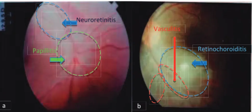
Figure 8. Additional findings. a: Neuroretinitis and papillitis due to
ocular toxoplasmosis b: Active retinochoroiditis with perivascular sheathing.
62
Figure 9. Retinal detachment as a complication of ocular
toxoplasmosis.
62
Figure 10. Bilateral macular compromise. Chorioretinal scars with an
atrophic center and hyperpigmented borders.
64
Figure 11. Bilateral macular compromise. Extensive tissue
destruction. Retinal tissue has been completely destructed, leading to visualization of the sclera in necrotic areas of the retinochoroidal scars.
64
Figure 12. Bilateral compromise. a,b: Extensive chorioretinal scars. b:
Optic nerve atrophy.
65
Figure 13. Bilateral macular compromise. Extensive and multiple
chorioretinal scars.
65
Figure 14. Bilateral, extensive, multiple, chorioretinal scars, both
central and peripheral.
66
Figure 15. Unilateral macular lesions in different patients. 66
Figure 16. Influence of virulence on the pathogenesis of OT. Different
clinical outcomes in OT in Europe and South America.
142
xxi European and South American patients. Toxoplasma strains, parasitic
load, protein recognition (IB), and cytokine/chemokine patterns were different between the populations.
143
Figure 18. Strain modulation of cytokine network in immune response
to Toxoplasma and virulence. Cytokine modulation of Type I, II, and III Toxoplasma strains and their virulence in a mouse model. Comparison of cytokines present in AH of Colombian and French patients.
147
Figure 19. Proposed dynamics of a Type I/III and atypical (South
American) ocular infection, in contrast to a Type II (European) ocular infection. Influence of virulence on intraocular immune response.
156
Figure 20. Local production of IL-17 in a mouse model of OT. Eyes
infected with PRU and LEF Toxoplasma strains. IL-17 is present in a progressive pattern (Days 1, 3, and 7) from the outer Ganglion Cells (GC) to the inner layers Retinal Pigmented Epithelium (RPE) of the retina with PRU and LEF strains.
170
Figure 21. Possible resident producer cells of IL-17A in a mouse
model of ocular toxoplasmosis (OT).
171
Figure 22. In vivo siRNA delivery to the vitreous in a mouse model of
ocular toxoplasmosis (OT). Sticky siRNA + in vivo jetPEI (24 hours). Preliminary results in the first step of siRNA delivery to the vitreous in a mouse model of OT. Transfected cells can be seen in the vitreous, which seem to be monocytes. There is probably one lymphocyte in the retina.
xxii
LIST OF TABLES
This list does not include the tables in the articles.
Table 1. Hypothesis of the implications of infecting strains in the
main differences in intraocular cytokine levels in AH samples of patients with active OT from Europe and South America.
1
2 Toxoplasmosis is caused by a ubiquitous apicomplexan parasite of warm-blooded animals, and is one of the more common parasitic zoonoses worldwide (Elmore et al., 2010). Felids are the key animal species in the life cycle of this parasite because they are the hosts that can excrete the environmentally resistant stage, the oocyst. Humans become infected congenitally or postnatally. Acquired infection could be due to ingestion of tissue cysts from undercooked meat, consuming food or drink contaminated with oocysts, or by accidentally ingesting oocysts from the environment (Elmore et al., 2010). However, only a small percentage of exposed adult humans or other animals develop clinical signs of disease. In pregnant women, the infection may be transmitted to the fetus and result in a severe infection and in immunocompromised hosts, a latent infection may be activated and cause clinical disease (Dubey and Jones, 2008). It is unknown whether the severity of toxoplasmosis in immunocompetent hosts is due to the parasite strain, host variability, or other factors. Recently, attention has been focused on genetic variability among Toxoplasma gondii isolates from sick and apparently healthy hosts (Dubey and Jones, 2008), but also on virulence differences among T. gondii strains (Lehmann et al., 2006).
Ocular toxoplasmosis (OT) is the most common cause of posterior uveitis (Holland, 2003). It can cause visual impairment and blindness (Holland, 2003; de-la-Torre, López-Castillo et al., 2009). It affects patient’s quality of life (de-la-Torre et al., 2011) and produces irreversible sequelae (Holland, 2003). Although OT is a typical recurrent disease, we still do not know how to avoid recurrences or why they occur (de-la-Torre, Rios-Cadavid et al., 2009). There is no ideal treatment and the treatments being applied have controversial efficacy (Stanford and Gilbert, 2009; de-la-Torre, Stanford et al., 2011).
Considering these circumstances, the main challenges we have today are to really understand the immunopathology of OT (Garweg and Candolfi, 2009),
3 to find out how to limit the damage, avoid sequelae, and prevent recurrences, and to develop a new treatment based on immunomodulation that should be more efficient than the current antibiotic-based one. Thus, it is essential to look for immune-based interventions supported by a better clinical and pathophysiological understanding that can lead to more effective strategies to prevent and treat OT. Treatments with cytokines or anti-cytokines could be considered, if we obtain a better understanding of the nature of the immune response. Several studies have shown that Th2 involvement in OT is important in the humoral response, and that Th1 plays an important role in limitation of parasite proliferation (Gaddi and Yap, 2007; Amadi-Obi et al., 2007). The role of Th17, at least in ocular infection by Type II strains, is probably related to development of retinal lesions (Sauer et al., 2012).
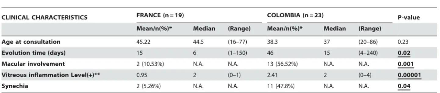
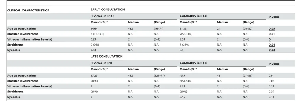
Confirmation of the differences in the clinical picture between Colombian and French patients suffering active OT, with higher severity in Colombian patients, the differences in the biological and immunological responses, and the different infecting strains in the Old and New World are an important input in the field of this neglected disease. Particularly for me, working in a South American country and seeing daily severe cases of OT in my uveitis clinic that seriously compromise the quality of life of our patients, this work inspires me to continue investigating this fascinating field where too much remains to be elucidated.
4
5
A. T. gondii
i. Discovery and history of T. gondii
It has been more than 100 years since T. gondii was initially described by Charles Nicolle and Louis Manceaux in 1908, while conducting Leishmania research at the Pasteur Institute in Tunis. They described a blood-borne unicellular parasite in the tissues of a small hamster-like rodent named Ctenodoactylus gundi. In parallel (1908), in Brazil, Alfonso Splendore identified the same protozoan in rabbit tissues (Weiss and Dubey, 2009). One year later, Nicole and Manceaux named the parasite in accordance with its morphology (toxo: arc or bow; plasma: life) and the animal in which it was discovered (the gundi). In retrospect, the correct name should be T. gundii (Dubey, 2008). The parasite was first found in laboratory animals. For the next 30 years, T. gondii-like organisms were found in several other hosts, mainly avian species (Dubey, 2002), although viable T. gondii was first isolated by Sabin and Olitsky (1937) and proven to be identical with to human isolate of T. gondii (Dubey, 2008).
Regarding studies of the complex protection against T. gondii, which involves innate and specific immunity, in the 1940s, humoral antibodies were found to kill extracellular but not intracellular tachyzoites (Sabin, 1948; Sabin et al., 1937). In the next 50 years, protective immunity was found to be mediated largely by immune lymphoid cells (Frenkel, 1967; Suzuki et al., 1988; Gazzinelli et al., 1991; Dubey, 2008).
The question of why some hosts develop clinical toxoplasmosis whereas most remain asymptomatic is unknown. During the 1980s and 1990s, methods were developed to recognize genetic differences among T. gondii isolates from humans and animals (Pfefferkorn et al., 1980; Darde et al., 1998; Tibayrene et al., 1991; Sibley et al., 1992; Howe et al., 1995; Dubey, 2008).
6 Mapping of T. gondii genes was achieved recently (Khan et al., 2005), and undoubtedly will help in the search for better antigens for diagnosis and protection, and mechanism of disease. Until recently, T. gondii was considered clonal, with very little genetic variability (Howe et al., 1995). Lehmann et al. (2006) performed the first in-depth study of genetic variability among more than 275 T. gondii isolates obtained worldwide from one host (free-range chicken) and in one laboratory (Dubey et al., 2002). They found geographic differences, with some isolates being confined to Brazil, whereas others were distributed worldwide. Phenotypically, T. gondii isolates from asymptomatic chickens from Brazil were mouse virulent (Dubey et al., 2002). This point is of interest because according to Dubey (2008), there is no non-pathogenic strain of T. gondii and virulence in mice may have no clinical relevance with respect to disease in humans and farm animals. T. gondii can cause several clinical syndromes including encephalitis, chorioretinitis, congenital infection, and neonatal mortality (Weiss and Dubey, 2009). Fifteen years after the description of T. gondii by Nicolle and Manceaux, a fatal case of toxoplasmosis in a child was reported by JankĤ (Weiss and Dubey, 2009).
In 1939, Wolf, Cowen, and Paige were the first to demonstrate the medical importance of T. gondii by conclusively identifying it as a cause of human disease in tissues of a congenitally infected infant in New York City, USA (Dubey, 2009). Its veterinary importance became known when in 1957, it was found to cause abortion storms in sheep in Australia (Hartley et al., 1957; Tenter et al., 2000; Dubey, 2008). The discovery of a T. gondii-specific antibody test, the Sabin-Feldman dye test, in 1948 led to the recognition that T. gondii is a common parasite of warm-blooded hosts with a worldwide distribution. Its life cycle was not discovered until 1970, when it was found that felids were its definitive host and an environmentally resistant stage (oocyst) was excreted in feces of infected cats (Dubey, 2008). The recent discovery of its common infection in certain marine wildlife (sea otters) indicates
7 contamination of our seas with T. gondii oocysts washed from land (Dubey, 2008).
ii. Parasite transmission and life cycle
T. gondii, an obligate intracellular parasite, is a facultatively heteroxenous, polyxenous protozoa that has developed several potential routes of transmission within and between different host species (Tenter et al., 2000). This cosmopolitan parasite infects the majority of warm-blooded animals including humans. Felids are its definitive hosts, in which the parasite completes the sexual cycle, representing the main reservoir of infection, by excreting oocysts, which are the environmentally resistant stage. The parasite propagates by the use of an asexual cycle in other mammals and in birds.
Nearly one-third of the human population has been exposed to this parasite (Halonen and Weiss, 2013). Transmission to humans occurs through ingestion of tissue cysts from undercooked meat, by accidentally consuming food or drink contaminated with oocysts, or by accidentally ingesting oocysts from the environment (Elmore et al., 2010).
Serological surveys indicate that T. gondii infections are common in wild carnivores, including pigs, bears, felids, fox, raccoons, and skunks. Clinical and subclinical toxoplasmosis have been reported for wild cervids, ungulates, marsupials, monkeys, and marine mammals. Southern sea otter populations have been severely impacted by Toxoplasma infections (Hill et al., 2005).
In the life cycle of T. gondii, there are three different infectious stages: tachyzoites, which facilitate expansion during acute infection; bradyzoites, which maintain chronic infection; and sporozoites, which are disseminated in the environment within oocysts (Dubey, 1998) (Figure 1).
8 Figure 1. Life cycle of Toxoplasma gondii. Modified from: Dubey JP, Lindsay DS, Speer CA. Structures of Toxoplasma gondii tachyzoites, bradyzoites, and sporozoites and biology
and development of tissue cysts. Clin Microbiol Rev. 1998 Apr;11(2):267-99.
All three stages are haploid; tachyzoites and bradyzoites divide asexually, while sporozoites are the product of meiosis. Sexual development only occurs within enterocytes of the feline gut, ultimately yielding diploid oocysts, which undergo meiosis after shedding (Figure 1).
Understanding the adaptations of these stages for various steps in the life cycle provides a support for considering the unique population structure of T. gondii.
9
1. Tachyzoites, bradyzoites, and tissue cysts
Humans and animals become infected mainly by ingesting bradyzoites or oocytes. After ingestion, both bradyzoites and sporozoites convert to tachyzoites inside tissues. The conversion of tachyzoites to bradyzoites and bradyzoites to tachyzoites is of biological and clinical significance because bradyzoites are less susceptible to chemotherapy, and reactivation of bradyzoites to tachyzoites is considered the cause of fatal toxoplasmosis in acquired immunodeficiency syndrome (AIDS) patients. Of all the methods currently available to assess stage conversion of T. gondii, feeding infective stages to cats is the most reliable method. Felidae, the definitive hosts of T. gondii, excrete oocysts 3–10 days after ingesting tissue cysts/bradyzoites, t 18 days after ingesting oocysts, and t 13 days after ingesting tachyzoites (Dubey, 1998).
Tachyzoites. The tachyzoite is the stage that Nicolle and Manceaux found in the gundi. This stage has also been called the trophozoite, the proliferative form, the feeding form, and the endozoite. It divides into two by a specialized process called endodyogeny (Dubey, 2008).
Bradyzoites and tissue cysts. The term “bradyzoite” was proposed by Frenkel (1973) to describe the stage encysted in tissues. Bradyzoites are also called cystozoites. Dubey and Beattie (1988) proposed that cysts should be called tissue cysts to avoid confusion with oocysts and pseudocysts. Jacobs, Remington, and Melton (1960a) first provided a biological characterization of cysts when they found that the cyst wall was destroyed by pepsin or trypsin, but the cystic organisms were resistant to digestion by gastric juices (pepsin-HCl), whereas tachyzoites were destroyed immediately. Thus, tissue cysts were shown to be important in the life cycle of T. gondii because
10 carnivorous hosts can become infected by ingesting infected meat (Dubey, 2008).
Bradyzoites and tissue cysts are an integral part of the life cycle of T. gondii, independent of immunity. There are no strains of T. gondii in nature that do not form tissue cysts. Tissue cysts develop and remain intracellular, and bradyzoites differ from tachyzoites with respect to location of the nucleus (central in tachyzoites, terminal in bradyzoites), amylopectin granule (numerous in bradyzoites, absent or few in tachyzoites), contents of rhoptries (honeycomb in tachyzoites, electron dense in older bradyzoites). Asexual and sexual stages are morphologically different from tachyzoites and bradyzoites, which also occur in the cat intestine (Dubey, 2008).
2. Asexual cycle
Toxoplasma is capable of infecting and replicating within virtually any nucleated mammalian or avian cell (Black and Boothroyd, 2000). Its life cycle is divided between feline and non-feline infections, which are correlated with sexual and asexual replication, respectively (Figure 2).
Tachyzoites multiply very quickly in a wide variety of nucleated host cells during the acute phase of infection. Parasite invasion is driven by actin-based motility, generating a parasitophorous vacuole (PV) derived from invagination of the host cell plasma membrane and secretion of parasite proteins (Dubey, 1998; Bradley and Sibley, 2007).
Within the PV, tachyzoites divide every 6–9 hours by a process called endodyogeny, in which daughter cells form internally within the mother cell (Dubey, 1998; Morrissette and Sibley, 2002). Rupture of the host cell leads to emergence of parasites that infect new host cells. Infection evokes strong
11 innate and adaptive immune responses that control parasite replication but do not eliminate the infection. In response to environmental stress, tachyzoites convert into a semidormant stage known as bradyzoites, which are contained within tissue cysts (Dubey et al., 1976; Dubey, 1998).
Figure 2. Sexual and asexual cycle of Toxoplasma gondii. From: Black MW, Boothroyd JC. Lytic cycle of Toxoplasma gondii. Microbiol Mol Biol Rev. 2000 Sep;64(3):607-23.
Tissue cysts form in a variety of cells, especially long-lived differentiated cells such as neurons and muscle cells, thus assuring long-term infection (Dubey, 1997). Histological evidence suggests that cysts turnover slowly in vivo, releasing bradyzoites into the surrounding tissue (Frenkel et al., 1987; Dubey, 1998). The subsequent inflammatory and cellular immune responses contain the infection, although cyst rupture also gives rise to daughter cysts. Following cyst rupture, conversion to tachyzoites can result in reactivation of latent infection, for example, in immunocompromised mice (Suzuki et al., 1988; Dubey, 1998). Similarly, reactivation of latent infection results in
12 toxoplasmic encephalitis, an important cause of opportunistic disease in immunodeficient patients (i.e., AIDS, transplant, and chemotherapy patients) (Dubey, 1998; Montoya and Liesenfeld, 2004; Sibley et al., 2009).
If first contracted during pregnancy, T. gondii may be transmitted vertically by tachyzoites that are passed to the fetus via the placenta. Horizontal transmission of T. gondii may involve three life-cycle stages, i.e., ingesting infectious oocysts from the environment, drinking contaminated water (Dubey, 2004; López-Castillo et al., 2005; Balasundaram et al., 2010; Ekman et al., 2012), or ingesting tissue cysts or tachyzoites that are contained in meat or primary offal (viscera) of many different animals (Balasundaram et al., 2010; Gómez-Marín et al., 2012). Transmission may also occur via tachyzoites contained in blood products, tissue transplants, or unpasteurized milk. However, it is not known which of these routes is more important epidemiologically.
In the past, the consumption of raw or undercooked meat, in particular of pigs and sheep, was regarded as a major route of transmission to humans. However, recent studies showed that the prevalence of T. gondii in meat-producing animals decreased considerably over the past 20 years in areas with intensive farm management. For example, in several countries of the European Union, prevalences of T. gondii in fattening pigs are now < 1%. Considering these data, it is unlikely that pork is still a major source of infection for humans in these countries. However, it is likely that the major routes of transmission are different in human populations with differences in culture and eating habits. In the Americas, recent outbreaks of acute toxoplasmosis in humans have been associated with oocyst contamination of the environment. Therefore, future epidemiological studies on T. gondii infections should consider the role of oocysts as potential sources of infection
13 for humans, and methods to monitor these are currently being developed (Tenter et al., 2000).
3. Sexual cycle
The sexual cycle of T. gondii starts when a domestic cat or any other member of the Felidae family ingests any of the infectious stages (tachyzoites, bradyzoites, or sporozoites). The parasite then infects the epithelial cells of the ileum, and initiates asexual development in a series of different morphological schizont stages (stages A to E) that show particular division characteristics. Gamete formation is likely imitated by merozoites released from stage D schizonts approximately 2 days post infection of the cat. The female macrogamete contains abundant organelles while the male microgamont harbors up to 21 microgametes. The male microgametes have a top end perforatorium organelle and flagella, which they employ to swim, penetrate, and fertilize mature female macrogametes to form zygotes. A number of layers of cyst wall are subsequently formed around the parasite, infected epithelial cells split, and oocysts are released into the intestinal lumen. Oocysts are excreted following defecation, and after that, sporulation occurs in the environment. Within 1 to 5 days post excretion, sporulated oocysts, containing two sporocysts harboring four sporozoites each, are ready to start a new cycle (Dubey et al, 1998).
The oocyst is the infectious stage subsequent to sexual recombination of the parasites (Dubey, 1998; Dubey et al., 1997). This stage is very resistant to all kind of disinfectants (Dubey et al., 1997), tremendously infective, and more pathogenic in mice compared with bradyzoites (Dubey, 1998). Oocysts may persist for years in the soil (Dubey, 2004), as well as in water (de Moura et al., 2006), and possibly also in other free-living microorganisms
14 (Winiecka-Krusnell et al., 2009). Thus, oocysts are probably widespread in nature where domestic and wild cats ramble (Dubey, 2004).
4. T. gondii proteins involved in gliding motility and host cell attachment, invasion, and egress
a. Resident surface proteins and lipids
Diverse surface proteins have been found to be involved in virulence, such as GPI lipid, P30, SAG1, and SAG3 (Striepen et al., 1997; Boothroyd et al., 1998; Dzierszinski et al., 2000; Lekutis et al., 2001). Some of them are differentially expressed during the life cycle but their role is still poorly understood. The only data gathered so far suggest a contribution to host cell attachment before invasion and to modulating the immune defense of the host (Boothroyd et al., 1998; Dubremetz and Lebrun, 2012). When purified and injected into mice, GPI lipid elicits a strong TNF response, mediated by interaction with TLR2 and TLR4, suggesting a significant effect on the immune response (Debierre-Grockiego et al., 2003; Debierre-Grockiego et al., 2007; Dubremetz and Lebrun, 2012).
A group of transmembrane proteases named rhomboids, some of which are located on the parasite surface, have the particularity of cleaving within the transmembrane domain of proteins. One of them, called ROM4, acts on the microneme protein AMA1 (Buguliskis et al., 2010), and has been shown to be needed for invasion, as interfering with this cleavage inhibits invasion (Parussini et al., 2012). ROM4 also controls indirectly the intracellular proliferation of tachyzoites (Santos et al., 2011; Dubremetz and Lebrun, 2012).
15
b. Transient surface proteins: MICs
Most MIC proteins are transiently expressed adhesins, i.e., surface proteins involved in binding specific ligands expressed on the surface of putative target cells, these ligands being either peptide sequences or glycans. In addition, at least one of them (thrombospondin-related anonymous protein, TRAP) is also a transmembrane protein connected to a unique actomyosin-based gliding motility motor located underneath the plasma membrane of the parasite (Opitz and Soldati, 2002; Dubremetz and Lebrun, 2012). In T. gondii, the TRAP ortholog TgMIC2 also proved to be essential for motility (Jewett and Sibley, 2004), due to its role in transducing the actomyosin motor power through the parasite membrane (Huynh and Carruthers, 2006; Dubremetz and Lebrun, 2012). The TgMIC2 companion TgMIC2-AP was itself found to play a significant role, as its deletion led to an 80% reduction in invasion capability, most likely due to a trafficking defect of the TgMIC2 protein to the parasite surface in the absence of TgM2-AP (Huynh et al., 2003).
Other microneme proteins have also been shown to modulate the infectivity of T. gondii. Soluble TgMIC1 protein is one of them, the deletion of which induces a 50% decrease in invasion, but only a slight decrease in virulence in vivo (Cerede et al., 2005; Dubremetz and Lebrun, 2012). TgMIC1 was shown to bind sialylated carbohydrates specifically through a microneme adhesive repeat (MAR) domain structure (Blumenschein et al., 2007), suggesting that its effect on invasion is probably through helping in the binding step preceding moving junction formation, like most of the adhesins found on the invasive stages (zoites) or in micronemes. The TgMIC3 protein is unessential for invasion in vitro, and a mutant without TgMIC3 has also a slight defect in virulence in vivo, but what is interesting is that MIC1 and MIC3 double deletion parasites fully lose virulence in vivo, although they do not show such a spectacular phenotype in vitro where they behave like the single TgMIC1 knockout (Cerede et al., 2005; Dubremetz and Lebrun, 2012). The reason for
16 this discrepancy between in vitro and in vivo results is not known, but could arise from either differences in invasion depending on cell type in mouse tissues, or from involvement of the MIC proteins in the immune response, such as described below for ROP proteins. MIC6 and MIC4 are involved in a complex with MIC1, MIC6 being the transmembrane escort that ensures targeting of its companion MIC1 to the micronemes, whereas MIC4 is also needed for the association between MIC6 and MIC1 (Reiss et al., 2001; Dubremetz and Lebrun, 2012). MIC8 is a transmembrane microneme protein that was initially thought to act as an escort for MIC3, but was later proven not to be; it was shown to be essential for invasion by taking part in the signalling cascade leading to rhoptry exocytosis (Kessler et al., 2008).
Apical membrane antigen 1 (AMA-1) is a microneme protein that was discovered in Plasmodium sp. 30 years ago and found in T. gondii more recently. It is conserved in all Apicomplexa, and was shown recently to be an important component of the moving junction during invasion (Alexander et al., 2005). In addition, it probably contributes to intracellular tachyzoite multiplication (Santos et al., 2011).
Another group of microneme proteins, perforins (pore-forming proteins), also affect intracellular development. The microneme protease TgSub1, which cleaves TgMIC2, MIC4, and M2AP after their translocation on the parasite surface, can be ablated without blocking parasite development. However, motility and invasion are strongly reduced, and virulence in mice is also considerably reduced (Dubremetz and Lebrun, 2012; Lagal et al., 2010).
c. Rhoptry neck proteins: RONs
RONs contribute to the formation of the Apicomplexa parasite structure that drives host cell invasion, the moving junction (Alexander et al., 2005; Lebrun et al., 2005). One of them, named TgRON8, can be deleted without
17 interrupting parasite development, yet deletion leads to decreased invasion and decreased virulence in mice (Straub et al., 2011; Dubremetz and Lebrun, 2012).
d. Rhoptry bulb proteins
Rhoptry bulb proteins could act on host cell gene expression control rather
than invasion. Toxofilin, which appears to modify the cortical actin skeleton of the host cell during invasion, may facilitate invasion (Lodoen et al., 2010; Delorme-Walker et al., 2012; Dubremetz and Lebrun, 2012).
5. T. gondii proteins involved in development and stage
differentiation
a. Dense granules
More than 20 proteins have been identified in T. gondii dense granules (DG), which are exocytosed during or after host cell invasion, to be targeted to either the vacuolar space, the parasitophorous vacuole membrane (PVM), or the cytosol of the cell. Their function is not clearly established, except for those with obvious enzymatic activities, such as the NTPases. Several of these are involved in specialized PV membranous structures such as the tubulovesicular network, or the host organelle-sequestering tubulo structures (HOSTs; Coppens et al., 2006; Travier et al., 2008).
Among DG proteins, only GRA1 is suggested to be essential, as its coding gene could not be deleted. Nevertheless, some morphological changes could be observed in the vacuole, small changes at in vitro development occur when GRA2 (Mercier et al., 1998) and GRA3 (Craver and Knoll, 2007) are deleted, virulence in mice is significantly attenuated. By contrast, GRA5 (Mercier et al., 2001) or GRA14 (Rome et al., 2008) deletion does not affect
18 virulence in mice. The GRA7 protein, which has been shown to be involved in the sequestration of host cell lysosomes into the PV (HOSTS; Coppens et al., 2006), is required for in vitro development in low-nutrient conditions, but no data have been reported on its contribution in vivo. GRA15, recently described, is an effector of the immune response (Dubremetz and Lebrun, 2012).
b. Cytoskeleton
The cytoskeleton is a major element of parasite shape and of gliding motility. T. gondii tachyzoites have a refined subplasmalemmal cytoskeleton that comprises a system of flattened vesicles underneath the plasmalemma, together with subpellicular microtubules extending from an apical ring that forms near the centriole at an early stage of endodyogeny. The protein
MORN1, despite its participation in the biogenesis of the cytoskeleton of
tachyzoites in building of the posterior end of the cytoskeleton, has been shown to be unnecessary. Parasites without MORN1 are nevertheless partly impaired in the last step of endodyogeny, with a negative impact on the production of infective parasites and resulting in decreased proliferation in vitro and attenuated virulence in mice (Heaslip et al., 2010; Dubremetz and Lebrun, 2012). TgPhIL1, a protein associated with the apical part of the inner complex, can be deleted, but the resulting parasites have a growth defect in vitro that translates into reduced proliferation and dissemination during mouse infection (Barkhuff et al., 2011; Dubremetz and Lebrun, 2012).
6. Cyst formation and parasite tissue burden
As acute infections by virulent strains in mice ordinarily lead to death, excluding, in this way, the possibility of a chronic phase characterized by cyst formation, the occurrence of this phase is generally considered a negative marker of virulence. Yet, cysts are necessary for transmission to both
19 intermediate and definitive hosts, and parasite factors modulating the ability to make cysts must consequently be considered as contributing to virulence. Bradyzoite-specific surface proteins are such virulence factors, as ablation of a cluster of genes coding for bradyzoite-specific SAG2-related proteins decreased the cyst numbers and persistence, impairing transmission (Saeij et al., 2008; Dubremetz and Lebrun, 2012). Ablation of the SRS9 gene coding a major bradyzoite protein related to SAG1 also induced a decrease in persistence of brain cysts, but it also led to an earlier reactivation in the intestine upon immunosuppression, suggesting organ-specific consequences for persistence, which might be associated with the immune response (Kim et al., 2007; Dubremetz and Lebrun, 2012).
7. Population structure and genotype differences
It is essential to consider the contribution of genetic variation among parasites to patterns of disease transmission and clinical manifestations. Focusing on the geographic component of this variation, it has been shown that most genotypes are locale-specific, but some are found across continents and are closely related to each other, indicating a recent radiation of a pandemic genotype. Furthermore, the geographic structure of T. gondii is extraordinary in having one population that is found on all continents except South America (SA), whereas other populations are generally confined to SA, and yet another population is found worldwide (Lehmann et al., 2006). There is an unusual global population structure: in North America and Europe, isolated strains fall predominantly into four largely clonal lineages, but in SA, there is great genetic diversity and the North American clonal lineages are rarely found (Minot et al., 2012). Type II, followed by HG12, Type III, and Type I strains, are the dominant clonotypes in North America and Europe, whereas clonality is largely absent in SA (Minot et al., 2012).
20 An additional issue has emerged recently when comparative clinical series were analyzed between continents. A comparative prospective cohort study of congenitally infected children in Brazil and Europe found that Brazilian children presented eye lesions that were larger, more numerous, and more likely to affect the part of the retina responsible for central vision, compared with their counterparts in Europe (Gilbert et al., 2008). Additionally, parasite genotyping indicates that a different parasite strain is responsible for disease in Europe and in SA (Gómez-Marín, 2009).
Differences between strains may be an explanation for the high incidence and rate of complications in South American children compared with those in Europe (Gómez-Marín, 2009). Previous and recent comparative data (Garweg et al., 2005; Dodds et al., 2008) found significant differences in immunological response between South American and European patients with OT. These results support the notion that South American patients should be treated differently to the standard European protocols (Sauer et al., 2011). In recent studies, cytokine assays of aqueous humor (AH) showed upregulation of inflammatory responses in European patients, notably IL-17, which we did not observe in Colombian patients. In a newly established mouse model of intraocular tachyzoite injection (Sauer et al., 2012), parasite multiplication and pathology showed similar differences between Type II and atypical T. gondii strains as in human infections. There seem to be strain-specific differences in IL-17 and IFN-Ȗ induction, and this could play an important role in the pathogenesis of this disease. These differences should be considered when thinking in perspectives of any possible immune-modulatory treatment in OT.
Serotyping of T. gondii in chronically infected pregnant women showed the predominance of Type II in the Old World and Types I and III in the New World. Homogenous genotype II results were found in Europe and Type I or
21 III were only found in Colombia (Peyron et al., 2006). Serotypes from immunocompetent individuals with various clinical presentations (including active toxoplasmic retinochoroiditis, pulmonary involvement, and altered general status, secondary to severe primary infection) and those from human immunodeficiency virus (HIV)-infected patients differed according to geographical origin, with a homogeneous distribution of serotype II in Europe and of serotypes I and III in SA, independent of the clinical presentation of the disease (Morisset et al., 2008).
An atypical multilocus genotype with one allele found only for isolates of French Guiana has been seen in severe acquired toxoplasmosis in immunocompetent adult patients in this region. This newly described form of toxoplasmosis, “Amazonian toxoplasmosis” (AT), is characterized by severe cases and atypical strains linked to a neotropical forest-based cycle, leading to disseminated toxoplasmosis with a possible trend toward life-threatening pneumonia. These atypical T. gondii strains, which are unrelated to archetypal clonal lineages (I, II, and III), have been reported more frequently over the last decade in areas other than Europe and North America (Carme et al., 2002; Carme et al., 2009; Demar et al., 2012). Genetic variation between Toxoplasma strains determines differences in virulence, modulation of host signaling pathways, growth, dissemination, and disease severity in mice and likely in humans (Minot et al., 2012).
B. Virulence
i. Introduction
T. gondii is a common parasite of animals and humans and can cause grave opportunistic infections. However, the majority of infections are asymptomatic, possibly because the organism has co-evolved with its many vertebrate hosts
22 and has developed multiple strategies to persist asymptomatically for the lifetime of the host (Hunter and Sibley, 2012).
Toxoplasma virulence is dependent on factors involved in either parasite-host cell interaction or host immune response. It is fundamentally defined in the mouse but little is known concerning human infection. The genetic dependence of virulence is a growing field that is benefiting from the recent development of research of the population structure of T. gondii (Dubremetz and Lebrun, 2012).
Over the past two decades, infection studies in the mouse, combined with forward genetic approaches aimed at disentangling the molecular basis of infection, have discovered that T. gondii virulence is mediated, in part, by secretion of effector proteins into the host cell during invasion. These virulence factors neutralize innate immunity and promote survival of the parasite (Hunter and Sibley, 2012).
T. gondii has long been considered a mild pathogen, compared with a fatal pest such as Plasmodium falciparum (Dubremetz and Lebrun, 2012). Except being considered a serious concern for pregnancy in a very limited number of countries such as France and Austria, it was mostly looked upon as a commensal in the human host, and producing essentially asymptomatic infection. With the AIDS epidemics, the concept of “opportunistic pathogen” emerged, and this parasite took an important place, leading to increasing medical and scientific interest (Dubremetz and Lebrun, 2012). The way in which the organism was viewed also changed with the turn of the 21st century, when T. gondii strains were capable of killing healthy humans in South American tropical areas (Darde et al., 1998). Additionally, while the chronic phase of the infection has been considered so far as completely innocuous, some authors are now suggesting that the existence of cysts in the brain
23 could be involved with mental disorders such as schizophrenia (Torrey and Yolken, 2003).
ii. Definition of virulence in T. gondii
T. gondii virulence has been defined by the number of tachyzoites needed to finally kill a mouse subsequent to an intraperitoneal injection. The lethal dose 100 (LD100), defined as the number of tachyzoites needed to kill 100% of BALB/c mouse, is the index most widely used to define the degree of virulence of a given strain. This can vary widely, from a high virulence, i.e., one to ten tachyzoites, to low virulence (> 1,000 tachyzoites). This definition has been very helpful for studying the T. gondii-mouse interaction, and is still extensively accepted. This topic is less apparent while analyzing other hosts, in particular man, where the criteria are not so simple to define and analyze (Dubremetz and Lebrun, 2012). In this case, some stages in pathology have been described, partially in relation to organ localization (eye, lung, and brain) or to septicemia, even though the association with specific virulence factors has not been recognized so far, and the epidemiological data obtained up to now are just linked to parasite strain differences (Boothroyd and Grigg, 2002).
T. gondii virulence definition is complicated by the fact that this parasite is ubiquitous, as it is able to infect a large variety of hosts. Within this host range, susceptibility to infection and acute disease is extremely variable, as mice can die in a few days and rats may be fully refractory, showing that not only parasitic factors, but also host factors are involved in virulence (Dubremetz and Lebrun, 2012).
Most of the virulence studies have been done on mouse models and consequently, T. gondii virulence is generally defined with respect to mouse infection, leading to much vagueness when defining virulence in other hosts, especially humans, which may be different to that in rodents. Up to recently,