RESEARCH OUTPUTS / RÉSULTATS DE RECHERCHE
Author(s) - Auteur(s) :
Publication date - Date de publication :
Permanent link - Permalien :
Rights / License - Licence de droit d’auteur :
Dépôt Institutionnel - Portail de la Recherche
researchportal.unamur.be
University of Namur
Capnocytophaga canimorsus Capsular Serovar and Disease Severity, Helsinki Hospital
District, Finland, 2000–2017
Hess, Estelle; Renzi, Francesco; Karhunen, Panu; Dol, Mélanie; Lefevre, Adrien ; Antikainen,
Jenny; Carlier, Elodie; Hästbacka, Johanna; Cornelis, Guy
Published in:
Emerging Infectious Diseases
DOI:
10.3201/eid2412.172060
Publication date: 2018
Document Version
Publisher's PDF, also known as Version of record
Link to publication
Citation for pulished version (HARVARD):
Hess, E, Renzi, F, Karhunen, P, Dol, M, Lefevre, A, Antikainen, J, Carlier, E, Hästbacka, J & Cornelis, G 2018, 'Capnocytophaga canimorsus Capsular Serovar and Disease Severity, Helsinki Hospital District, Finland, 2000–2017', Emerging Infectious Diseases, vol. 24, no. 12, pp. 2195-2201.
https://doi.org/10.3201/eid2412.172060
General rights
Copyright and moral rights for the publications made accessible in the public portal are retained by the authors and/or other copyright owners and it is a condition of accessing publications that users recognise and abide by the legal requirements associated with these rights. • Users may download and print one copy of any publication from the public portal for the purpose of private study or research. • You may not further distribute the material or use it for any profit-making activity or commercial gain
• You may freely distribute the URL identifying the publication in the public portal ?
Take down policy
If you believe that this document breaches copyright please contact us providing details, and we will remove access to the work immediately and investigate your claim.
We assembled a collection of 73 Capnocytophaga cani-morsus isolates obtained from blood cultures taken from patients treated at Helsinki University Hospital (Helsinki, Finland) during 2000–2017. We serotyped these isolates by PCR and Western blot and attempted to correlate pathogen serovar with patient characteristics. Our analyses showed, in agreement with previous research, that 3 C. canimorsus serovars (A–C) caused most (91.8%) human infections, de-spite constituting only 7.6% of isolates found in dogs. The 3 fatalities that occurred in our cohort were equally repre-sented by these serovars. We found 2 untypeable isolates, which we designated serovars J and K. We did not detect an association between serovar and disease severity, im-mune status, alcohol abuse, or smoking status, but dog bites occurred more frequently among patients infected with non-A–C serovars. Future research is needed to confirm serovar virulence and develop strategies to reduce risk for these infections in humans.
C
apnocytophaga canimorsus is a gram-negative,rod-shaped, usually commensal bacteria of dog and cat oral flora that causes rare but potentially severe in-fections in humans (1,2). Even with administration of adequate antimicrobial therapy, C. canimorsus–induced septicemia can progress to a debilitating disease or sep-tic shock and can cause a mortality rate as high as 30%. Annual incidence of C. canimorsus infections has been estimated at 0.5–0.67 cases/1 million persons (3,4), but in a retrospective study, a prevalence of 4.1 cases/1 mil-lion persons was estimated (5); this discrepancy prob-ably resulted from the choice of diagnostics. The clinical manifestation of C. canimorsus infection might be mild,
with influenza-like symptoms and intestinal complaints (1), a disease severity not always reaching the threshold for a blood culture. Moreover, C. canimorsus is a fas-tidious and slow-growing organism, rendering its culture and isolation difficult (2).
Human exposure to a dog’s oral flora can occur through a bite or scratch or even through just being in close prox-imity to the animal (1,5). Although splenectomy, asplenia, alcohol abuse, smoking, and advanced age are often de-scribed as predisposing factors for severe illness caused by this bacterium, up to 40% of patients have no obvious risk factor (1); thus, C. canimorsus should not be considered exclusively an opportunistic pathogen.
C. canimorsus is enveloped by a lipooligosaccharide
and a capsule consisting of units of the same O antigen but assembled by different polymerases (6). The capsule confers to C. canimorsus resistance to the bactericidal ef-fects of human serum and phagocytosis by macrophages (6). One study showed that despite the seemingly vast rep-ertoire of capsular serovars among C. canimorsus isolates from dog mouths, 3 serovars (A, B, and C) are associated with most human infections (7). However, this finding was from a study carried out with just 25 isolates from patients worldwide. To validate this finding, we evaluated the se-rovars present in a collection of 73 isolates from patients treated at Helsinki University Hospital (Helsinki, Finland) during 2000–2017.
Materials and Methods Study Setting
HUSLAB (Helsinki) is a central laboratory that offers microbiological services to the whole Helsinki Hospi-tal District, which encompasses the city of Helsinki and surrounding municipalities. The laboratory maintains a
Capnocytophaga canimorsus
Capsular Serovar and Disease
Severity, Helsinki Hospital District,
Finland, 2000–2017
Estelle Hess,1 Francesco Renzi,1 Panu Karhunen,1 Mélanie Dol, Adrien Lefèvre,
Jenni Antikainen, Elodie Carlier, Johanna Hästbacka,2 and Guy R. Cornelis2
Author affiliations: University of Namur, Namur, Belgium (E. Hess, F. Renzi, M. Dol, A. Lefèvre, E. Carlier, G.R. Cornelis); University of Eastern Finland, Kuopio, Finland (P. Karhunen); University of Helsinki and Helsinki University Hospital, Helsinki, Finland (J. Antikainen, J. Hästbacka)
DOI: https://doi.org/10.3201/eid2412.172060
1These first authors contributed equally to this article. 2These authors were co–principal investigators.
frozen archive of bacterial isolates obtained from pa-tient blood cultures. For the purposes of this study, we searched laboratory records for blood cultures positive for C. canimorsus during 2000–2017; a corresponding frozen bacteria isolate could be found for 78 patients. Of these frozen isolates, we could grow and analyze 73. To correlate analyses with clinical data, we searched patient journals, electronic patient records, and laboratory da-tabases for patient characteristics, clinical information, and laboratory data. We recorded patient age, sex, con-current medical conditions, medications administered, immune status, lifestyle factors, and type of contact with dogs (bitten, contact but not bitten, or not known), whenever the information was available. Of the clini-cal data, we recorded the level of care, length of stay in the hospital, complications, 30-day and 1-year mortality rates, and registered coagulation and fibrinolysis labora-tory variables. We analyzed partial thromboplastin time according to the Owren method (8).
The Administrative Department of Helsinki Hos-pital District and Helsinki City College of Social and Health Care gave approval for obtaining this data from patient medical records. Because only data registers were used for acquiring data, obtaining informed con-sent from patients was waived.
Bacterial Isolates and Growth Conditions
We cultured C. canimorsus bacterial isolates (Table 1) obtained from HUSLAB, which were originally obtained from blood samples of patients in Finland, as described previously (9). In brief, we incubated aerobic and an-aerobic blood culture bottles with BacT/ALERT 3D (bio-Mérieux, Marcy l’Etoile, France) for 6 days or until the cultures became positive. We used Gram staining and cultivated all positive samples on chocolate agar, fas-tidious anaerobe agar, or heart infusion agar plates. For
serotyping, we grew bacteria on heart infusion agar plates (BD Difco, Franklin Lakes, NJ, USA) supplemented with 5% sheep blood (Oxoid, Basingstoke, UK) and 20 µg/mL gentamicin (Sigma-Aldrich, Darmstadt, Germany) for 48 h at 37°C with 5% CO2.
C. canimorsus Identification by 16S rDNA Sequencing
We extracted genomic DNA directly from blood culture bottles or by boiling of a single colony (online Tech-nical Appendix Table 1, https://wwwnc.cdc.gov/EID/ article/24/12/17-2060-Techapp1.pdf). We used 4 different amplification methods involving 8 different primers to se-quence 16S rDNA from bacterial isolates (online Techni-cal Appendix Tables 2, 3). When >1 primer was used to sequence a PCR product, we obtained the consensus se-quence using Bioedit (https://bioedit.software.informer. com), and we analyzed sequences using BLAST (https:// blast.ncbi.nlm.nih.gov/Blast.cgi).
Antisera Production and Adsorption
The production of antisera to serovars A–I has previ-ously been described (7). We produced rabbit polyclonal anti-J (against isolate H12) and anti-K (against isolate H24) likewise (7). Immunizations were carried out at the Centre d’Economie Rurale (Aye, Belgium). The Centre d’Economie Rurale animal welfare committee approved our animal handling protocols and procedures. We ad-sorbed anti-J and anti-K sera with a mixture of 25 iso-lates from patients (Cc1–Cc25; online Technical Appen-dix Table 4) to obtain polyclonal antibodies specifically recognizing J or K capsular serovars. We performed ad-sorptions by incubating 250 µL of antiserum with 6 × 10⁹ paraformaldehyde-fixed bacteria on a rotating wheel
at room temperature for >2 hours. We removed bacteria by successive centrifugations. We repeated the incuba-tions and centrifugaincuba-tions 4 times. We performed capsular
Table 1. Capsular typing of 73 Capnocytophaga canimorsus isolates from patient blood samples, Helsinki Hospital District, Finland, 2000–2017*
Isolates ABC A B C D E PCR typing† A B C D E Western blot typing‡ F G H I Serovar H11, H16, H23, H37, H39, H42, H48, H52, H56, H60, H62, H70, H74, H75, H76, H78, H80 + + + – – – + – – ND ND ND ND ND ND A H3, H4, H5, H6, H9, H14, H22, H25, H26, H30, H35, H38, H49, H50, H53, H55, H57, H58, H63, H65, H67, H68, H69, H71, H72, H73, H79 + – + – – – – + – ND ND ND ND ND ND B H27 + – + – – – + + – ND ND ND ND ND ND B H1, H7, H8, H10, H13, H15, H17, H18, H19, H20, H28, H29, H33, H34, H36, H43, H44, H45, H46, H47, H51, H59 + – – + – – – – + ND ND ND ND ND ND C H41, H64 – – – – + – – – – + ND ND ND ND ND D H31 – – – – – + – – – ND + ND ND ND ND E H21 – – – – – – – – – – – – – – + I H12, H24 – – – – – – – – – – – – – – – NT
*ND, not done; NT, nontypeable.
†We performed PCR capsular typing using the oligonucleotides given in online Technical Appendix Table 2 (https://wwwnc.cdc.gov/EID/article/24/12/17-2060-Techapp1.pdf). Results were interpreted as done previously (7): isolates positive for PCR ABC, A, and B were typed as A, isolates positive for PCR ABC and B were typed as B, and isolates positive for PCR ABC and C were typed as C.
typing of C. canimorsus by Western blot, ELISA, and PCR as previously described (7).
Statistical Analysis
We expressed categorical data as counts and percentages and continuous data as medians and interquartile ranges. We compared categorical data between groups by Fisher exact test. We assumed continuous data were nonnormally distributed and analyzed data using Mann-Whitney U-test for 2 groups and Kruskal-Wallis nonparametric test for >3 groups. Because of the retrospective nature of the study, many data points were unavailable for many cases (data were more complete for severely ill patients and less com-plete for mildly ill patients), so we provided the number of patients included in each analysis. We considered p val-ues <0.05 statistically significant and performed analyses using SPSS version 22 (https://www.ibm.com/analytics/ spss-statistics-software).
Results
Capsular Typing Collection of 73 Isolates from Finland
We identified the 73 isolates originating from Helsinki University Hospital (Table 1) as C. canimorsus through 16S rDNA sequencing (online Technical Appendix Tables 1, 3). We subjected isolates to a PCR designed to detect capsular serovars A, B, and C (7); 67 of 73 isolates were ABC positive (Table 1; online Technical Appendix Figure 1). We also typed these 67 strains us-ing A-, B-, and C-specific PCR tests (7). To validate the PCR typing results, we performed Western blot analy-ses with polysaccharide samples of the 73 isolates using antiserum specifically recognizing A, B, or C capsular serovars (online Technical Appendix Figure 2). This analysis confirmed the PCR typing results and interpre-tation of all isolates tested, except H27. According to Western blot analyses, isolate H27 could be considered
serovar A or B, but in agreement with the PCR results, we considered this isolate a B capsular serovar only. In short, 91.8% (67/73) of isolates tested were serovars A (n = 17), B (n = 28), or C (n = 22).
We then subjected isolates to PCR analyses for the detection of capsular types D and E, which have previ-ously been detected among C. canimorsus isolates from human infections (7). Two isolates were serovar D and 1 serovar E (online Technical Appendix Figure 1), findings that were confirmed by Western blot analyses (Table 1; online Technical Appendix Figure 2). We tested the 3 re-maining nontypeable (non-A–E) isolates by Western blot for capsular types F–I, which have only been detected in isolates obtained from dogs (7). Isolate H21 was typed as serovar I, leaving only 2 strains (H12 and H24) not typed of the 73 tested.
We raised rabbit antisera against H12 and H24 bacte-ria and adsorbed antisera with related bactebacte-ria strains. The 2 new antisera recognized only the capsule of the isolate against which they were raised, indicating the 2 isolates be-longed to 2 new serovars, which we named J and K (online Technical Appendix Figure 2). Thus, the 73 C. canimorsus isolates from the Helsinki University Hospital collection comprised 8 serovars (Figure 1, panel A); A, B, and C dom-inated (91.8%), consistent with the findings of the previous study involving 25 worldwide isolates (Figure 1, panel B) (7). The distribution of serovars A (p = 0.071), B (p = 0.47), C (p = 0.20), D (p = 0.27), E (p = 0.45), and I–K (p = 1) was not significantly different between the 2 collections (all p values analyzed by Fisher exact test; Figure 1, panel C).
Screening of Dog Isolates for Capsular Serovars J and K
We next tested for the prevalence of the J and K capsu-lar serovars in a previously described collection of C.
canimorsus isolates obtained from mouths of healthy dogs
(7,10). We screened these 52 dog isolates by ELISA using
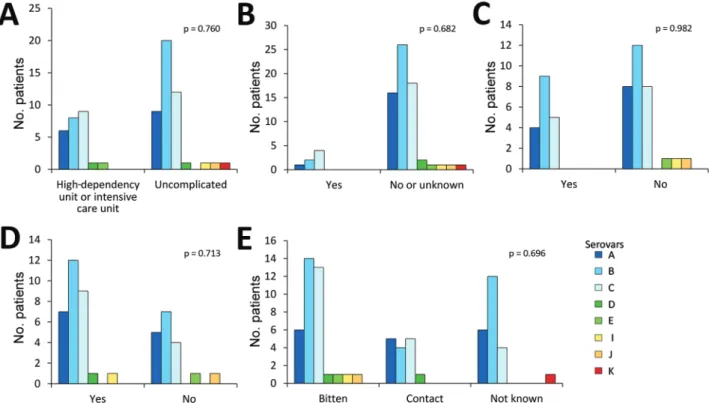
Figure 1. Prevalence of capsular serovars among Capnocytophaga canimorsus isolates from patients and dogs. A) Prevalence among
73 isolates from patients in Helsinki, Finland, 2000–2017. B) Prevalence among 25 isolates acquired from patients worldwide. C) Prevalence among pooled samples (n = 98). D) Prevalence among 52 isolates from dog mouths, Switzerland and Belgium. Percentages do not add up to 100% because of rounding. A portion of the data presented in panels B and D were previously published (7).
the antisera we produced. Although no isolates reacted with the anti-K serum, isolate CcD35 from a dog in Switzerland reacted with the anti-J serum (Figure 1, panel D; online Technical Appendix Table 5). We confirmed this result by Western blot analysis of the polysaccharidic structures (online Technical Appendix Figure 2), which showed that capsular serovar J is thus not limited to Finland.
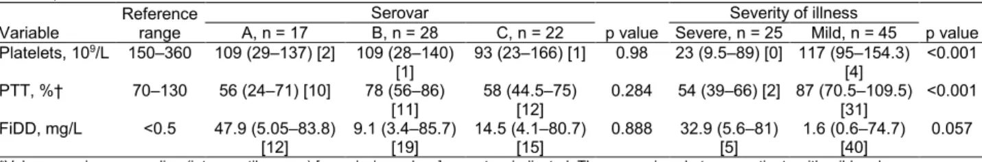
Correlation between Disease Severity and Capsular Type
We also tested the association between serovar and disease severity. For this investigation, the level of care was used as a surrogate; patients treated in a regular ward or who had only visited the emergency department were regarded as having a mild course of disease, and patients treated in a high-dependency or intensive care unit were regarded as severely ill. No statistically significant difference could be found in the proportions of any serovar between patients with mild and severe disease (p = 0.76; Figure 2, panel A). Among the 73 cases of C. canimorsus infection included in this study, 3 were fatal (Table 2). The isolates from these 3 patients were serovars A (H80), B (H26), and C (H28). Extensive amputations were reported in 6 cases, among which included the nonsurviving patient infected with the capsular B isolate H26. The 5 other capsular types associ-ated with amputations were A (n = 2, H48 and H56), B (n = 1, H79), and C (n = 2, H46 and H59). Therefore, capsular
serovars A, B, and C are all capable of causing severe dis-ease in humans.
We looked for an association between capsular serovar and patient immune status or lifestyle factors but found no statistically significant link between serovar and immune compromised state (p = 0.682), alcohol abuse (p = 0.982), or smoking (p = 0.713) (Figure 2, panels B–D). We defined patients as immune compromised if they had been on im-mune suppressive medication or had recently received che-motherapy, had a concurrent medical condition associated with impaired immunity or active cancer, or had undergone splenectomy. One of the 2 splenectomized patients had a severe course of disease, but both survived.
Severe C. canimorsus infections are often associated with purpura or petechiae, disseminated intravascular co-agulation, and gangrene of extremities (1). In particular, coagulation disorders were found to be associated with 94% of patients having C. canimorsus–induced septic shock in a 10-year retrospective study in Helsinki (5). In our study, no statistically significant association could be found between coagulation and fibrinolysis laboratory variables (platelet count, partial thromboplastin time, fi-brin D-dimers) and capsular serovars (Table 3). Given the low number of cases associated with some serovars, we could assess only the 3 dominant serovars (A, B, and C). We compared coagulation and fibrinolysis disorder mark-ers between patients with mild and severe clinical course.
Figure 2. Association between Capnocytophaga canimorsus capsular serovar and various patient factors, Helsinki, Finland, 2000–2017.
A) Disease severity (n = 70); B) immune compromised (n = 73); C) alcohol abuse (n = 49); D) smoking status (n = 48); and E) contact with dogs (n = 73). Fisher exact test was used for statistical analysis.
As expected, the analyzed variables were more affected in patients with a severe course of infection (Table 3). Of note, deviating coagulation and fibrinolysis variables were frequently present in patients with mild courses of disease, further strengthening the previously reported close association of C. canimorsus infection and coagula-tion disorders.
Correlation between Type of Contact with Dog and Capsular Type
The type of contact with dogs did not differ among infec-tions with any of the dominant serovars, but 4 of the 5 patients infected with serovar D, E, I, or J had been bitten (Figure 2, panel E). The contact type was not known for the patient infected with the serovar K isolate, the fifth rare serotype.
Discussion
In this study, we analyzed 73 C. canimorsus isolates ob-tained from patients treated at Helsinki University Hospi-tal. All isolates were serotyped and found to be endowed with a capsular polysaccharide (CPS), further confirming the commonality of the presence of a CPS in C.
canimor-sus isolates (6,7). We confirmed the high prevalence of
capsular serovars A, B, and C among isolates from human infections; 67 (91.8%) of 73 isolates were typed as 1 of these 3 serovars. No significant difference was found in the prevalence of these serovars between this collection of 73 isolates from Finland and a previously studied collection of 25 isolates obtained from cases worldwide (7). Among the 98 C. canimorsus isolates from these 2 studies, 89 (90.8%) were capsular types A, B, or C. Our data confirmed that se-rovars A, B, and C are significantly more common among clinical isolates than dog isolates (4/52; 7.6%), suggesting these serovars are more virulent than the others. Our data also confirmed that serovars A, B, and C are present in dif-ferent geographic areas.
Besides the A, B, and C serovars, the Helsinki collec-tion contained 2 other serovars: 2 isolates of serovar D and 1 of serovar E. This observation is of high interest because
serovars D and E were previously isolated from patients in the United States (n = 1), Belgium (n = 1), and Switzerland (n = 1) (7). Thus, although serovars D and E represent only 4.1% and 2%, respectively, of the total clinical isolates in this study, these serovars should be considered virulent and taken into account in prophylaxis.
One patient in our cohort was infected with a se-rovar I strain. This sese-rovar had not been encountered before among humans but was found in dogs (1 in Bel-gium and 1 in Switzerland) (7). These findings suggest that not only serovars A–E but also rare serovars are widely distributed.
Last, we describe 2 new capsular serovars, J and K, each with a limited (1%) prevalence in human infections. We tested these 2 new antisera against our collection of iso-lates obtained from dogs in Switzerland and Belgium (10) and found 1 C. canimorsus isolate had a J-type CPS. Thus, using the 11 antisera we have that are specific to serovars
Table 2. Patient demographics, clinical characteristics, and contact with dogs, Helsinki Hospital District, Finland, 2000–2017* Characteristic patients† No. Value Age, y, median (IQR) 73 55 (48.3–
64.8) Sex 73 M 38 (52.1) F 35 (47.9) Immune compromised 73 7 (9.6) Smoking 48 30 (62.5) Alcohol abuse 49 18 (36.7) Contact with dog 73
Not known 21 (28.8) Contact but not bitten 15 (20.5) Bitten 37 (50.7) Disease severity 70
Regular ward or emergency
department 45 (64.3) High surveillance unit 11 (15.7) Intensive care unit 14 (20.0) Length of hospital stay, d, median (IQR) 62 6 (3–13.3) Deaths at day 30 73 3 (4.1) Deaths at 1 y 61 4 (6.6) Amputation 73 6 (8.2)
*Values are no. (%) patients except as indicated. IQR, interquartile range. †Because of missing data, number of patients in each category varied.
Table 3. Coagulation and fibrinolysis laboratory variables, by Capnocytophaga canimorsus serovar and disease severity, Helsinki, Finland, 2000–2017*
Variable Reference range A, n = 17 Serovar B, n = 28 C, n = 22 p value Severe, n = 25 Severity of illness Mild, n = 45 p value Platelets, 109/L 150–360 109 (29–137) [2] 109 (28–140) [1] 93 (23–166) [1] 0.98 23 (9.5–89) [0] 117 (95–154.3) [4] <0.001 PTT, %† 70–130 56 (24–71) [10] 78 (56–86) [11] 58 (44.5–75) [12] 0.284 54 (39–66) [2] 87 (70.5–109.5) [31] <0.001 FiDD, mg/L <0.5 47.9 (5.05–83.8) [12] 9.1 (3.4–85.7) [19] 14.5 (4.1–80.7) [15] 0.888 32.9 (5.6–81) [5] 1.6 (0.6–74.7) [40] 0.057
*Values are given as median (interquartile range) [no. missing values] except as indicated. The comparison between patients with mild and severe courses of disease was defined by the level of care they needed. Patients with mild disease were those who were treated in a regular ward or the emergency department, and patients with severe disease were those treated in high surveillance or intensive care units. FiDD, fibrin D-dimers; PTT, partial thromboplastin time.
†PTT was analyzed according to the Owren method (8). PTT was calculated as the ratio of the result (in seconds) from normal plasma to the result (in seconds) from the patient sample x 100.
A–K, which identified 98 human clinical isolates, we can only type 36.5% (19/52) of our collection of dog isolates from Switzerland and Belgium. This finding, again, rein-forces the hypothesis of the existence of a large repertoire of CPS serovars in C. canimorsus among dog isolates.
Because C. canimorsus extensively deglycosylates hu-man N-linked glycoproteins from cell surfaces (11–13), a given blood group might be a predisposing factor for C.
canimorsus infection, but further research is needed to
in-vestigate an association between blood type and serovar. Blood group information was available for 55 patients in our cohort, and we found no enrichment in any blood groups among patients infected with C. canimorsus com-pared with the blood group distribution of the population of (data not shown).
The availability of clinical records associated with the isolates typed in this study gave us the opportunity to investigate the link between capsular serovar and disease severity, patient immune status, lifestyle, or type of contact with dogs. When comparing the most prevalent capsular types (A, B, and C) found in these 73 clinical isolates, we found no significant correlation between disease severity and capsular type. In the previous C. canimorsus capsular typing study, the authors suggested that strains belonging to capsular types of lower prevalence, like D and E, might preferentially infect immunocompromised patients (7); we could not draw such a conclusion here. In addition, alcohol abuse or smoking status could not be linked to infection by a specific capsular type. Alcohol abuse, smoking status, and immune suppression all were not significantly associ-ated with disease severity or the 30-day mortality rate (data not shown), although the relatively low sample size and missing data preclude us from drawing conclusions regard-ing this matter.
The capsular serovars less frequently isolated in hu-man infections, such as E, I, and J, were mainly found in patients who had been bitten, which could suggest that these serovars are less virulent than serovars A–D, perhaps requiring a deeper inoculation to provoke an infection. Un-fortunately, the information on dog exposure was missing for the patient infected with the serovar K strain.
Two patients included in this study were reportedly bitten on the same day by the same dog. The isolates from these 2 patients (H44 and H46) were both typed as capsu-lar serovar C, suggesting that the same strain infected both patients. The 2 patients had a severe form of the infection, requiring treatment in an intensive care unit. This observa-tion of 2 patients being infected by the same dog has not been reported previously and gives an indication of the epi-demiology of disease.
The observation of so few cases of C. canimorsus in-fection is indeed striking, considering that up to 74% of dogs carry C. canimorsus bacteria (14). We hypothesize
that only a few C. canimorsus strains are virulent in hu-mans, and few dogs carry these dangerous strains. In-deed, the 3 most prevalent serovars in human infection (A–C), represent only 7.6% of the C. canimorsus iso-lates from dogs (7), suggesting that a minority of dogs represent a risk for humans. This disease might be pre-ventable in humans by identifying the dogs that carry these dangerous serotypes and specifically vaccinating them to eliminate the pathogen or drastically reduce pathogen shedding.
This work was financed by grant SOC 1510582 from the Belgian Walloon Region.
About the Author
Dr. Hess is a researcher at the Research Unit in the Biology of Microoganisms at the University of Namur in Belgium and an immunologist with a research interest in host– pathogen interactions.
References
1. Butler T. Capnocytophaga canimorsus: an emerging cause of sepsis, meningitis, and post-splenectomy infection after dog bites. Eur J Clin Microbiol Infect Dis. 2015;34:1271–80. http://dx.doi.org/ 10.1007/s10096-015-2360-7
2. Butler T, Weaver RE, Ramani TK, Uyeda CT, Bobo RA, Ryu JS, et al. Unidentified gram-negative rod infection. A new disease of man. Ann Intern Med. 1977;86:1–5. http://dx.doi.org/10.7326/ 0003-4819-86-1-1
3. Pers C, Gahrn-Hansen B, Frederiksen W. Capnocytophaga
canimorsus septicemia in Denmark, 1982–1995: review of 39
cases. Clin Infect Dis. 1996;23:71–5. http://dx.doi.org/10.1093/ clinids/23.1.71
4. van Dam AP, Jansz A. Capnocytophaga canimorsus infections in The Netherlands: a nationwide survey. Clin Microbiol Infect. 2011;17:312–5. http://dx.doi.org/10.1111/j.1469-0691. 2010.03195.x
5. Hästbacka J, Hynninen M, Kolho E. Capnocytophaga canimorsus bacteremia: clinical features and outcomes from a Helsinki ICU cohort. Acta Anaesthesiol Scand. 2016;60:1437–43. http://dx.doi.org/10.1111/aas.12752
6. Renzi F, Ittig SJ, Sadovskaya I, Hess E, Lauber F, Dol M, et al. Evidence for a LOS and a capsular polysaccharide in
Capnocytophaga canimorsus. Sci Rep. 2016;6:38914.
http://dx.doi.org/10.1038/srep38914
7. Hess E, Renzi F, Koudad D, Dol M, Cornelis GR. Identification of virulent Capnocytophaga canimorsus isolates by capsular typing. J Clin Microbiol. 2017;55:1902–14. http://dx.doi.org/10.1128/ JCM.00249-17
8. Owren PA. Thrombotest a new method for controlling anticoagulant therapy. Lancet. 1959;274:754–8. http://dx.doi.org/ 10.1016/S0140-6736(59)90857-8
9. Tissari P, Zumla A, Tarkka E, Mero S, Savolainen L, Vaara M, et al. Accurate and rapid identification of bacterial species from positive blood cultures with a DNA-based microarray platform: an observational study. Lancet. 2010;375:224–30. http://dx.doi.org/ 10.1016/S0140-6736(09)61569-5
10. Renzi F, Dol M, Raymackers A, Manfredi P, Cornelis GR. Only a subset of C. canimorsus strains is dangerous for humans. Emerg Microbes Infect. 2016;5:e29. http://dx.doi.org/10.1038/emi.2016.43
11. Mally M, Shin H, Paroz C, Landmann R, Cornelis GR.
Capnocytophaga canimorsus: a human pathogen feeding at
the surface of epithelial cells and phagocytes. PLoS Pathog. 2008;4:e1000164. http://dx.doi.org/10.1371/journal.ppat.1000164 12. Manfredi P, Renzi F, Mally M, Sauteur L, Schmaler M, Moes
S, et al. The genome and surface proteome of Capnocytophaga
canimorsus reveal a key role of glycan foraging systems in host
glycoproteins deglycosylation. Mol Microbiol. 2011;81:1050–60. http://dx.doi.org/10.1111/j.1365-2958.2011.07750.x
13. Renzi F, Manfredi P, Mally M, Moes S, Jenö P, Cornelis GR. The N-glycan glycoprotein deglycosylation complex (Gpd) from
Capnocytophaga canimorsus deglycosylates human IgG. PLoS
Pathog. 2011;7:e1002118. http://dx.doi.org/10.1371/journal. ppat.1002118
14. Suzuki M, Kimura M, Imaoka K, Yamada A. Prevalence of
Capnocytophaga canimorsus and Capnocytophaga cynodegmi in
dogs and cats determined by using a newly established species-specific PCR. Vet Microbiol. 2010;144:172–6. http://dx.doi.org/ 10.1016/j.vetmic.2010.01.001
Address for correspondence: Guy R. Cornelis, Unité de Recherche en Biologie des Microorganismes, Université de Namur, 61 rue de Bruxelles, 5000 Namur, Belgium; email: [email protected]
Sources
1. Brenner DJ, Hollis DG, Fanning GR, Weaver RE.
Capnocytophaga canimorsus sp. nov. (formerly
CDC group DF-2), a cause of septicemia follow-ing dog bite, and C. cynodegmi sp. nov., a cause of localized wound infection following dog bite. J Clin Microbiol. 1989;27:231–5.
2. Leadbetter ER, Holt SC, Socransky SS.
Capnocytophaga: new genus of gram-negative gliding
bacteria. I. General characteristics, taxonomic considerations and significance. Arch Microbiol. 1979;122:9–16. http://dx.doi.org/10.1007/BF00408040
F
rom the Greek kapnos (“smoke”) for its dependence on carbon dioxide, which is a largecomponent of smoke, Capnocytophaga canimorsus (Latin canis, “dog,” and morsus, “bite”) are gram-negative, facultatively anaerobic, rod-shaped bacteria that are part of the normal oral microbiota of dogs and cats. The genus was proposed to distinguish these bacteria from
Cytophaga spp. (Greek kytos, “cell,” and phagein, “eat”), which also exhibit gliding motility. C. canimorsus was previously known as CDC group DF-2 (dysgonic fermenter type 2) and was
first isolated from a man who had ex-perienced multiple dog bites and devel-oped septicemia and meningitis. C.
cani-morsus remains a
major cause of sep-ticemia in persons, particularly those who are asplenic or immunocompro-mised, who are bit-ten by dogs or cats.
Capnocytophaga canimorsus [kapʺno-si-tofʹǝ-gǝ kanʺǝ-morʹsǝs]
etymologia
Address for correspondence: Ronnie Henry, Centers for Disease Control and Prevention, 1600 Clifton Rd NE, Mailstop E28, Atlanta, GA 30329-4027, USA; email: [email protected]
DOI: https://doi.org/10.3201/eid2412.ET2412
Paul de Vos, Cats
Fighting in a Larder
1630–1640. Oil on
canvas. Museo Nacional del Prado. https://www. museodelprado.es/ coleccion/galeria-on-line/galeria-on-line/ obra/pelea-de-gatos-en-una-despensa/, Public Domain, https://commons. wikimedia.org/w/index. php?curid=39117357 Ronnie Henry
Article DOI: https://doi.org/10.3201/eid2412.172060
Capnocytophaga canimorsus Capsular
Serovar and Disease Severity, Helsinki
Hospital District, Finland, 2000–2017
Technical Appendix
Technical Appendix Table 1. Description of method and sample used for 16S rDNA identification of 73 Capnocytophaga
canimorsus isolates from study, Helsinki, Finland, 2000–2017
Isolate ID Method used Sample used H1 UNamur Isolated colony H3 UNamur Isolated colony H4 UNamur Isolated colony H5 HUSLAB method 3 Directly from blood culture bottle H6 HUSLAB method 3 Directly from blood culture bottle H7 HUSLAB method 3 Directly from blood culture bottle H8 HUSLAB method 3 Directly from blood culture bottle H9 HUSLAB method 3 Isolated colony H10 HUSLAB method 3 Isolated colony H11 HUSLAB method 3 Directly from blood culture bottle H12 UNamur Isolated colony H13 HUSLAB method 3 Directly from blood culture bottle H14 HUSLAB method 3 Directly from blood culture bottle H15 HUSLAB method 3 Directly from blood culture bottle H16 HUSLAB method 3 Directly from blood culture bottle H17 HUSLAB method 3 Directly from blood culture bottle H18 HUSLAB method 3 Directly from blood culture bottle H19 HUSLAB method 3 Isolated colony H20 HUSLAB method 3 Directly from blood culture bottle H21 UNamur Isolated colony H22 HUSLAB method 3 Isolated colony H23 HUSLAB method 3 Isolated colony H24 UNamur Isolated colony H25 HUSLAB method 3 Directly from blood culture bottle H26 HUSLAB method 3 Isolated colony H27 HUSLAB method 3 Isolated colony H28 HUSLAB method 3 Isolated colony H29 HUSLAB method 3 Isolated colony H30 HUSLAB method 3 Isolated colony H31 HUSLAB method 3 Directly from blood culture bottle H33 HUSLAB method 3 Isolated colony H34 HUSLAB method 3 Isolated colony H35 HUSLAB method 3 Directly from blood culture bottle H36 HUSLAB method 3 Directly from blood culture bottle H37 HUSLAB method 3 Directly from blood culture bottle H38 HUSLAB method 3 Directly from blood culture bottle H39 HUSLAB method 3 Isolated colony H41 HUSLAB method 3 Directly from blood culture bottle H42 HUSLAB method 3 Directly from blood culture bottle H43 HUSLAB method 3 Directly from blood culture bottle H44 HUSLAB method 2 Isolated colony H45 HUSLAB method 2 Isolated colony H46 HUSLAB method 2 Isolated colony H47 HUSLAB method 2 Directly from blood culture bottle H48 HUSLAB method 2 Directly from blood culture bottle H49 HUSLAB method 1 Isolated colony H50 HUSLAB method 1 Isolated colony H51 HUSLAB method 1 Directly from blood culture bottle H52 HUSLAB method 1 Directly from blood culture bottle H53 HUSLAB method 1 Directly from blood culture bottle
Page 2 of 7
Isolate ID Method used Sample used H55 HUSLAB method 1 Isolated colony H56 HUSLAB method 1 Directly from blood culture bottle H57 HUSLAB method 1 Directly from blood culture bottle H58 HUSLAB method 1 Directly from blood culture bottle H59 HUSLAB method 1 Directly from blood culture bottle H60 HUSLAB method 1 Isolated colony H62 HUSLAB method 1 Directly from blood culture bottle H63 HUSLAB method 1 Isolated colony H64 HUSLAB method 1 Isolated colony H65 HUSLAB method 1 Directly from blood culture bottle H67 HUSLAB method 1 Directly from blood culture bottle H68 HUSLAB method 1 Isolated colony H69 HUSLAB method 1 Directly from blood culture bottle H70 UNamur Isolated colony H71 UNamur Isolated colony H72 UNamur Isolated colony H73 UNamur Isolated colony H74 HUSLAB method 1 Directly from blood culture bottle H75 HUSLAB method 1 Isolated colony H76 UNamur Isolated colony H78 HUSLAB method 1 Directly from blood culture bottle H79 HUSLAB method 1 Isolated colony H80 HUSLAB method 1 Isolated colony
Technical Appendix Table 2. Oligonucleotides used for typing Capnocytophaga canimorsus isolates, Helsinki, Finland, 2000–2017 Name Sequence 5-3 Reference 533R TTACCGCGGCTGCTGGCAC (11) FD1 mod AGAGTTTGATCYTGGYTYAG (11) CLSI-F TTGGAGAGTTTGATCMTGGCTC (12) Forward Bosshard AGAGTTTGATCMTGGCTCAG (12) Reverse Bosshard GTATTACCGCGGCTGCTG (12) 27F AGAGTTTGATCCTGGCTCAG (13,14) 1100R GGGTTGCGCTCGTTG (13,14) 685R TCTACGCATTTCACCGCTAC (13,14) SeroA-fw CATACCATGGGAAAAAAAGTACCAATAGTTTTTATATTTAACC (10) SeroA-rev CCGCTCGAGTCATTTTTTTATCTTTTTTAATATATTCCAC (10) SeroB-fw CATACCATGGGAATTAACAAAATTCTAATAG (10) SeroB-rev CCGCTCGAGTTATTTTTTATTTTCATTAG (10) SeroC-fw GGCGTATATCGTTGCTATTTTGTATG (10) SeroC-rev CTATTAATATTTTCATTGTACACCACTTC (10) SeroD-fw GATTTAAAAAATATAGTATTTTAGGAATTATCG (10) SeroD-rev CTATACTTGTTCCCACTTTTTAGTTTC (10) SeroE-fw GGAGGAGGAAAAGTATTATTAGATTATC (10) SeroE-rev CTATTCATAATTCTTAAAGATACTTATCAATTC (10) SeroABC-fw CTTGGTTAGGTAAAGTTGCCTTAC (10) SeroABC-rev CAACATTTCTCCCATCTTATAATCCC (10) Technical Appendix Table 3. Description of 16S rDNA sequencing methods used for Capnocytophaga canimorsus isolates, Helsinki, Finland, 2000–2017
Category
Method name
HUSLAB method 1 HUSLAB method 2 HUSLAB method 3 UNamur Forward primer 533R CLSI F CLSI F 27F Reverse primer FD1mod Reverse Bosshard Reverse Bosshard 1100R DNA polymerase AmpliTaq Gold
(Applied Biosystems, Waltham, MA, USA)
AmpliTaq Gold (Applied Biosystems.
Waltham, MA, USA)
MolTaq 16S (Molzym, Bremen,
Germany)
Takara PrimeSTAR (Clontech, Kasatsu, Japan) Amplification program 94°C for 10 min. 35 cycles of 94°C for 15 s, 55°C for 15 s, and 72°C for 30 s 95°C for 10 min. 30 cycles of 94°C for 15 s, 64°C for 15 s, and 72°C for 30 s 95°C for 2 min. 40 cycles of 94°C for 15 s, 64°C for 15 s, and 72°C for 30 s
5 cycles of 94°C for 30 s, 60°C for 2 min, and 72°C for 3 min with a reduction of annealing temperature of 1.5°C/cycle. 30 cycles of 94°C for 30s,
52°C for 90 s, and 72°C for 3 min. Final elongation at 72°C for 10 min. Sequencing
primer(s)
Technical Appendix Table 4. Capnocytophaga canimorsus strains used in study, Helsinki, Finland, 2000–2017*
Isolate ID Collection History and geographic origin Reference Cc1 BCCM/LMG 11511; CCUG 17234;
strain P810; strain SSI P810
BCCM/LMG <CCUG Sweden <W.Frederiksen <J.Ursing. Malmö. Sweden
(1) Cc2 CCUG 70775 G. Wauters and M. Delmee. Cliniques Universitaires St Luc. Brussels.
Belgium
(2) Cc3 – G. Wauters and M. Delmee <Sint-Jan Hospital. Brugges. Belgium (3) Cc4 CCUG 70776 J. Schrenzel. Hopitaux Universitaires de Genève. Switzerland (4) Cc5 BCCM/LMG 28512. CCUG 70777 G. Wauters and M. Delmee <Clinic of Libramont. Libramont. Belgium (5) Cc6 CCUG 70778 KU Leuven. Leuven. Belgium (6) Cc7 – G. Wauters and M. Delmee. <KU Leuven. Leuven. Belgium (5) Cc8 – M. Delmee <Liège. Belgium (6) Cc9 BCCM/LMG 11510. CCUG 12569.
CDC A3626
BCCM/LMG. CCUG <R. Weaver. CDC. Atlanta. Georgia <Virginia. USA
(7) Cc10 BCCM/LMG 11541. CCUG 24741.
ATCC 35978. CDC C8936
BCCM/LMG. MCCM. ATCC <R. Weaver. CDC. Atlanta. Georgia <California Health Department. California. USA
(7) Cc11 BCCM/LMG 11551. CCUG 70779.
MCCM 01373
BCCM/LMG <MCCM <A. von Graevenitz. Unersität Zurich. Switzerland
(7) Cc12 ATCC 35979. CDC 7120. CCUG
53895
ATCC <R.Weaver. CDC Atlanta Georgia <California Health Dept. <San Antonio Community Hospital. California. USA
(8) Cc13 – F.S. Stals. Laurentius Ziekenhuis. Roermond. The Netherlands (9) Cc14 – R. Jarsumbeck. Medizinisches labor Ostsachsen. Dresden. Germany (6) Cc15 – K. Mühlemann. University Hospital Bern. Switzerland (6) Cc16 – G. Glupczynski. Centre Hospitalier Universitaire Mont Godinne <D.
Olivier. Hopital Univ. Erasme. Brussels. Belgium
(6) Cc17 – G. Glupczynski. Centre Hospitalier Universitaire Mont Godinne <D.
Olivier. Hopital Univ. Erasme. Brussels. Belgium
(6) Cc18 – G. Glupczynski. Centre Hospitalier Universitaire Mont Godinne <D.
Olivier. Hopital Univ. Erasme. Brussels. Belgium
(6) Cc19 – A. Magnette. Centre Hospitalier Universitaire Mont-Godinne <M
Delmée <Clinique Saint Pierre. Ottignies. Belgium
(6) Cc20 CCUG 55909 CCUG <E. Ek. Blood Department. PHLS. Göteborg. Sweden <UK
National External Quality assessment. Colindale. London. UK
(10) Cc21 CCUG 60839 CCUG <E. Ek. Blood Department. PHLS. Göteborg. Sweden (10) Cc22 CCUG 20318 CCUG <W. Frederiksen. Statens Seruminstitut. Copenhagen.
Denmark
(10) Cc23 CCUG 48899 CCUG <V. Roux and D. Raoult. Marseille. France (10) Cc24 CCUG 67384 CCUG <PHLS. Uddevalla <Trollhätten. Sweden (10) Cc25 CCUG 66222 CCUG <I. Adlerberth. Blood Department. PHLS. Sahlgrenska
University Hospital. Göteborg. Sweden
(10) CcD3-CcD106† – Switzerland (6) CcD113-CcD131† – Belgium (6)
*All human isolates listed were from human septicemia cases except Cc4, which was isolated from a prosthetic aortitis case. ATCC, American Type Culture Collection; BCCM/LMG, Belgian Co-ordinated Collections of Micro-organisms, Laboratory of Microbiology, UGent; CCUG, Culture Collection University of Gothenburg; CDC, Centers for Disease Control and Prevention; MCCM, Medical Culture Collection Marburg; PHLS, Public Health Laboratory Services.
Page 4 of 7
Technical Appendix Table 5. Capsular serotyping of Capnocytophaga canimorsus dog isolates by ELISA* Isolate Serovar J Serovar K Mean SD Mean SD H12 100 0 6.2 0.6 H24 10 2.2 100 0 Cc5 9.4 2.1 6.1 0.4 Cc6 9.6 2.3 6.4 0.5 Cc9 10 2.6 6.4 0.6 Cc12 9.7 2.3 7.5 1.0 Cc4 10 2.9 7.4 1.4 CcD3 6.9 0.7 5.5 0.4 CcD5 7.2 1.3 5.9 0.3 CcD6 7.6 1.8 6.4 0.8 CcD10 7.1 1.2 7.2 0.4 CcD13 6.4 1.2 5.7 0.5 CcD16 6.9 1.5 6.2 0.4 CcD18 8.4 2.1 6.9 0.7 CcD20 9.6 2.3 6.9 0.8 CcD25 6.6 1.1 6.0 0.4 CcD33 7.2 1.7 7.1 1.4 CcD34 8.7 2.0 6.4 1.9 CcD35 43.5 4.0 5.8 0.3 CcD37 6.2 1.3 5.6 0.2 CcD39 6.7 1.5 5.7 0.3 CcD40 7.4 1.8 7.0 1.3 CcD43 7.4 1.3 5.9 0.3 CcD44 6.3 1.2 5.4 0.2 CcD47 7.0 1.9 5.8 0.3 CcD51 7.6 2.0 6.4 1.0 CcD52 8.3 1.5 7.0 1.9 CcD53 7.6 1.9 6.3 0.4 CcD57 7.3 1.9 7.2 3.0 CcD58 9.9 4.1 6.8 0.8 CcD63 7.2 1.1 5.8 0.2 CcD68 8.0 2.8 5.8 0.4 CcD69 8.6 3.1 5.6 0.4 CcD71 8.8 2.0 5.7 0.4 CcD73 7.1 1.4 6.9 1.3 CcD76 6.8 1.3 5.4 0.2 CcD77 7.6 1.6 6.7 0.5 CcD80 7.3 1.4 6.2 0.6 CcD81 7.6 0.6 8.2 2.3 CcD84 7.6 1.0 7.1 0.5 CcD89 8.5 1.3 8.0 0.7 CcD96 7.6 1.0 12.4 8.3 CcD101 9.3 1.8 12.2 8.7 CcD104 7.6 0.8 10.8 7.3 CcD105 8.7 1.5 8.4 2.9 CcD106 9.5 1.8 11.2 2.9 CcD113 7.5 1.1 11.2 7.5 CcD115 7.5 1.5 11.8 8.2 CcD116 8.0 1.6 10.1 5.8 CcD117 8.1 1.8 12.8 8.6 CcD118 7.2 1.1 11.6 7.3 CcD119 7.6 1.5 11.5 7.4 CcD120 7.4 1.6 12.4 9.6 CcD122 8.8 1.6 11.5 8.8 CcD124 7.2 1.2 12.2 9.4 CcD126 7.3 1.3 11.3 8.2 CcD129 8.1 1.6 9.4 3.8 CcD130 8.4 1.6 14.7 10.2 CcD131 7.1 1.8 12.2 9.2
*Capsular serotyping was determined by ELISA on entire heat-killed bacteria. The following sera were used: anti-H12 adsorbed with human isolates Cc1 to Cc25 (J antiserum) and anti-H24 adsorbed with human isolates Cc1 to Cc25 (K antiserum). Isolates Cc5, Cc6, Cc9, Cc12, and Cc4, which were serovars A–E, respectively, were used as negative controls. The readout of the ELISA was absorbance but results are expressed here as percentage of reactivity calculated with respect to the absorbance value obtained for the capsular type strain. Values are the mean and SD of at least 3 independent experiments.
Technical Appendix Figure 1. Capsular typing of Capnocytophaga canimorsus isolates from patients,
Helsinki, Finland, 2000–2017, by PCR. PCR detection of capsular serovars A–E with oligonucleotides given in Technical Appendix Table 2. The capsular type of each serovar described in reference (10) were used as positive controls: Cc5, Cc6 (6), Cc9 (7), Cc12 (American Type Culture Collection 35979) (8), and Cc4 (4) for A, B, C, D, and E serovars, respectively. For PCR ABC, Cc5 was used as a positive control. A strain of another capsular type and no DNA were used as negative controls. Three strains were identified by 16S rDNA sequencing to be a member of other dog-hosted Capnocytophaga species and were removed from our study and from this figure by cutting out the according lanes. Ctrl, control.
Page 6 of 7
Technical Appendix Figure 2. Capsular serotyping of Capnocytophaga canimorsus isolates from
patients, Helsinki, Finland, 2000–2017, by Western blot. Western blot analysis of proteinase-K treated lysates of C. canimorsus isolates by using A–K antisera. Cc5, Cc6, Cc9, Cc12, Cc4, CcD37 (6), CcD63 (6), CcD101 (6), and CcD129 (6) were used as positive controls for capsular serovars A–I, respectively. Numbers correspond to molecular mass (kDa). Three strains were identified by 16S rDNA sequencing to be members of other dog-hosted Capnocytophaga species and were removed from our study and from this figure by cutting out the according lanes.
References
1. Heltberg O, Busk HE, Bremmelgaard A, Kristiansen JE, Frederiksen W. The cultivation and rapid enzyme identification of DF-2. Eur J Clin Microbiol. 1984;3:241–3. PubMed
http://dx.doi.org/10.1007/BF02014893
2. Hantson P, Gautier PE, Vekemans MC, Fievez P, Evrard P, Wauters G, et al. Fatal Capnocytophaga
canimorsus septicemia in a previously healthy woman. Ann Emerg Med. 1991;20:93–4. PubMed http://dx.doi.org/10.1016/S0196-0644(05)81130-8
3. Vanhonsebrouck AY, Gordts B, Wauters G, Van Landuyt HW. Fatal septicemia with Capnocytophaga
canimorsus in a compromised host. A case report with review of the literature. Acta Clin Belg.
1991;46:364–70. PubMedhttp://dx.doi.org/10.1080/17843286.1991.11718192
4. Rougemont M, Ratib O, Wintsch J, Schrenzel J, Hirschel B. Capnocytophaga canimorsus prosthetic aortitis in an HIV-positive woman. J Clin Microbiol. 2013;51:2769–71. PubMed
5. Shin H, Mally M, Kuhn M, Paroz C, Cornelis GR. Escape from immune surveillance by
Capnocytophaga canimorsus. J Infect Dis. 2007;195:375–86. PubMed http://dx.doi.org/10.1086/510243
6. Renzi F, Dol M, Raymackers A, Manfredi P, Cornelis GR. Only a subset of C. canimorsus strains is dangerous for humans. Emerg Microbes Infect. 2016;5:e29. PubMed
http://dx.doi.org/10.1038/emi.2016.43
7. Vandamme P, Vancanneyt M, van Belkum A, Segers P, Quint WG, Kersters K, et al. Polyphasic analysis of strains of the genus Capnocytophaga and Centers for Disease Control group DF-3. Int J Syst Bacteriol. 1996;46:782–91. PubMedhttp://dx.doi.org/10.1099/00207713-46-3-782
8. Butler T, Weaver RE, Ramani TK, Uyeda CT, Bobo RA, Ryu JS, et al. Unidentified gram-negative rod infection. A new disease of man. Ann Intern Med. 1977;86:1–5. PubMed
http://dx.doi.org/10.7326/0003-4819-86-1-1
9. Kleijnen-Grebien B, Boorsma S, Stals FS, van Schelven R. Fatal case of sepsis with Capnocytophaga
canimorsus after a minor dog bite [in Dutch]. Ned Tijdschr Geneeskd. 2008;152:1882–5. PubMed
10. Hess E, Renzi F, Koudad D, Dol M, Cornelis GR. Identification of virulent Capnocytophaga
canimorsus isolates by capsular typing. J Clin Microbiol. 2017;55:1902–14. PubMed http://dx.doi.org/10.1128/JCM.00249-17
11. Kotilainen P, Jalava J, Meurman O, Lehtonen OP, Rintala E, Seppälä OP, et al. Diagnosis of meningococcal meningitis by broad-range bacterial PCR with cerebrospinal fluid. J Clin Microbiol. 1998;36:2205–9. PubMed
12. Edwards KJ, Logan JM, Langham S, Swift C, Gharbia SE. Utility of real-time amplification of selected 16S rRNA gene sequences as a tool for detection and identification of microbial signatures directly from clinical samples. J Med Microbiol. 2012;61:645–52. PubMed http://dx.doi.org/10.1099/jmm.0.041764-0
13. Johnson JL. Similarity analysis of rRNAs. In: Gerhard P, Murray RGE, Wood NR, Krieg NR, editors. Methods for general and molecular bacteriology. Washington, DC (USA): American Society for Microbiology; 1994. p. 683–700.
14. Lane DJ. 16S/23S rRNA sequencing. In: Stackebrandt EG, Goodfellow M, editors. Nucleic acid techniques in bacterial systematics. New York (USA): John Wiley & Sons; 1991. p. 115–74.