HAL Id: tel-00447178
https://tel.archives-ouvertes.fr/tel-00447178
Submitted on 14 Jan 2010
HAL is a multi-disciplinary open access
archive for the deposit and dissemination of sci-entific research documents, whether they are pub-lished or not. The documents may come from teaching and research institutions in France or abroad, or from public or private research centers.
L’archive ouverte pluridisciplinaire HAL, est destinée au dépôt et à la diffusion de documents scientifiques de niveau recherche, publiés ou non, émanant des établissements d’enseignement et de recherche français ou étrangers, des laboratoires publics ou privés.
Food quality monitoring and analytical techniques
optimization of some aliments within plant-animal
correlation Contaminated aliments effects on the
detoxication enzymes
Radu Corneliu Duca
To cite this version:
Radu Corneliu Duca. Food quality monitoring and analytical techniques optimization of some ali-ments within plant-animal correlation Contaminated aliali-ments effects on the detoxication enzymes. Médicaments. Université Paris Sud - Paris XI, 2009. Français. �tel-00447178�
FACULTE DE MEDICINE PARIS-SUD
………
N° attribue par la bibliothèque
THESE
pour obtenir le grade de
DOCTEUR DE L’UNIVERSITE PARIS XI
Champ disciplinaire : Pharmacologie et biologie cellulaires et moléculaires
Ecole Doctorale de rattachement :
Signalisations, neurosciences, endocrinologie, reproduction
présentée et soutenue publiquement
par
Radu-Corneliu DUCA
le 30 juin 2009
Food quality monitoring and analytical techniques optimization of some
aliments within plant-animal correlation
Contaminated aliments effects on the detoxification enzymes
Titre :
Directeur de thèse : Dr. DELAFORGE Marcel
Codirecteur de thèse : Pr. VLADESCU Luminita
JURY
Président :
Pr. TAOUIS Mohamed
Rapporteur :
Pr. RICHERT Lisiane
Rapporteur :
Dr. TARANU Ionelia
Examinateur :
Dr. GALTIER Pierre
Directeur de thèse : Dr.
DELAFORGE
Marcel
I dedicate my present work to my family, especially to my
mother, who supported me unconditionally in all my
demarches and goals, and to my dear wife who provided me
a moral support and had sufficient patience to be close to
me even from faraway.
Abstract
Zearalenone (Zen) is a secondary metabolite biosynthesised through a polyketide pathway by several
Fusarium strains (Fusarium graminearum, F. culmorum, F. equiseti, and F. crookwellense). It is a
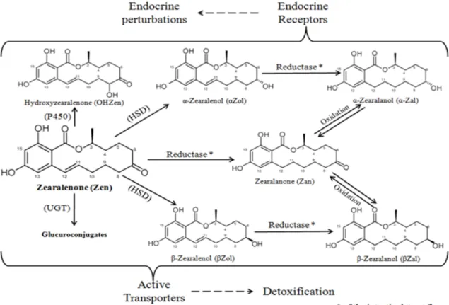
non-steroidal estrogen or mycoestrogen and is frequently named phytoestrogen. Zen is a regular contaminant of cereal crops worldwide (Bennett and Klich, 2003). Zen resists to most common treatments occurred during food manufacturing and despite its non-steroidal structure, binds to estrogen receptors resulting in functional and morphological alterations in reproductive organs. It interacts not only with both types of estrogen receptors (Celius et al., 1999; Shier et al., 2001; Yu et al., 2004; Takemura et al., 2007), but also with the substrates for a number of hepatic enzymes. Zen is well-absorbed and is able to reach intracellular targets. The important disparity concerning the effects of Zen in animal species could in part result from the differences in their hepatic enzymes profile (Gaumy et al., 2001; Cavret and Lecoeur, 2006). Zen metabolism is complex, dominated by conjugation reactions (considered as detoxification pathways) and reduction reactions which correspond to biological activation (Gaumy et al., 2001).
Our main objectives were the elucidation of the effects of contaminated aliments (especially with zearalenone) on the enzymes of detoxification (especially CYPs P450) and the understanding of Zearalenone effects on different species. In the available literature the effect of Zen on the expression and activity of detoxification enzymes is limited to in vitro experiments, to our knowledge in vivo researches has not been reported. In order to establish the zearalenone in vivo effects on the detoxification enzymes we performed several experiments: on rat, a classical animal model for the biological studies existing a large documentation within the specialized literature on the xenobiotics effects and in particularly on Zen effects, and on chicken, one of the species considered most resistant and “used” to Zearalenone presence. Complementary approaches have been used to determine zearalenone effects on the detoxification enzymes: a) development of analytical tools for the mycotoxins pharmacokinetics studies; b) determination of the in vivo effects of zearalenone on the detoxification enzymes expression and its becoming within animal organism c) in vitro zearalenone molecular mechanisms – the direct effects on hepatic detoxification enzymes d) species specificity and human risk assessment.
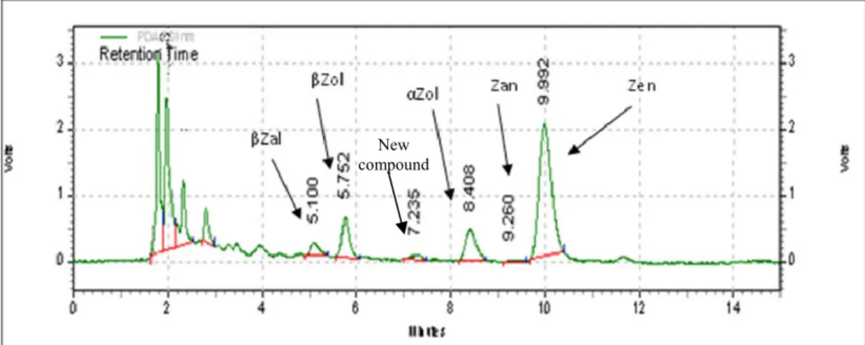
Important stimulatory effects on P-gp mRNA expression were observed, upon in vivo Zearalenone rat treatments, suggesting that P-glycoprotein might be implicated in the detoxification path of zearalenone. P-glycoprotein implication in Zen transport has been determined in Caco2 cell lines by Videmann et al., in 2008. The Zearalenone presence induces early and rapid metabolic responses, especially for CYP2C7, which could have an important role within Zen’s detoxification pathway in rats. An influence of Zearalenone treatments on the mRNA expression and enzyme activities of the P450 isoforms CYP2B2 and 3A was also stated. CYP2C7 and CYP3A1 are rat homologues of human CYP2C8 and CYP3A4, respectively (Nelson, 1999), cytochromes implication in the formation of a new 8-hydroxy Zen metabolite. The in vivo occurrence of this 8-hydroxy Zen metabolite was also determined. Zearalenone becoming within rat and poultry was assessed using new developed methods: HPLC-DAD and LC-MS (using enriched 13C Zen as internal standard). In rats, Zearalenone is rapidly
eliminated in the urine within the first 6 hours after administration; about 60% (47.8 ±14.3 µg) of the total amount of urinary eliminated Zen (79.8 ±23.9 µg), and the overall urinary eliminated Zen and metabolites are about 4% of the administrated Zen. In poultry muscle samples the levels of α-Zol (13.42 µg/kg) are higher than the JEFCA accepted level (2 µg/kg) and are not suitable for human consumption. A worldwide risk assessment of human zearalenone exposure, taking in to account the eating habits, was done resulting in an important and constant human health risk. Also the necessity of regulation changes concerning the acceptable maximum level of zearalenone in cereals, a 5 µg/kg we consider to be acceptable.
Résumé
Zéaralénone (Zen) est un métabolite secondaire de type polykétide biosynthétisé par différentes
espèces de Fusarium (Fusarium graminearum, F. culmorum, F. equiseti, et F. crookwellense). Il s'agit d'un oestrogène non-stéroïdien (ou mycoestrogène) souvent nommé phytoestrogène. La Zen est un contaminant de cultures de céréales dans le monde entier (Bennett et Klich, 2003). Elle résiste à la plupart des traitements qui ont lieu au cours de la fabrication des denrées alimentaires. En dépit de sa structure non-stéroïdienes, le zen est capable de se lier aux récepteurs des oestrogènes et provoque des altérations morphologiques et fonctionnelles des organes de reproductions. Elle interagit non seulement avec les deux types de récepteurs aux œstrogènes (Celius et al., 1999; Shier et al., 2001; Yu et al., 2004; Takemura et al., 2007), mais aussi avec les substrats dans un certain nombre d'insuffisance des enzymes hépatiques. La Zen est bien absorbée et est capable d'atteindre des cibles intracellulaires. La disparité importante des effets de la Zen entre les différentes espèces animales pourrait en partie due à la différence entre leur profil des enzymes hépatiques (Gaumy et al., 2001; Cavret et Lecoeur, 2006). Le métabolisme de la Zen est complexe, il est dominé par des réactions de conjugaison (considérées comme des voies de détoxication) et des réactions de réduction qui correspondent à l'activation biologique (Gaumy et al., 2001).
Nos principaux objectifs étaient l'élucidation des effets des aliments contaminés (en particulier avec le zéaralénone) sur les enzymes de détoxication (en particulier CYP P450) et la compréhension des effets de la zéaralénone sur les différentes espèces. Dans la littérature l’effet de la Zen sur l'expression et sur l'activité des enzymes de détoxication est limité à des expériences in vitro, à notre connaissance aucune recherche in vivo n’a encore été publiée. En vue d'établir les effets in vivo de la zéaralénone sur les enzymes de détoxication nous avons effectué plusieurs expériences: tout d’abord sur le rat, qui est un modèle animal classique pour les études biologiques et sur lequel on trouve une large documentation sur les effets des xénobiotiques et en particulier sur les effets de la Zen, et ensuite sur le poulet, l'une des espèces considérée comme la plus résistante et «habituée» à la présence de la Zéaralénone. Des approches complémentaires ont été utilisées pour déterminer les effets de la zéaralénone sur les enzymes de détoxication: a) Le développement d'outils d'analyses pour des études de la pharmacocinétique des mycotoxines; b) la détermination in vivo des effets de la zéaralénone sur l'expression des enzymes de détoxication et de son devenir dans l'organisme animal c) du mécanisme moléculaire in vitro de la zéaralénone - les effets directs sur les enzymes de détoxification hépatique d) la spécificité des espèces et l'évaluation des risques humains.
D’importants effets stimulants sur l’expression d'ARNm de la P-gp ont été observés in vivo chez le rat traitait avec Zéaralénone, ce qui suggère que la P-glycoprotéine pourrait être impliqué dans la voie de détoxication de la zéaralénone. L’implication de la P-glycoprotéine dans le transport de la Zen a été déterminé dans des lignées cellulaires Caco2 par Videmann et al., en 2008. La présence de zéaralénone induit des réponses métaboliques précoces et rapides, en particulier pour le CYP2C7, il pourrait jouer un rôle important dans la voie de détoxication de la Zen chez le rat. Une influence des traitements par la Zéaralénone sur l’expression des RNAm et sur l’activité des isoformes de P450, CYP2B2 et 3A a également été réalisée. CYP2C7 et CYP3A1 sont respectivement les homologues chez le rat des formes CYP2C8 et CYP3A4 humaine (Nelson, 1999), ces cytochromes sont impliqués dans la formation d'un nouveau métabolite le 8-hydroxy Zen. In vivo l'apparition de ce métabolite (le 8-hydroxy Zen) a également été démontrée. Le devenir dans le rat et le poulet de la Zéaralénone a été évalué en utilisant les nouvelles méthodes développées: HPLC-DAD et LC-MS (en utilisant comme étalon interne Zen 13C enrichi). Chez le rat, la zéaralénone est rapidement éliminée. Elle est retrouvée
dans les urines, pendant les 6 premières heures après l'administration : environ 60% (47,8 ± 14,3 mg) du montant total de la zéaralénone urinaire éliminé (79,8 ± 23,9 mg) et la Zen et ses métabolites éliminés par l’urine sont d'environ 4% de la zéaralénone administrés. Dans les échantillons de muscle de poulet les niveaux d'α-Zol (13,42 mg / kg) sont plus élevés que le niveau JEFCA accepté (2 mg / kg). Ces volailles sont donc impropres à la consommation. A l'échelle mondiale l'évaluation des risques de l'exposition de l'homme à la zéaralénone, en prenant en compte les habitudes alimentaires, a été réalisée et il en résulte un important et constant risque pour la santé humaine. Aussi la nécessité d’apporter des modifications au règlement concernant la teneur maximale acceptable de la zéaralénone dans les céréales a était surligniez, et nous considérons comme acceptable la valeur 5 µg/kg.
First of all, I pay special thanks to my two Ph-D thesis supervisors: prof. dr. Luminita VLADESCU and dr. Marcel DELAFORGE (DR CNRS) for their implication, guidance and support. Thank you so much for making possible this experience, namely my co-directed joint thesis with a mix of Romanian and French ways of living and working, and for all the things that you been teaching me all along.
Secondly, I want to show all my gratitude to the French and Romanian teams and friends, thanking them for their direct support at a working and humanly level.
From the French team arise the name of Frederique Bravin, my colleague (CEA Sacaly) and dear friend, which have made much easier the long days of laboratory work and support me in overcoming several encounter problems. I thank Francois Andre for his energetic presence, helpful advices and for the “taxi-driving” in the late evenings. Also I thank Sandrine Blondel, Paola Difabio, Jamel Meslamani and Silvan Pitarque for their friendship that made the last two years of my thesis more agreeable, in laboratory and not only. Special thank I pay to Nicolas Loiseau (INRA Toulouse), who enabled my encounter with Marcel Delaforge and the current work development.
From the Romanian team I want to thank dr. Rodica Diana Criste (INCDBNA-Balotesti) which support and advise me all along my young research experience. Special thanks I pay to Ionelia Taranu that made possible my first 6 month French experience within INRA Toulouse, which proved to be the first break in developing the current thesis. Between my Romanian colleagues I want to thank for their support and help to Cristina Tabuc, Daniela Marin, Arabela Untea, Panaite Tatiana, Denisa Racheru and Catalin Dragomir. To prof. dr. Irinel Adriana Badea and prof. dr. Iulia Gabriela David (University of Bucharest) I thank for their support concerning the experiments and the elucidation of the oxidation-reduction mechanism.
Thirdly, I thank the Service of Academic and Scientific Cooperation, the French Embassy in Romania (CROUS) and the French Minister of Foreign Affairs (EGIDE) for support with travel and living expenses of my French incursion and the French AFSSET for the financial support of the laboratory expenses.
Last but not the least, I pay the dearest and special thanks to my family, especially to my mother, who supported me unconditionally in all my demarches and goals, and to my wife who provided me a moral support and had sufficient patience to be next to me even from faraway. I dedicate my present work to the two of them and thank them for their support.
Abstract... i
Acknowledgments ... iii
Table of Contents ...v
List of abbreviations ... ix
Introduction ...1
Chapter 1 - Bibliographical study on Zearalenone State of the art ...7
1.1. Mycotoxins ...9
1.1.1. General overview...11
1.1.1.1. Occurrence ...12
1.1.1.2. Human and animal disorders ...14
1.1.1.3. Risk assessment and legislative aspects ...16
1.2. Zearalenone...19
1.2.1. Occurrence (cereals to feed and food) ...20
1.2.2. Human and animal health disorders ...31
1.2.2.1 Clinical signs associated with exposure to zearalenone contaminated feeds in farm animals...33
1.2.2.2 Human pathologies associated with exposure to zearalenone ...39
1.2.2.3 Toxicological reference values ...40
1.2.3. Fate in the body ...43
1.2.3.1. Absorption ...43
1.2.3.2. Distribution ...43
1.2.3.3. Biotransformation ...44
1.3. Enzymes implicated within detoxification pathway ...50
1.3.1. Metabolism, detoxification and transport of xenobiotics ...50
1.3.1.1. Phase I...51
1.3.1.2. Phase II: Conjugation ...57
1.3.1.3. Transport...57
1.4. Zearalenone biotransformation and effects on the hepatic enzymes ...59
1.4.1. Hepatic biotransformations of zearalenone ...59
1.4.2. Effects of zearalenone on the activity and expression of liver drug metabolizing enzymes ...61
Chapter 2 - Development of analytical tools for studies of mycotoxins pharmacokinetics...63
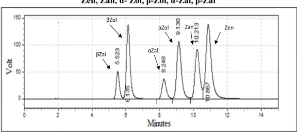
2.1. HPLC assessment of Zen and metabolites in biological samples – Method development and validation ...65
2.1.1. Material and methods ...65
2.1.1.1. Instrumentation ...65
2.1.1.2. Chemicals and Solvents...66
2.1.1.3. Sample collection and preparation...66
2.1.2. Results and discussion ...69
2.1.2.1. Method development ...69
2.1.2.2. Method validation ...72
v
2.1.2.3. Zearalenone determination within chicken biological samples...732.1.3. Conclusion ...74
2.2. Study on the oxidation - reduction equilibriums involving Zearalenone ...75
2.2.1. Study on the oxidation - reduction equilibriums involving Zen and Ce (IV)...75
Table of Contents
2.2.1.2. Studies of oxidation - reduction equilibriums involving Zen and Ce (IV)
by HPLC ...79
2.2.2. Voltammetric behaviour of zearalenone...86
2.2.2.1. Voltammetric behaviour of Zen...87
2.2.2.2. Possibilities of voltammetric quantitative determination of Zen...91
2.2.2.2. Possibilities of voltammetric quantitative determination of Zen...91
2.3. LC-MS assessment of Zen by using 13C enriched Zearalenone as internal standard ...94
2.3.1. Mass spectrometry - brief reminder...95
2.3.2. Zearalenone determination in different matrices ...96
2.3.2.1 Determination of Zen in extracts of cereal ...96
2.3.2.2. Determination of natural Zen contained in extracts of treated animals...101
Chapter 3 - In vivo effects of zearalenone on the expression of detoxification enzymes and its bio-transformation within the animal organism ...105
3.1. In vivo effects of Zearalenone on enzymes expression (cellular signalization)...107
3.1.1. Zearalenone effects on the P450 amounts. ...110
3.1.1.1. Zearalenone effects on P450 amounts in treated rats ...110
3.1.1.2. Zearalenone effects on the P450 amounts of treated chickens ...112
3.1.1.3. Zearalenone effects on the P450 amounts – Conclusions ...113
3.1.2. Determination of the expression of mRNA encoding the detoxification enzymes, by quantitative Reverse Transcriptase Polymerase Chain Reaction (RT-PCR)...114
3.1.2.1. Zearalenone and classical P450 inducers effects on mRNA gene expression ...116
3.1.2.2. Early effects of zearalenone on mRNA gene expression ...118
3.1.2.3. Effects of zearalenone on mRNA gene expression - Conclusions ...122
3.2. Zearalenone presence and bio-transformation in the living animal...123
3.2.1. Zearalenone and its metabolites in treated rats ...124
3.2.1.1. Assessment of the Zearalenone within rat organism (blood, urine and liver samples)...124
3.2.1.2 Zearalenone becoming within rats’ organism - Conclusions ...126
3.2.2. Zearalenone bio-transformation within poultry organism ...127
3.2.2.1. Assessment of the Zearalenone in chicken organism (blood, meat and liver samples)...127
3.2.3. Effects of Zearalenone on chicken meat quality ...130
3.2.3.1. Determination of the gross chemical composition of the broiler meat...130
3.2.2.3 Zearalenone becoming within poultry organism – Conclusions ...132
Chapter 4 - In vitro zearalenone molecular mechanisms – direct effect on hepatic detoxification enzymes...135
4.1. Assessment of Zearalenone metabolism on different species and expressed human P450 ...137
4.1.1. Zearalenone metabolism on treated rats ...138
4.1.1.1 P450 mono-oxygenases activities measured at pH 7.4 on treated rats ...138
4.1.1.2 In vitro zearalenone metabolism at pH 4.5 on treated rats ...141
4.1.1.2 In vitro zearalenone metabolism at pH 4.5 on treated rats ...141
vi
4.1.2. Zearalenone metabolism in treated poultry...1414.1.2.1 P450 mono-oxygenases activities measured at pH 7.4 in treated chickens..141
4.1.2.2 In vitro zearalenone metabolism at pH 4.5 on treated chickens ...142
4.1.3. Zearalenone metabolism in human microsomes, expressed human P450 and
other species microsomes ...143
4.1.3.1. Zearalenone metabolism in human and other species microsomes ...144
4.1.3.2. Zearalenone metabolism on expressed human P450 ...145
4.1.3.3 Zearalenone metabolism on human microsomes, expressed human P450 and other species microsomes - Conclusions ...147
4.2. Interactions of Zearalenone and its metabolites on the enzymatic metabolism of reference compounds ...148
4.2.1. X-ROD and 7-BFC metabolism in treated rats microsomes ...149
4.2.1.1. Metabolic activity of resorufin derivates (X-ROD) on Zen and classical inducers treated rat microsomes ...149
4.2.1.2. Metabolic activity of the 7-Benzyloxy-4-(trifluoromethyl)-coumarin (7- BFC) on Zen and classical inducers treated rat microsomes ...151
4.2.1.3. X-ROD and 7-BFC metabolism in treated rats microsomes - Conclusions 152 4.2.2. X-ROD and 7-BFC metabolism on treated poultries...153
4.2.2.1. Metabolic activity of resorufin derivates (X-ROD) on Zen and classical inducers treated chicken microsomes ...153
4.2.2.2. Metabolic activity of the 7-Benzyloxy-4-(trifluoromethyl)-coumarin (7-BFC) on Zen and classical inducers treated chicken microsomes...154
4.2.2.3 X-ROD and 7-BFC metabolism on treated poultries - Conclusions ...155
4.3. Interactions of Zearalenone and its metabolites on steroid metabolism ...157
4.3.1. Estradiol metabolism ...157
4.3.1.1. Estradiol metabolism on treated rats...157
4.3.1.2. Estradiol metabolism in treated poultry...158
4.3.1.3. Estradiol metabolism in human and other species...160
4.3.1.4. Estradiol metabolism – Conclusions: ...160
4.3.2. Testosterone metabolism...162
4.3.2.1. Testosterone metabolism in treated rats ...162
4.3.2.2. Testosterone metabolism in treated poultry...163
4.3.2.3. Testosterone metabolism in human and other species...164
4.3.2.4. Testosterone metabolism – Conclusions ...165
Chapter 5 - Species specificity and human risk assessment...167
5.1. In vitro zearalenone metabolism effect on the metabolism of reference compounds and species specificity (IC50 assessment)...169
5.1.1. Zearalenone metabolism effect on ethoxyresorufin O-deethylase metabolic activity...170
5.1.1.1. Zearalenone metabolism effect on ETR O- deethylase metabolic activity, using rat microsomes and human expressed forms ...170
5.1.1.2. Zearalenone metabolism effect on ETR O-deethylase metabolic activity, using rat, human and pig microsomes and human expressed forms...171
5.1.2. Zearalenone metabolism effect on the metabolic activity of 7-benzyloxy-4-(trifluoromethyl)-coumarin O-debenzylase ...172
vii
5.1.2.1. Zearalenone metabolism effects on 7-BFC O-debenzylase metabolic activity, using rat microsomes ...1725.1.2.2. Zearalenone metabolism effect on 7-BFC O-debenzylase metabolic activity, using pig microsomes ...173
5.1.2.2. Zearalenone metabolism effect on 7-BFC O-debenzylase metabolic activity, using microsomes with a high content of Cyp3A...173
Table of Contents
5.1.3. In vitro zearalenone metabolism effect on the metabolism of reference
compounds and species specificity (IC50 assessment) - Conclusions...174
5.2. Zearalenone and its metabolites effect on human – risk assessment...175
5.2.1. Zearalenone risk assessment – general overview...175
5.2.2. European zearalenone risk assessment ...178
5.2.3. Worldwide zearalenone risk assessment...179
Conclusions...181
Bibliography ...189
Annex 1 - Articles
Annex 2 - Comunications
ABC ATP-Binding Cassette
ABCB1 P-gP: P-glycoprotein
ABCC, MRP Multidrug resistance associated protein;
ABCG2, BCRP Breast Cancer Resistance Protein
Ac Ac Acid Acetic
ACN Acetonitrile
AD Added Dose
ADP Adenosine Di Phosphate
AFSSA Agence Française de Sécurité Sanitaire des Aliments
ATP Adenosine Tri Phosphate
b.w. Body weight
CEA Commissariat de l’Énergie Atomique
Clof Clofibrate COMT Catechol-O-Methyl-Transferase CYP Cytochrome P450 CYP19 Aromatase DM Dexamethasone DON Deoxynivalenol
DSV Direction des Sciences du Vivant
E2 Estradiol
ER Estrogenic Receptor
FAO Food and Agriculture Organization
FB 1,2,3 Fumonisin B1, B2, B3
FDA Food & Drug Administration
G6P Glucose-6-phosphate;
G6PDH Glucose-6-Phosphate Dehydrogenase
GC Gas Chromatography
GSH Glutathione
HIF1B, ARNT Aryl Hydrocarbon Receptor Nuclear Translocator
HPLC High Performance Liquid Chromatography
HSD Hydroxy Steroid Dehydrogenase
HSD17B 17-beta hydroxysteroid dehydrogenase
HSD3B 3-beta hydroxysteroid dehydrogenase
i.p., IP: intra-peritonealy
IAC Immunoaffinity Column
IARC Internal Agency for Research on Cancer
ID50 Inhibition Dose for 50% of exposed individuels
ix
INRA Institut National de Recherche Agronomique
IUPAC International Union of Pure and Applied Chemistry
JECFA Joint FAO/WHO Expert Committee on Food Additives
LOAEL Low Observed Adverse Effect Level
MRP Multi Drug Resistance Associated Protein
MS Mass Spectroscopy
NADP(H) Nicotinamid Adenine Dinucleotide Phosphate
(Hydrogenized or reduce form)
NCI National Cancer Institute
NOAEL No Observed Adverse Effect Level
NR1C1, PPARA Peroxisome Proliferative Activated Receptor Alpha
NR1I2, PXR Pregnane Xenobiotic Receptor
NR2B1, RXRA Retinoid Xenobiotic Receptor Alpha
NR3A1, ESR1 Estrogen Receptor
OH-Zen Hydroxy-Zearalenone
OMS World Health Organisation
p.o., PO per os
PB Phenobarbital
PBS Phosphate Buffer Saline
qRT-PCR quantitative real time PCR
SCF Scientific Committee on Food
SG NADPH Generator System
SPE Solid Phase Extraction
TDI Tolerated Daily Intake
TMS Tri Methyl Silan
TST Testosterone
UDP Uridine Di Phosphate
UGT UDP – glucuronosyl transferases
UV Ultra Violet Zan Zearalanone Zen Zearalenone α-Zal α-Zearalanol α-Zol α-Zearalenol β-NF β-Naphtoflavone β-Zal β-Zearalanol β-Zol β-Zearalenol
FID Flame Ionization Detector
MeOH Methanol
DAD Diode Array Detector
λ Wave length
CV Cyclic Voltammetry
DPV Differential Pulse Voltammetry
x
Introduction
Understanding the influence of environmental factors1 of synthetic or natural origin for health is a vast field of investigation. This implies to precisely quantify the acute or chronic exposure to such factors, to identify the role of each component in question and to quantify their contribution in the genesis or aggravation of many diseases, especially multifactorial ones and with unknown aetiologies. Knowledge of the interaction of these factors with other environmental determinants of health, whether behavioural, social or genetic, is a challenge for the scientific research. The challenge is to understand the means available to protect the health of population, and the conditions that must be met for an action to have the desired effect. National authorities shall ensure control over the commercialization, use, dissemination, and presence of certain products. For example: residues and degradation products in the environment or in food, chemicals, but also pesticides, additives, residues of medicinal products for human or veterinary use, or environmental contaminants (dioxins, polycyclic aromatic hydrocarbons, mycotoxins). The continuous human exposure to these compounds should be below levels likely to have adverse effects on consumer health (INRA, 2004)
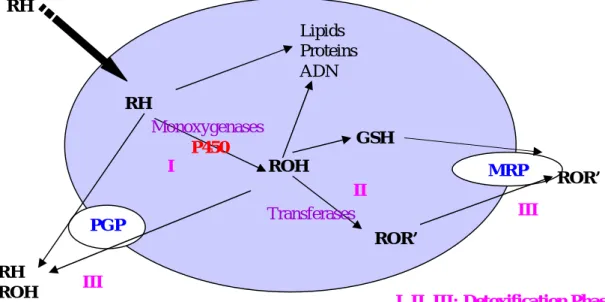
Humans and animals have evolved different enzymatic systems to convert xenobiotics and drugs into hydrophilic metabolites, more easily eliminated via the bile and urine. These biotransformation reactions take place mainly in the liver, which expresses the most prominent class of biotransformation enzymes, but other organs including the lungs, kidneys and intestines may contribute to the overall xenobiotic biotransformation. Differences in biotransformation enzyme activities alter the systemic bioavailability and subsequently the efficacy of drugs; they may also provide protection against certain xenobiotics and environmental pollutants, but can increase the toxicity of others (Snawder and Lipscomb, 2000).
Among the food contaminants, some have hormone-mimetic properties. There may be estrogenic properties (similar to female hormones) or anti-androgenic (preventing the action of male hormones). These substances, called xenoestrogens, may act on the endocrine system of animals and humans (Afsset, 1996-2000). Among them, zearalenone, produced by fungi of the Fusarium family (filamentous fungi) is currently researched in
3
1
In epidemiology, environmental factors are those determinants of disease that are not transmitted genetically. Stress, physical and mental abuse, diet, exposure to toxins, pathogens, radiation and chemicals found in almost all personal care products and household cleaners are common environmental factors that determine a large segment of non-hereditary disease.
Introduction
animal feeds and meat products. This mycotoxin has been studied in this co-directed thesis; researches have been conducted in France at CEA Saclay in the Department of Life Sciences2, under the supervision of Marcel Delaforge and in Romania in the Laboratory of
Chemistry and Animal Physiology, INCDBNA3 and the Department of Analytical
Chemistry4, University of Bucharest under the supervision of Luminita Vladescu. Our main objectives were the elucidation of the effects of contaminated aliments (mainly with zearalenone) on the enzymes of detoxification (especially CYPs P450) and the understanding of Zearalenone effects on different species.
Why Zearalenone?
The interest for this mycotoxin arises from the high human exposure to it and from its toxicological properties:
9 It is a regular contaminant of cereal crops worldwide;
9 It resists to most common treatments occurring during food manufacturing; 9 It is well-absorbed and it is able to reach intracellular targets;
9 It binds to estrogen receptors resulting in functional and morphological alterations in reproductive organs;
9 Its biological and chemical properties may be feared, zearalenone presents a risk to human health by disrupting the hormonal balance;
9 Its effects on the expression and activity of detoxification enzymes are limited to in
vitro experiments.
To elucidate the effect of zearalenone on the detoxification enzymes (especially CYPs P450) and to understand Zen effect on different species, we have to answer several key questions:
9 Can we develop new technological tools to track traces of zearalenone and its derivatives in biological matrices?
9 Can we characterize zearalenone metabolisation and bio-transformation within the animal organism?
4
2Institute of Biology and Technologies of Saclay (IBiTec-S), the Service of Bioenergetics, Structural Biology and Mechanisms (SB2SM) and more precisely the Laboratory of Oxidant Stress and Detoxification (LSoD)
3
National Institute of Research & Development for Biology and Animal Nutrition - Balotesti 4
9 Does zearalenone affect the hepatic detoxification enzymes? If yes, which are its in
vivo effect and transformation, and its molecular mechanism?
9 Are there species specificities linked to Zen metabolism? If yes, are humans undergoing a risk?
In order to answer to these questions, in the next sections we present:
1. The state of the art presentation of zearalenone concerning its occurrence and effects on animal / human organism.
2. The new analytical tools for zearalenone pharmacokinetics studies
3. The in vivo effect of zearalenone on the detoxification enzymes expression and its bio-transformation within the animal organism
4. The in vitro molecular mechanisms of zearalenone and the direct effect on the hepatic detoxification enzymes
5. The species specificity and human risk assessment
Introduction
Bibliographical study on Zearalenone
State of the art
9
1.1. Mycotoxins
Among the secondary metabolites1 of fungi, mycotoxins are toxic compounds that are difficult to define simply and comprehensively. Mycotoxins are natural compounds of low molecular weight, secondary metabolites of filamentous fungi belonging to the strains Aspergillus, Penicillium and Fusarium (Table 1.1).
Table 1.1. Mycotoxins and fungi found associated with producing feed and food (Risks assessment associated with the presence of mycotoxins in feed and food chain. AFSSA 2006)
Mycotoxins Major producing molds
Mycotoxins regulated or ongoing regularization Aflatoxins B1, B2, G1, G2 Ochratoxin A Patulin Fumonisins B1, B2, B3 Trichothecenes (DON, NIV) Zearalenone
Aspergillus flavus, A. parasiticus, A. nomius Penicillium verrucosum, Aspergillus
ochraceus,
Aspergillus carbonarius
Penicillium expansum, Aspergillus clavatus Byssochlamys nivea
Fusarium verticillioides, F. proliferatum Fusarium graminearum, F. culmorum F. crookwellense, F. sporotrichioides F. poae, F. tricinctum, F. acuminatum Fusarium graminearum, F. culmorum F. crookwellense.
Other mycotoxins
Ergot alkaloids (the ergot of rye) Citrinin
Alternaria toxins (alternariol, alternariol methyl ether ...) Acid cyclopiazonique Sterigmatocystin Sporidesmines Stachybotryotoxines Toxins endophytes (ergovaline, lolitrem B) Phomopsines Toxins tremorgenes
Claviceps purpurea, C. paspali, C. africana Aspergillus terreus, A. carneus, A. niveus Penicillium verrucosum, P. citrinum, P. expansum
Alternaria alternata, Alternaria solani Aspergillus flavus, A. versicolor, A. tamarii Penicillium dont P. camemberti
Aspergillus nidulans, A. versicolor, A. flavus Pithomyces chartarum
Strachybotrys chartarum
Neotyphodium coenophialum, N. lolii Phomopsis leptostromiformis
Penicillium roquefortii, P. crustosum, P. puberrelum
Aspergillus clavatus, A. fumigatus
If mycotoxins are of fungal origin, not all the toxic compounds produced by fungi are mycotoxins; the factors of importance being the target and the concentration of the
1
Secondary metabolites are organic compounds that are not directly involved in the normal growth,
development or reproduction of organisms. Unlike primary metabolites, absence of secondary metabolites results not in immediate death, but in long-term impairment of the organism's survivability/fecundity or aesthetics, or perhaps in no significant change at all.
10
metabolite. Thus, fungi products that are toxic to bacteria (such as penicillin) are usually named antibiotics and ones that are toxic to plants are called phytotoxic (Bennett and Klich, 2003). There is a possible application of such products in agriculture: some herbicides may be selective (tentoxine), or total. Others may be peptide compounds, like -amantinine produced by certain Amanitas, which is an inhibitor of eukaryotes RNA polymerase II (Labia and Michelot, 1988). Mycotoxins may develop on the plant, in the field or during storage, and are potentially toxic to human and animals.
More than 12 000 secondary metabolites have been identified from the AntiBase in 2008, but only thirty having toxic properties of a real concern. Toxins are natural contaminants of many food of plant origin: mainly cereals, but also fruits, nuts, almonds and feed compounds, manufactured for use as food and feed (Charmley, 1995; Underhill, 1996; Charmley, 2000).
It should be noted that certain aspects and effects of fungal secondary metabolites are of great interest to man and may be the subject of scientific research:
- As has been reported, the phytotoxicity was used in the agricultural industry; - Certain toxic effects have been studied and found to have a real therapeutic value: this is the case for ergot. Medicine has benefited using it vasodilator effects. Today derived of ergot molecule are used in particular in the treatment of migraine crises or Alzheimer.
However, the balance toxicity / therapeutic properties is sometimes weak. Indeed, the reactivity of these compounds is closely related to their structure: the pharmacophore2and toxicophore3 layouts may be close. The reactive part of the molecule recognized by the pharmacophore (or another part of the molecule) may be unstable and the source of more reactive compounds (radicals or epoxy). Toxicity then covers the first therapeutic effects. This applies, for example, to cyclosporine, which, despite immunosuppressive properties, has proven to be toxic for the kidney at high concentrations.
Thus, it is important to keep in mind that 90% of medicines come from natural compounds and very few are entirely of synthetic nature. If mycotoxins, with their large number and low doses are still unclear, it remains true that alongside the pathogenic
2
A pharmacophore is an ensemble of steric and electronic features that is necessary to ensure the optimal supramolecular interactions with a specific biological target and to trigger (or block) its biological response.
3
A toxicophore is a feature or group within a chemical structure that is thought to be responsible for
11
effects is now necessary to take into account that some have a certain therapeutic potential once adverse reactions identified and controlled. These can be limited for example by hemisyntheses in order to inactivate the toxicophore.
1.1.1. General overview
Mycotoxins are toxic secondary metabolites produced by fungi (molds). These fungal toxins are diverse on a chemical level - they belong to different chemical families - and their molecular weight varies from approximately 200 to 500. There are hundreds of known mycotoxins, but few have been researched and there are appropriate analysis methods only for a smaller number (Whitlow, 2001).
The fungi that produce them, can affect crops in the field, during handling or storage. From a practical point of view, a mycotoxin is a fungal metabolite that causes side effects in animals or humans who are exposed. Exposure usually occurs by consumption of contaminated foods, including foods for humans or livestock. The mycotoxicoses are diseases caused by exposure to foods contaminated by mycotoxins (Nelson et al., 1993). Mycotoxins produce various biological effects in animals, including liver toxicity and renal anomalies of central nervous system, estrogenic responses, respiratory diseases by inhalation (invasive aspergillosis) and other effects.
We distinguish among the mycotoxins, considered important from a food and health point of view (e.g. among the products tested and are subject to standards and recommendations): aflatoxins, ochratoxin A, patulin, fumonisins, zearalenone and trichothecenes (including deoxynivalenol and T-2 toxin) (Charmley, 1994). It should be noted that the toxicity may vary widely from one toxin to another and that the risk is not always the toxin itself, but also its metabolites. Historically, the oldest known mycotoxicosis in France is the ergotism. From the Middle Ages hallucinogenic effects produced by the ingestion of a parasite of rye, rye ergot or Claviceps purpurea, have been described. The symptoms took the form of delirium, prostration, severe pain, abscesses, and gangrene of the extremities, leading to severe and incurable disabilities. Epidemics have plagued the eighth and the sixteenth century due to the people miserable alimentation, especially the consumption of flour contaminated with sclerotia4 of the fungus. In France, the last episode occurred in 1951 in Pont Saint-Esprit in the Gard.
4
A sclerotium (plural sclerotia) is a compact mass of hardened mycelium stored with reserve food material that, in some higher fungi such as ergot, becomes detached and remains dormant until a favorable
12
Today, we know that the "Fire of St. Anthony" is attributable to certain alkaloids produced by ergot. In addition, around 1900, Japanese researchers link specific clinical manifestations with the ingestion of moldy rice. In U.R.S.S. between 1942 and 1947 scores of people die (up to 10% in some communities) following consumption of wheat and millet contaminated with a microorganism of the Fusarium strains.
In the early 60s, the characterization of aflatoxin will be the starting point for systematic research on mycotoxins and their effects. The fact that aflatoxins have proved to be the most potent natural carcinogens, is not foreign to this sudden interest. Since then, the list of recognized molds capable of producing toxins continues to grow. In fact, toxigenic molds can grow in all climates, on all the media solid or liquid, if there are nutrients and humidity. This explains the great variety of foods and environmental substrates involved. Nevertheless, cereals have the greatest risk factor in view of their consumption and frequency of contamination. In the 80s, the Food and Agriculture Organization (FAO) estimated that at least 25% of the grains produced in the world were contaminated by mycotoxins. This phenomenon is even more important because it is becoming more important: with the warming climate, geographical areas that previously had no (or little) fungal contamination, become favourable to the development of mycotoxin-producing strains.
In addition to the health issues posed by the contamination of mycotoxins, inherent socio-economic aspects emerge recently. If, in order to export their grains, the developing countries are submitted to the sanitary regulations concerning food and feed of the developed countries, the local people remain largely exposed to a mycotoxic risk through uncontrolled local markets. This is the case of South Africa (oral presentation by Gordon Shephard at the XIIth International Congress of IUPAC on mycotoxins and phycotoxins, held from 21 to 25 May 2007 in Istanbul).
Chronic effects (repeated exposure to low or very low doses) are the most feared because of dietary habits and the power of persistence of these toxins. In addition, a food or other media can contain multiple mycotoxins simultaneously.
1.1.1.1. Occurrence
In the light of research conducted in North America, it is likely that the main types of fungi producing mycotoxins are Aspergillus, Fusarium and Penicillium (Whitlow, 2001). Several species of these fungi produce mycotoxins in food. Molds are fungi that grow into multicellular colonies, unlike yeasts that are unicellular fungi. Molds can grow and
13
produce mycotoxins before or after harvest or during storage, transport, processing or alimentation. The proliferation of molds and production of mycotoxins are associated with extreme weather, inadequate storage practices, poor quality food and bad alimentation conditions. In general, environmental conditions (heat, water, insect damages), stress and predispose plants to mycotoxins contamination in the field. Temperature, moisture content and insect activity are the main factors influencing the contamination of feed grains and feed by mycotoxins after harvest (Coulombe 1993). Molds proliferates in temperatures ranging between 10 and 40°C, pH may be between 4 and 8, and moisture content higher than 0.7 aw(water activity5). While yeasts require free water, molds can grow on a dry surface (Lacey, 1991). They can also grow on foods that contain more than 12 or 13% humidity. In the wet foods such as cheese, molds (e.g.
Penicillium roqueforti) will develop in the presence of oxygen and a suitable pH. Since
most molds are aerobic, the very wet media exclude adequate oxygen supply and can prevent mold growth. The most favourable conditions for their proliferation may not coincide with optimal conditions for the formation of mycotoxins in the laboratory. For example, it was observed that molds of the Fusarium strain proliferate between 25 and 30°C without producing important quantities of mycotoxins, while at temperatures close to freezing point, a large quantity of mycotoxins are produced when fungi present a minimal growth (Joffe, 1986). The fungicide treatments in the fields could reduce the proliferation of mold, and thus the production of mycotoxins, but stress or shock caused by the fungicide on the fungus could stimulate the production of mycotoxins (Boyacioglu et al. 1992; Ceynowa and Gareis, 1994). Compared to the Fusarium species, the
Aspergillus strains are developing normally in environments where water activity has low
values and temperatures are high. Therefore Aspergillus flavus and aflatoxins frequently infest maize grown under stress conditions caused by heat and dryness in the warm climates. Aflatoxin contamination is favoured by the damage inflicted by insects (before and after harvest) that creates openings for the fungi penetration. The species of the genus
Penicillium are widespread and proliferate in the presence of a relatively low water and
low temperature. Because Aspergillus and Penicillium requires only low water activity to grow, they are considered as storage fungi (Christensen et al., 1977).
5
Water activity is a dimensionless quantity used to represent the energy status of the water in a system. It
is defined as the vapor pressure of water above a sample divided by that of pure water at the same temperature; therefore, pure distilled water has a water activity of exactly one. It is widely used in food science as a simple, straightforward measure of the dryness of food; foods typically have an optimum water activity at which they have the longest shelf life
14
1.1.1.2. Human and animal disorders
Most toxicity studies are related to the ingestion of contaminated food, but inhalation and dermal exposure are also caused signs of toxicity. Thus the toxic effects of several mycotoxins, including those that occur most commonly, are well documented in several animal species and humans (Trenholm, 1988; 1996-1997). The toxicity of these natural contaminants can be direct or indirect. Some mycotoxins have a very strong acute toxicity following exposure to a single high dose. In all cases, the chronic effects (repeated exposure to low or very low doses) are the most feared because of the persistence of these toxins often resistant to temperature and process technology used in the food industry (Sorensen and Elbaek, 2005). Toxicity is variable (Table 1.2), some mycotoxins are known or suspected to be carcinogens (aflatoxins, ochratoxin A, fumonisins). Some mycotoxins exert hepatotoxicity (aflatoxins), others are estrogenic (zearalenone), immunotoxic, hematotoxic (patulin, trichothecenes and fumonisins) or dermonecrotic (trichothecenes) (Bunger, 2004).
Table 1.2. Major mycotoxins identified or suspected effects and the cellular and molecular mechanisms of action identified experimentally (Assessment of risks associated with the mycotoxins presence in food and feed chain. AFSSA, 2006)
Toxin Effects Cellular mechanisms of action and
molecular Aflatoxin B1 + M1 Hepatotoxicity Genotoxicity Carcinogenicity Immunomodulation
Formation of DNA adducts Lipid peroxidation
Bioactivation by cytochrome P450 Combination with GS-transferases
Ochratoxin A
Nephrotoxicity Genotoxicity Immunomodulation
Nephrotoxicity
Impact on protein synthesis. Inhibition of ATP production Detoxification by peptidases
Patulin Neurotoxicity
In vitro mutagenesis Indirect inhibition of enzymes
Trichothecenes (T-2 toxin, DON ...) Haematotoxicity Immunomodulation Dermal toxicity
Induction of apoptosis in progenitor haematopoietic and immune cells Impact on protein synthesis Alteration of immunoglobulin
Zearalenone Fertility and Reproduction
Binding to estrogen receptors Bioactivation by reductases
Conjugation to glucuronyltransferases
Fumonisin B1
Lesion of the central nervous system
Hepatotoxicity Genotoxicity Immunomodulation
Inhibition of the synthesis of ceramide Alteration of the report sphinganine / sphingosine
15
Table 1.3. Toxic effects of some mycotoxins on humans organs
Mycotoxin Liver Digestive tube Nervous system Rein Endocrines glands Skin Blood Aflatoxin B1 + + + + + Ochratoxin A + + + + Vomitoxin + + + + + T2-toxin + + + + + Zearalenone + + Fumonisin B1
Mycotoxins exert their effects through 3 main mechanisms:
(1) Reducing the amount of nutrients available to the animal. This effect is the result of a multi-factorial process. First, there may be an alteration of nutrient content of foods during the molding process. The proliferation of mold can reduce the nutrient such as vitamins, and the content of amino acids such as lysine (Kao and Robinson, 1972). Thus, the mold tends to reduce the energy value of feed. Secondly, some mycotoxins, because of qualitative considerations (taste, smell, etc.) reduce food consumption and therefore nutrient. Thirdly, irritation of the digestive system caused by mycotoxins can reduce the absorption of nutrients. Fourthly, some mycotoxins disrupt the normal metabolism of nutrients: like in the case of protein synthesis inhibited by T-2 toxin.
(2) Effects on the endocrine and exocrine glands. The effect of zearalenone on the reproductive performance, because of its estrogenic action, is an example. The estrogenic effects of zearalenone result from the mycotoxin and its derivatives affinity for the estrogen receptors of the animal (Kiang et al., 1978).
(3) Immunosuppression. The effects of mycotoxins on the immune system have been studied (Sharma, 1993). Trichothecenes, such as DON and T-2 toxin, reduce immunity by inhibition of protein synthesis and thus cell proliferation. Some mycotoxins have a cytotoxic action in vitro on lymphocytes. Corticosteroids produced in response to stress also reduce immune function.
Some of the symptoms observed in mycotoxicoses may be secondary in nature, e.g. they result from an opportunistic illness because of the immunosuppression. Thus, the growth and diversity of the symptoms are confusing and the diagnosis is difficult (Hesseltine, 1986; Schiefer, 1990). In fact, the diagnosis is also complicated by the lack of relevant
16
researches, the lack of feed analysis, by non-specific symptoms and interactions with other stress factors. Against this background, it should develop means of prevention strategies including agronomics (good agricultural practices including the selection of varieties, cultivation, processing plant ...), improving the conditions for harvesting and storage and improved monitoring throughout the food chain. The case of organic production method restricts the use of fungicide treatments, but focuses on techniques against contamination by mycotoxins such as crop rotation, tillage or low nitrogen inputs. The available data for contamination by mycotoxins of products from organic farming, even limited, show variable rates of infection but may not be relieved of significant differences with those of products of conventional agriculture (Bellon et al., 2000).
1.1.1.3. Risk assessment and legislative aspects
Results of contamination, generally recognized as of vegetal origin, mycotoxins are therefore a current problem concerning the quality and safety of feed and food stuffs. Risk assessment of mycotoxins remains difficult due to several reasons:
ü this risk is depending of natural factors, which are independent of humans control (especially weather conditions),
ü fungal contamination is hard to control,
ü there may be multiple mycotoxins present due to the ability of the same mold to produce several mycotoxins (AFSSA, 2006).
Numerous studies have demonstrated that several mycotoxins may be found in the same feed (Hagler et al., 1984). For example, Abbas et al. have demonstrated that Fusarium species isolated from maize in Minnesota have produced multiple mycotoxins (Abbas et al., 1989). Given that animals are fed a mixture of feeds and molds, which produce a variety of mycotoxins, thus several mycotoxin interactions are possible.
Because of their partial degradation in the rumen, mycotoxins are less toxic to cattle than for most other animals. Nevertheless, mycotoxins are not completely degraded and some degradation products are toxic (Kiessling et al., 1984). Dietary factors known to interact with mycotoxins include nutrients such as fats, proteins, fibres, vitamins and minerals (Smith et al. 1971; Brucato et al. 1986; Coffey et al. 1989). The agglomerating agents used in feeds (clay) and other additives, such as glucomannan, bind some mycotoxins and thus reduce exposure of the animal (Diaz et al., 1999). Galvano et al. have recently studied the interaction of mycotoxins with dietary factors such as antioxidants, medicinal
17
herbs, plants extracts and minerals and biological agglomerating agents (Galvano et al., 2001). Some of these dietary factors modify the response of animals to mycotoxins, which has the effect of generating mixed reactions in the field, but also to provide data in order to modulate the toxicity to animals. Thus, the alimentary approaches appear promising in order to protect animals against the effects of mycotoxins and prevent the risk of mycotoxins that can contaminate food for human consumption.
Some of the factors that make diagnosis difficult also contribute to the difficulty of setting safety thresholds. These include the lack of research, sensitivity differences between animal species, the imprecision of the sampling and analysis, the large number of mycotoxins potential interactions with other mycotoxins and interactions with the stress generated by the environment and production (Hamilton, 1984; Schaeffer and Hamilton, 1991). The effects of mycotoxins are also modulated by factors such as gender, age, diet and duration of exposure. It is therefore impossible to provide specific guidance as to the concentrations of mycotoxins, which produce a mycotoxicosis on the ground. The recommendations provide hazardous concentrations of mycotoxins, rather than the lower concentrations of mycotoxins that have been associated with mycotoxicosis (Table 1.4). In this context, in Europe, from the 1st July 2006, a regulation fixing the fusariotoxin acceptable rates (deoxynivalenol, zearalenone and fumonisins) in cereal products used for human consumption (Regulation No. 856 / 2005 of 6 June 2005: Kyprianou, 2005) was introduced. These rates reflect the threshold levels of exposure estimated from the levels of contamination of cereals and toxicity studies conducted in animals (JECFA, 2000).
18
Table 1.4. European rules in force for the limitation of the levels of mycotoxins in foodstuffs for human (No. 466 of 8/03/2001, OJ L77, No. 472, 12/03/2002, OJ L75; No. 257, 12/02/2002, OJ L41)
Mycotoxins Commodity Maximum
limit (µg/kg)
Aflatoxin B1
• Peanuts, nuts and dried, and derivatives • Peanuts subjected to physical treatment before consumption or ingredients • Nuts, dried and subjected to physical treatment before consumption or ingredients • Cereals and derivatives, direct or
ingredients
• Cereals subjected to physical treatment before consumption or ingredients
2
8
5
2
2
Aflatoxin B1 • Spices (pepper, pepper, paprika, nutmeg,
ginger, saffron) 5
Aflatoxin M1 • Milk 0,05
Zearalenone
• Cereals and derived products, grains, raw • Vegetable oils
50 200
Ochratoxin A
• Cereals (including rice and buckwheat) and derived products, grains, raw
• Derivatives and grain consumption • Raisins
5 3 10
Patulin
• Fruit juice (apple) and fruit nectar • spirit drinks, cider and fermented beverages
• Products based on pieces of apple (compote and mashed)
• Apple juice and products made from pieces of apple (compote and puree) for infants and young children
50
50
25
19
1.2. Zearalenone
Zearalenone is a macrocyclic lactone derived from resorcyclic acid (Resorcyclic Acid Lactone, hence the acronym RAL) of molecular formula C18H22O5. Its formula is shown in Figure 1.1. Its scientific name is (-)-(3S, 11E) -3, 4, 5, 6, 9, 10 –hexahydro -14, 16 – dihydroxy – 3 – methyl - 1H – 2 -benzoxacyclotétradécin-1, 7 (8H)-dione (Merk Index, 1996).
Zearalenone is a natural form of the trans- isomer and an S configuration with its methyl group in position C3 (Kuo et al., 1967). It is thermostable and resistant to a temperature of 120°C for 4 hours (Trenholm, 1982).
- - - -zearalanol
--ZAL), which can be co-produced with the ZEN by fungi and is can end up with the ZEN in infected grains.
15 13 OH O H 12 10 O 3 4 9 8 6 5 O O 11 15 13 OH O H 12 10 O 3 4 9 8 6 5 OH O 11
Figure 1.1. Zearalenone (ZEN) Figure 1.2. -Zearalanol ( -ZAL)
Whether zearalenone is a source of toxicity in food
-zearalanol (Figure 1.2) present a significant hazard in terms of health. It is commonly
used in the United States since 1969 as a growth promoter (Ralgro® ND
-http://www.ralgro.com/) to improve the rate of fattening cattle. Use of this product has been banned within the European Union in 1989 but still permitted in North America and New Zealand (Gaumy et al., 2001). Extensive experiments on rat, dog and monkey have -zearalanol is an estrogeno-mimetic, whose main effects are changes in the mammary glands or organs of the reproductive system.
The study of physical and chemical properties of zearalenone suggests that this compound has ideal characteristics for a wide distribution within tissues. The major difference between species sensitivity to the zearalenone effects could partly result from metabolic differences.
20
1.2.1. Occurrence (cereals to feed and food)
Zearalenone is a mycotoxin produced by fungi of the genus Fusarium, particularly F.
graminearum, F. semitectum, F. equiseti, F. crookwellense and F. culmorum but all
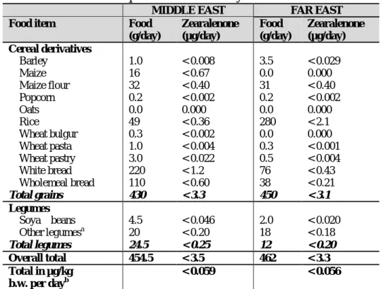
strains are not producing zearalenone (Kuiper-Goodman et al., 1987). This mycotoxin is a natural contaminant of cereals, especially wheat and maize, but also some fruits and vegetables (bananas, beans, nuts ...) and can also be found in animal products such as milk, liver and eggs through contaminated feed. Contamination by zearalenone is a global phenomenon; the fungus producer develops easily in any type of weather but preferentially at low temperatures. An overview on the cereals and feedstuffs Zearalenone occurrence is summarized within the figure 1.3; as well as an estimation of human Zen daily intake depending on the different eating habits is presented in the figure 1.4. The Zen is produced by strains of Fusarium in Australia, Europe and North America (Vesonder et al., 1991), New Zealand (di Menna et al., 1997), the Philippines, Thailand and Indonesia (Yamashita et al., 1995). The Zen also has been detected in food in South America (Dalcero et al. 1997; Molto et al., 1997), Africa (Doko et al., 1996), Taiwan, China and Russia (Ueno et al., 1986).
From a quantitative point of view, the Zen and its metabolites were found in various cereals and derived products in Europe at rates ranging from a few micrograms per kg to 8000 µg/kg (Placinta et al., 1999). The estimated average intake for the French population is of 30 ng/kg bw6/day for adults aged of 15 years or more; and of 70 ng/kg bw/day in children aged from 3 to 14 years. The vector contributing the most to this exposure is constituted, in more than 60%, of cereals by-products and more particularly the breakfast cereals (between 12 and 23%)7.
EUROPE
Available data in Europe indicate that maize is the most prominent cereal at risk with high incidence and high levels of contamination with Zen, however wheat, oats, as well as soybean products have been found to be contaminated occasionally with Zen (EC, 2004). In 1988, Tanaka et al. published an overview on data about the occurrence of Zen from 19 countries including some European countries (Germany, Italy, Poland and UK).
6
bw: body weight
7
Study conducted within the Met@risk (Methods for Food Risk Analysis) unit of INRA for a period of 11 months on a French population (from southern, eastern and western areas of France) consisting in: 2492 individues from which: 1474 adults of 15 years old or more, 1018 children of 3 to 14 years old, and a selection of 338 foodstuffs.
21
The paper reported the contamination of wheat, barley, maize, oat, sorghum, rye and rice by Zen. Placinta et al. (1999) reported the contamination of samples of wheat, barley, oat, rye and feeds from Bulgaria, Germany, Finland, Netherlands, Norway and Poland by Zen at levels from few µg/kg to 8 mg/kg. Germany seems to be the European country where more data can be found about Zen in cereals are more available in Europe. Surveys of cereals and derivatives for several years confirmed their contamination with Zen (Muller et al., 1997a,b ; Schneweis et al., 2002; Schollenberger et al., 2005, 2006). In Yugoslavia, Zen was found at high levels (up to 10 mg/kg) in corn (Balzer et al., 1977) and in dairy cattle feeds (Skrinjar et al., 1995). In Poland, the contamination of wheat by Zen was confirmed by Perkowski et al. (1990). Zakharova et al. (1995) reported a low contamination of cereal crop from Russia in 1993 and 1994 by Zen. The contamination of wheat by Zen was also found in Bulgaria (Vrabcheva et al., 1996). In Hungary, Fazekas et al. (1996) reported the contamination of mouldy and stored corn with ZEN that ranged between 0.01 and 11.8 mg/kg. Cereals from Finland (oats, barley) have been found to contain Zen together with deoxynivalenol (DON) and 3-acetyldeoxynivalenol (3-ADON) (Hietaniemi and Kumpulainen, 1991). In the Netherlands, the occurrence of Zen was reported in wheat (Tanaka et al., 1990) and in feed ingredients (Veldman et al., 1992). In the United Kingdom, Zen was detected in corn and ingredients of animal feeding stuffs (maize and maize products) by Scudamore et al. (1998). In Scotland, according to Gross and Robb (1975), high contamination of barley stored (for 3 month to about one year) with Zen was detected and levels varied between 2.1 and 26.5 mg/kg. In Italy, the contamination of corn was found with Zen was reported from studies of Pietri et al. (2004) and Visconti and Pascale (1998). In Slovakia, Labuda et al. (2005) reported the contamination of poultry feed mixtures with Zen with the co-occurrence of DON, 3-ADON, 15-acetyldeoxynivalenol (15-ADON), T-2 and HT-2 toxins. Concerning recent data on human exposure in Europe to ZEN, the occurrence of the toxin was reported in 32% of mixed cereal samples (n = 4918) from nine European countries. The distribution showed that much of this contamination was in maize and wheat grains. A high incidence of Zen was found in samples of oat from Finland (47% of samples containing >0.2 mg/kg with a maximum level of 1.31 mg/kg being reported) and high incidence of Zen in wheat from France (16% of samples containing >0.2 mg/kg with a maximum level of 1.817 mg/kg being reported). Raw maize was the food commodity with the highest level of Zen, results reported the contamination of 14% of maize with a levels >0.2 mg/kg, the highest level (6.492 mg/kg) was reported in a sample of maize from Italy (SCOOP, 2003).
22
An overview on the cereals and feedstuffs Zearalenone occurrence in Europe is summarized within the table 1.5; as well as an estimation of human Zen daily intake depending on the different eating habits is presented in the table 1.6 (Zinedine et al., 2007; JECFA 2000, 2001; FAO, 2004 )
Table 1.5. European occurrence of zearalenone within cereals and feedstuffs
EUROPE Barley Corn Maize Wheat Oats Feeds ZEN Mean 4,83 1.66 3.49 0.62 0.03 0.20
(mg/kg) Max. 26.5 10 11.8 8.4 0.09 1.8
Table 1.6. European predicted human daily intake of zearalenone
EUROPE
Food item Food
(g/day) Zearalenone (µg/day) Cereal derivatives Barley 20 < 0.16 Maize 0.0 0.000 Maize flour 8.8 < 0.11 Popcorn 0.2 < 0.002 Oats 2.0 < 0.010 Rice 12 < 0.087 Wheat bulgur 0.0 0.000 Wheat pasta 1.3 < 0.005 Wheat pastry 1.0 < 0.007 White bread 120 < 0.66 Wholemeal bread 59 <0.33 Total grains 240 < 1.4 Legumes Soya beans 0.0 0.000 Other legumesa 12 <0.12 Total legumes 12 < 0.12 Overall total 252 < 1.5 Total in µg/kg bw per dayb < 0.025 a
The zearalenone value for tinned beans was used for other legumes b
Based on 60 kg b.w. NORTH AMERICA
In Canada, high concentrations of Zen (up to 141 mg/ kg) were reported in corn for animals (Funnell, 1979). Monitoring of Canadian foods (wheat, barley, soybeans, corn, corn-based foods and grain crops) by Stratton et al. (1993) and Scott (1997) reported also the presence of Zen at different levels. Recently, Zen was detected in infant cereal foods from the Canadian retail market (Lombaert et al., 2003). In the USA, an earlier investigation by Shotwell et al. (1977) showed the contamination of wheat with Zen. Corn from USA was found to be contaminated by Zen (Bennett et al., 1985; Bagneris et al., 1986; Hooshmand and Klopfenstein, 1995). High level values of Zen related to mouldy corn samples were reported by Abbas et al. (1988) and Park et al. (1996). The
23
contamination of sorghum and mouldy sugar beet root was also reported by Bagneris et al. (1986) and by Bosch and Mirocha (1992) respectively. On some occasions, phenomenally high concentrations of Zen have been reported, e.g. 2900 mg/ kg in a food sample from the USA (Pittet, 1998).
An overview on the cereals and feedstuffs Zearalenone occurrence in North America is summarized within the table 1.7; as well as an estimation of human Zen daily intake depending on the different eating habits is presented in the table 1.8 (Zinedine et al., 2007; JECFA 2000, 2001; FAO, 2004 )
Table 1.7. North American occurrence of zearalenone within cereals and feedstuffs
NORTH AMERICA Barley Corn Wheat Sorghum Feeds
ZEN Mean 0.13 3.88 0.13 0.76 47
(mg/kg) Max. 0.21 21.4 0.21 1.48 141
Table 1.8. North American predicted human daily intake of zearalenone
NORTH AMERICA
Food item Food
(g/day) Zearalenone (µg/day) Cereal derivatives Barley 4.7 < 0.039 Maize 0.0 0.0 Maize flour 0.0 0.0 Popcorn 49 0.49 Oats 320 < 1.6 Rice 20 < 0.15 Wheat bulgur 64 <0.24 Wheat pasta 117 < 0.48 Wheat pastry 5.3 < 0.039 White bread 76 < 0.43 Wholemeal bread 38 <0.22 Total grains 315 < 1.6 Legumes Soya beans 0.0 0.0 Other legumesa 13 <0.53 Total legumes 13 < 0.53 Overall total 328 < 2.2 Total in µg/kg bw per dayb < 0.037 a
The zearalenone value for tinned beans was used for other legumes b