HAL Id: inserm-00842001
https://www.hal.inserm.fr/inserm-00842001
Submitted on 5 Jul 2013
HAL is a multi-disciplinary open access
archive for the deposit and dissemination of
sci-entific research documents, whether they are
pub-lished or not. The documents may come from
teaching and research institutions in France or
abroad, or from public or private research centers.
L’archive ouverte pluridisciplinaire HAL, est
destinée au dépôt et à la diffusion de documents
scientifiques de niveau recherche, publiés ou non,
émanant des établissements d’enseignement et de
recherche français ou étrangers, des laboratoires
publics ou privés.
mouse model of Emery-Dreifuss muscular dystrophy
caused by lamin A/C gene mutation.
Antoine Muchir, Young Kim, Sarah Reilly, Wei Wu, Jason Choi, Howard
Worman
To cite this version:
Antoine Muchir, Young Kim, Sarah Reilly, Wei Wu, Jason Choi, et al.. Inhibition of extracellular
signal-regulated kinase 1/2 signaling has beneficial effects on skeletal muscle in a mouse model of
Emery-Dreifuss muscular dystrophy caused by lamin A/C gene mutation.. Skeletal Muscle, BioMed
Central, 2013, 3 (1), pp.17. �10.1186/2044-5040-3-17�. �inserm-00842001�
R E S E A R C H
Open Access
Inhibition of extracellular signal-regulated kinase
1/2 signaling has beneficial effects on skeletal
muscle in a mouse model of Emery-Dreifuss
muscular dystrophy caused by lamin A/C gene
mutation
Antoine Muchir
1,2,3*, Young Jin Kim
1,2,4, Sarah A Reilly
1,2,5, Wei Wu
1,2, Jason C Choi
1,2and Howard J Worman
1,2*Abstract
Background: Autosomal Emery-Dreifuss muscular dystrophy is caused by mutations in the lamin A/C gene (LMNA)
encoding A-type nuclear lamins, intermediate filament proteins of the nuclear envelope. Classically, the disease manifests as
scapulo-humeroperoneal muscle wasting and weakness, early joint contractures and dilated cardiomyopathy with
conduction block; however, move variable skeletal muscle involvement can be present. Previously, we demonstrated
increased activity of extracellular signal-regulated kinase (ERK) 1/2 in hearts of Lmna
H222P/H222Pmice, a model of autosomal
Emery-Dreifuss muscular dystrophy, and that blocking its activation improved cardiac function. We therefore examined the
role of ERK1/2 activity in skeletal muscle pathology.
Methods: Sections of skeletal muscle from Lmna
H222P/H222Pmice were stained with hematoxylin and eosin and histological
analysis performed using light microscopy. ERK1/2 activity was assessed in mouse tissue and cultured cells by
immunoblotting and real-time polymerase chain reaction to measure expression of downstream target genes.
Lmna
H222P/H222Pmice were treated with selumetinib, which blocks mitogen-activated protein kinase/extracellular
signal-regulated kinase kinase 1/2 that activates ERK1/2, from 16 to 20 weeks of age to assess the effects of treatment
on muscle histology, ERK1/2 activity and limb grip strength.
Results: We detected enhanced activation of ERK1/2 in skeletal muscle of Lmna
H222P/H222Pmice. Treatment with
selumetinib ameliorated skeletal muscle histopathology and reduced serum creatine phosphokinase and aspartate
aminotransferase activities. Selumetinib treatment also improved muscle function as assessed by in vivo grip
strength testing.
Conclusions: Our results show that ERK1/2 plays a role in the development of skeletal muscle pathology in
Lmna
H222/H222Pmice. They further provide the first evidence that a small molecule drug may be beneficial for
skeletal muscle in autosomal Emery-Dreifuss muscular dystrophy.
Keywords: Muscular dystrophy, Nuclear envelope, Lamin, Selumetinib, Mitogen-activated protein kinase
* Correspondence:a.muchir@institut-myologie.org;hjw14@columbia.edu
1
Department of Medicine, College of Physicians and Surgeons, Columbia University, 630 West 168th Street, New York, NY 10032, USA
2Department of Pathology and Cell Biology, College of Physicians and
Surgeons, Columbia University, New York, NY, USA
Full list of author information is available at the end of the article
© 2013 Muchir et al.; licensee BioMed Central Ltd. This is an Open Access article distributed under the terms of the Creative Commons Attribution License (http://creativecommons.org/licenses/by/2.0), which permits unrestricted use, distribution, and reproduction in any medium, provided the original work is properly cited.
Muchir et al. Skeletal Muscle 2013, 3:17
Background
Emery-Dreifuss muscular dystrophy (EDMD) is classically
characterized clinically by a triad of: (1) slowly progressive
muscle weakness and wasting in a scapulo-humeroperoneal
distribution; (2) early contractures of the elbows, ankles,
and posterior neck; and (3) dilated cardiomyopathy with
conduction defects [1,2]. Contractures are usually the first
clinical sign of the disease occurring in the first decade of
life. During the second decade of life, the slowly progressive
muscle weakness and wasting typically begin. At the end of
the second decade, most patients develop evidence of
car-diomyopathy [3-5].
EDMD can be inherited in a X-linked or autosomal
fash-ion. X-linked EDMD is caused by mutations in EMD
en-coding emerin [6]. Emerin is an integral protein of the
inner nuclear membrane [7,8]. The majority of autosomal
dominant and less frequent recessive cases are caused by
mutations in LMNA [9,10]. LMNA encodes two major
somatic cell polypeptides, lamin A and lamin C, which are
components of the nuclear lamina, a meshwork of
inter-mediate filaments on the inner aspect of the inner nuclear
membrane [11-14]. While the classical EDMD phenotype
was first attributed to EMD and LMNA mutations, it is
now apparent that the same mutations in these genes can
cause dilated cardiomyopathy with more variable skeletal
muscle involvement [6,9,15-21]. Intriguingly, LMNA
muta-tions (different than those leading to myopathy) can also
cause partial lipodystrophy, peripheral neuropathy, or
accel-erated aging disorders such as Hutchinson-Gilford progeria
syndrome [22].
Despite the relatively recent advances in understanding
the genetics of EDMD and related myopathies, the
patho-genic mechanisms leading to striated muscle damage are
only poorly understood. One useful small animal model to
study pathogenesis and evaluate potential therapeutic
inter-ventions in autosomal EDMD is the Lmna
H222P/H222Pmouse [23]. Starting at approximately 16 weeks, male
Lmna
H222P/H222Pdevelop progressive dystrophic pathology
in several skeletal muscle groups. Later, they have
progres-sive accumulation of connective tissue in skeletal muscle.
Lmna
H222P/H222Pmice also develop dilated cardiomyopathy
with conduction system abnormalities and significant
car-diac fibrosis.
We have previously shown that Lmna
H222P/H222Pmice
have increased activity of the mitogen-activated protein
kinase extracellular signal-regulated kinase (ERK) 1/2 in
cardiac muscle [24]. This increased ERK1/2 activity occurs
prior to the onset of overt tissue pathology, suggesting that
it plays a primary pathogenic role. Treatment of
Lmna
H222P/H222Pmice with drugs that inhibit
mitogen-activated protein kinase/extracellular signal-regulated
kin-ase kinkin-ase (MEK) 1/2, the kinkin-ase that activates ERK1/2,
leads to improved left ventricular ejection fraction [25,26],
decreased cardiac fibrosis [26,27] and prolonged survival
[27]. While these results strongly suggest that abnormal
ERK1/2 activation contributes to the development of
car-diomyopathy in Lmna
H222P/H222Pmice, its pathogenic role
in affected skeletal muscles is unknown.
Based on our findings in heart, we hypothesize that
abnor-mal activation of ERK1/2 is similarly involved in the
patho-genesis of skeletal muscular dystrophy in the Lmna
H222P/H222Pmouse model of EDMD. In the present study, we
demon-strate increased activation of ERK1/2 in affected skeletal
muscle these mice. We further show that treatment with
the MEK1/2 inhibitor selumetinib ameliorates pathological
changes and improves function. These results suggest that
MEK1/2 inhibitors may be beneficial in treating both
car-diac and skeletal muscle disease in patients with EDMD.
Methods
Mice
Lmna
H222P/H222Pmice were bred and genotyped as
pre-viously described [23]. Mice were fed chow and housed
in a disease-free barrier facility with 12 h/12 h light/dark
cycles. The Institutional Animal Care and Use
Commit-tee at Columbia University Medical Center approved the
use of animals and the study protocols.
Drug treatment protocol and harvesting of muscle
samples
Selumetinib (Selleck Chemicals) was dissolved in dimethyl
sulfoxide (DMSO) (Sigma) at a concentration of 0.5 mg/
mL to allow for intraperitoneal injections in mice. The
pla-cebo control consisted of the same volume of DMSO.
Selumetinib was delivered at a dose of 1 mg/kg daily by
in-traperitoneal injection using a 27 5/8-gauge syringe starting
when mice were 16 weeks of age and continuing until 20
weeks of age. At the end of the study, mice were sacrificed
and hindlimb and diaphragm muscles dissected. Part of
each dissected muscle was frozen in liquid nitrogen and
stored at -80°C for biochemical analysis. The remaining
muscle was rapidly frozen in isopentane pre-chilled by
li-quid nitrogen for cryostat sectioning.
Histology
Frozen pieces of quadriceps femoris, diaphragm, and tibialis
anterior were mounted in Tissue-Tek (Fisher Scientific) and
10 μm sections cut on a cryostat. Sections were stained with
hematoxylin and eosin for histological analysis.
Representa-tive sections were photographed using a Microphot SA
(Nikon) light microscope attached to a Spot RT Slide
cam-era (Diagnostic Instruments). Images were processed using
Adobe Photoshop CS (Adobe Systems).
Osmotic shock of C2C12 cells stably expression wild-type
and H222P lamin A
Generation of stable C2C12 cells expressing wild-type
and H222P lamin A has been described previously [28].
These cells were maintained at 37°C with 5% CO
2and
subcultured at approximately 60% to 70% confluence in
Dulbecco’s modified Eagle’s medium supplemented with
10% fetal bovine serum (Invitrogen). To assess the
im-pact of osmotic shock on the activation of ERK1/2, cells
were treated with D-sorbitol (600 mM) for 1 h and
pro-teins were harvested in RIPA extraction buffer (Cell
Sig-naling Technology) as previously described [24].
Quantitative real-time reverse transcription-polymerase
chain reaction (RT-PCR)
Total RNA was extracted using the RNeasy isolation
kit (Qiagen). Total RNA was used to synthesize cDNA
using
SuperScript
First-strand
Synthesis
System
(Invitrogen) according to the manufacturer’s
instruc-tions. For each replicate in each experiment, RNA from
tissue samples of different animals was used. Primers
were designed corresponding to mouse RNA
se-quences using Primer3 [29]. Real-time quantitative
RT-PCR reactions contained HotStart-IT SYBR green
qPCR Master Mix (Affymetrix), 200 nM of each primer
and 0.2 μL of template in a reaction volume of 25 μL.
Amplification was carried out using the ABI 7300
Real-time PCR System (Applied Biosystems). Relative
levels of mRNA expression were calculated using the ΔΔC
Tmethod [30] and individual expression values were
normal-ized by comparison to Gapdh mRNA.
Protein extraction and immunoblotting
Skeletal muscle was homogenized in RIPA extraction
buffer (Cell Signaling Technology) as previously
de-scribed [24]. Extracted proteins were separated by
SDS-polyacrylamide gel electrophoresis, transferred to
nitro-cellulose membranes, and blotted with primary
anti-bodies against ERK1/2 and phosphorylated ERK1/2 (Cell
Signaling Technology). Secondary antibodies were
horse-radish peroxidate-conjugated (GE Healthcare). Recognized
proteins were visualized by enhanced chemiluminescence
(GE Healthcare). To quantify results, the immunoblots
were scanned and band densities calculated using ImageJ64
(Applied Imaging). Signals obtained for phosphorylated
ERK1/2 were normalized to those for total ERK1/2.
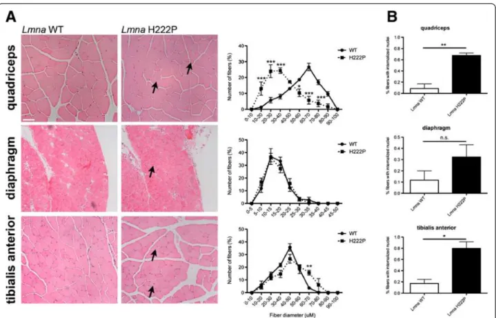
Figure 1Skeletal muscle pathology in LmnaH222P/H222Pmice. (A) Representative micrographs of hematoxylin and eosin-stained sections of quadriceps, diaphragm, and tibialis anterior muscles from 20-week-old male LmnaH222P/H222Pmice (Lmna H222P). Similar sections from wild-type mice (Lmna WT) are shown for comparison. Scale bar = 50 mm. Arrows indicate internalized nuclei. To the right of each pair of micrographs, quantitative analyses of muscle fiber diameter (Feret’s diameter) are shown for wild-type (circles, sold line) and LmnaH222P/H222Pmice (squares, dashed line). Values are means ± SEM for n = 3 mice per group; **P <0.005, ***P <0.0005. (B) Bar graphs showing percentages of fibers in specified muscle groups with internalized nuclei. Values shown are means ± SEM for between 500 and 1,000 nuclei analyzed in tissue samples 3 per group; *P <0.05, **P <0.005, n.s. not significant.
Muchir et al. Skeletal Muscle 2013, 3:17 Page 3 of 10
Serum biochemistry
Serum was separated from mouse blood and stored at -80°C
for 3 to 9 months until analyzed. Creatine phosphokinase
(CPK) and aspartate aminotransferase (AST) activities were
measured using an Analyst III Analyzer (Hemagen
Diagnos-tics) in the Comparative Pathology Laboratory at Columbia
University Medical Center. CPK and AST activities have
been shown to be stable in rodent serum stored for up to
360 days at -70°C [31].
Limb grip strength measurements
Lmna
H222P/H222Pmice treated with DMSO or selumetinib
were subjected to limb grip strength testing using a
hori-zontally positioned grip strength meter (Bioseb). Mice
were lowered by the tail towards the grid on the apparatus.
Upon grasping the grid with their limbs, mice were pulled
backward in the horizontal plane. The procedure was
re-peated consecutively three times and the peak tension of
the three pulls was recorded as the grip strength value.
Each animal was subjected to a total of two serial trials of
three pulls each with 20 s of rest in between.
Statistics
Values for real-time quantitative RT-PCR, scanned
im-munoblots, internalized nuclei, serum CPK and AST
ac-tivities, and grip strength were compared using an
unpaired Student t-tests. Values for Feret’s diameter
were compared using two-way ANOVA. Statistical
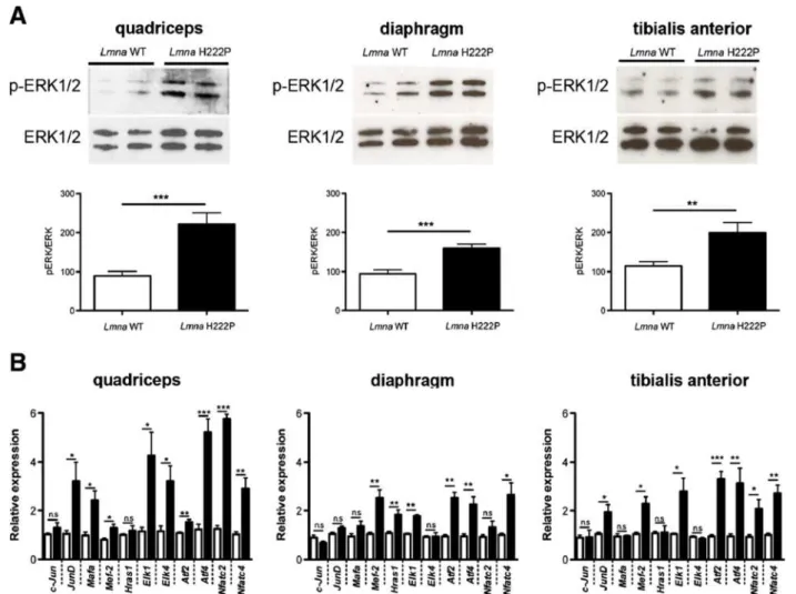
Figure 2Increased ERK1/2 activity in skeletal muscle of LmnaH222P/H222Pmice. (A) Immunoblots showing phosphorylated ERK1/2 (p-ERK1/2) and total ERK1/2 in protein extracts from quadriceps, diaphragm, and tibialis anterior muscles of 20-week-old male wild-type (Lmna WT) and LmnaH222P/H222P(Lmna H222P) mice. Each lane contains protein extracts from a different mouse. The bar graph shows means ± SEM values of pERK1/2 normalized to total ERK1/2 from scanned band densities of five immunoblots from n = 5 different mice per group. **P <0.005, ***P <0.0005. (B) Differential expression of 11 selected genes in the ERK1/2 signaling pathway analyzed using real-time quantitative RT-PCR in quadriceps, diaphragm, and tibialis anterior muscles of 20-week-old male wild-type and LmnaH222P/H222Pmice. White bars show relative RNA expression levels in skeletal muscles from wild-type mice Lmna+/+mice and black bars in skeletal muscles from LmnaH222P/H222Pmice. Values are means ± SEM for n = 5 mice per group; the real-time quantitative RT-PCR was performed in triplicate on each different RNA sample. *P <0.05, **P <0.005, ***P <0.0005, n.s. not significant.
analyses were performed using Prism (GraphPad
Software).
Results and discussion
Dystrophic skeletal muscle pathology in Lmna
H222P/H222Pmice
Arimura et al. [23] previously reported progressive
dys-trophic changes in skeletal muscle starting at 16 weeks male
Lmna
H222P/H222Pmice. Their non-quantitative
histopatho-logical analysis included descriptions of a wide variation in
fiber size, an increased number of atrophic, hypertrophic,
and lobulated fibers, some regenerative fibers and a
men-tion that some fibers had internalized nuclei. We therefore
carefully quantified myofiber diameters and internalized
nuclei in histological sections of quadriceps, diaphragm,
and tibialis anterior muscle of male wild-type and
Lmna
H222P/H222Pmice at 20 weeks of age. Compared to
wild-type mice, quadriceps and tibialis anterior from the
Lmna
H222P/H222Pmice exhibited a wider variation in fiber
size (Figure 1A). In quadriceps, there was a clear shift
to-wards smaller fiber diameters, consistent with the
pres-ence of greater numbers of atrophic and regenerative
fibers. Both of these muscle groups also had an increased
percentage of fibers with internal nuclei, which is observed
during regeneration (Figure 1A,B). At this age, however,
diaphragm did not show significant differences between
Lmna
H222P/H222Pand wild-type mice (Figure 1A,B).
Abnormal ERK1/2 signaling in skeletal muscle of
Lmna
H222P/H222Pmice
Hearts of Lmna
H222P/H222Pmice and human subjects
with autosomal EDMD have increased activity of ERK1/
2, which likely plays a role in pathogenesis of
cardiomy-opathy [24-27]. We hypothesized that a similar increased
activation of this signaling pathway occurs in skeletal
muscle. We therefore examined ERK1/2 activity in
skel-etal muscle from 20-week-old male Lmna
H222P/H222Pmice. Immunoblotting with antibody against
phosphory-lated (activated) ERK1/2 demonstrated a two-fold
in-crease in activity in quadriceps, diaphragm, and tibialis
anterior of Lmna
H222P/H222Pmice compared to wild type
mice (Figure 2A). We then used quantitative real-time
PCR to measure expression of downstream ERK1/2
tar-get genes, several of which are members of the ETS
fam-ily of transcription factors that are phosphorylated by
ERK1/2 and positively autoregulate their transcriptional
activity [24,32,33]. Of 11 targets genes assessed, we
detected significantly increased expression of mRNAs
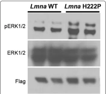
Figure 3Cultured myoblasts expressing H222P lamin A have greater ERK1/2 activity after osmotic shock than those expressing wild-type lamin A. One hour after osmotic shock with D-sorbitol, protein extracts from C2C12 cells stably expressing flag-tagged wild-type lamin A (Lmna WT) and flag-flag-tagged H222P lamin A (Lmna H222P) were analyzed by immunoblotting. Blots were probed with antibodies against phosphorylated ERK1/2 (pERK1/2), total ERK1/2 (ERK1/2) and Flag. The immunoblot shown is representative of three separately performed experiments.
Figure 4Increased ERK1/2 activity in skeletal muscle of LmnaH222P/H222Pmice at 16 weeks of age. Immunoblot showing phosphorylated ERK1/2 (p-ERK1/2) and total ERK1/2 in protein extracts from quadriceps of 16-week-old male wild-type (Lmna WT) and LmnaH222P/H222P(Lmna H222P) mice. Each lane contains protein extracts from a different mouse. The bar graph shows means ± SEM values of pERK1/2 normalized to total ERK1/2 from scanned band densities of three immunoblots from n = 3 different mice per group. *P <0.05.
Muchir et al. Skeletal Muscle 2013, 3:17 Page 5 of 10
for nine in quadriceps, six in diaphragm, and seven in
tibi-alis anterior of Lmna
H222P/H222Pmice compared to
wild-type controls (Figure 2B). Among these, Mef-2, Elk1, Atf2,
Atf4, and Nfatc-4 showed significantly increased
expres-sion in the three skeletal muscles examined. These data
demonstrate that ERK1/2 is hyperactivated in the skeletal
muscles of Lmna
H222P/H222Pmice. Increased ERK1/2
acti-vation in diaphragm at an age before there is any
detect-able histological abnormalities is consistent with its
increased activity in heart prior to the onset of detectable
pathological signs of cardiomyopathy [24]. This suggests
that increased ERK1/2 signaling is involved in the
patho-genesis of dystrophic skeletal muscle pathology.
Stress-induced activation of ERK1/2 in cultured myoblasts
stably expressing H222P lamin A
We have previously shown that transient transfection of
C2C12 mouse myoblasts with cDNA encoding H222P
prelamin A or other variants associated with striated
muscle disease have increased ERK1/2 activity compared
to those transfected with a cDNA encoding wild-type
prelamin A [24]. However, stably transfected C2C12 cells
expressing H222P lamin A do not have increased ERK1/
2 activity at baseline but do after glucose depravation or
treatment with 5-aminoimidazole-4-carboxyamide
ribo-nucleoside [28]. This led us to hypothesize that
physio-logical stress, such as that associated with manipulations
necessary for transient transfection or induced by altered
energy metabolism, is necessary to increase ERK1/2
ac-tivity in myoblasts expressing lamin A variants. We
fur-ther tested this hypothesis by subjecting the same cells
stably expressing lamin A H222P that do not have
base-line elevation in ERK1/2 [28] to osmotic shock. One
hour after an osmotic shock with 600 mM D-sorbitol,
cells expressing flag-tagged H222P lamin A had a greater
activity of ERK1/2 compared to those expressing
flag-tagged wild-type lamin A (Figure 3). This result provided
additional support for a model in which alterations in
the nuclear lamina associated with striated muscle
dis-ease lead to abnormalities in the activities of cellular
stress-responsive signaling pathways [24,34,35]. The
re-quirement of a stress to hyperactivate ERK1/2 in cells
expressing the H222P lamin A may also at least in part
explain why striated muscle, a tissue repeatedly under
mechanical strain, is preferentially affected by LMNA
mutations generating certain A-type lamin variants.
Blocking ERK1/2 activity with selumetinib has beneficial
effects on skeletal muscle in Lmna
H222P/H222Pmice
Given the enhanced ERK1/2 activity in skeletal muscle
of Lmna
H222P/H222Pmice that develop muscular
dys-trophy, we hypothesized that it may contribute to
path-ology. To test this hypothesis, we set up experiments to
determine if inhibiting ERK1/2 signaling would prevent
the progression of muscular dystrophy. At 16 weeks of
age, ERK1/2 activity was elevated in quadriceps muscle
of Lmna
H222P/H222Pmice compared to wild-type mice, as
assessed by immunoblotting with antibody against
phos-phorylated kinase (Figure 4).
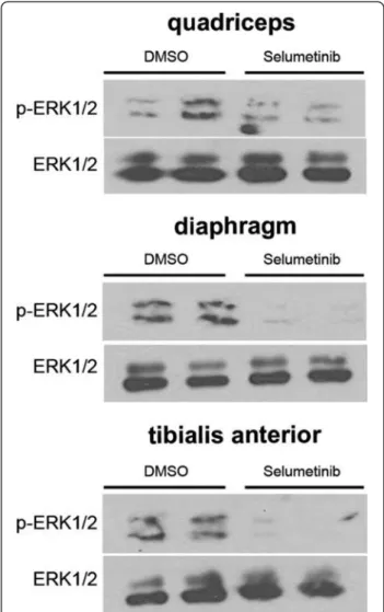
We administered the MEK1/2 inhibitor selumetinib
to male Lmna
H222P/H222Pmice by giving daily
intraper-itoneal injections (1 mg/kg) starting at 16 weeks of age.
After 4 weeks of treatment, the mice had reduced
phosphorylated ERK1/2 in quadriceps, tibialis anterior,
and diaphragm compared to placebo-treated mice.
This demonstrated that systemically administered
selumetinib inhibited ERK1/2 signaling in skeletal
muscle (Figure 5).
Figure 5Selumetinib inhibits ERK1/2 phosphorylation in skeletal muscles from LmnaH222P/H222Pmice. Representative immunoblots using antibodies against phosphorylated ERK1/2 (pERK1/2) and total ERK1/2 (ERK1/2) to probe proteins extracts from quadriceps, diaphragm, and tibialis anterior from 20-week-old male LmnaH222P/H222Pmice treated with selumetinib or DMSO for 4 weeks. The immunoblot shown is representative of three separately performed experiments.
Figure 6 (See legend on next page.)
Muchir et al. Skeletal Muscle 2013, 3:17 Page 7 of 10
Following 4 weeks of treatment with selumetinib, there
was significantly reduced expression of embryonic
my-osin heavy chain (Myh3) mRNA in quadriceps,
dia-phragm, and tibialis anterior of Lmna
H222P/H222Pmice
(Figure 6A). This represented a partial reversal of
embry-onic myosin expression that typically occurs in dystrophic
muscle [36,37]. While quadriceps from DMSO-treated
mice had 0.52% fibers (4/772 from three mice) with
inter-nalized nuclei (Figure 6A, arrows), there were none
detected in 571 fibers from three mice in the
selumetinib-treated mice (and 1/604 fibers from three wild-type mice as
shown in Figure 1B). DMSO treatment did not impact
myofiber diameter compared to untreated Lmna
H222P/H222Pmice; however, mice treated with selumetinib had a greater
myofiber diameter in quadriceps compared to those treated
with DMSO (Figure 6B). Between 16 and 20 weeks of age,
there was a significant increase in serum CPK activity in
Lmna
H222P/H222Pmice treated with DMSO; however, CPK
activity did not significantly increase in the mice that
re-ceived selumetinib and at 20 weeks it was significantly
lower than in those that received DMSO (Figure 6C).
Mean serum AST activity was also significantly lower in
the selumetinib-treated mice compared to the
placebo-treated mice at 20 weeks of age (data not shown). To
de-termine if selumetinib improved skeletal muscle function
in Lmna
H222P/H222Pmice, we evaluated limb grip strength.
At 20 weeks of age, mean grip strength was significantly
greater in selumetinib-treated Lmna
H222P/H222Pmice than
in DMSO-treated mice (Figure 6D). Hence, selumetinib
improved skeletal muscle dystrophic pathology and
im-proved function in Lmna
H222P/H222Pmice.
Conclusions
We have shown increased activity of ERK1/2 in skeletal
muscle of the Lmna
H222P/H222Pmouse model of
auto-somal EDMD and that blocking its activity ameliorates
pathology. These results are in accordance with a
grow-ing body of research providgrow-ing evidence that alterations
in various cellular signaling pathways, including ERK1/2,
are involved in the pathogenesis of muscular dystrophy
[38]. In addition to autosomal EDMD, ERK1/2 has been
implicated as contributing to skeletal or cardiac muscle
pathology in mdx [39-41], γ-sarcoglycan-deficient [42,43],
and Lama2
Dy-w[44] mice, respective small animal models
of Duchenne, limb girdle type 2C, and a form of congenital
muscular dystrophy. ERK1/2 activity is also abnormally
in-creased in hearts of mice with emerin deficiency, which is
the genetic alteration in X-linked EDMD [45].
Blocking increased ERK1/2 signaling activity with
selumetinib had beneficial effects on skeletal muscle
func-tion in Lmna
H222P/H222Pmice. Previously, we obtained
similar results with respect to the cardiomyopathy that
oc-curs in these mice [24-27]. In a human clinical trial,
selumetinib has been reported to promote muscle gain in
patients with cholangiocarcinoma [46]. As oral selumetinib
and other orally bioavailable MEK1/2 inhibitors with
en-couraging safety profiles are currently in clinical
develop-ment for other indications [47,48], pilot trials in patients
with EDMD and possibly other muscular dystrophies
should be considered.
Abbreviations
AST:Aspartate aminotransferase; CPK: Creatine phosphokinase; DMSO: Dimethyl sulfoxide; EDMD: Emery-Dreifuss muscular dystrophy; ERK: Extracellular signal-regulated kinase; LMNA: Lamin A/C gene; MEK: Mitogen-activated protein kinase/extracellular signal-regulated kinase kinase; RT-PCR: Reverse transcription-polymerase chain reaction.
Competing interests
AM and HJW are inventors on a pending patent application (PCT/US09/ 42614) on methods for treating and/or preventing cardiomyopathies by ERK and JNK inhibition filed by the Trustees of Columbia University in the City of New York.
Authors’ contributions
AM conceived of the study, bred mice, treated mice with drugs, carried out experiments measuring ERK1/2 activity in mouse tissue and cells, assessed skeletal muscle pathology and grip strength in mice, and drafted the manuscript. YJK carried out experiments on measuring ERK1/2 activity in mouse tissue and cells and assessing skeletal muscle pathology in mice. SAR assisted with isolating skeletal muscle form mice and participated in experiments measuring ERK1/2 activity in mouse tissue. WW bred mice, drew blood from mice, and assisted in treating mice with drugs. JCC generated stable cell lines expressing H222P and wild type lamin A. HJW helped conceive the study, supervised and coordinated all of the research, and wrote the final manuscript. All of the authors read and approved the final manuscript.
(See figure on previous page.)
Figure 6Selumetinib from 16 to 20 weeks of age improves skeletal muscle pathology and function in LmnaH222P/H222Pmice. (A) Expression of Myh3 in LmnaH222P/H222Pmice measured using real-time quantitative RT-PCR. White bars show relative RNA expression levels in skeletal muscles of DMSO-treated (white bars) and selumetinib-treated (black bars) mice. Values are means ± SEM for n = 5 mice per group; the real time RT-PCR was performed in triplicate with the different RNA sample; *P <0.05. (B) Representative micrographs of hematoxylin and eosin-stained sections of quadriceps from 20-week-old male LmnaH222P/H222Pmice (Lmna H222P) treated for 4 weeks with DMSO or selumetinib. Scale bar = 50 mm. Arrows indicate internalized nuclei. To the right of the micrographs, quantitative analyses of muscle fiber diameter (Feret’s diameter) are shown for mice treated with DMSO (circles, solid line) and selumetinib (squares, sold line). Values are means ± SEM for n = 3 mice per group; **P <0.005, ***P <0.0005. (C) Serum CPK activities in LmnaH222P/H222Pmice at 16 weeks (16 W) and 20 weeks (20 W) of age that were treated with DMSO (white bars) or selumetinib (black bars). Values are means ± SEM for n = 7 DMSO-treated mice and n = 15 selumetinib-treated mice; **P <0.005, n.s. not significant,#P <0.05. (D) Grip strength (force) in Newtons (N) in LmnaH222P/H222Pmice at 20 weeks of age that were treated with DMSO (circles) (n=6) or selumetinib (squares) (n=8). Each circle and square represents a measurement from an individual mouse; the longer horizontal bar are means and the shorter horizontal bars ± SEM; **P <0.05.
Acknowledgements
We thank Dr. Gisèle Bonne (Institut de Myologie) for providing LmnaH222P/H222P mice and Dr. Michio Hirano (Columbia University) for assistance with grip strength testing. This work was supported by grants from the National Institutes of Health/National Institute of Arthritis and Musculoskeletal and Skin Diseases (AR048997) and Los Angeles Thoracic and Cardiovascular Foundation to HJW and a grant from the Association Franaçise Contre les Myopathies to AM. Author details
1Department of Medicine, College of Physicians and Surgeons, Columbia
University, 630 West 168th Street, New York, NY 10032, USA.2Department of
Pathology and Cell Biology, College of Physicians and Surgeons, Columbia University, New York, NY, USA.3Current address: Therapie des maladies du
muscle strie, Institut de Myologie UM76 - UPMC Univ. Paris 6, U974 – Inserm, UMR7215 - CNRS G.H., F-75651 Paris, Cedex 13, France.4Current address:
Stony Brook University School of Medicine, Stony Brook, NY 11794, USA.
5Current address: Tufts University School of Medicine, Boston, MA 02111,
USA.
Received: 3 January 2013 Accepted: 22 April 2013 Published: 1 July 2013
References
1. Emery AEH, Dreifuss FE: Unusual type of benign X-linked muscular dystrophy. J Neurol Neurosurg Psychiatry 1966, 29:338–342. 2. Emery AE: X-linked muscular dystrophy with early contractures and
cardiomyopathy (Emery-Dreifuss type). Clin Genet 1987, 32:360–367. 3. Waters DD, Nutter DO, Hopkins LC, Dorney ER: Cardiac features of an
unusual X-linked humeroperoneal neuromuscular disease. N Engl J Med 1975, 293:1017–1022.
4. Bécane HM, Bonne G, Varnous S, Muchir A, Ortega V, Hammouda EH, Urtizberea JA, Lavergne T, Fardeau M, Eymard B, Weber S, Schwartz K, Duboc D: High incidence of sudden death of conduction system and myocardial disease due to lamins A/C gene mutation. Pacing Clin Electrophysiol 2000, 23:1661–1666.
5. van Berlo JH, de Voogt WG, van der Kooi AJ, van Tintelen JP, Bonne G, Yaou RB, Duboc D, Rossenbacker T, Heidbüchel H, de Visser M, Crijns HJ, Pinto YM: Meta-analysis of clinical characteristics of 299 carriers of LMNA gene mutations: do lamin A/C mutations portend a high risk of sudden death? J Mol Med 2005, 83:79–83.
6. Bione S, Maestrini E, Rivella S, Mancini M, Regis S, Romeo G, Toniolo D: Identification of a novel X-linked gene responsible for Emery-Dreifuss muscular dystrophy. Nat Genet 1994, 8:323–327.
7. Nagano A, Koga R, Ogawa M, Kurano Y, Kawada J, Okada R, Hayashi YK, Tsukahara T, Arahata K: Emerin deficiency at the nuclear membrane in patients with Emery-Dreifuss muscular dystrophy. Nat Genet 1996, 12:254–259.
8. Manilal S, Nguyen TM, Sewry CA, Morris GE: The Emery-Dreifuss muscular dystrophy protein, emerin, is a nuclear membrane protein. Hum Mol Genet 1996, 5:801–808.
9. Bonne G, Di Barletta MR, Varnous S, Bécane HM, Hammouda EH, Merlini L, Muntoni F, Greenberg CR, Gary F, Urtizberea JA, Duboc D, Fardeau M, Toniolo D, Schwartz K: Mutations in the gene encoding lamin A/C cause autosomal dominant Emery-Dreifuss muscular dystrophy. Nat Genet 1999, 21:285–288.
10. Raffaele Di Barletta M, Ricci E, Galluzzi G, Tonali P, Mora M, Morandi L, Romorini A, Voit T, Orstavik KH, Merlini L, Trevisan C, Biancalana V, Housmanowa-Petrusewicz I, Bione S, Ricotti R, Schwartz K, Bonne G, Toniolo D: Different mutations in the LMNA gene cause autosomal dominant and autosomal recessive Emery-Dreifuss muscular dystrophy. Am J Hum Genet 2000, 66:1407–1412.
11. McKeon FD, Kirschner MW, Caput D: Homologies in both primary and secondary structure between nuclear envelope and intermediate filament proteins. Nature 1986, 319:463–468.
12. Fisher DZ, Chaudhary N, Blobel G: cDNA sequencing of nuclear lamins A and C reveals primary and secondary structural homology to
intermediate filament proteins. Proc Natl Acad Sci USA 1986, 83:6450–6454. 13. Aebi U, Cohn J, Buhle L, Gerace L: The nuclear lamina is a meshwork of
intermediate-type filaments. Nature 1986, 323:560–564.
14. Lin F, Worman HJ: Structural organization of the human gene encoding nuclear lamin A and nuclear lamin C. J Biol Chem 1993, 268:16321–16326.
15. Fatkin D, MacRae C, Sasaki T, Wolff MR, Porcu M, Frenneaux M, Atherton J, Vidaillet HJ Jr, Spudich S, De Girolami U, Seidman JG, Seidman C, Muntoni F, Muehle G, Johnson W, McDonough B: Missense mutations in the rod domain of the lamin A/C gene as causes of dilated cardiomyopathy and conduction-system disease. N Engl J Med 1999, 341:1715–1724. 16. Muchir A, Bonne G, van der Kooi AJ, van Meegen M, Baas F, Bolhuis PA, de
Visser M, Schwartz K: Identification of mutations in the gene encoding lamins A/C in autosomal dominant limb girdle muscular dystrophy with atrioventricular conduction disturbances (LGMD1B). Hum Mol Genet 2000, 9:1453–1459.
17. Bonne G, Mercuri E, Muchir A, Urtizberea A, Becane HM, Recan D, Merlini L, Wehnert M, Boor R, Reuner U, Vorgerd M, Wicklein EM, Eymard B, Duboc D, Penisson-Besnier I, Cuisset JM, Ferrer X, Desguerre I, Lacombe D, Bushby K, Pollitt C, Toniolo D, Fardeau M, Schwartz K, Muntoni F: Clinical and molecular genetic spectrum of autosomal dominant Emery-Dreifuss muscular dystrophy due to mutations of the lamin A/C gene. Ann Neurol 2000, 48:170–180.
18. Brodsky GL, Muntoni F, Miocic S, Sinagra G, Sewry C, Mestroni L: Lamin A/C gene mutation associated with dilated cardiomyopathy with variable skeletal muscle involvement. Circulation 2000, 101:473–476. 19. Astejada MN, Goto K, Nagano A, Ura S, Noguchi S, Nonaka I, Nishino I,
Hayashi YK: Emerinopathy and laminopathy clinical, pathological and molecular features of muscular dystrophy with nuclear envelopathy in Japan. Acta Myol 2007, 26:159–164.
20. Ura S, Hayashi YK, Goto K, Astejada MN, Murakami T, Nagato M, Ohta S, Daimon Y, Takekawa H, Hirata K, Nonaka I, Noguchi S, Nishino I: Limb-girdle muscular dystrophy due to emerin gene mutations. Arch Neurol 2007, 64:1038–1041.
21. Lu JT, Muchir A, Nagy PL, Worman HJ: LMNA cardiomyopathy: cell biology and genetics meet clinical medicine. Dis Model Mech 2011, 4:562–568. 22. Worman HJ, Fong LG, Muchir A, Young SG: Laminopathies and the long strange
trip from basic cell biology to therapy. J Clin Invest 2009, 119:1825–1836. 23. Arimura T, Helbling-Leclerc A, Massart C, Varnous S, Niel F, Lacène E, Fromes
Y, Toussaint M, Mura AM, Keller DI, Amthor H, Isnard R, Malissen M, Schwartz K, Bonne G: Mouse model carrying H222P-Lmna mutation develops muscular dystrophy and dilated cardiomyopathy similar to human striated muscle laminopathies. Hum Mol Genet 2005, 14:155–169. 24. Muchir A, Pavlidis P, Decostre V, Herron AJ, Arimura T, Bonne G, Worman HJ:
Activation of MAPK pathways links LMNA mutations to cardiomyopathy in Emery-Dreifuss muscular dystrophy. J Clin Invest 2007, 117:1282–1293. 25. Muchir A, Shan J, Bonne G, Lehnart SE, Worman HJ: Inhibition of
extracellular signal-regulated kinase signaling to prevent cardiomyopathy caused by mutation in the gene encoding A-type lamins. Hum Mol Genet 2009, 18:241–247.
26. Wu W, Muchir A, Shan J, Bonne G, Worman HJ: Mitogen-activated protein kinase inhibitors improve heart function and prevent fibrosis in cardiomyopathy caused by mutation in lamin A/C gene. Circulation 2011, 123:53–61. 27. Muchir A, Reilly SA, Wu W, Iwata S, Homma S, Bonne G, Worman HJ:
Treatment with selumetinib preserves cardiac function and improves survival in cardiomyopathy caused by mutation in the lamin A/C gene. Cardiovasc Res 2012, 93:311–319.
28. Choi JC, Wu W, Muchir A, Iwata S, Homma S, Worman HJ: Dual specificity phosphatase 4 mediates cardiomyopathy caused by lamin A/C (LMNA) gene mutation. J Biol Chem 2012, 287:40513–40524.
29. Koressaar T, Remm M: Enhancements and modifications of primer design program Primer3. Bioinformatics 2007, 23:1289–1291.
30. Ponchel F, Toomes C, Bransfield K, Leong FT, Douglas SH, Field SL, Bell SM, Combaret V, Puisieux A, Mighell AJ, Robinson PA, Inglehearn CF, Isaacs JD, Markham AF: Real-time PCR based on SYBR-Green I fluorescence: an alternative to the TaqMan assay for a relative quantification of gene rearrangements, gene amplifications and micro gene deletions. BMC Biotechnol 2003, 3:18.
31. Cray C, Rodriguez M, Zaias J, Altman NH: Effects of storage temperature and time on clinical biochemical parameters from rat serum. J Am Assoc Lab Anim Sci 2009, 48:202–204.
32. Seth A, Papas TS: The c-ets-1 proto-oncogene has oncogenic activity and is positively autoregulated. Oncogene 1990, 5:1761–1767.
33. Tohgo A, Pierce KL, Choy EW, Lefkowitz RJ, Luttrell LM: Beta-Arrestin scaffolding of the ERK cascade enhances cytosolic ERK activity but inhibits ERK-mediated transcription following angiotensin AT1a receptor stimulation. J Biol Chem 2002, 277:9429–9436.
Muchir et al. Skeletal Muscle 2013, 3:17 Page 9 of 10
34. Lammerding J, Schulze PC, Takahashi T, Kozlov S, Sullivan T, Kamm RD, Stewart CL, Lee RT: Lamin A/C deficiency causes defective nuclear mechanics and mechanotransduction. J Clin Invest 2004, 113:370–378. 35. Dauer WT, Worman HJ: The nuclear envelope as a signaling node in
development and disease. Dev Cell 2009, 17:626–638.
36. Chen YW, Zhao P, Borup R, Hoffman EP: Expression profiling in the muscular dystrophies: identification of novel aspects of molecular pathophysiology. J Cell Biol 2000, 151:1321–1336.
37. Haslett JN, Sanoudou D, Kho AT, Bennett RR, Greenberg SA, Kohane IS, Beggs AH, Kunkel LM: Gene expression comparison of biopsies from Duchenne muscular dystrophy (DMD) and normal skeletal muscle. Proc Natl Acad Sci USA 2002, 99:15000–15005.
38. Burks TN, Cohn RD: Role of TGF-β signaling in inherited and acquired myopathies. Skeletal Muscle 2011, 1:19.
39. Kumar A, Khandelwal N, Malya R, Reid MB, Boriek AM: Loss of dystrophin causes aberrant mechanotransduction in skeletal muscle fibers. FASEB J 2004, 18:102–113.
40. Lang JM, Esser KA, Dupont-Versteegden EE: Altered activity of signaling pathways in diaphragm and tibialis anterior muscle of dystrophic mice. Exp Biol Med 2004, 229:503–511.
41. Smythe GM, Forwood JK: Altered mitogen-activated protein kinase signaling in dystrophic (mdx) muscle. Muscle Nerve 2012, 46:374–383. 42. Griffin MA, Feng H, Tewari M, Acosta P, Kawana M, Sweeney HL, Disher DE:
γ-sarcoglycan deficiency increases cell contractility, apoptosis and MAPK pathway activation but does not affect adhesion. J Cell Sci 2005, 118:1405–1416.
43. Barton ER: Impact of sarcoglycan complex on mechanical signal transduction in murine skeletal muscle. Am J Physiol Cell Physiol 2006, 290:C411–C419.
44. Kumar A, Yamauchi J, Girgenrath T, Girgenrath M: Muscle-specific expression of insulin-like growth factor 1 improves outcome in Lama2Dy-w mice, a model for congenital muscular dystrophy type 1A. Hum Mol Genet 2011, 20:2333–2343.
45. Muchir A, Pavlidis P, Bonne G, Hayashi YK, Worman HJ: Activation of MAPK in hearts of EMD null mice: similarities between mouse models of X-linked and autosomal dominant Emery Dreifuss muscular dystrophy. Hum Mol Genet 2007, 16:1884–1895.
46. Prado CM, Bekaii-Saab T, Doyle LA, Shrestha S, Ghosh S, Baracos VE, Sawyer MB: Skeletal muscle anabolism is a side effect of therapy with the MEK inhibitor: selumetinib in patients with cholangiocarcinoma. Br J Cancer 2012, 106:1583–15836.
47. Chapman MS, Miner JN: Novel mitogen-activated protein kinase kinase inhibitors. Expert Opin Investig Drugs 2011, 20:209–220.
48. Trujillo JI: MEK inhibitors: a patent review 2008–2010. Expert Opin Ther Pat 2011, 21:1045–1069.
doi:10.1186/2044-5040-3-17
Cite this article as: Muchir et al.: Inhibition of extracellular signal-regulated kinase 1/2 signaling has beneficial effects on skeletal muscle in a mouse model of Emery-Dreifuss muscular dystrophy caused by lamin A/C gene mutation. Skeletal Muscle 2013 3:17.
Submit your next manuscript to BioMed Central
and take full advantage of:
• Convenient online submission
• Thorough peer review
• No space constraints or color figure charges
• Immediate publication on acceptance
• Inclusion in PubMed, CAS, Scopus and Google Scholar
• Research which is freely available for redistribution
Submit your manuscript at www.biomedcentral.com/submit