HAL Id: hal-02898087
https://hal.sorbonne-universite.fr/hal-02898087
Submitted on 13 Jul 2020
HAL is a multi-disciplinary open access
archive for the deposit and dissemination of sci-entific research documents, whether they are pub-lished or not. The documents may come from teaching and research institutions in France or abroad, or from public or private research centers.
L’archive ouverte pluridisciplinaire HAL, est destinée au dépôt et à la diffusion de documents scientifiques de niveau recherche, publiés ou non, émanant des établissements d’enseignement et de recherche français ou étrangers, des laboratoires publics ou privés.
140 Years of the Leçons sur l’histologie du système
nerveux: the pioneering description of the nodes of
Ranvier
Otto Jesus Hernandez Fustes, Cláudia Suemi Kamoi Kay, Paulo Lorenzoni,
Renata Dal-Pra Ducci, Jean-Gaël Barbara, Lineu Werneck, Rosana Herminia
Scola
To cite this version:
Otto Jesus Hernandez Fustes, Cláudia Suemi Kamoi Kay, Paulo Lorenzoni, Renata Dal-Pra Ducci, Jean-Gaël Barbara, et al.. 140 Years of the Leçons sur l’histologie du système nerveux: the pioneering description of the nodes of Ranvier. Arquivos de Neuro-Psiquiatria, Academia Brasileira de Neurologia - ABNEURO, 2019, 77 (10), pp.749-751. �10.1590/0004-282X20190119�. �hal-02898087�
749 https://doi.org/10.1590/0004-282X20190119
HISTORICAL NOTE
140 Years of the Leçons sur l’histologie du
système nerveux: the pioneering description
of the nodes of Ranvier
140 Anos da Leçons sur l’histologie du système nerveux: a descrição pioneira dos nodos
de Ranvier
Otto Jesus HERNANDEZ FUSTES1, Cláudia Suemi Kamoi KAY1, Paulo José LORENZONI1, Renata Dal-Pra
DUCCI1, Jean-Gaël BARBARA2, Lineu Cesar WERNECK1, Rosana Herminia SCOLA1
In recent years, significant advances have been made in the knowledge of the pathophysiology of immune-mediated neuropathies, especially in the role of the nodal region, giving rise to the so-called nodopathies1.
These nodopathies may point directly to the site of nerve injury; circumventing the apparent paradox that axonal lesions may be reversible and have a good prognosis. Some patients may be classified as having a demyelinating neu-ropathy, and others as having an axonal neuropathy; the potential reversibility in neuropathies traditionally thought to be characterized only by axonal degeneration opens a therapeutic window and stimulates research for timely tar-geted treatments1.
This year marks 140 years on from the first description of the nodal region by French physician, physiologist and anato-mist, Louis Ranvier, a student of Claude Bernard.
LOUIS ANTOINE RANVIER
Louis Antoine Ranvier was born in Lyon, in 1835, where he took up medical studies at the École Préparatoire de
Médecine et de Pharmacie, then moved to Paris in 1860,
after he succeeded in the highly competitive examination for internship at Parisian hospitals. During his medical training, Ranvier became acquainted with normal and pathological anatomy, and soon turned to microscopy as a means for fur-ther studies on tissues2,3,4,5.
Between 1860 and 1865, Cornil and Ranvier devoted much of their time to microscopy. Besides observing tumors and other pathological tissues, Ranvier focused on bone prep-arations, which led him to study cartilage and bone lesions for his medical thesis. By 1865, they had started collaborating on epithelial tumors.
1Universidade Federal do Paraná, Hospital de Clínicas, Departamento de Clínica Médica, Serviço de Neurologia, Serviço de Doenças Neuromusculares,
Curitiba PR, Brasil.
2Neuroscience Paris Seine, Sorbonne Universités, UPMC Université Paris 06, Institut de biologie Paris-Seine, Paris, França.
Otto Jesus Hernández Fustes https://orcid.org/0000-0003-0778-5376; Cláudia Suemi Kamoi Kay https://orcid.org/0000-0003-0173-0809; Paulo José Lorenzoni https://orcid.org/0000-0002-4457-7771; Renata Dal-Prá Ducci https://orcid.org/0000-0002-1673-5074; Jean-Gaël Barbara https://orcid.org/0000-0002-2778-9369; Lineu Cesar Werneck https://orcid.org/0000-0003-1921-1038; Rosana Herminia Scola https://orcid. org/0000-0002-3957-5317
Correspondence: Otto J. H. Fustes; Hospital de Clínicas da UFPR; Rua General Carneiro, 181; 80060-900 Curitiba PR; E-mail: otto.fustes@hc.ufpr.br Conflict of interest: There is no conflict of interest to declare.
Received 16 November 2018; Received in final form 24 June 2019; Accepted 28 June 2019.
ABSTRACT
This paper reviews aspects of the life and work of Professor Louis Ranvier 140 years after the publication of Leçons sur l’histologie du
système nerveux, published in 1878, and shows the importance of the histological description of myelinated fibers of the nodes of Ranvier.
Keywords: Histology; peripheral nervous system diseases; Ranvier’s nodes. RESUMO
Os autores apresentam uma revisão sobre aspectos da vida e obra do Professor Louis Ranvier 140 anos após a publicação de seu livro
Leçons sur l’histologie du système nerveux publicado em 1878 e mostra a importância da descrição histológica nas fibras mielínicas dos
nodos de Ranvier.
750 Arq Neuropsiquiatr 2019;77(10):749-751
From 1866 to 1867, Cornil and Ranvier’s one-semes-ter course in microscopy had no equivalent in France. The course was published in three parts, as an authoritative man-ual, two years later. It was translated into English, with notes and additions both in England and the United States. It rep-resented a well-written and useful modern textbook for med-ical students interested in normal and pathologmed-ical histol-ogy. Their collaboration ended when Ranvier agreed to join Claude Bernard at the Collège de France.
Ranvier was influenced by Virchow’s extension of cellu-lar theory to pathology. In Ranvier’s introductions to studies on cartilage and bone, Virchow’s observations were empha-sized. While Cornil further investigated pathological tissues, Ranvier focused on normal histology. He was not only con-cerned with cell theory, but also, as a student of Bernard, with the development, nutrition and functions of normal tissues.
THE NODE
In 1853, Virchow used the term myelin to refer to the large sheath mass involving some axons. The term oligoden-drocyte was suggested by Pío del Río-Hortega, who observed that this cell had a shorter length and less branching when compared with astrocytes and also suggested that, like the neurolemmocyte, it formed myelin, a theory proven years later by electron microscopy6.
In 1871, from his laboratory in the College of Paris, Ranvier went further, looking for new techniques to visualize “invisible” structures and to be able to explain questions of physiologi-cal type7. The sciatic nerve of frogs, dissociated (nerve trunks)
and fixed in osmic acid, showed visible strangulations that had already been referred to in other works, but it was Ranvier who described them for the first time. We refer to the “Ranvier nodes” as the narrowing observed in the modulated nerve fibers, at intervals of 1 mm, due to the interruption of myelina-tion. But for Ranvier, histology did not end with technique and description, but had also to give way to the study of functions.
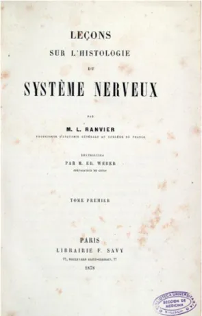
In 1878, he wrote in his Lessons on Histology of the
Nervous System (Figure 1) that myelin acted as an electrical
insulation, and was interrupted at various points along the axon8. These interruptions were therefore called the nodes of
Ranvier (Figure 2). The importance of these nodes was unrav-eled by two research groups during the 1940s: Tasaki and Takeuchi, in 19419; and Huxley and Stämpfli in 194910.
In myelinated fibers, depolarization (due to ion exchange) can only occur in the nodes of Ranvier, where the dielectric envelope (myelin) is interrupted. This mode of impulse propa-gation in myelinated fibers was deciphered in 1925, by Lillie11.
In 1897, Ranvier and Balbiani founded the Archives
d’anatomie microscopique, the first French journal devoted
exclusively to microscopic studies.
Between 1875 and 1890, people like Malassez, Louis de Sinéty, Maurice Debove, Renaut, Babinski, William Nicati,
Figure 1. Cover of the book Leçons sur l’histologie du système
nerveux. Paris, 1878.
Figure 2. A page from Leçons sur l’histologie du système
751
Hernandez Fustes OJ et al. Nodes of Ranvier
Albarran and Suchard, among others trained in Ranvier’s laboratory.
In 1900, Ranvier was isolated from the scientific com-munity and retired to Thélys, where he spent the following 22 years on activities unrelated to science. Ranvier died in Vendranges, Loire, on 22 March 1922, leaving a legacy that transcended time, still serving as an inspiration today.
Ranvier’s contributions were emphasized by Ramon y Cajal: “In my systematic explorations through the realms of microscopic anatomy […] I examined [the Nervous System] eagerly in various animals, guided by the books of Meynert, Huguenin, Luys, Schwalbe and, above all, the incomparable works of Ranvier, of whose ingenious technique I made use with conscientious determination”12.
References
1. Uncini A, Kuwabara S. Nodopathies of the peripheral nerve: an emerging concept. J Neurol Neurosurg Psychiatry. 2015 Nov;86(11):1186-95. https://doi.org/10.1136/jnnp-2014-310097 2. Barbara JG. History of neuroscience: Louis Ranvier (1835-1922).
IBRO History of Neuroscience. 2007 [cited 2018 Mar 10]. Available from: http://ibro.org/wp-content/uploads/2018/07/Ranvier-Louis. pdf
3. Barbara JG. Biological generality and general anatomy from Xavier Bichat to Louis Antoine Ranvier. In: Chemla K, Chorlay R, Rabouin D, editors. The Oxford handbook of generality in mathematics and the sciences. Oxford: Oxford UP; 2016. p. 359-84.
4. Barbara JG. Louis Ranvier (1835-1922): the contribution of microscopy to physiology and the renewal of French general anatomy. J Hist Neurosci. 2007 Oct-Dec;16(4):413-31. https://doi. org/10.1080/09647040600685503
5. Barbara JG. Louis Ranvier, l’anatomie générale microscopique et les recherches sur les cellules nerveuses. In: Barbara JG, Clarac F, editors. Le cerveau au microscope: la neuroanatomie française aux XIXe et XXe siècles. Paris: Hermann; 2017. p. 71-88.
6. Mendes PB, Melo SR. Origem e desenvolvimento da mielina no sistema nervoso central: um estudo de revisão. Rev Saúde Pesq. 2011 Jan-Abr.;4(1):93-9.
7. Ranvier L. Contributions à l’histologie et à la physiologie des nerfs périphériques. Comptes Rendus de l’Académie des Sciences. 1871;73:1168-71.
8. Ranvier ML. Leçons sur l’histologie du système nerveux. Paris: Librairie F. Savy; 1878.
9. Tasaki I, Takeuchi T. Der am Ranvierschen Knoten entstehende Aktionsstrom und seine Bedeutung für die Erregungsleitung. Pflügers Archiv ges Physiol. 1941;244:696-711.
10. Huxley AF, Stämpfli R. Evidence for saltatory conduction in peripheral myelinated nerve fibers. J Physiol. 1949;108(3):315-39. https://doi.org/10.1113/jphysiol.1949.sp004335
11. Lillie RS. Factors affecting transmission and recovery in the passive iron nerve model. J Gen Physiol. 1925 Mar;7(4):473-507. https://doi.org/10.1085/jgp.7.4.473
12. Ramón y Cajal S. Recuerdos de mi vida, historia de mi labor científica. Madrid: Aianza Universidade; 1995.
