Assessment of regional analgesia in clinical
practice and research
Michele Curatolo*, Steen Petersen-Felix* and Lars Arendt-Nielsen† *Department of Anaesthesiology, University Hospital of Berne, Inselspital, Switzerland and †Laboratory for Experimental Pain Research, Centre for Sensory–Motor Interaction, University of Aalborg, Denmark
Assessment of pain and sensory function during regional analgesia contributes to a better understanding of the mechanisms underlying the action of drugs and techniques, and provides information on the effectiveness of regional analgesia in daily practice. Sensory tests only partially mimic clinical pain, mainly because they are artificial and reproduce only a part of the complex experience of pain. Therefore information gained by sensory tests should not be uncritically generalized to clinical pain conditions. Studies using experimental pain models are not in competition with studies performed under clinical conditions, but complement them. In order to mirror clinical pain, experimental models ideally stimulate muscles and viscera, induce peripheral and central sensitization, and evoke temporal and spatial summation. These methods are available, but are underused. Test modalities used in clinical practice have limited validity. In recent years almost no research has been performed to develop better test modalities that are suitable for daily practice.
Introduction
Regional analgesia is routine practice in anaesthesia and pain management. The possibility of providing analgesia at the site of pain, with minimal or no impairment of other functions, is the main rationale behind it. Sur-gical analgesia, postoperative pain management and the diagnosis and treatment of chronic malignant and non-malignant pain are the fields of application of regional analgesia techniques.
Two professional categories are interested in assessing pain and sen-sory function in regional analgesia: the clinician and the researcher. Basically, the clinician would like to know whether intervention will produce a good outcome for the patient. An example of outcome is the absence of pain during orthopaedic surgery performed under epidural block. In this case, assessment of sensory function is expected to provide predictive information on the success of the block and therefore to help Accepted: December 14, 2004 Correspondence to: Dr Michele Curatolo, Department of Anaesthesiology, Division of Pain Therapy Inselspital, 3010 Berne, Switzerland. E-mail: [email protected]
in making decisions on the anaesthetic management. The researcher is interested in investigating the analgesic effect of new drugs before they are used in patients, the combination of different drugs or the mecha-nisms of action of analgesics.
While there is little doubt about the usefulness of assessing the effects of regional analgesia on sensory function and pain, important questions need to be addressed. How can we measure pain? Can we predict a suc-cessful or unsucsuc-cessful block based on preoperative sensory assess-ments? Do experimental pain tests predict the efficacy of new drugs or techniques in the clinical settings?
The main aims of this review are to describe the methods used for sen-sory assessment and to evaluate the relationship between tests and clinical pain. Ultimately, we discuss what tests should be used, what applications are suitable and what information can be or cannot be expected from assessment of pain and sensory function. In this sense, this paper
updates the information provided in a previous review.1
General aspects
Pain and related parameters need to be assessed in all patients with acute and chronic pain. Patients undergoing regional analgesia are not an
exception. Aspects related to pain include psychological evaluation,2
assessment of function and disability3 and quality of life.4 Obviously,
not all measurements need to be made in all patients after regional anal-gesia; the choice depends on the pain condition that is treated and the type of intervention. The assessment of ongoing pain and related factors5 is beyond the scope of this paper.
An extended and more advanced approach than simply assessing ongoing clinical pain is to impose additional standardized stimuli on the patient and evaluate the response under controlled conditions. This approach is termed quantitative sensory testing and allows a better understanding of the mechanisms involved in pain transduction, trans-mission and perception under normal and pathophysiological conditions.
Pain researchers and clinicians have a dream—to be able to measure pain objectively. However, this dream will probably never come true. Pain remains subjective in nature. Furthermore, it is a multidimensional sensory and emotional experience which is unlikely to be completely represented or described by a number. Thus any measurement or set of measurements will provide only partial information about the complex events and mechanisms that are involved in clinical pain. Nevertheless, important information can be gained by pain and sensory assessment, provided that the clinician and the researcher understand the characteristics of the tests that they employ.
Methods for assessing regional analgesia
An ideal test has the following characteristics:1 it delivers reproducible stimuli;
2 it can be applied to all body parts without causing damage; 3 it selectively activates pain receptors;
4 it elicits different and discriminatory pain intensities and qualities; 5 it can induce temporal and spatial summation (see below);
6 it provides a positive correlation between stimulus intensity and pain intensity;
7 it is easy to apply and control in laboratory and clinical settings; 8 it mirrors clinical pain mechanisms.
Unfortunately the ideal test does not exist, and hence the most ade-quate test should be selected based on its characteristics and the specific application.
As pain is a multidimensional perception, the reaction to a single standardized experimental stimulus of a given modality can only represent a very limited fraction of the entire pain experience. Therefore it is man-datory to combine different stimulation and assessment approaches to gain advanced differentiated information about the nociceptive system under normal and pathophysiological conditions.
Stimulus modalities
Stimuli can have phasic properties (short-lasting, milliseconds to few seconds) or tonic properties (long lasting, many seconds to minutes). The various pain induction techniques are summarized in Table 1. It can be seen that most methods have been developed for cutaneous applications and therefore may not reliably mimic clinical pain, which usually involves deeper structures. Only in more recent years have quantitative
experimental methods for the assessment of muscle6 and visceral7 pain
been developed.
Most studies have used electrical stimulation, but this technique is non-selective and bypasses the receptors by depolarizing the afferent nerve fibre. However, bypassing the nociceptors allows the exploration of pain reactivity without the influence of peripheral nociception. Thus investigations using electrical stimulation may be interesting when the excitability of the central nervous system is explored.8
The current perception threshold,9 which is based on the theory that
used for sensory assessment. The frequencies normally used are 5, 250
and 2000 Hz which have been claimed to activate C, Aδ and Aβ fibres,
respectively. However, there is no electrophysiological evidence that this differentiated nerve fibre activation can be achieved in humans.
Lasers are probably the most selective heat stimulators.10 The
advant-age is that the laser radiation can be applied without touching the skin and hence does not contaminate the stimulation by mechanosensitive input. The disadvantage is that lasers are usually difficult to operate, expensive and not very mobile.
Chemical stimuli are difficult to repeat over time. However, some of them (e.g. capsaicin) induce peripheral and central sensitization. Therefore
Table 1 Pain induction modalities
Electrical
Transcutaneous (through contact electrodes)
Intracutaneous (through electrodes that penetrate the skin) Tooth pulp
Intramuscular (through stimulating needles)
Transmucosal (through contact electrodes: oral or visceral stimulation)
Heat
Radiant heat (laser, light, infrared) Contact thermode (skin stimulation)
Circulating hot water (possible stimulation of skin, muscle or viscera)
Cold
Cold pressor test (immersion of hand in ice water) Contact thermode (skin stimulation)
Evaporation of gas (e.g. ether applied to the skin) Menthol (applied to the skin)
Circulating cold water (possible stimulation of muscle or viscera)
Mechanical
Brushing/stroking
Pinprick (e.g. with needle or von Frey hair) Pinch
Impact stimuli
Pressure (generally with a pressure algometer) Distension (e.g. using a balloon to distend viscera)
Ischaemia
Tourniquet (applied to arm or leg to induce ischaemic pain and pressure pain) Exercise (e.g. lifting a weight repeatedly or exercising with a hand-grip trainer)
Chemical
Capsaicin (activation of nociceptor of skin, muscle or viscera) Mustard oil (induction of inflammatory reaction of the skin) Melittin (active substance of bee venom applied to the skin) Hypertonic saline (5% solution injected into muscle)
Bradykinin, serotonin, substance P and other algogenic substances (injected into muscle) Carbon dioxide (stimulation of the nasal mucosa)
Glutamate (injected into muscle to induce hyperalgesia) Nerve growth factor (injected into muscle)
Glycerol (applied to viscera)
these tests may produce experimental conditions that are closer to clinical
pain compared with methods that do not induce sensitization.1
Assessment modalities
The methods used to assess experimentally induced pain are summarized in Table 2.
Psychophysical methods
Psychophysical tests require awake subjects who fully understand the instructions, which is usually the case during regional analgesia. Psycho-physical methods can be divided into response- and stimulus-dependent methods.
Response-dependent methods rely on how the person evaluates intensity or unpleasantness of the stimulus on a given scale (e.g. the visual ana-logue scale).
Stimulus-dependent methods are based on adjustment of the stimulus intensity until a predefined response, typically a threshold, is reached. Thresholds are of great value because they can be assessed reliably, easily and simply. The pain threshold is the least amount of physical energy necessary to elicit pain, whereas the pain tolerance threshold is the least amount of physical energy that produces unbearable pain. Thresholds demonstrate where the pain range starts (pain threshold) and where it ends (pain tolerance threshold). However, there is no information about pain perception between these range delimiters as obtained using stimulus– response functions.
The diffuse noxious inhibitory control (DNIC) paradigm is based on the inhibition of pain that is produced by the application of another
painful stimulus.11 This is accomplished by measuring the pain response
Table 2 Pain assessment modalities
Psychophysical
Visual analogue scale (VAS), verbal rating scale (VRS) or numerical rating scale (NRS) Pain detection and tolerance thresholds
Diffuse noxious inhibitory control (DNIC)
Electrophysiological
Microneurography
Excitatory or inhibitory reflexes Evoked potentials
Electroencephalogram Magnetoencephalogram
Imaging techniques
Positron emission tomography (PET)
Single-photon emission computed tomography (SPECT) Functional magnetic resonance imaging (FMRI)
to a phasic stimulus before, during and after the application of a tonic painful stimulus. Impairment of the inhibition of the pain response is assumed to reflect altered endogenous pain modulation. Thus this test modality explores the complex phenomena of endogenous pain modula-tion, which is probably important in clinical pain. Indeed, DNIC may have a good correlation with clinical outcomes: the greater the DNIC,
the lesser the pain and physical impairment.11 To our knowledge, this
paradigm has not yet been applied to questions related to regional analgesia.
Electrophysiological methods
Microneurography is the direct recording of nerve fibre activity via needle electrodes inserted percutaneously.12 The method allows the identification
of the different classes of fibres and nociceptors that are activated by dif-ferent types of peripheral stimulation in humans.
The nociceptive withdrawal reflex is a spinal reflex of the lower limb
that is elicited by a painful somatic stimulus.13 This stimulus can be
applied as a single stimulus or repeated (temporal summation). Either the reflex threshold or the reflex amplitude to a fixed supra-threshold stimulus can be assessed. The stimulus threshold required to elicit the electromyographic response usually corresponds to the stimulus threshold
eliciting a subjective pain perception.14 The generation of a withdrawal
reflex is initiated by the nociceptive input, but extensive processing takes place within the spinal cord. Other afferent inputs, descending activity and the excitability of the neurons in this pathway modulate the generation of the spinal nociceptive reflex.
The potentials evoked by painful stimulation have been widely used in pain research. However, evoked potentials are the result of a general activation of the sensory system and are not specific correlates of pain perception.15 It is unclear whether they reflect the inhibition of nocicep-tive transmission after regional analgesia. Indeed, it has been repeatedly shown that there is poor correlation between analgesic effect and effect on evoked potentials.1
The electroencephalogram and the magnetoencephalogram (measure-ment of the magnetic signals from the brain) reflect unspecific activity of brain structures, and until now have been of limited value in the evaluation of pain after regional analgesia.
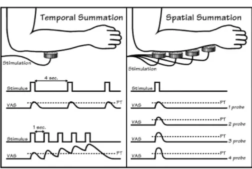
Temporal and spatial summation
Temporal summation occurs when a stimulus causes exaggerated per-ceptions of pain when repeated at certain frequencies (Fig. 1). In animal studies, repeated stimulation increases the excitability of spinal cord neurons which persists after discontinuing the peripheral stimulation.
This phenomenon is probably an important mechanism for the induction
and maintenance of acute and chronic pain syndromes in humans.16
Temporal summation can be induced by repeated thermal17 or
electrical13 stimulation of the skin. It has also been elicited by
stimul-ating muscles6 or viscera18 in humans. Temporal summation can be
measured by either psychophysical or electrophysiological responses. In the latter case, electromyographic recordings of the nociceptive
reflex after repeated stimulation of the sural nerve are performed.13
Five electrical stimuli at a frequency of 2 Hz are applied through sur-face electrodes to the innervation area of the sural nerve. An increased reflex amplitude is observed during the stimulation at a current inten-sity that corresponds to the stimulus inteninten-sity causing a subjective increase in pain perception.13
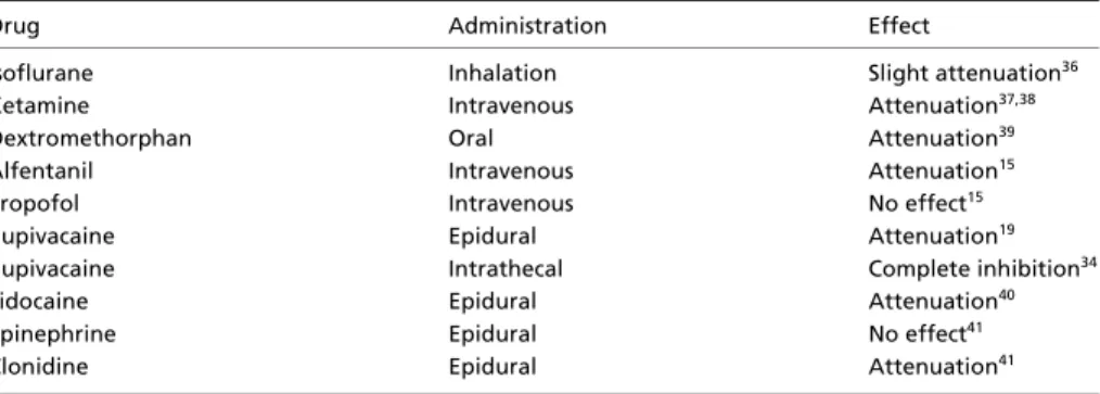
Temporal summation is a very potent mechanism which is difficult to block pharmacologically and cannot be adequately blocked by epidural analgesia19 or general anesthesia20 (Table 3).
Fig. 1 Temporal and spatial summation induced by heat stimulation. For temporal summation, repetition of the stimulus every 4 s results in a pain sensation of constant intensity (VAS, visual analogue scale for pain intensity). However, repetition of the stimulus at higher frequency (every 1 s) produces a progressive increase in pain sensation as the result of central summation phenomena. For spatial summation, increasing the stimulation area by increasing the number of stimulating probes results in increased pain intensity, although the intensity of the stimulus remains constant. Reproduced with permission from Curatolo et al. Anesthesiology 2000; 93: 1517–30.
Spatial summation occurs when pain perception increases with increasing area at which the stimulus is applied, although the stimulus
intensity remains constant.21 It is observed after application of heat,
cold, pressure or pinprick.1 The mechanisms underlying spatial
summa-tion are poorly understood. The N-methyl-D-aspartate (NMDA)
recep-tor antagonist ketamine inhibits heat pain arising from a large area
more effectively than it inhibits heat pain arising from a small area.22
This suggests that the NMDA receptor may be involved in spatial summation.
Imaging techniques
Functional neuroimaging has fundamentally changed our knowledge about the cerebral representation of pain. For the first time it has been possible to delineate the functional anatomy of different aspects of pain in the brain.
Positron emission tomography (PET)23 and single-photon emission
computed tomography (SPECT)24 require the injection of water or glucose
labelled with a radio-isotope into the blood stream flowing towards the brain. Radio-isotopes with a short half-life accumulate for a brief period of time in the active areas of brain and can be localized by scanners sen-sitive to the transient increase in radio-activity (Fig. 2). The temporal and spatial resolution is not as good as with functional magnetic reso-nance imaging (FMRI) (see below), but PET and SPECT also allow assess-ment of the concentration of ligands and receptors that are relevant to pain transmission in the brain.
FMRI25 is based on the different magnetic properties of the different
tissues in the brain. Red blood cells loaded with oxygen (oxygenated haemoglobin) present with different magnetic properties than unloaded ones (deoxygenated haemoglobin), and active areas in the brain have higher levels of oxygenated haemoglobin. This blood oxygenation level dependent signal allows the detection of active areas with good spatial and temporal resolution by magnetic field scanners.
Table 3 Pharmacological modulation of temporal summation in humans
Drug Administration Effect
Isoflurane Inhalation Slight attenuation36 Ketamine Intravenous Attenuation37,38 Dextromethorphan Oral Attenuation39 Alfentanil Intravenous Attenuation15
Propofol Intravenous No effect15
Bupivacaine Epidural Attenuation19
Bupivacaine Intrathecal Complete inhibition34
Lidocaine Epidural Attenuation40
Epinephrine Epidural No effect41
Implications for research
The main applications of sensory testing for research are to investigate mechanisms of action of regional analgesia techniques, to study new drugs and combinations of drugs, and to compare drugs and techniques. Given regional analgesia techniques modulate different pain pathways and mechanisms. If we assume that different stimulus paradigms activate differ-ent mechanisms or pathways, we should be able to determine which mecha-nisms or pathways are involved and to what degree. For instance, if the activation of mechanism/pathway A is predominantly inhibited by the drug or technique employed, the target of the specific intervention can be clarified. While the above assumption is the framework for a mechanism-based approach of sensory testing, this concept can be implemented only par-tially; selectivity is still a major problem of sensory tests. Most stimulation modalities activate different nerve fibres and probably involve multiple
pain mechanisms,1 and many of these mechanisms are still poorly
under-stood. Experimental pain studies are frequently performed on healthy vol-unteers. However, the nociceptive system of healthy subjects is very different from that of pain patients. Indeed, pain states are associated with plasticity changes in both the peripheral and the central nervous system which profoundly affect the processing of applied sensory stimuli.16 Even when sensory tests are performed on pain patients, the pain stimuli applied
Fig. 2. PET after cutaneous laser stimulation (top) and electrical intramuscular stimulation (bottom) in healthy volunteers.42 The maps are superimposed on a magnetic resonance image. Note the different, although overlapping, activation of brain structures after cutaneous and intramuscular stimulation. Reproduced with permission from Svensson
are artificial and may not resemble clinical pain. Most of the experimental measurements are short, harmless, predictable, easily tolerated and involve little stress. Therefore they exhibit marked differences to clinical pain.
Thus it is important to employ human experimental models that can simulate pathophysiological conditions. An example may be the use of experimental stimuli after previous application of capsaicin26 or induction
of burn injury27 (experimental hyperalgesia), inducing temporal and spatial summation,10,19 or stimulating muscle6 or viscera.18 Experimental
inves-tigations of such mechanisms in relation to regional analgesia have not yet been performed extensively, probably because of the sophisticated technology involved and their invasive nature.
Despite the above limitations, the experimental approach to the assess-ment of regional analgesia has important positive features which can be summarized as follows.
1 The same stimulus can be repeated over time. This allows the assessment of the time course of the drug action. Clinical pain is not repeatable and there is a large time variation.
2 The same stimulus can be applied to different body areas, thus allowing the assessment of the spread of block.
3 The variability associated with the type of painful stimulus is avoided. Var-iability is usually large in studies performed under clinical conditions. 4 The volunteer can be used as his or her own control and so studies can be
performed with a much smaller number of participants.
5 Differentiated responses to the activation of different structures (skin, muscle, viscera) can be recorded (multitissue sensory testing approach).
6 Differentiated responses to different stimulus modalities can be assessed (multimodal sensory testing approach).
7 Pain sensitivity at normal and injured regions can be compared quantitatively.
Because of these characteristics, quantitative sensory tests have an important role in regional analgesia research, provided that one is fully aware of the information that can and cannot be gained by them. Experi-mental pain research is not an alternative to research performed under clinical conditions; rather, it complements it.
Clinical implications
Traditional clinical testingThe main aims of sensory tests for clinical purposes are to assess the spread of sensory block and to predict the effectiveness of the regional analgesia technique in individual patients.
Assessing the spread of sensory block provides information on the site of effect of the technique employed, which is important for deciding further
management.1 For instance, normal sensation of pinprick or cold after
injection of a local anaesthetic clearly indicates inadequate position of the needle or catheter and warrants either supplemental block or change to general anaesthesia. Unilateral spread of block or inadvertent intrathecal injection after epidural analgesia can be detected, thereby avoiding pain during surgery or severe complications resulting from injection of high doses in the subarachnoid space, respectively.
Most of the assessment methods that have been described in this paper require expensive apparatus and substantial expertise. Moreover, there is very little scientific data and clinical experience on the value of these tests for clinical purposes. Thus, despite the major technological improvements and the substantial increase in experimental data on sensory assessments, simple methods such as pinprick, cold and touch are still used in clinical practice.
There is evidence that block of touch, pinprick or cold sensation does
not necessarily imply block of nociception during regional analgesia.28
The occurrence of pain after a regional block that produces complete absence of touch, pinprick or cold sensation may puzzle the anaesthesi-ologist. However, in view of the characteristics of these sensory tests, this phenomenon is hardly surprising. First, different nociceptors and fibres are activated by these tests and the surgical stimulus. For instance, while absence of touch sensation implies an effect of the block
on Aβ fibres, pain is transmitted by Aδ and C fibres. Moreover,
although block of pinprick sensation indicates an effect on Aδ fibres,
this does not mean that all the functions that are mediated by these fibres are blocked. The intensity of the stimulus is a determinant of the efficacy of regional block. The simple sensory tests are weak stimuli that are easier to block than strong surgical stimuli. As mentioned above, analgesia to brief stimuli, applied to small areas, does not imply analgesia to long-lasting stimuli, applied to wide areas (temporal and spatial summation). Finally, testing the skin does not give information on drug effects on deep structures that contribute to pain in clinical conditions.
After epidural analgesia, the larger the number of dermatomes that are hyposensitive to pinprick, the lower is the incidence of pain during29 and
after28 surgery. Therefore the spread of sensory block may be a useful
clinical indicator of the efficacy of the regional block. However, the spread, as assessed by pinprick or cold stimulation, can explain only a limited part of the variability of the efficacy of epidural analgesia.28,29 In a study of postoperative pain, the slope of the curve describing the rela-tionship between number of dermatomes that were hyposensitive to pin-prick and relief of postoperative pain was very shallow, indicating a modest
quantitative impact of spread on pain relief.28 Moreover, only 2–5% of the variability of postoperative pain intensity could be explained by the
spread of sensory block in a multivariate analysis.28 This means that
adequate analgesia cannot be predicted based only on the determination of the spread of sensory block. Other factors, partly unknown, play an important role. For instance, the depth of the sensory block at the site of surgery, which cannot be quantified in clinical practice, may play an important role.
An important limitation of the tests used in clinical practice is that the response is usually confined to either block or no block of sensation, i.e. the response is not quantitative. The same response to pinprick stimulation (e.g. absence of sensation) is obtained at dermatomes displaying differ-ent degrees of sensory block as measured by pain threshold after laser
stimulation.10 Therefore these simple qualitative tests have a low
dis-criminative power with regard to the depth of sensory block after regional analgesia.
Assessing the effects of opioids
Epidural fentanyl injected at the lumbar level produced a higher degree of analgesia to heat at the lumbar dermatomes than at sacral or thoracic
dermatomes.30 This suggests that the effect of epidural fentanyl on the
peripheral nerves or dorsal root ganglions may be more important than is usually believed. In fact, if penetration through the meninges and action at the spinal cord receptors were the main mechanisms explaining regional analgesia after epidural fentanyl, the depth of analgesia at the sacral dermatomes would be at least equally strong. Conversely, lumbar epidural morphine attenuates heat pain up the trigeminal dermatomes, reflecting rostral spread of this hydrophilic drug after penetration into
the subarachnoid space.31
It is clinically relevant that opioids frequently have no effect on
pin-prick or cold sensation.32 Thus opioid-based epidural analgesia cannot
be assessed reliably by these tests.
The study by Eichenberger and colleagues,30 mentioned above, also
employed an intramuscular pain model (injection of hypertonic saline) which proved sensitive for detecting the analgesic action of epidural fen-tanyl. A study of intravenous remifentanil found a much deeper analgesic effect of this opioid on intramuscular than on cutaneous electrical
pain.33 This suggests that opioids may have a stronger effect on deep
pain than on skin pain in clinical conditions, and this difference could also apply to epidural opioids. Translated to surgical pain, one can assume that pain on surgical incision does not necessarily imply pain during surgical stimulation of deep tissues.
Differences between different anaesthetic techniques
Do different regional anaesthetic techniques work to the same extent? To our knowledge, there is no direct comparison of different techniques in terms of depth of sensory block, as assessed experimentally in a dose– response manner. We performed two different studies analysing the
effects of 20 ml bupivacaine 0.5% epidurally19 and 18 mg bupivacaine
intrathecally34 on repeated electrical stimulation. The median pain
threshold in the epidural study increased from 6 mA at baseline to 23 mA at 60 min. In the intrathecal study, the median pain threshold increased from 4 mA at baseline to 60 mA, which was the maximum current intensity allowed, at 40 min. Despite the limitations regarding the comparison between two different studies and the use of single doses, the data suggest that the sensory block is stronger with spinal than with epidural analgesia, an impression that is shared by most anaesthesiologists. It is noteworthy that the measurements were made at the dermatome S1, which is thought to be particularly resistant to epidural blockade.
Importance of temporal and spatial summation
An important concept in regional analgesia is that the duration of the applied stimulus (temporal summation) and the area where the stimulus is applied (spatial summation) are determinants of the effects of regional block.10,19 Analgesia to a brief stimulus is not necessarily a guarantee of
analgesia to a long-lasting stimulus. This finding is clinically relevant, since clinical pain is mostly ongoing rather than brief in duration. The different effect on the two stimulation modalities is probably due to the fact that brief stimuli, although not perceived as painful, are not com-pletely blocked by epidural local anaesthetics and arrive at the spinal cord. At this level, they sum and eventually evoke pain if they are applied for a longer time. In contrast, spinal anaesthesia with 18 mg bupivacaine inhibited pain after both single and repeated electrical
stim-ulation, indicating complete inhibition of temporal summation.34 This
may be the result of a strong block of the sensory input, which is there-fore unable to sum in the spinal cord to evoke a pain sensation.
Spatial summation after epidural10 and intrathecal35 administration of
bupivacaine 0.5% was studied by comparing the response to stimulation using one needle with the response to stimulation using 10 needles applied simultaneously. In both studies, pain was evoked with 10 needles in patients in whom stimulation with one needle did not evoke pain. Therefore a stimulus applied to a small area, although not evoking a pain sensation, may not be completely blocked and will arrive at the spinal cord. When the same stimulus is applied to a wider area, it can spatially
sum in the spinal cord and be perceived as painful. This finding also has clinical relevance, since clinical pain is rarely confined to very small areas. It is noteworthy that many sensory tests used for clinical and experimental purposes are of brief duration and are applied to small areas. This probably contributes to the discrepancy between sensory tests and clinical pain.
Conclusions
Sensory tests have an important role in regional anaesthesia and pain research. They have substantially increased our understanding of the laws regulating the action of drugs and techniques that are used daily in clinical practice. Studies conducted using experimental pain cannot be expected to provide the same information as clinical trials. They have limitations, mostly due to their artificial nature and the unidimensional assessment procedure. However, experimental tests can provide information that cannot be gained in clinical trials and can reduce the gap between animal research and studies conducted under clinical con-ditions. Thus sensory assessment is not an alternative to clinical pain assessment. Rather, the two approaches complement each other. Tests that more closely mimic clinical pain are available, but are still underused. In clinical practice, sensory tests provide useful information on the efficacy of regional analgesia, but have limited validity. Unfortunately, almost no progress on assessment of regional analgesia for clinical pur-poses has been made in the last decade, largely because of the paucity of published research.
References
1 Curatolo M, Petersen-Felix S, Arendt-Nielsen L. Sensory assessment of regional analgesia in humans. A review of methods and applications. Anesthesiology 2000; 93: 1517–30
2 Turk DC. The role of psychological factors in chronic pain. Acta Anaesthesiol Scand 1999; 43: 885–8
3 Robinson JP, Evaluation of function and disability. In Loeser JD (ed.) Bonica’s Management
of Pain (3rd edn). Philadelphia, PA: Lippincott–Williams and Wilkins, 2001; 342–62
4 Ware JE Jr, Kosinski M, Gandek B et al. The factor structure of the SF-36 Health Survey in 10 countries: results from the IQOLA Project. International Quality of Life Assessment. J Clin
Epidemiol 1998; 51: 1159–65
5 Chapman CR, Syriala KL, Measurement of pain. In Loeser JD (ed.) Bonica’s Management of
Pain (3rd edn). Philadelphia, PA: Lippincott–Williams and Wilkins, 2001; 310–28
6 Graven-Nielsen T, Arendt-Nielsen L, Svensson P, Jensen TS. Quantification of local and referred muscle pain in humans after sequential i.m. injections of hypertonic saline. Pain 1997;
69: 111–17
7 Drewes AM, Schipper KP, Dimcevski G et al. Multi-modal induction and assessment of allodynia and hyperalgesia in the human oesophagus. Eur J Pain 2003; 7: 539–49
8 Banic B, Petersen-Felix S, Andersen OK et al. Evidence for spinal cord hypersensitivity in chronic pain after whiplash injury and in fibromyalgia. Pain 2004; 107: 7–15
9 Park R, Wallace MS, Schulteis G. Relative sensitivity to alfentanil and reliability of current perception threshold vs von Frey tactile stimulation and thermal sensory testing. J Peripher
Nerv Syst 2001; 6: 232–40
10 Arendt-Nielsen L, Øberg B, Bjerring P. Quantitative assessment of extradural bupivacaine analgesia. Br J Anaesth 1990; 65: 633–38
11 Edwards RR, Ness TJ, Weigent DA, Fillingim RB. Individual differences in diffuse noxious inhibitory controls (DNIC): association with clinical variables. Pain 2003; 106: 427–37 12 Torebjörk HE, Hallin RG. C-fibre units recorded from human sensory nerve fascicles in situ.
A preliminary report. Acta Soc Med Ups 1970; 75: 81–4
13 Arendt-Nielsen L, Brennum J, Sindrup S, Bak P. Electrophysiological and psychophysical quantification of central temporal summation of the human nociceptive system. Eur J Appl
Physiol 1994; 68: 266–73
14 Chan CWY, Dallaire M. Subjective pain sensation is linearly correlated with the flexion reflex in man. Brain Res 1989; 479: 145–50
15 Petersen-Felix S, Arendt-Nielsen L, Bak P, Fischer M, Zbinden AM. Psychophysical and electro-physiological responses to experimental pain may be influenced by sedation: comparison of the effects of an hypnotic (propofol) and an analgesic (alfentanil). Br J Anaesth 1996; 77: 165–71 16 Woolf CJ, Salter MW. Neuronal plasticity: increasing the gain in pain. Science 2000; 288:
1765–69
17 Price DD, Hu JW, Dubner R, Gracely RH. Peripheral suppression of first pain and central summation of second pain evoked by noxious heat pulses. Pain 1977; 3: 57–68
18 Arendt-Nielsen L, Drewes AM, Hansen JB, Tage-Jensen U. Gut pain reactions in man: an experimental investigation using short and long duration transmucosal electrical stimulation.
Pain 1997; 69: 255–62
19 Curatolo M, Petersen-Felix S, Arendt-Nielsen L, Fischer M, Zbinden AM. Temporal summa-tion during extradural anaesthesia. Br J Anaesth 1995; 75: 634–35
20 Petersen-Felix S, Arendt-Nielsen L, Bak P, Fischer M, Bjerring P, Zbinden AM. The effects of iso-flurane on repeated nociceptive stimuli (central temporal summation). Pain 1996; 64: 277–81 21 Nielsen J, Arendt-Nielsen L. Spatial summation of heat induced pain within and between
dermatomes. Somatosens Mot Res 1997; 14: 119–25
22 Arendt-Nielsen L, Nielsen J, Petersen-Felix S, Schnider TW, Zbinden AM. Effect of racemic mixture and the (S+)-isomer of ketamine on temporal and spatial summation of pain. Br J
Anaesth 1996; 77: 625–31
23 Petrovic P, Ingvar M. Imaging cognitive modulation of pain processing. Pain 2002; 95: 1–5 24 Di Piero V, Ferracuti S, Sabatini U, Pantano P, Cruccu G, Lenzi GL. A cerebral blood flow
study on tonic pain activation in man. Pain 1994; 56: 167–73
25 Davis KD. The neural circuitry of pain as explored with functional MRI. Neurol Res 2000; 22: 313–7
26 Petersen KL, Jones B, Segredo V, Dahl JB, Rowbotham MC. Effect of remifentanil on pain and secondary hyperalgesia associated with the heat-capsaicin sensitization model in healthy vol-unteers. Anesthesiology 2001; 94: 15–20
27 Gustorff B, Anzenhofer S, Sycha T, Lehr S, Kress HG. The sunburn pain model: the stability of primary and secondary hyperalgesia over 10 h in a crossover setting. Anesth Analg 2004; 98: 173–7
28 Curatolo M, Kaufmann R, Petersen-Felix S, Arendt-Nielsen L, Scaramozzino P, Zbinden AM. Block of pinprick and cold sensation poorly correlate with relief of postoperative pain during epidural analgesia. Clin J Pain 1999; 15: 6–12
29 Curatolo M, Orlando A, Zbinden AM, Scaramozzino P, Venuti FS. A multifactorial analysis to explain inadequate surgical analgesia following extradural block. Br J Anaesth 1995; 75: 274–81
30 Eichenberger U, Giani C, Petersen-Felix S, Graven-Nielsen T, Arendt-Nielsen L, Curatolo M. Lumbar epidural fentanyl: segmental spread and effect on temporal summation and muscle pain. Br J Anaesth 2003; 90: 467–73
31 Angst MS, Ramaswamy B, Riley ET, Stanski DR. Lumbar epidural morphine in humans and supraspinal analgesia to experimental heat pain. Anesthesiology 2000; 92: 312–24
32 Coda BA, Cleveland Brown M, Schaffer R et al. Pharmacology of epidural fentanyl, alfentanil, and sufentanil in volunteers. Anesthesiology 1994; 81: 1149–61
33 Curatolo M, Petersen-Felix S, Gerber A, Arendt-Nielsen L. Remifentanil inhibits muscular more than cutaneous pain in humans. Br J Anaesth 2000; 85: 529–32
34 Curatolo M, Petersen-Felix S, Arendt-Nielsen L, Zbinden AM. Spinal anaesthesia inhibits central temporal summation. Br J Anaesth 1997; 78: 88–89
35 Arendt-Nielsen L, Anker-Møller E, Bjerring P, Spangsberg N. Onset phase of spinal bupi-vacaine analgesia assessed quantitatively by laser stimulation. Br J Anaesth 1990; 65: 639–42 36 Petersen-Felix S, Arendt-Nielsen L, Bak P et al. Analgesic effect in humans of subanaesthetic
isoflurane concentrations evaluated by experimentally induced pain. Br J Anaesth 1995; 75: 55–60
37 Arendt-Nielsen L, Petersen-Felix S, Fischer M, Bak P, Bjerring P, Zbinden AM. The effect of N-methyl-D-aspartate antagonist (ketamine) on single and repeated nociceptive stimuli: a
placebo-controlled experimental human study. Anesth Analg 1995; 81: 63–68
38 Guirimand F, Dupont X, Brasseur L, Chauvin M, Bouhassira D. The effects of ketamine on the temporal summation (wind- up) of the R(III) nociceptive flexion reflex and pain in humans.
Anesth Analg 2000; 90: 408–14
39 Price DD, Mao J, Frenk H, Mayer DJ. The N-methyl-D-aspartate receptor antagonist
dex-tromethorphan selectively reduces temporal summation of second pain in man. Pain 1994; 59: 165–74
40 Curatolo M, Petersen-Felix S, Arendt-Nielsen L et al. Adding sodium bicarbonate to lidocaine enhances the depth of epidural blockade. Anesth Analg 1998; 86: 341–7
41 Curatolo M, Petersen-Felix S, Arendt-Nielsen L, Zbinden AM. Epidural epinephrine and cloni-dine: segmental analgesia and effects on different pain modalities. Anesthesiology 1997; 87: 785–94
42 Svensson P, Minoshima S, Beydoun A, Morrow TJ, Casey KL. Cerebral processing of acute skin and muscle pain in humans. J Neurophysiol 1997; 78: 450–60