Stability of Heartbeat Interval Distributions in
Chronic High Altitude Hypoxia
M. MEYER, 1'3 A. RAHMEL, 3 C. MARCONI, 2 B. GRASSI, 2 P. CERRETELLI, 1'2 AND J.E. SKINNER 4 D(partement de Physiologie, CMU, Genkve, Switzerland 1
Istituto di Tecnologie Biomediche Avanzate, CNR, Milano, Italy 2
Department of Physiology, Max Planck Institute for Experimental Medicine, GOttingen, Germany 3 Cardiovascular Division, Totts Gap Laboratories, Bangor, Pennsylvania, USA 4
Abstract--Recent studies of nonlinear dynamics of the long-term variability of heart rate have identified nontrivial long-range correlations and scale-invariant power-law character- istics (1/f noise) that were remarkably consistent between individuals and were unrelated to external or environmental stimuli (Meyer et al., 1998a). The present analysis of complex nonstationary heartbeat patterns is based on the sequential application of the wavelet trans- form for elimination of local polynomial nonstationary behavior and an analytic signal approach by use of the Hilbert transform (Cumulative Variation Amplitude Analysis). The effects of chronic high altitude hypoxia on the distributions and scaling functions of car- diac intervals over 24 hr epochs and 4 hr day/nighttime subepochs were determined from serial heartbeat interval time series of digitized 24 hr ambulatory ECGs recorded in 9 healthy subjects (mean age 34 yrs) at sea level and during a sojourn at high altitude (5,050 m) for 34 days (Ev-K2-CNR Pyramid Laboratory, Sagarmatha National Park, Nepal). The results suggest that there exists a hidden, potentially universal, common structure in the heterogeneous time series. A common scaling function with a stable Gamma distribution defines the probability density of the amplitudes of the fluctuations in the heartbeat interval time series of individual subjects. The appropriately rescaled distributions of normal sub- jects at sea level demonstrated stable Gamma scaling consistent with a single scaled plot (data collapse). Longitudinal assessment of the rescaled distributions of the 24 hr record- ings of individual subjects showed that the stability of the distributions was unaffected by the subject's exposure to a hypobaric (hypoxic) environment. The rescaled distributions of 4 hr subepochs showed similar scaling behavior with a stable Gamma distribution indicat- ing that the common structure was unequivocally applicable to both day and night phases and, furthermore, did not undergo systematic changes in response to high altitude. In contrast, a single function stable over a wide range of time scales was not observed in patients with congestive heart failure or patients after cardiac transplantation. The func- tional form of the scaling in normal subjects would seem to be attributable to the underly- ing nonlinear dynamics of cardiac control. The results suggest that the observed Gamma scaling of the distributions in healthy subjects constitutes an intrinsic dynamical property of normal heart function that would not undergo early readjustment or late acclimatization to extrinsic environmental physiological stress, e.g., chronic hypoxia.
Key words--antonomic nervous system, cardiac rhythm, electrocardiography, heart rate, high altitude, Hilbert transform, non-linear dynamics, scaling properties, wavelet trans- form.
Address for correspondence: Professor Michael Meyer, M.D., Ph.D., Department of Physiology, Max Planck Institute for Experimental Medicine, Hermann-Rein-Strage 3, D-37075 G0ttingen, Germany; e-mail: [email protected]
Integrative Physiological and Behavioral Science, October-December 1998, Vol. 33, No. 4, 344-362. 3 4 4
NON-LINEAR DYNAMICS OF HEART RATE AT ALTITUDE 345
I n t r o d u c t i o n
PHYSIOLOGICAL SYSTEMS TYPICALLY EXHIBIT complex physiological variability that would appear as irregular or random. These fluctuations have traditionally been attributed to extrinsic changes in the environment of the organism or to changes in the level of activity. However, recent evidence suggests that physiological systems are invariably nonlinear, and nonlinearity of control systems could lead to nonlinear and nonstationary behavior, i.e., fluctuations that are generated
intrinsically.
The analysis of physiological variability by signal processing techniques derived from nonlinear dynamics would help elucidate the underlying mechanisms about how the organism or its subsystems are operating.The study of the response of an individual to a low-oxygen environment has a long history. It is generally accepted that
acclimatization
constitutes an indispensable require- ment when man is chronically exposed to a hypoxic environment, e.g., during a sojourn at high altitude. Substantial complex readjustments of the pulmonary, cardiovascular, and metabolic systems are known to occur that would enable a person to climb mountains well above 8,000 meters without supplemental oxygen, but many of these "adaptations" are not completely understood (CerreteUi, 1980; Cerretelli and Hoppeler, 1996; Ueda et al., 1992; Ward etal., 1990).Recent studies of nonlinear dynamics in signal processing of the long-term variability of heart rate have identified nontrivial long-range correlations (memory) and scale-invariant power-law characteristics
(1/f
noise) of the heartbeat interval patterns that were unrelated to extemal stimuli and remarkably consistent between individuals (Peng et al., 1993, 1995; Meyer et al., 1998a, 1998b). These findings have been associated with the fractal scaling behavior and long-range correlation properties typically exhibited by physical systems near a critical point of phase transition, i.e., far away from equilibrium (Bak and Creutz, 1994; Beran, 1994; Stanley, 1971). The physiological significance of the long-range power-law or1/f
behavior of normal cardiac interbeat time series has not been established. One attractive possibility is that the long-term correlations reflect the nonlinear interaction between different components of the control system that operate on different time scales. Recent statistical analyses of heartbeat interval dynamics of normal (innervated) subjects and heart transplant (noninnervated) patients has suggested that the neuroautonomic car- diac control system was responsible for the genuine self-similar fractal scaling properties of the fluctuating interval patterns (Meyer et al., 1998a, 1998b). However, these studies have revealed that heart rate scaling properties remained unchanged when normal subjects were chronically exposed to a hypobaric (hypoxic) high altitude (5,050 m) environment and inaugurated the concept that the heart, in terms of neuroautonomic control, ispre-
adapted
to hypoxia.In the present study, the analysis of complex nonstationary heartbeat interval fluctua- tions in subjects exposed to chronic high altitude hypoxia is based on a novel alternative method referred to as Cumulative Variation Amplitude Analysis that involves the wavelet transform and an analytic signal approach by use of the Hilbert transform. The major objectives were: 1) to quantify the distributions and scaling functions of the variations in the heartbeat interval fluctuations over extended 24 hr epochs and shorter 4 hr day/night- time subepochs; 2) to assess the intrinsic stability or otherwise extrinsic adaptive changes that, upon exposure to a hypoxic environment, would be expected to evoke differential responses that rely on instantaneous strategies in the acute phase and on acclimatization in the chronic phase; and 3) to evaluate the mechanisms underlying the scaling functions of heart rate dynamics.
346 M E Y E R E T A L .
Methods
Subjects and Data Acquisition
Nine subjects (7 males, 2 females; mean age ___ SD, 33.7 + 4.6 yrs) volunteered for this study, which was approved by the ethical review boards of the institutions involved. A written informed consent was obtained from each subject. Heartbeat interval time series data were obtained from ambulatory Holter ECG recorders during sojourn for 34 days at the Italian EvK2-CNR high-altitude Pyramid Laboratory (Sagarmatha National Park, Khumbu Valley, Nepal, 5,050 m - 16,568 ft; barometric pressure -420 mmHg, 02 partial pressure -79 mm Hg, arterialized blood 02 saturation -83%) located in the vicinity of the Everest Base Camp. A more detailed account of the experimental procedures is presented in our previous report (cf. Meyer et al., 1998a) and the altitude profile and locations are shown in Fig. 6 (see Results). Although mostly the same time series data sets were used in this study, the present study is new and does not duplicate previous findings. Additional data were collected in normal subjects from the University of Geneva, Switzerland, and in cardiac patients from La Pitie-Salpetri~re Hospital, Pads, France, and Riuniti Hospital, Bergamo, Italy.
Analysis of Temporal Evolution of Non-Stationary Heartbeat Interval Patterns
Under free-running conditions the heartbeat interval time series is highly nonstationary and exhibits extreme complexity as reflected by the emergence of patchy patterns varying with time. Here the terms "complexity" and "patterns" are not defined in a rigorous sense, rather they are used to signify the difficulty of quantitatively describing or understanding the irregularity of the time series. The fluctuations in the heart rate pattern may represent: 1) uncorrelated (white) noise superimposed on a basically regular process; 2) short-range correlations such that the current value is influenced only by its most recent predecessors but otherwise random fluctuations over the long run; 3) fluctuations generated by the underlying nonlinear dynamics of the system itself; and 4) trivial changes of physiological or background conditions (unsteady state). A novel analysis, termed Cumulative Variation Amplitude Analysis, has recently been introduced by Ivanov et al. (1996, 1998) to study the subtle structure of nonstationary physiological time series. The approach is based o n the sequential application of the wavelet and Hilbert transform analysis.
Wavelet transform. The
wavelet transform represents a time-scale analysis of the signal which is closely related but different from the conventional windowed Fourier transform. Actually, a wavelet constitutes a "small wave" that wiggles around the time axis. The wavelet transformW~s(a,b)
of a signals(t)
is defined as the sum over all time of the signal multiplied by scaled, shifted versions of the wavelet function ~.W~s= f s ( t ) + ly(t-b)dt
(1)..** "x/a a
where the analytic wavelet gt has a width of the order of the scale factor (a) and is located at position b. The result of the wavelet transform are many wavelet coefficients W~, which are a function of scale and position. The wavelet transform (W~) can be interpreted as the
detail
properties contained in the signal at any given scale a. If the resolution is defined as1/a,
then the resolution increases as the scale decreases. The size of the revealed details isNON-LINEAR DYNAMICS OF HEART RATE AT ALTITUDE 347
proportional to the size of the domain in which the wavelet or wavelet function V/is not too close to zero. For small a, the Ig functions are effectively nonzero at only small regimes and show good localizations, hence short-time domains (or high-frequency components) can be detected. For large a, however, the wavelet eliminates most of the entire frequency content of the signal, thus loosing information of the intrinsic dynamics or fine structure of the system. For nonstationary signals, local polynomial behavior (trends) can be eliminated by the wavelet transform given an appropriate choice of the analytic wavelet ~ (Arneodo et al., 1995; Daubechies, 1992; Kaiser, 1994; Muzy et al., 1994). In the present analysis, n th order derivatives of the Gaussian probability density function were considered as analytic wavelets. The wavelets I//(n) with n = 1 and n = 2 are:
t . e -t2/2
V/(~) = (2)
i//(2) (t 2 - 1). e -tz/2
=
(3)
The wavelet transform of s with respect to V (n) is proportional by s" to the n th derivative of the average of s at scale a. Thus, the n th derivative :(t) reveals the n th order detail of s at t. V :1) is orthogonal to segments of the signal with different constant local averages but is not orthogonal to linear, non-constant trends. ~2) and higher-order derivatives are not or- thogonal to both linear and nonlinear trends and hence are less suited when nonlinear dynamics is expected underlying the original signal.
Hilbert transform. A convenient way in signal processing to define the amplitude and phase of an arbitrary signal is established by the analytic signal approach based on the Hilbert transform. This approach unambiguously returns the instantaneous amplitude (A(t)) and instantaneous phase (0(t)) of a signal (s(t)) by construction of the analytic signal (S(t)) which is a complex function of time (Gabor, 1964; Panter, 1965; Rabiner and Gold, 1975; Smith and Mersereau, 1992):
S(t) = ~(t) + is(t) = A ( t ) e i~(O (4)
where the function g (0 is the Hilbert transform of s(t) and PV refers to the Cauchy Principal Value integration (Arfken, 1985):
+oo
~(t) = n-IPV ~ s(z) d'r
(5)
The instantaneous amplitude, A ( t ) = - ~ s 2 ( t ) + ~2(t), and phase, r = t a n - l ( g (t)ls(t)), are thus uniquely defined from eq.(4). The instantaneous amplitudes ( A ( t ) ) of the wavelet transform signal (see above) represent the cumulative variations of the original signal over an interval proportional to the wavelet scale a. Neither the wavelet transform nor the Hilbert transform require stationarity of the data.
The sequential steps of the analysis and the intermediate results of the operations are shown in Figure 1. A segment of a 24 hr heartbeat interval time series is displayed (upper
348 M E Y E R E T A L .
cr
0.5
o 2 4 ~ 8 ~o 0 1000 2000 3000
x ~ 0 4
o5i . , Jilll, illlliihi, la i j 0.4 9 9 9
~ o g o ~ - o 5 ~ ' ~-~--~'. o 2 4 6 8 ~o 0 1 0 0 0 2 0 0 0 3 0 0 0 xlO" . . . . 0 . 4 9 9 9 0 . 3 ~ 02 o~ O. 0 o 2 4 9 8 1c~ 0 tO00 2000 3000
N u m b ~ of'Heartbeats x lo' Number of Heartbeats
FIG. 1. Segment of original heartbeat interval time series ( R - R ) n plotted against consecutive beat number for a period of -24 hr (104 beats) (upper left). Wavelet transform W~R-R) of R-R time series (first derivative of the Gaussian probability function ~(l) used as analytic wavelet, wavelet scale (a = 8) (middle left). Instanta- neous amplitudes A(t) of the wavelet transform coefficients obtained by the Hilbert transform (lower left). The analysis of a 1 hr nighttime subsegment (indicated by rectangle in upper leftpanel) is shown in the right panels of figure, respectively.
left) along with an 1 hr nighttime subsegment (indicated by rectangle in upper left and displayed in upper right of figure) where nonstationarity resulting from changes in the level of physical activity is minimized. Nonstationarity is evident over both long- and short-time scales. The wavelet transform (Wds)s) of the segments is shown in the middle panels. Nonstationarities due to constants and linear trends have been eliminated by the wavelet transform using a wavelet scale a = 8 that relates to the Fourier wavelength by / t = 2 n n - ~ T l ~ - l a n d best probes patterns of ~40 s duration. The orthogonality of Gaussian ~(1) to segments of the time series with different local averages results in fluc- tuations of the wavelet transform coefficients around zero with high peaks occurring at the positions of sharp transitions in the wavelet values. The larger peaks thus indicate the boundaries between segments with different local averages in the original signal, whereas smaller variations represent fluctuations within a given regime. The presence of consecu- tive linear trends in the heartbeat interval time series results in fluctuations of the wavelet transform coefficients around different non-zero levels corresponding to the magnitudes of the slopes of the linear trends. The instantaneous amplitudes (A(t)) of the sequence of the wavelet transform coefficients obtained by the Hilbert transform are shown in the lower panels. A(t) reflects the cumulative variations of the beat-by-beat intervals over an interval proportional to the wavelet scale a.
NON-LINEAR DYNAMICS OF HEART RATE AT ALTITUDE 30 25 20 r~ 10 5 0 0 0.1 0.2 0.3 0.4 0.8 ~::~0.6 X v Q.. 0.4 0.2 1 2 3 4 5 x P rrlax 349
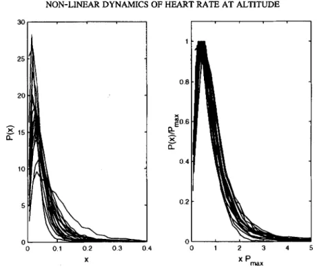
FIG. 2. Probability distributions P(x) of the instantaneous amplitudes of heartbeat interval fluctuations x = A(t) derived from 24 hr ECG recording for a group of healthy adult subjects (N = 20) (left). Same probability distributions after rescaling P(x) by P,n~x and x by llP,~x (right). The rescaled probability distributions show "data collapse" onto a single scaled plot.
Probability distributions. The distributions of the instantaneous amplitudes (A(t)) are well fit by a generalized homogeneous function, the Gamma distribution, which, with parameters a (shape) and b (scale), has density:
P ( x I a,b) = 1 . xa,le_X/b (6)
baF(a)
where b - y / x 0' F(a) is the Gamma function, ), is the fitting parameter and Zo is the peak position o f P, Pmax. A generalized homogeneous function f (a,b) has the form:
f(~k~ a, ~,kZb) = V ( a , b) (7)
where /1, is a parameter and kj and k2 are two arbitrary exponents (scaling powers). By defmition, f ( a , b ) is valid for all positive & and eq. (6) satisfies eq. (7) with kl = -1 and k2 = 1 (Stanley, 1971). Generalized homogeneous functions are functions describing physical systems near their critical point. One of the important properties of these functions is that of "data collapse", i.e., instead, for each value of b, data falling on a family of curves, data collapse onto a single curve given by the scaling function:
P(u) - P(a,b) (8)
350 M E Y E R E T A L . 30 1 25 0.8 20 j o . 6 0.4 10 5 0.2 0 0 0 0.1 0.2 0.3 0.4 0 X 1 2 3 4 x P ~ x
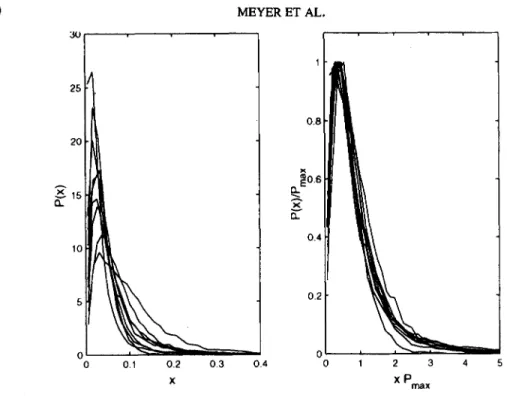
FIG. 3. Probability distributions (unscaled, left; rescaled, right) of 24 hr heartbeat interval time series of healty subjects (N = 9) prior to sojourn at high altitude. Sea level measurements (Milan, 122 m).
where, by defining the scaled variable u--ba, the number of independent variables is reduced. The probability distributions (P(x)) of the instantaneous amplitudes (A(t)) were normalized to unit area, and P(x) was rescaled by P,,~ and x by 1/Pmax. Rescaling is equivalent to scaling according to eq.(8) as P(x) - P(a,b) and PmaxOt b.
The probability distributions (P(x)) of the normalized (to unit area) instantaneous car- diac interbeat amplitudes (x = A(t); 24 hr recordings, -104 beats) for a group of normal healthy subjects from the University of Geneva (N -- 20, 12 males, 8 females; mean age + SD, 30.2 + 4.2 yrs) are shown in Figure 2 (left). Inspection of the distribution function reveals marked differences between the subjects indicated by the different mean values and standard deviations (widths) of the distributions. Figure 2 (right) shows the same probabil- ity distributions after rescaling with superposition of the data points on an almost single curve. The rescaled distributions for the group of healthy subjects thus conform to a single scaled plot (data collapse). The sequential application of the wavelet transform and the Hilbert transform extracts dynamical properties of the heartbeat interval time series that are hidden in the cumulative fluctuations. The Gamma scaling of the distributions of the amplitudes of the cumulative variations is characteristic for normal healthy subjects, and the absence of data collapse viz. lack of stable Gamma scaling would be indicative of abnormal cardiac dynamics (see Discussion). For healthy subjects, stable Gamma distribu- tions were obtained for a wide range of wavelet transform scales (a = 4 to 24) and most consistent results were obtained for a = 8, which probes the collective properties of pat- terns in the time series with ~40 s duration.
NON-LINEAR
DYNAMICS OF HEART RATE AT ALTITUDE
25
20
x 0"
0
.
4
~
0.2
o;
oi,
" 0.2 0.3 " - 0.4 ~ 0 1 2 3 4 5 X X Pmax 351Fro. 4. Cumulative amplitude probability distributions (unscaled, left; rescaled, right) of serial 24 hr heart- beat interval time series in an individual subject (#8, n = 23) during sojourn at 5,050 m altitude for 34 days (see Subjects and Experimental Protocol).
Experimental Time Series
The cumulative variation amplitude analysis using a wavelet transform scale of a = 8 was applied to the experimental heartbeat interval time series of the full-length 24 hr epochs. Typically, the data length ranged from 90,000 to 135,000 data points as a result of varying daily activities (trekking during ascent or descent and laboratory work during sojourn at altitude). A total of 180 24 hr ambulatory Holter recordings were analyzed. In addition, 4 hr subsets from 1:00 AM (PM) through 5:00 AM (PM) were selected from the original time series to determine the distributions of nighttime and daytime subepochs that were 12 hrs apart and differed in the subjects' physical activity. The data length of these segments was 13,000 to 17,000 data points.
R e s u l t s
Figure 3 (left) shows the results of the cumulative variation amplitude analysis (wavelet scale a = 8) of 24 hr heartbeat interval series of the 9 healthy subjects at sea level (Milan, 122 m a.s.1.). The rescaled distributions (Figure 3, fight) demonstrate stable G a m m a scal- ing consistent with a single scaled plot (data collapse) in agreement with previous findings in another group of healthy subjects (cf. Figure 2).
Figure 4 shows the cumulative amplitude probability distribution plots (unscaled, left; rescaled, fight) of serial 24 hr heartbeat interval time series (n = 23) from an individual
352 25 2O 15 5 25 2O 15 10 $ 0 MEYER ET AL. 0 1 X 2 0 3 ( ,o a ,,~ night-time
~
~
0 4 0 1 2 3 4 5 1 2 3 4 5 X Pmax x Pmax ~,o8o
a day-time06
~oe
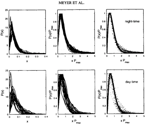
0 0 1 0 2 0 3 0 4 0 1 2 3 4 5 1 2 3 4 X X P m ~ x PFIG. 5. Cumulative amplitude probability distributions (unscaled, left; rescaled, middle) of 4 hr nighttime (upper panels) and 4 hr daytime (lower panels) subepochs in an individual subject (#8, n = 23) sojourning at high altitude. The apparent truncation at 1.0 in the middle panels results from rescaling. The solid line (right panels) is a fit of the rescaled distributions to a stable Gamma distribution (see text for details).
subject (#8) during sojourn at high altitude, i.e., during ascent to 5,050 m altitude, a sojourn at 5,050 m for 34 days, and return to sea level. Inspection of the unscaled distribu- tion functions of the amplitude of the cumulative variations (Figure 4, left) reveals marked differences as a result of changes in the level of physical activity and environmental hypoxic stress. In contrast, the rescaled distributions (Figure 4, right) demonstrate "data collapse", i.e., the data conform to a single scaled plot that is well approximated by a homogeneous Gamma distribution defined with a single parameter (cf. eq. 8). These re- suits demonstrate that there exists a hidden, potentially universal, common structure in the (unscaled) heterogeneous time series. A common scaling function with a stable Gamma distribution defines the probability density Of the (rescaled) amplitudes of the fluctuations in the heartbeat interval time series for an individual subject, irrespective of nonstationarity or varying physiological conditions. Moreover, the stability of the distributions is unaf- fected by the subject's exposure to a hypobaric (hypoxic) environment. The results suggest that the observed scaling of the distributions for healthy subjects constitutes an intrinsic property of normal heartbeat dynamics that would lack any adaptation to extrinsic environ- mental physiological stress, e.g., chronic hypoxia.
The results of the daytime and ,nighttime 4 hr subepochs in the same subject are summa- rized in Figure 5. The rescaled distributions (middle panels) demonstrate similar scaling behavior with a Gamma distribution that applies to both night and day phases and, again,
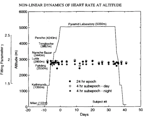
NON-LINEAR DYNAMICS OF HEART RATE AT ALTITUDE 6000 353 2.5 2- E Q_ cr) i- LL 1.5 5000 4000 3000 2000 I00C C -20 Periche (4243m) I Tengboche l (3867m) / Namche Bazar / - J (3440m) t Lukla / (zssom) ti ~tt, l~ 9 9 Pakding ,r.y, _ 9 (2550rn) ] e 9 K a t h r n a n d u , . J (1350m) / 0 9 M i l a n ~ I I -10 0 Pyramid Laboratory (5050m) 0 5 9 9 24 hr epoch 4 hr subepoch - day " - ~ 4 hr subepoch - night Subject #8 L i I I i 10 20 30 40 50 Days
FIG. 6. Fitting parameter ~/of individual Gamma distributions of 24 hr, 4 hr daytime and 4 hr nighttime heartbeat interval time series of a subject (#8) during acclimatization to high altitude (5,050 m). The locations and altitudes of the measurements are indicated. The arrival at the Pyramid Laboratory is arbitrarily assigned as day 0. A more detailed description of the experimental protocol and methods of data acquisition is given in a
previous report (cf. Meyer et al., 1998a).
neither daytime nor nighttime sequences show any systematic changes in response to high altitude exposure. Although the day-phase distributions show a larger variance compared to the night-phase distributions, the results indicate that the apparently universal scaling behavior of heartbeat dynamics holds not only for the night phase but also for the day phase. The solid line (right panels) is a fit of the rescaled distributions to a stable Gamma distribution. Whereas the daytime distributions follow the Gamma distribution over almost the entire range, the nighttime distributions show a systematic deviation in the tails, i.e., deviation from the exponential form of the decay. The slower decay in the tails of the nighttime distributions indicates higher probability of larger variations in the cardiac dy- namics compared with the daytime dynamics and is suggestive of an enhanced complexity of cardiac dynamics during nocturnal episodes (see Discussion). However, the characteris- tics of the nighttime distributions would not be reflected in the distributions of the full- length 24 hr epochs that mostly (~70%) constitute daytime characteristics.
A comprehensive representation of the cumulative variation amplitude analysis of the serial 24 hr heartbeat interval time series and its 4 hr day/nighttime subsets of subject #8 along with the altitude profile and locations, covering sea level, ascent, sojourn at 5,050 m for 34 days, and return from altitude, is shown for the fitting parameter (y) of the indi- vidual Gamma distributions in Figure 6. The fitting parameters were unaffected by the subject being exposed to environmental hypoxic stress and no differences were apparent when the data from the initial stages of exposure to hypoxia were compared with those of
354 MEYER ET AL.
TABLE 1
Scaling Properties of Nonstationary Heartbeat Interval Time Series 24 hr epoch 4 hr subepoeh 4 hr subepoch
Subject n Daytime Nighttime
#1 18 1.77 + 0.09 1.85 + 0.05 1.69 + 0.10 #2 22 1.65 + 0.09 1.77 + 0.10 1.74 + 0.11 #3 22 1.72+0.10 1.75+0.10 1.76+0.10 #4 22 1.67+0.10 1.71 +0.10 1.79+0.12 #5 16 1.61 + 0.09 1.77 + 0.09 1.76 + 0.13 #6 17 1.77 + 0.09 1.76 + 0.09 1.74 + 0.11 #7 23 1.70 + 0.11 1.85 + 0.08 1.66 + 0.07 #8 23 1.79 + 0.13 1.86 + 0.10 1.80 + 0.07 #9 17 1.74 + 0.I0 1.79 + 0.10 1.89 + 0.05 Group mean 1.71 + 0.06 1.79 + 0.10 1.76 + 0.05
Note: Values of individual subjects are means + SD of fitted Gamma distributions with fitting parameter ~of serial measurements averaged over time of the experimental protocol.
the later stages. No systematic directional changes of the Gamma scaling of the distribu- tions were observed in any of the 9 subjects that were exposed to chronic hypoxia. The group mean averages + SD of the Gamma distributions with fitting parameter 7 are com- piled in Table 1.
The lack of evidence of any effects of hypoxia on heartbeat interval dynamics in terms of potential readjustments during the early phase or acclimatization in the late phase of sojourn at high altitude is supported by the analysis of the 5 subjects who were monitored simultaneously (see Meyer et al., 1998a; Methods, Experimental Protocol). The data base consists of 6 data sets each for the early and late phases, respectively. In the early phase, the subjects were monitored continuously for 6 days (separated by 10 min breaks), whereas
in the late phase the recordings were performed over a period of 16 days (separated by 48 hr intervals). There were no differences in the stable Gamma scaling behavior of cardiac dynamics in any of the 5 subjects (Kolmogorow-Smirnov test), no matter whether the early vs. late comparison was based on the full-length 24 hr epochs or 4 hr day/night subepochs.
The stable scaling properties observed a) in normal subjects at sea level (intersubject comparison), b) in normal subjects sojourning at high altitude, and c) during daytime and nighttime episodes (intrasubject comparison) are suggestive of a common structure o f heartbeat interval dynamics that would not be explained by physical activity or environ- mental hypoxic stress. The present findings indicate that nonstationarity (patchiness) due to unsteady physiological conditions was unlikely to account for the nonstationary 24 hr heartbeat interval time series and their ensuing scaling properties.
D i s c u s s i o n
The present study presents a dynamical analysis of heart rate fluctuations in normal subjects during long-term (> 14 days) exposure to high (> 5,000 m) altitude hypoxia. The analysis of the scaling properties of long-term 24 hr heartbeat interval time series has revealed the following important findings:
1. The distributions of the long-term fluctuations in normal subjects exhibit a single function which is stable over a wide range of time scales. The observed scaling
NON-LINEAR DYNAMICS OF HEART RATE AT ALTITUDE 355
property appears to reflect the underlying intrinsic nonlinear cardiac dynamics and is reminiscent of a wide class of physical systems with universal scaling properties (Stanley, 1971; Vicsek, 1992).
2. The stable scaling properties of the dynamics are essentially unaffected by the physi- ologic challenge of chronic exposure to high altitude hypoxia, i.e., the intrinsic heart- beat dynamics do not undergo early readjustment or late acclimatization.
3. The analysis of the rescaled distributions of the 4 hr daytime and 4 hr nighttime subepochs selected as subsets of the full-length 24 hr time series showed similar scaling behavior with a stable Gamma distribution indicating that the common struc- ture was unequivocally applicable to both day and night phases and, furthermore, was unaffected in response to high altitude exposure.
4. The stability of the scaling form suggests that the underlying dynamical mechanisms of heart rate regulation in health have similar statistical properties on different time scales and hence exhibit self-similar fractal dynamics.
5. In contrast, a single function stable over a wide range of time scales was not observed in cardiac disease, i.e., in patients with congestive heart failure or patients after cardiac transplantation
(see below).
The effects of high altitude hypoxia on the long-term variability of heart rate has recently been addressed by nonlinear signal processing (Meyer et al., 1998a). This analy- sis, in part based on the same data base as the present study, has identified nontrivial long- range correlations (memory-like effects) and scale-invariant fractal power-law characteris- tics ( I / f noise) of the heartbeat patterns that were remarkably consistent between individu- als and were unaffected by external or environmental stimuli (chronic hypoxia). The fractal scaling exponent (a) that was calculated by the Detrended Fluctuation Analysis (DFA - algorithm, cf. Meyer et al., 1998a) was -1.0 (0.99 + 0.04, mean + SD) indicating genuine self-similar fractal dynamics that were not the consequence of summation over random variables or artifacts of nonstationarity due to different levels of physical activity. The lack of a characteristic time scale along with the absence of any effects of exposure to chronic hypoxia on scaling properties has led to the concept that neuroautonomic cardiac control underlying heart rate dynamics was
preadapted
to hypoxia, whereas the heart rate patterns in disease, approaching random walk-like fluctuations ( a -1.5), were interpreted as evidence that the heart wasunadapted
to its environment. Here the difference in the time series of cardiac intervals in health and disease was attributed to thetime ordering
rather than to the distribution of interbeat variations. In the present study the underlying concept of analysis was that different dynamical patterns, reflected in thedistribution
of interbeat variations, can be observed on different time scales that correspond to the time scales of the operating physiological processes (provided that masking effects of nonstationarities have been eliminated). To that end, the results of the present analysis suggesting that the stable Gamma scaling of the distributions in healthy subjects consti- tutes an intrinsic dynamical property of normal heart function that would not undergo readjustment or adaptation to extrinsic environmental physiological stress is perfectly in line with our previous findings (cf. Meyer et al., 1998a). It is important to emphasize that the present algorithmic approach which is based on the wavelet transform and on an analytic signal approach by the Hilbert transform is substantially different from the modi- fied random-walk analysis used in the previous study.356 MEYER ET AL. 0 2 4 6 8 10 x 10" 0 2 4 6 8 10 x lo"
j
O2 2O 10 5 0 0 0 . 1 0 2 0 . 3 0 4 x ,,0.8 02 L ~ ~ = ~ %- 0 2 4 6 8 l o 0 1 2 3 4 5 N u m b e r of P o i n t s x ~o' X PmaxFro. 7. Cumulative variation amplitude analysis of artificial genuine stationary fractal time series. Original time series (upper left), wavelet transform (middle left), Hilbert transform (lower left). Cumulative amplitude probability distributions (right panels) for 10 realizations of the fractal process (see text for details).
Non-Stationarity and Heart Rate Dynamics
The identification of hidden dynamical patterns in biological time series analysis and its limitations are closely associated with and complicated by the inherent characteristics of the biological system under study. Specifically, biological systems are expected to adjust continuously to their internal and external environment. These environments are changing on different time scales while, simultaneously, the adjusting mechanisms available to biological systems also operate on different time scales. Thus, biological systems may be assumed to be highly nonequilibrium systems characterized by continuous changes on different time scales. Moreover, relevant experimental time series may require long peri- ods of time relative to the time scales on which the system under study is operating.
Biological systems typically consist of many components that ultimately determine the temporal evolution of the system. Only few independent and dependent variables are generally assumed to be underlying a biological time series whereas the remaining are interpreted as stochastic perturbation with little relevance for the dynamical evolution of the system. However, "stochastic fluctuations" of the recorded variable are often found to be large relative to the "intrinsic fluctuations" yielding a low signal-to-noise ratio of the data set. To further complicate these issues, biological systems may exhibit sudden transi- tions between quantitatively different states from complex to stereotypic oscillatory pat- terns.
The 24 hr heartbeat time series of the present study were highly nonstationary, with heart rate ranging from 40/min (nighttime supine resting conditions) to about 200/min
NON-LINEAR DYNAMICS OF HEART RATE AT ALTITUDE 357
(daytime physical activity up to exhaustion). In the present analysis, the anticipated intrin- sic dynamics of a complex nonlinear system were assumed to be superimposed by extrin- sic sources, i.e., nonstationarity due to nonsteady state physiological or environmental conditions that could give rise to highly nonstationary behavior. The general concept here is that the exceedingly complicated heartbeat interval time series can be decomposed by the wavelet transform given an appropriate choice of the analytic wavelet and visualized in a spectrum of scales after elimination of local polynomial behavior (trends). Although these "trends," e.g., due to varying physical activity, may be physiologically important, the associated strain-related uncorrelated responses would obscure the intrinsic dynamics of the system itself. The wavelet transform yielding a cumulative measure of the variations in the time series over a region proportional to the wavelet scale thus facilitates the identifica- tion of the intrinsic properties of the dynamics embedded in a nonstationary time series.
In order to further support the concept that the stable Gamma scaling of the distributions was an intrinsic property of normal cardiac dynamics, the cumulative variation amplitude analysis was applied to artificial genuine stationary fractal time series. Figure 7 (left) shows a genuine time series (with the same statistical properties as the heartbeat interval time series of Figure 1) generated by a stationary fractal process with long-range power- law correlations
(llf
dynamics, scaling coefficient ct = 1.0) and the results of the cumula- tive variation amplitude analysis (right) for 10 realizations of the fractal process. Similarly to the experimental time series (Figure 1) the genuine time series displays patchy patterns changing with time and "drift-like" or "trend-like" fluctuations at all time scales. However, the underlying process is stationary, i.e., has a unique fractal dimension. By definition, a long-range correlated times series consists of drift-like or trend-like fluctuations at all time scales that are the consequence of the built-in long-range power-law correlations rather than the result of nonstationarity of the underlying fractal mechanism. The rescaled ampli- tude distributions, after wavelet transform, reveal some similarity with the results of the experimental time series, but a more rigorous assessment shows that the distributions follow a Raleigh probability distribution (lack of an exponential tail) which presents a special case of the generalized Gamma distribution:P(xla, b,A)=
r(a)A (
b
A)ab_le-(A)b(9)
where, the parameters (a, b, A) assigned to particular values, a number of well-known probability functions appear as s~ecial cases (a = a, b = l, A = l: elementary Gamma distribution; a = l, b = 2, A = q2cr : Raleigh distribution function). Similarly, tracking the intrinsic dynamics of the original heartbeat interval time series is facilitated by construct- ing Phase-Randomized Surrogate data from the original time series (cf. Kantz and Schreiber, 1997; Kaplan and Glass, 1995; Theiler et al., 1992). Surrogate data have two important properties in that the process underlying their generation is both stationary and linear (absence of any dynamical nonlinearities). The mean, variance, and power spectrum of the surrogate data are identical to that of the test data. Realizations of phase-randomized surrogate data derived from the original time series result in a Raleigh distribution of the rescaled probability distributions of the amplitudes of the variations after wavelet transfor- mation. These findings point at the important role of nonlinear phase correlations in the original cardiac interbeat series that are preserved in the cumulative variation amplitude analysis.
358 MEYER ET AL.
Complexity, Scaling, and Fracta! Dimension
The rescaled distributions of the 4 hr daytime subepochs were found to follow a Gamma distribution over almost the entire range whereas the distributions of the 4 hr nighttime subepochs showed a systematic deviation in the tails from the exponential form of the decay. These findings are indicative of a higher probability of larger variations in the nocturnal cardiac dynamics as compared to the daytime dynamics. The emergence of "strange" heart rate patterns potentially associated with the onset of periodic breathing at altitude has previously been described (Lipsitz et al., 1995; Malconian et al., 1990; Yamamoto et al., 1993) and various types of highly irregular heart rate oscillations other than the normal daytime pattern were also observed in the present data base. Our previous statistical analysis of heartbeat interval dynamics at high altitude has identified long-range correlations and power-law scaling in the normal heartbeat fluctuations. Specifically, in the Detrended Fluctuation Analysis, the 4 hr nighttime subepochs had lower scaling coeffi- cients (~ = 0.86) compared to the 4 hr daytime subepochs (~ = 0.96), and the fractal dimension calculated by the Point-Correlation Dimension (PD2i) was increased from 2.6 to 3.8, respectively (cf. Meyer et al., 1998a). The scaling behavior of the nocturnal epi- sodes reflected in the tails of the rescaled distributions would point at an increased com- plexity of the intrinsic cardiac dynamics, but the physiological significance for the control of heart rate remains unclear. In patients with cardiopulmonary instability caused by ob- structive sleep apnoea, Ivanov et al. (1996; 1998) found a dramatic alteration in the scaling pattern in the rescaled distributions as a result of transient pathologic low-frequency heart rate oscillations associated with periodic breathing. However, it is important to emphasize that the low-frequency oscillations during nocturnal episodes at altitude that were poten- tially associated with the onset of periodic breathing had a probability distribution that was clearly different from the group of patients with obstructive sleep apnoea. It would seem that the cumulative variation amplitude analysis could pick up salient scaling features of cardiac regulation that may help distinguish between physiologic and pathologic modes.
Origin of Intrinsic Scaling Properties of Heartbeat Fluctuations
This study has established that the scaling properties of heart rate constitute an intrinsic feature of normal cardiac dynamics emerging from nonlinear interactions (nonlinear phase correlations) between different components of the system. The control mechanisms under- lying heart rate and its fluctuations are acting over a wide range of time scales that are not independent. This scaling behavior calls for an organizational principle for coordinating these time scales, which, in physiological conditions, is not badly locked to any single mode.
The cardiac rhythm results from the spontaneous activity of the sino-atrial node which is further modulated by the activity of the sympathetic and parasympathetic components of the autonomic nervous system and by other cardiac and extracardiac factors. The precise mechanisms of interaction have not been identified but offer the possibility of interaction of several negative or mixed feedback systems with delays that are known to produce periodic, quasiperiodic, or even chaotic dynamics (Glass and Malta, 1990). Even though the present experiments and analysis did not specifically address the origin of the intrinsic scaling properties of heartbeat fluctuations, preliminary results from patients with cardiac disease may provide some clues that will help put the role of the autonomic nervous system into perspective with respect to the scaling behavior of heartbeat interval fluctuations.
*C 13.
t 0 0
NON-LINEAR DYNAMICS OF HEART RATE AT ALTITUDE
CTL CHF 100, 80 60 40 20 0
&,
01 x 13.. 80 60k
013 0.4 0 X X 0.4 359 1 0.8 ~:. Eo.6 x 0.4 0.2 0 I 2 3 4 x P max 0.8 x ~ " 0.4 0.2 1 2 3 4 x P maxFIc. 8. Cumulative amplitude probability distributions (unscaled, upper; rescaled, lower) of 24 hr heartbeat interval time series of a group of normal subjects (CTL, n = 9, left) and a group of patients with congestive heart failure (CHF, n = 11, right).
Figure 8 shows the probability distributions of nonstationary cardiac intervals (24 hr Holter recordings) of a group of normal subjects (CTL, left) and a group o f patients with congestive heart failure (CHF, right). Whereas the normal subjects demonstrate G a m m a scaling stable over a wide range of time scales characteristic for healthy heart rate dynam- ics, the distributions of the cardiac patients do not conform to a single scaled plot. The subjects with cardiac disease show more individual probability distributions which fail to exhibit the characteristic data collapse.
A straightforward approach as to the significance of autonomic control for the hidden time-ordered scaling features is facilitated by studies in recipients of a cardiac transplant (HTR). Notably, the transplanted allograft is deprived of its neuroautonomic control, and the recurrence of autonomic control due to reinnervation remains an open issue. Figure 9 shows the probability distributions of two patient groups who underwent cardiac transplan- tation in different locations. The HTR-Paris group (left) shows a dramatic alteration in the scaling pattem with stereotypic single-mode locking. Similarly, a subgroup of the HTR- Bergamo group, i.e., patients early (< 2 yrs) after transplantation, shows the same pattern (middle) while the remaining, i.e., patients late (> 2 yrs) after transplantation, demonstrate an intermediate pattern, with some of the individual distributions approaching the normal pattern (right).
These findings appear to suggest two important aspects of cardiac regulation. In the intact heart, control of heart rate is governed by the extrinsic nervous system, i.e., the branches of the central autonomic nervous system, and the intrinsic cardiac nervous system located at the base of the heart containing efferent post-ganglionic sympathetic and para-
360 100 80 6O x 40 H T R - Paris
L
01 0.2 0 3 0 4 X O8 ~'*04 o o 1 2 3 4 x P MEYER ET AL. H T R - B e r g a m o early'i
L
o ' ' 0 1 0 2 0 3 X x 0 8:~
~" o41. x Pma~ H T R - B e r g a m o late lOOr 4O o O l 0 2 0 3 X 1 0 8~
E 0 6 O 2 0 x Pm~Fro. 9. Cumulative amplitude probability distributions (unscaled, upper; rescaled, lower) in patients after cardiac transplantation, HTR - Paris (n = 25), heart transplant recipients operated at La Pitie-Salpetri&e Hospital, Paris, 1981-1990; HTR-Bergamo (n = 10, 13), heart transplant recipients operated at Riuniti Hospi- tal Bergamo, 1987-1995 (see text for details).
sympathetic neurons, local circuit neurons and afferent neurons (Murphy et al., 1994; Skinner and Kresh, 1996). The stable Gamma scaling of the heartbeat interval distributions is attributable to the activity of the extrinsic and intrinsic nervous system of the heart. Breakdown (in CHF patients) or absence (in early HTR patients) of autonomic control results in a dramatic change of the pattern of the distributions that easily allows for distinction between normal and pathologic modes of heartbeat control. We have previously argued, based on conventional linear analysis (power spectrum analysis), that the trans- planted heart remains denervated indefinitely (cf. Meyer et al., 1992). This conclusion was based on the analysis of the (older) HTR-Paris data, and the results of the present scaling analysis do in fact support our previous hypothesis. However, the additional data from the (more recent) HTR-Bergamo group would call for revision in that functional recurrence of heart rate control late after cardiac transplantation is likely to occur and may be due to reinnervation of the transplanted allograft b y t h e extrinsic nervous system, but preliminary evidence suggests that the post-transplant recovery of heart rate fluctuations may be due to reorganization (self-organization) of viable intrinsic cardiac neurons (Meyer et al., 1996). The reason for the discrepancy between the HTR-Paris and HTR-Bergamo groups remains unclear and is expected to be attributable to clinical differences in the early induction of immunosuppressive therapy and the ensuing effects on the viability of intrinsic cardiac neurons rather than to differences in cardiosurgical procedures. Although the Gamma scaling behavior of heart rate fluctuations constitutes an essential feature of normal cardiac function, the transplanted heart which, in terms of its heart rate control is deprived of its
NON-LINEAR DYNAMICS OF HEART RATE AT ALTITUDE 361
scaling features, may well provide for a sufficient cardiac output by compensatory mecha- nisms of cardiac contractility, and neuroautonomic denervation per se is unlikely to ac- count for the impaired exercise capacity of cardiac transplant recipients (Grassi et al.,
1997; Meyer et al., 1992).
Summary and Perspectives
The cumulative variation amplitude analysis of heartbeat intervals in health and disease has revealed the following important features:
1. The distributions of the fluctuations in the heartbeat intervals in normal subjects can be described by a single function (Gamma distribution) stable over a wide range of time scales.
2. Under physiological conditions, these distributions reflecting the nonlinear dynamics underlying the control of heart rate remain stable and are unaffected by environmen- tal stress, e.g., chronic high altitude hypoxia, suggesting that the heart is preadapted to systemic (arterial) hypoxia.
3. The characteristic scaling patterns are the result of the activity (i.e., nonlinear interac- tion) of the components of the neuroautonomic control system of the heart.
4. Pathologic modes of cardiac regulation may be identified from the scaling properties of heartbeat interval time series.
5. Analysis of heartbeat scaling properties may help detect the recurrence of cardiac control and its significance in long-term survivors of cardiac transplantation.
Acknowledgments
Supported by Swiss National Science Foundation, Grant no. 32-040397.94, and by American National Insti- tutes of Health, Grant no. NS27745-06.
References
Arfken, G. (1985). Mathematical Methods for Physicists, 3rd ed., pp. 401-403. Orlando, FL: Academic Press. Arneodo, A., Bacry, E., Graves, P.V., and Muzy, J.F. (1995). Characterizing long-range correlations in DNA
sequences from wavelet analysis. Physical Review Letters, 74: 3293-3296.
Bak, P., and Creutz, M. (1994). Fractals and self-organized criticality. In A. Bunde and S. Havlin (eds.),
Fractals in Science, pp. 27-47. New York: Springer.
Beran, J. (1994). Statistics for Long-Memory Processes. New York: Chapman & Hall.
Cerretelli, P. (1980). Gas exchange at high altitude. In J.B. West (ed.), Pulmonary Gas Exchange, pp. 97-147. New York: Academic Press.
Cerretelli, P., and Hoppeler, H. (1996). Morphologic and metabolic response to chronic hypoxia: The muscle system. In Handbook of Physiology, Section IV; Environmental Physiology, Vol. II, pp. 1155-1181. New York: Oxford University Press.
Daubechies, I. (1992). Ten Lectures on Wavelets. Philadelphia: SIAM Press.
Gabor, D. (1964). Theory of Communication. Journal of the Institute of Electrical Engineers, 93: 429--457. Glass, L., and Malta, C.P. (1990). Chaos in multi-looped negative feedback systems. Journal of Theoretical
Biology, 145: 217-223.
Grassi, B., Marconi, C., Meyer, M., Rieu, M., and Cerretelli, P. (1997). Gas exchange and cardiovascular kinetics upon different exercise protocols in heart transplant recipients. Journal of Applied Physiology, 82:
1952-1962.
Ivanov, P.Ch., Rosenblum, M.G., Peng, C.-K., Mietus, J., Havlin, S., Stanley, H.E., and Goldberger, A.L. (1996). Scaling behavior of heartbeat intervals obtained by wavelet-based time series analysis. Nature, 383: 323-327.
362 MEYER ET AL.
Ivanov, P.Ch., Rosenblum, M.G., Peng, C.-K., Mietus, J.E., Havlin, S., Stanley, H.E., and Goldberger, A.L. (1998). Scaling and universality of heart rate variability distributions. Physica A, 249: 587-593.
Kaplan, D.T., and Glass, L. (1995). Understanding Nonlinear Dynamics. New York: Springer. Kaiser, G. (1994). A Friendly Guide to Wavelets. Boston: Birkhauser.
Kantz, H., and Schreiber, T. (1997). Nonlinear Time Series Analysis. Cambridge: Cambridge University Press. Lipsitz, L.A., Hashimoto, F., Lubowsky, L.P., Mietus, J., Moody, G., Appenzeller, O., and Goldberger, A.L.
(1995). Heart rate and respiratory rhythm dynamics on ascent to high altitude. British Heart Journal, 74: 390-396.
Malconian, M., Hultgren, H., Nitta, M., Anholm, J., Houston, C., and Fails, H. (1990). The sleep electrocardio- gram at extreme altitude (operation Everest II). American Journal of Cardiology, 65: 1014-1020.
Meyer, M., Marconi, C., Grassi, B., Rieu, M., Cerretelli, P., and Cabrol, C. (1992). Adjustment of cardiac output to step exercise in heart transplant recipients. Applied Cardiopulmonary Pathophysiology, 4: 213- 223.
Meyer, M., Marconi, C., Ferretti, G., Fiocchi, R., Cerretelli, P., and Skinner, J.E. (1996). Heart rate variability in the human transplanted heart: Nonlinear dynamics and QT vs. RR-QT alterations during exercise suggest a return of neurocardiac regulation in long-term recovery. Integrative Physiological and Behavioral Sci- ence, 31: 289-305.
Meyer, M., Rahmel, A., Marconi, C., Grassi, B., Skinner, J.E., and Cerretelli, P. (1998a). Is the heart pre- adapted to hypoxia? Evidence from fractal dynamics of heartbeat interval fluctuations at high altitude (5,050 m). Integrative Physiological and Behavioral Science 33: 9--40.
Meyer, M., Marconi, C., Rahmel, A., Grassi, B., Ferretti, G., Skinner, J.E., and Cerretelli, P. (1998b). Fractal dynamics of heartbeat interval fluctuations in health and disease. In F. Orsucci (ed.), The Complex Matters of the Mind. pp. 105-128. Singapore: World Scientific.
Murphy, D.A., O'Blenes, S., Hanna, B.D., and Armour, J.A. (1994). Functional capacity of nicotine-sensitive canine intrinsic cardiac neurons to modify the heart. American Journal of Physiology, 266:R1127-R1135. Muzy, J.F., Bacry, E., and Arneodo, A. (1994). The multifractal formalism revisited with wavelets. Interna-
tional Journal of Bifurcation and Chaos, 4: 245-302.
Panter, P. (1965). Modulation, Noise and Spectral Analysis. New York: MacGraw-Hill.
Peng, C.-K., Mietus, J., Hausdorff, J.M., Havlin, S., Stanley, H.E., and Goldberger, A.L. (1993). Long-range anti-correlations and non-Gaussian behavior of the heartbeat. Physical Review Letters, 70:1343-1346. Peng, C.-K., Havlin, S., Stanley, H.E., and Goldberger, A.L. (1995). Quantification of scaling exponents and
crossover phenomena in nonstationary heartbeat time series. Chaos, 5: 82-87.
Rabiner, R., and Gold, B. (1975). Theory and Application of Digital Signal Processing. Englewood, NJ: Prentice-Hall.
Skinner, J.E., and Kresh, J.Y. (1996). Self-organization in a simple biological system: The intrinsic cardiac nervous system. In G. Mayer-Kress, H. Kantz, J. Kurths (eds.), Nonlinear Techniques in Physiological Time Series Analysis. New York: Springer.
Smith, M.J., and Mersereau, R.M. (1992). Introduction to Digital Signal Processing: A Computer Laboratory Textbook. New York: Wiley.
Stanley, H.E. (1971). Introduction to Phase Transitions and Critical Phenomena. New York: Oxford Univer- sity Press.
Theiler, J., Eubank, S., Longtin, A., Galdrikian, B., and Farmer, J.D. (1992). Testing for nonlinearity in time series: the method of surrogate data. Physika D, 58: 77-94.
Ueda, G., Reeves, J.T., and Sekiguchi, M. (1992). High Altitude Medicine. Matsumoto: Shinshu University Press.
Vicsek, T. (1992). Fractal Growth Phenomena (2nd. ed.). Singapore: World Scientific Publishing.
Ward, M.P., Milledge, J.S., and West, J.B. (1990). High Altitude Medicine and Physiology. Philadelphia: University of Philadelphia Press.
Yamamoto, Y., Hughson, R.L., Sutton, J.R., Houston, C.S., Cymerman, A., Fallen, E.L., and Kamath, M.V. (1993). Operation Everest II: An indication of deterministic chaos in human heart rate variability at simu- lated extreme altitude. Biological Cybernetics, 69: 205-212.