ORIGINAL ARTICLE
Antenatal and postnatal ultrasound in the evaluation
of the risk of vesicoureteral reflux
Serge Grazioli&Paloma Parvex&Laura Merlini&
Christophe Combescure&Eric Girardin
Received: 21 December 2009 / Revised: 15 April 2010 / Accepted: 16 April 2010 / Published online: 4 June 2010 # IPNA 2010
Abstract Antenatal hydronephrosis (ANH) is a frequent anomaly detected on fetal ultrasound scans. There is no consensus recommendation for the postnatal follow-up and/or the necessity to perform a voiding cystourethrography (VCUG) to diagnose vesicoureteral reflux (VUR). We conducted a cohort/non-randomized trial of 121 patients with ANH, defined as an anterior posterior diameter (APD)≥5 mm after the 20th week of gestation, to evaluate the ability of the antenatal and postnatal ultrasonography results to predict VUR. All infants had two successive ultrasounds at 5 days and 1 month, respectively, after birth. A VCUG was performed at 6 weeks in children with a persistent APD≥5 mm and/or an ureteral dilatation observed on at least one of two postnatal ultrasounds. In total, 88 patients had VCUG and nine had VUR, with five having high-grade reflux (>grade II). The risk of VUR increased significantly with the degree of APD detected on the postnatal ultrasound scan (p=0.03). The odds ratios were 5.0 [95% confidence interval (CI) 0.5– 51.2] for APD = 7–9 mm and 9.1 (95% CI 1.0–80.9) for APD≥10 mm. The results of this study show that among
our patient cohort antenatal ultrasound was not predictive of reflux. There was, however, a relation between the importance of the postnatal renal pelvis diameter and the risk of VUR. A cut-off of 7 mm showed a fair ability of ultrasonography to predict VUR and a cut-off of 10 mm enabled all severe refluxes in the 88 patients who had a VCUG to be diagnosed.
Keywords Hydronephrosis . Nephrology . Prenatal diagnosis . Ultrasonography . Vesicoureteral reflux
Abbreviations
APD anterior posterior diameter RPD renal pelvic dilatation UTI urinary tract infection VCUG voiding cystourethrography VUR vesicoureteral reflux
Introduction
Antenatal hydronephrosis (ANH) accounts for 50% of anomalies detected by prenatal ultrasound [1], with a 2.5–
5% prevalence in the second trimester [2]. Although the presence of a renal pelvic dilatation (RPD) is often a physiological and transient state, it can be associated with urinary tract anomalies [3,4].
However, even if the presence of antenatal dilatation is an important determinant for the management of congenital renal diseases, the relationship between the degree of antenatal and postnatal pelvic dilatation and the risk of the patient to present with vesicoureteral reflux (VUR) remains unclear. Indeed, the use of the postnatal ultrasound for the screening of VUR remains controversial, and there is no
S. Grazioli
:
P. Parvex:
E. Girardin (*)Department of Pediatric, Pediatric Nephrology Unit, University Hospital of Geneva, University of Geneva, 6 Willy-Donzé Street,
1211 Geneva, Switzerland e-mail: eric.girardin@hcuge.ch L. Merlini
Department of Pediatric, Pediatric Radiology Unit, University Hospital of Geneva, University of Geneva, Geneva, Switzerland
C. Combescure
Division of Clinical Epidemiology,
University Hospital of Geneva, University of Geneva, Geneva, Switzerland
clear recommendation in the literature on the indication to perform a voiding cystourethrography (VCUG), which is currently the gold standard for the detection of VUR [5–9]. This situation generates a relevant number of invasive and irradiating clinical examinations associated with a signifi-cant parental anxiety [10].
Many studies have focused on RPD and VUR, but only a few have specifically focused on the relation between the degree of the APD and the risk of VUR and, in particular, high grade VUR (>grade II) responsible for long-term renal morbidity [6,11–15]. In this prospective study, we analyzed the relationship between the antenatal and postnatal ante-roposterior diameter of RPD and the presence of reflux.
Patients and methods
Patients
We prospectively followed all children referred to our nephrology unit with the diagnosis of ANH between May 2004 and May 2006. This study was approved by the local University Ethics Committee.
The Swiss Maternity Health Care Program offers all pregnant women the opportunity to have an ultrasound examination on two occasions during the second and third trimester. The aim of the ultrasound performed during the second trimester is to detect fetal anomaly, including uro-nephrologic malformations. The ultrasound of the third trimester is performed routinely for evaluation of amniotic fluid and fetal behavior. Women in whom an anomaly was detected on the first ultrasound receive an ultrasound during the third trimester that specifically focuses on control of the anomaly. In this group of women, the largest anteroposterior diameter (APD) is taken into consideration for inclusion.
The inclusion criterion of our study was the presence of a pelvic dilatation with an APD≥5 mm measured on the antenatal renal ultrasound scan performed at our hospital after the 20th week of gestation. We excluded patients with non-urological congenital anomalies, duplex system, renal cysts, chromosomal anomalies, missing antenatal data or incomplete follow-up.
All patients had two successive neonatal ultrasound examinations of the urinary tract, namely, on day 5 and at 1 month after birth. The infants were categorized according to the APD as follows: normal (0–4 mm), mild (5–9 mm), moderate (10–15 mm) and severe (>15 mm) [16]. Only patients in whom abnormalities persisted on one or both postnatal ultrasound examinations had a VCUG at 6 weeks after birth [9]. Criteria for an abnormal postnatal ultrasound included pelvic diameter ≥5 mm, calyceal or ureteral dilatation (ureteral diameter >2 mm), pelvic or ureteral wall thickening, absence of the corticomedullary
differen-tiation and signs of renal dysplasia (small kidney, thinned or hyperechogenic cortex and cortical cysts) [17]. VUR was graded according to the International Reflux Study Com-mittee classification [18]. Because of the benign evolution of grade I and II VUR [6], we classified the VUR into two groups: mild-moderate (I–II) and severe (III–V). Patients with bilateral VUR or bilateral dilatation were categorized into the higher grade of VUR or dilatation.
Antibiotic prophylaxis using amoxicillin (10 mg/kg/day) was started at birth and replaced by trimethroprim (2 mg/kg/day) at 3 months. The antibiotic prophylaxis was discontinued in patients without VUR on VCUG and in those with RPD <10 mm. The follow-up in the pediatric nephrology unit was stopped at 1 month in children with two normal consecutive ultrasound scans and in children without VUR based on the results of the VCUG. The children with persistence of a dilatation were followed with repeated ultrasounds at 3, 6, 12 and 24 months after birth.
The clinical course at 1 year was recorded for all children either by means of the clinical follow-up in the pediatric nephrology unit or a phone questionnaire for patients whose follow-up was stopped at 1 month. To reduce the number of false negative reports due to memory failure, we also reviewed the medical charts of all the children for the presence of urinary tract infection (UTI) and the need of a surgical procedure. As our hospital is the referral pediatric center in the region, the information contained in these charts was comprehensive.
Imaging
The postnatal ultrasound examinations were performed by trained pediatric radiologists using an Acuson Sequoia machine with 6-MHz convex and 8-MHz linear transducers.
Statistical methods
A descriptive analysis was performed (frequencies for the qualitative variables and mean, median for the quantitative variables). For the antenatal and postnatal examinations, the proportions of VUR were assessed according to the ultrasound results and categorized into three classes: ≤6 mm, 7–9 mm and ≥10 mm. The 95% confidence intervals (CIs) were assessed using the exact method of Clopper–Pearson. The increase of the risk of VUR according to the antenatal and postnatal RPD was checked using a linear trend test (Cochran–Armitage test). The ultrasound scans of boys and girls were compared using a Mann–Whitney test. Spearmann’s coefficient correlation was used to assess the antenatal and postnatal ultrasound scans. The statistical analysis was performed using SPSS ver. 16 (SPSS, Chicago, IL) and StatXact ver. 4.0.1 (Cytel, Cambridge, MA). A p value <0.05 was considered to be significant.
Results
From May 2004 through to May 2006, 143 children with ANH were referred to our clinic for treatment. Twenty-two cases (15%) were excluded from our study (7 for incomplete follow-up, 12 for incomplete antenatal data, 1 for kidney malformation, 1 for parental refusal for VCUG, 1 for Down syndrome). The remaining 121 newborns, all with ANH, were included (84 boys and 37 girls; male to female ratio 2:1). Among these 121 newborns, the antenatal ultrasound revealed that 76 (63%) had a mild dilatation, 37
(31%) had a moderate dilatation and eight (7%) had a severe dilatation. ANH was detected more often in boys (70%,n=85) than in girls (30%, n=36) (p <0.01).
The first postnatal ultrasound showed a normalization of the RPD in 28 patients (23%) and persistence in 93 children, with a majority of the children having a mild degree of RPD. Of the 93 children with persistent RDP, 32 (34%) showed an increase in their RPD, seven (8%) showed stabilization of the RDP and the majority (54, 58%) showed a regression in comparison with their RDP on the antenatal ultrasound. The findings on the postnatal ultrasound scan were weakly correlated to those on the antenatal ultrasound scan (Spearman's rho=0.5, p<0.01).
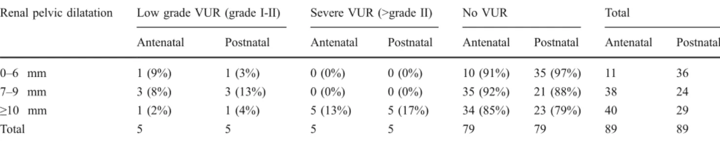
Among the 121 children, 88 had a VCUG in accordance with our protocol, and a VUR was identified in nine (10%) patients (Fig. 1). The distribution of the grade of the VUR according to the degree of pelvic dilatation is given in Table 1. A cut-off of 7 mm for APD was found to be of diagnostic value in selecting for children at risk for VUR, with a sensitivity of 90% (95% CI 55.5–99.8) and a specificity of 44.3% (95% CI 33.1–55.9). The positive and predictive values were 17% (95% CI 8.1–29.8) and 97.2% (95% CI 85.5–99.90), respectively. With a cut-off at 10 mm, sensitivity was 60% (95% CI 26.2–87.8) and specificity was 70.9% (95% CI 59.6–80.6). The positive and predictive values were 20.7% (95% CI 8.0–39.7) and 80% (95% CI 68.7–88.6), respectively. Among the 89 children who had a VCUG, all of the children with a severe VUR had a postnatal renal pelvic dilatation≥10 mm.
We found no significant difference in the mean antenatal and postnatal APD between boys and girls: 9.5 vs. 9.3 mm for the antenatal APD (p=0.63) and 8 vs. 7.4 mm for the postnatal APD (p=0.24). Among the 67 boys and 22 girls who had a VCUG, there were no difference in the number of VUR episodes (boys 10.4%, n=7 vs. girls 13.6%, n=3; p=0.70) and in the severity of the VUR (boys 57%, n=4 vs. girls 33%, n=1; p=1). The proportion of children with VUR increased with increasing postnatal RPD, as shown by the linear trend test (p=0.03). The odds ratios were 5.0 (95% CI 0.5–51.2) for children with a postnatal RPD 7–
Fig. 1 Distribution of the newborns with antenatal hydronephrosis
(ANH). VCUG Voiding cystourethrography, VUR vesicoureteral
reflux,US ultrasound scan, UTI urinary tract infection
Table 1 Distribution of the vesicoureteral reflux according to antenatal and postnatal renal pelvic dilatation in patients having a voiding cystourethrography
Renal pelvic dilatation Low grade VUR (grade I-II) Severe VUR (>grade II) No VUR Total
Antenatal Postnatal Antenatal Postnatal Antenatal Postnatal Antenatal Postnatal
0–6 mm 1 (9%) 1 (3%) 0 (0%) 0 (0%) 10 (91%) 35 (97%) 11 36
7–9 mm 3 (8%) 3 (13%) 0 (0%) 0 (0%) 35 (92%) 21 (88%) 38 24
≥10 mm 1 (2%) 1 (4%) 5 (13%) 5 (17%) 34 (85%) 23 (79%) 40 29
Total 5 5 5 5 79 79 89 89
9 mm compared to the children with a postnatal RPD 0– 6 mm, and 9.1 (95% CI 1.0–80.9) for children with a postnatal RPD ≥ 10 mm. Although we observed that all children with severe VUR had antenatal RPD≥ 10 mm, no correlation between VUR and antenatal RPD was found, and the odds ratios were 0.9 (95% CI 0.1–9.2) for children with antenatal RPD 7–9 mm and 1.8 (95% CI 0.2–16.4) for children with antenatal RPD≥ 10 mm.
Of the 121 children, 117 were followed for 1 year and underwent clinical or telephone screening for the presence of UTI. This group includes the 88 children (66 boys and 22 girls) who benefitted from a VCUG and the 29 (19 boys and 14 girls) with normal findings on the two ultrasounds after birth who did not have VCUG. Of these 117 children, ten had an UTI (7 boys and 3 girls), with the UTI occurring in two patients on antibiotic prophylaxis for VUR, in seven patients with a normal VCUG and in one patient (1 boy) followed without VCUG who had an UTI at 22 months, with the VCUG showing a grade II VUR but without renal scars, as shown on the dimercaptosuccinic acid (DMSA) scan. Among the 88 patients with a persistent RPD on the postnatal ultrasound, seven children needed surgery (8%; 5 boys and 2 girls). The mean age at surgery was 8 months and ranged from 6 days of life to 36 months. The surgical procedure included pyeloplasty in two cases, nephrostomy followed by nephrectomy in two cases, nephrostomy followed by kidney rotation in one case and ureteral re-implantation in two cases.
Discussion
Although the relationship between the degree of ANH and the risk of obstructive urinary tract anomalies is well documented [5], the correlation between the severity of APD and the risk to present with VUR is still unclear. The incidence of VUR in normal children is estimated at 9% [19], while studies that have included VCUG at birth estimate that VUR varies from 4 to 30% in children with ANH [15, 20]. Many studies have demonstrated a lack of correlation between the degree of pelvic dilatation and the risk of VUR, leading many authors to recommend performing a VCUG on all children with ANH regardless of the degree of postnatal dilatation [5,15,21,22].
Recent studies have also raised the question about the necessity to diagnose all cases of reflux, given that it is becoming evident that only high grade VUR are clinically relevant in terms of a high risk of UTI and renal scars in this patient population [23].
Children with persistent minor RPD without calyceal or ureteral dilatation seem not to be at risk for VUR [15]; indeed, in a prospective study of VCUG in children, Ismaili et al. showed that ultrasound is a valuable tool for the
screening of children at risk for severe VUR and that two normal postnatal ultrasound scans rarely coexist with abnormal VCUG findings [9]. Kapadia et al. observed no pyelonephritis at 2 years of age among children with postnatal RPD <10 mm followed without a VCUG [24].
Taking into account this strategy, we decided to perform a VCUG only in children with persistence of a dilatation with APD ≥5 mm on one of both postnatal ultrasounds. One limitation of this protocol is that no assumption can be made regarding the presence or absence of VUR among those children followed without a VCUG, which is why only those 88 children who had had a VCUG were considered in our analysis. However, we recorded UTI in the whole group at the 1-year follow-up, and only one child presented an UTI, in whom cystography revealed a low grade VUR (grade II). Even if the antenatal ultrasound represents an excellent screening tool for the detection of urinary tract anomalies, this study confirmed the conclusion drawn in a number of recent reports that the degree of antenatal pelvic dilatation is a poor predictor of VUR [15,
25]. Our results are similar to those of Walsh et al. who found a 17% correlation between the positive predictive value for the detection of VUR in children with antenatal APD >5 mm [26]. When comparing the result of the antenatal and postnatal ultrasounds we found a weaker correlation, possibly partially explained by the fact that ANH may be transient, with a normalization or a reduction of the renal pelvic dilatation in the majority of children: 67% of the children in our study cohort presented a reduction or a normalization of their RPD.
Interestingly, we observed a relation between the RPD on the postnatal ultrasound and the risk of VUR: the latter increased significantly with greater RPD on the postnatal ultrasound. Our results show that with a cut-off of 7 mm on the postnatal ultrasound, none of the ten cases of VUR were detected, and 35 children would avoid a VCUG (40% of the 88 children who had a VCUG). If we consider only high grade VUR, which is important to detect because of its clinical significance, we observed that with a cut-off of 10 mm on the postnatal ultrasound, we could have diagnosed all severe VUR cases, with only four low grade VUR cases being missed. These findings are in contradic-tion with the series of Phan et al. [21] in which the postnatal ultrasound was unable to predict the presence of VUR. This difference can possibly be explained by the fact that in the study of Phan et al., the comparison was based on a single ultrasound. Indeed, there is a high variability in the measure of APD in the same patient depending on the state of hydration or the degree of bladder filling, and this variability can account for false–negative results [27, 28]. In our series, three children with VUR (1 grade I and 2 grade II) had a normal first postnatal ultrasound with an APD <5 mm (performed at a mean age of 3.7 days) and
would have been missed if we had not performed a second ultrasound at 4 weeks. This result underlines the importance of relying on two ultrasound scans at different time intervals for the detection of VUR.
Our results show that the application of a protocol which includes a selection of children for VCUG on the basis of the results of two postnatal ultrasounds represents a safe strategy for children with ANH and avoids unnecessary invasive and irradiating examinations. Indeed, among the 29 children (25%) with two normal postnatal ultrasound scans followed without VCUG, only one had a UTI with a low grade VUR, and renal scars were absent according to the DMSA scan. This result is similar to those of several studies who demonstrated that normal results on two postnatal ultrasounds rarely coexist with a severe reflux [9,21,22,24,29,30].
Conclusion
The results of this study show that the results on an antenatal ultrasound were not predictive of reflux. However, we did find a relation between postnatal RPD in infants with antenatal RPD and the risk to present VUR. A cut-off for RPD of 7 mm showed a reasonable ability of ultrasound results to predict VUR and a cut-off of 10 mm was a tool that enabled all cases of severe reflux to be diagnosed.
Acknowledgments The authors acknowledge the contributions of
Mrs. Cécile Delhumeau, who provided assistance with the statistical aspects of our study and Dr. Eric Antonelli who transmitted the antenatal data.
The authors have indicated they have no financial relationships or conflict of interest relevant to this article to disclose.
References
1. Roth JA, Diamond DA (2001) Prenatal hydronephrosis. Curr
Opin Pediatr 13:138–141
2. Sairam S, Al-Habib A, Sasson S, Thilaganathan B (2001) Natural history of fetal hydronephrosis diagnosed on mid-trimester
ultrasound. Ultrasound Obstet Gynecol 17:191–196
3. Grignon A, Filion R, Filiatrault D, Robitaille P, Homsy Y, Boutin H, Leblond R (1986) Urinary tract dilatation in utero: classifica-tion and clinical applicaclassifica-tions. Radiology 160:645–647
4. Corteville JE, Gray DL, Crane JP (1991) Congenital hydro-nephrosis: correlation of fetal ultrasonographic findings with
infant outcome. Am J Obstet Gynecol 165:384–388
5. Lee RS, Cendron M, Kinnamon DD, Nguyen HT (2006) Antenatal hydronephrosis as a predictor of postnatal outcome: a
meta-analysis. Pediatrics 118:586–593
6. Upadhyay J, McLorie GA, Bolduc S, Bagli DJ, Khoury AE, Farhat W (2003) Natural history of neonatal reflux associated with prenatal hydronephrosis: long-term results of a prospective study.
J Urol 169:1837–1841, discussion 1841; author reply 1841
7. Farhat W, McLorie G, Geary D, Capolicchio G, Bagli D, Merguerian P, Khoury A (2000) The natural history of neonatal
vesicoureteral reflux associated with antenatal hydronephrosis. J
Urol 164:1057–1060
8. Ismaili K, Avni FE, Wissing KM, Hall M (2004) Long-term clinical outcome of infants with mild and moderate fetal pyelectasis: validation of neonatal ultrasound as a screening tool to detect significant nephrouropathies. J Pediatr 144:759– 765
9. Ismaili K, Avni FE, Hall M (2002) Results of systematic voiding cystourethrography in infants with antenatally diagnosed renal
pelvis dilation. J Pediatr 141:21–24
10. Malone PS (1996) Antenatal diagnosis of renal tract anomalies: has it increased the sum of human happiness? J R Soc Med
89:155P–158P
11. Marra G, Oppezzo C, Ardissino G, Dacco V, Testa S, Avolio L, Taioli E, Sereni F (2004) Severe vesicoureteral reflux and chronic renal failure: a condition peculiar to male gender? Data from the
ItalKid Project. J Pediatr 144:677–681
12. Knudson MJ, Austin JC, McMillan ZM, Hawtrey CE, Cooper CS (2007) Predictive factors of early spontaneous resolution in
children with primary vesicoureteral reflux. J Urol 178:1684–
1688
13. Penido Silva JM, Oliveira EA, Diniz JS, Bouzada MC, Vergara RM, Souza BC (2006) Clinical course of prenatally detected
primary vesicoureteral reflux. Pediatr Nephrol 21:86–91
14. Gonzalez E, Papazyan JP, Girardin E (2005) Impact of vesicoure-teral reflux on the size of renal lesions after an episode of acute
pyelonephritis. J Urol 173:571–574, discussion 574-575
15. Coplen DE, Austin PF, Yan Y, Dicke JM (2008) Correlation of prenatal and postnatal ultrasound findings with the incidence of vesicoureteral reflux in children with fetal renal pelvic dilatation. J Urol 180:1631–1634, discussion 1634
16. Wong DC, Anderson PA, Macken M, Jackson JR (1999) Congenital hydronephrosis who requires intervention? Can J Urol 6:812–818
17. Avni EF, Ayadi K, Rypens F, Hall M, Schulman CC (1997) Can careful ultrasound examination of the urinary tract exclude
vesicoureteric reflux in the neonate? Br J Radiol 70:977–982
18. Lebowitz RL, Olbing H, Parkkulainen KV, Smellie JM, Tamminen-Mobius TE (1985) International system of radiographic grading of vesicoureteric reflux. International Reflux Study in Children. Pediatr
Radiol 15:105–109
19. Sargent MA (2000) What is the normal prevalence of vesicoureteral
reflux? Pediatr Radiol 30:587–593
20. Persutte WH, Koyle M, Lenke RR, Klas J, Ryan C, Hobbins JC (1997) Mild pyelectasis ascertained with prenatal ultrasonogra-phy is pediatrically significant. Ultrasound Obstet Gynecol 10:12–18
21. Phan V, Traubici J, Hershenfield B, Stephens D, Rosenblum ND, Geary DF (2003) Vesicoureteral reflux in infants with isolated
antenatal hydronephrosis. Pediatr Nephrol 18:1224–1228
22. Estrada CR Jr (2008) Prenatal hydronephrosis: early evaluation.
Curr Opin Urol 18:401–403
23. Parvex P, Willi JP, Kossovsky MP, Girardin E (2008) Longitudinal analyses of renal lesions due to acute pyelonephritis in children
and their impact on renal growth. J Urol 180:2602–2606,
discussion 2606
24. Kapadia H, Lidefelt KJ, Erasmie U, Pilo C (2004) Antenatal renal pelvis dilatation emphasizing vesicoureteric reflux: two-year follow-up of minor postnatal dilatation. Acta Paediatr
93:336–339
25. Anderson NG, Wright S, Abbott GD, Wells JE, Mogridge N
(2003) Fetal renal pelvic dilatation—poor predictor of familial
vesicoureteral reflux. Pediatr Nephrol 18:902–905
26. Walsh G, Dubbins PA (1996) Antenatal renal pelvis dilatation: a predictor of vesicoureteral reflux? AJR Am J Roentgenol
27. Persutte WH, Hussey M, Chyu J, Hobbins JC (2000) Striking findings concerning the variability in the measurement of the fetal
renal collecting system. Ultrasound Obstet Gynecol 15:186–190
28. Wiener JS, O'Hara SM (2002) Optimal timing of initial postnatal ultrasonography in newborns with prenatal hydronephrosis. J Urol 168:1826–1829
29. Moorthy I, Joshi N, Cook JV, Warren M (2003) Antenatal hydronephrosis: negative predictive value of normal postnatal
ultrasound—a 5-year study. Clin Radiol 58:964–970
30. Lidefelt KJ, Herthelius M, Soeria-Atmadja S (2009) Antenatal renal pelvis dilatation: 2-year follow-up with DMSA scintigraphy. Pediatr Nephrol 24:533–536