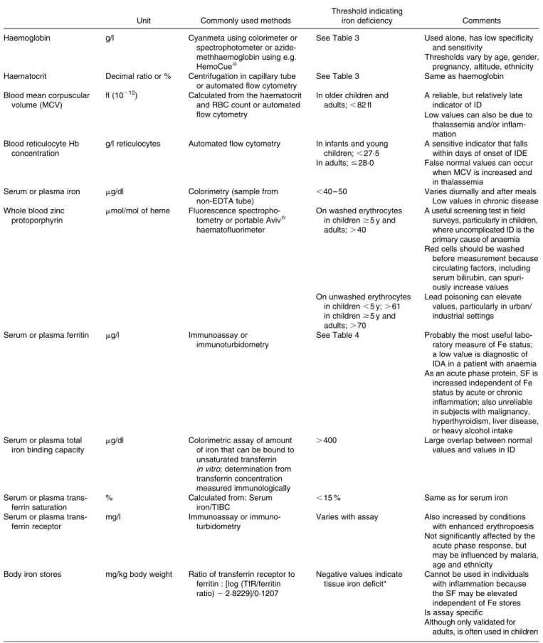
Methods to assess iron and iodine status
8
0
0
Texte intégral
Figure
+2
Documents relatifs