Comparison of Fas(Apo-1/CD95) and
perforin-mediated cytotoxicity in primary
T lymphocytes
Bente Lowin, Chantal Mattman, Michael Hahne and Jiirg Tschopp
Institute of Biochemistry, University of Lausanne, BIL Research Center, Chemin des Boveresses 155, 1066 Epalinges, Switzerland
Keywords: Apo-1/CD95 ligand, apoptosis, biosynthesis, cytotoxic T cells, Fas ligand, granzymes, kinetics, membrane damage, perforin
Abstract
Cytolytic T lymphocytes kill target cells by two independent cytolytic mechanisms. One pathway depends on the polarized secretion of granule-stored proteins including perforin and granzymes, causing target cell death through membrane and DNA damage. The second cytolytic effector system relies on the interaction of the Fas ligand (FasL) on the effector cell with its receptor (Fas) on the target cell, leading to apoptotic cell death. Using mixed lymphocyte culture (MLC)-derived primary T lymphocytes of perforin-knockout and gld (with non-functional FasL) mice, the molecular basis of the two killing mechanisms was compared. The activity of both pathways was dependent on extracellular Ca2 +. Incubation of MLC-stimulated primary T cells with protein synthesis
inhibitors prior to TCR triggering impaired FasL cell surface expression and abolished cytolytic activity, although the cells exhibited an intracellular pool of FasL. The perforin-dependent mechanism induced cell death more rapidly, although both pathways ultimately showed similar killing efficiencies. Both pathways induced comparable levels of DNA degradation, but Fas-induced membrane damage was less pronounced. We conclude that upon TCR triggering FasL may be recruited in part from pre-existing intracellular stores. However, efficient induction of target cell death still depends on the continuous biosynthesis of FasL molecules.
Introduction
Cytotoxic T lymphocytes (CTL) play a crucial role in the clearance of viral infections, tumor protection and graft rejec-tion. Recently, the use of knockout mice has demonstrated the presence of two independent cytotoxic pathways in CTL (1,2). One mechanism consists of the directional release of cytotoxic granule-stored effector molecules upon specific interaction with a target cell, eventually leading to cell lysis and apoptosis (3). Perforin, a granule protein capable of forming transmembrane pores in a Ca2+-dependent manner, is responsible for membrane damage (4,5), but fails to induce nuclear damage (6). Apoptosis is brought about by granzyme A or B, two granule proteins which belong to the family of serine proteases (7-10).
A second cytotoxic mechanism used by CTL depends on the interaction of the Fas ligand (FasL) and its cognate receptor (11). Fas-based killing can function in the absence of extracellular Ca2 + (11) and represents the major killing pathway of CD4+ CTL (12,13). FasL is a 40 kDa class II
transmembrane protein belonging to the tumor necrosis factor family, and is expressed in a variety of different tissues particularly spleen, thymus, testis, kidney and lung (14). In addition to its role in CTL-mediated cytotoxicity, Fas is implic-ated in immune homeostasis as evidenced by the analysis of mutant mouse strains for FasL (gld) and Fas (Ipr) (15). With age, these mice accumulate a large amount of non-malignant CD4~CD8~ T cells in their spleen and lymph nodes, and develop autoimmune disease pathology similar to systemic lupus erythematosus. Various studies examining the steps of T cell development regulated by the Fas/Apo-1 system have suggested that Fas is involved in the clonal deletion of lymphocytes in the periphery (16,17).
In the present study, we compared the mechanisms of perforin- and Fas-mediated killing. In our experimental system, Fas- and perforin-dependent pathways induced rapid cell death via apoptosis. Although FasL could be detected inside the CTL before TCR engagement, killing was dependent on
Corresponding author, J. Tschopp
de novo protein synthesis, suggesting either the need for continuous ligand synthesis or the production of a putative FasL-activating protein
Methods
Cell lines and mice
A murine B lymphoma cell line, A20 (H-2d, ATCC TIB 208), a murine fibrosarcoma clone, WEHI-164 clone 13 (H-2d' ATCC CRL 1751), and an embryonic fibroblast cell line, 3T3.A31 (H-2d, ATCC CRL 6588), were maintained in DMEM (Gibco, Basel, Switzerland) supplemented with 5% FCS, 10 mM HEPES (pH 7.4) and 5x10~5 M |}-mercaptoethanol To gener-ate concanavalin A (Con A) blasts, spleen cells from DBA/2 mice (H-2d) were freed from red blood cells and activated in complete DMEM supplemented with Con A (4 |ig/ml, Sigma) for 3 days. Alloreactive CTL were generated in a 5 day mixed lymphocyte culture (MLC) as previously described (18). Responder spleen cells (2 5xi06/ml) were obtained from adult (5- to 6-week-old) homozygous perfonn-deficient mice (19) and C57/B6 gld mice (H-2b; Jackson Laboratory, Bar Harbor, ME). Stimulator cells were irradiated spleen cells (2.5x10⁶/ml, 3000 rad; 1 rad = 0 01 Gy) from DBA/2 mice (Iffacredo, Grenoble, France). All cells were cultured in a humid atmosphere containing 5% CO2 at 3°C.
Cytotoxicity assay for MLC
Cytotoxicity assays were performed as previously described (20) To measure membrane damage, A20 cells or Con A blasts (106 cells in DMEM with 5% FCS) were labeled with 100 p.Ci [51Cr]sodium chromate (Amersham, Zurich, Switzer-land) for 1 h in a final volume of 200 |xl. For DNA degradation, target cells (2X10⁶) were labeled overnight with 2.5-5 nCi [3H]thymidine (Amersham). 3T3.A31 fibroblasts and WEHI-164 fibrosarcoma cells were labeled with [51Cr]sodium chro-mate (2 (iCi/well) in 96-well plates for 12-16 h (21). Cytotoxicity was measured in a 7 h assay. For the inhibitor studies, MLC were preincubated with the various metabolic inhibitors (actinomycin D 2.5 ng/ml; cycloheximide 2.5 ng/ml; emetine 5 ng/ml) for 45 min and either washed or directly mixed with labeled target cells. All inhibitors were obtained from Sigma (Buchs, Switzerland). In order to preactivate CTL, cells were incubated for 3 h in a mixture of phorbol 12-myristate 13-acetate (PMA; Sigma; final concentration of 5 ng/ml) and ionomycin (Sigma; 0.5 ng/ml final).
Flow cytometnc analysis
Primary MLC were taken at day 6 of stimulation and were centrifuged for 20 min at 2000 r.p.m. over a Ficoll-Hypaque cushion (Pharmacia, Location?) to eliminate dead cells. Washed buffy coat cells were stained with affinity-purified polyclonal antibodies against the FasL (PE62; M. Hahne etal, submitted for publication) followed by a fluorescein-coupled donkey anti-rabbit IgG antibody (Dianova, Hamburg, Germany) The samples were analyzed on a FACScan cyto-meter (Becton Dickinson, Mountain View, CA).
Immunofluorescence microscopy and Western blot analysis Primary MLC at day 6 of stimulation were enriched for T cells by depletion of B220+ cells on a FACS cell sorter Remaining
cells were cultured for 4 h in either the presence or absence of PMA (0.5 ng/ml) and ionomycin (0.5 ng/ml) Subsequently, cells were washed with PBS and plated onto glass slides (pretreated with polylysine at 50 ng/ml for 30 min) Cells were fixed and permeabilized in methanol at -20°C for 5 mm. Slides were then incubated with the polyclonal antibody anti-PE62 (MAP-RGQSCNNQPLNHKVYMRNSKYPEDL), affinity-purified against the FasL peptide PE82 (GQSCNNQPL-NHKVYMRNSKYPEDK). As a specificity control, the specific peptide PE82 (100 ng/ml) or an unrelated peptide PE58 (EDEMPKTLYVGNLS) were added during the first antibody incubation. Bound antibody was visualized with a donkey anti-rabbit FITC-conjugated secondary antibody (Dianova). Confocal microscopy was performed using the Zeiss LSM confocal laser system. Specimens were observed under epifluorescence illumination with a 488 nm scanning argon beam. For Western blotting, 2X10⁶ sorted T lymphocytes were lysed in 50 nl of cell lysis buffer (0.5% Triton X-100, 300 mM NaCI in 50 mM Tris-HCI, pH 7.6) containing 1 mM phenylmethylsulfonyl fluoride The postnuclear fraction was separated on a 10% SDS-PAGE and transferred to nitrocellu-lose FasL was detected with the polyclonal antibody anti-PE62 (10 n9/m|) an c l secondary anti-rabbit IgG antibody coupled to horseradish peroxidase (Sigma). Specificity con-trols were performed by adding either the specific peptide (PE82, 100 ng/ml) or an unrelated peptide (PE58, 100 ng/ml) during the first antibody incubation. Blots were developed using the Enhanced Chemiluminescence kit from Amersham
Results
Fas-mediated cytotoxicity is dependent on extracellular calcium
Perforin-mediated cytotoxicity is strictly dependent on extra-cellular calcium, which is required for TCR-regulated granule exocytosis as well as the lytic activity of perforin itself (22). In contrast, Fas-based cytotoxicity of T cell clones has been reported to work in the absence of Ca2 + (1,10,17,23). In primary T cells, however, depletion of extracellular calcium with Mg2+-EGTA (3 mM/5 mM) abolished perforin- as well as Fas-mediated killing (Fig. 1). Fas-mediated cytotoxicity in primary MLC is specific and dependent on TCR engagement (1). To investigate if inhibition of TCR signaling, a calcium-dependent event, was responsible for the abrogation of Fas-mediated cytotoxicity, we preincubated effector cells for 3 h with PMA plus ionomycin. These stimuli have been widely used to up-regulate FasL surface expression (14). In contrast to the perforin-mediated killing activity, Fas-mediated killing was partially restored by this treatment, suggesting that calcium is needed for the TCR signal-induced surface expres-sion of FasL, but not for the interaction of the ligand with its receptor.
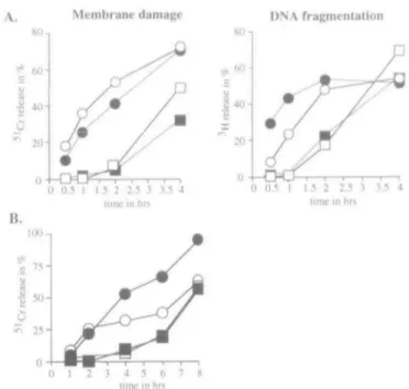
The Fas-based pathway kills targets with a lag period Perform-dependent killing is a rapid event due to the produc-tion and storage of readily accessible cytotoxic effector molecules prior to target cell contact. In order to compare the kinetics of perforin- and Fas-dependent killing pathways, target cells were incubated with g/d-derived or
perforin-Membrane damage DNA fragmentation
10 30 E : T ratio
10 30 E : T ratio
Fig. 1. EGTA inhibits Fas- and perforin-mediated cytotoxicity. The
cytotoxic activity of MLC (5 days after stimulation) derived from perforin-deficient and g/dmice was assessed in a standard 4 h 51
Cr-release assay against A20 target cells in the absence (squares) or presence (diamonds) of Mg2+-EGTA. Effector cells were also
preactivated for 3 h with PMA plus ionomycm and then assayed for their cytolytic activity (circles). E, effector, T, target.
80-, 80-i 0 0.5 I 1.5 2 2.5 3 3.5 4 lime in hrs 0 0.5 I 1.5 2 2.5 3 3.5 4 lime in hrs 2 3 4 5 6 7 8 lime in hrs
Fig. 2. Fas-mediated killing shows slower kinetics, but similar
efficiency as the perforin-mediated pathway. (A) The cytotoxic activity of MLC derived from perforin-deficient (squares) and gld mice (circles) was measured in a standard 4 h 51Cr- and [3
H]thymidine-release assay against A20 cells (open symbols) and Con A blasts (filled symbols). Radioactivity released into the supernatant was measured at the indicated time points. (B) As in (A), but either WEHI-164 fibrosarcoma cells (filled symbols) or 3T3.A31 fibroblasts (open symbols) were used as target cells 51Cr release was measured at
indicated time points up to 8 h
deficient CTL at a 30:1 effectontarget cell ratio (Fig. 2). At the indicated time points, membrane and DNA damage was measured. Hematopoietic target cells, i.e. A20 B lymphomas and Con A blasts, were rapidly killed via the perforin-mediated pathway, leading to significant membrane and DNA damage within the first 2 h (Fig. 2A). In contrast, killing by Fas showed a lag period of up to 2 h during which only a minor fraction of targets were killed. In particular, membrane damage was significantly delayed Even at the endpoint of the assay (4 h), where both mechanisms showed comparable efficiency in DNA fragmentation, Fas-induced membrane damage was still inferior to perforin-mediated cytolysis (30-50% of reduction). Similar results were obtained when fibroblast lines were used as target cells (Fig. 2B). Again, the perforin-dependent pathway induced cell death more rapidly, although differences in membrane damage were less pronounced for 3T3.A31 target cells.
Fas-mediated cytotoxicity requires de novo RNA and protein synthesis
Perforin-mediated cytotoxicity is independent of de novo RNA and protein synthesis, since preformed cytotoxic effector molecules are already stored in cytoplasmic granules of activated T cells. To analyze the requirement for an intact metabolic machinery during Fas-mediated killing, cytotoxicity tests with A20 cells as targets were performed in the presence of either actinomycin D, an RNA synthesis inhibitor, or the
two protein synthesis inhibitors cycloheximide and emetine. While perforin-mediated cytotoxicity was only slightly inhibited by either of the treatments (Fig. 3A), complete inhibition of Fas-mediated killing was observed, suggesting that both de novo RNA and protein synthesis is a prerequisite for this lytic activity. To demonstrate that the added inhibitors act on the effector and not on the target cells, perforin-deficient CTL were pretreated with either actinomycin D or emetine for 1 h and washed three times before testing their cytolytic activity. As shown in Fig. 3(B), the results were essentially the same as before, pinpointing the effect of the metabolic inhibitors to the effector cells.
Treatment of T lymphocytes with PMA plus ionomycin has been shown to increase FasL-specific mRNA and to up-regulate cell surface expression (14). Effector cells were incubated with these stimuli prior to the cytotoxicity test to investigate if such a pre-activation step could substitute for the need of de novo RNA and protein synthesis. After 3 h of incubation with PMA plus ionomycin, metabolic inhibitors were added and cytotoxic activity was determined. Killing activity of g/d-derived CTL remained unchanged (Fig. 3C). In contrast, the actinomycin D-induced inhibition of Fas-mediated killing was rescued by the prior PMA plus ionomycin treatment. Continuous de novo protein synthesis, however, was still needed for efficient killing.
rerlonn MJ gld (hasL-j
+ PMA/ionomvcin
10 30 E : T ratio
Fig. 3. Fas- and perforin-mediated killing can be discriminated by
metabolic inhibitors The cytotoxic activity of MLC derived from perform-deficient and g/dmice was assessed in a standard 4 h 51
Cr-release assay against A20 target cells in the absence (squares) or presence of actinomycm D (diamonds), cycloheximide (circles) and emetine (triangles) In (A), metabolic inhibitors were included during the whole assay. In (B), only effector cells were pretreated with the inhibitors for 1 h prior to the assay. In (C), effector cells were preactivated with PMA plus lonomycin for 3 h Subsequently, the inhibitors were added and cytotoxicity assays were performed after 1 h of incubation E, effector, T, target
Fig. 4. Efficient FasL surface expression requires protein synthesis
MLC-denved lymphocytes were stained with an affinity-purified polyclonal antibody FasL (PE62) followed by donkey anti-rabbit coupled to FITC and analyzed by flow cytometry Cells were analyzed after 4 h of incubation in the absence or presence of PMA plus lonomycin In the last panel, cells were preincubated with cycloheximide for 1 h prior to activation. An isotype control was included
need a second signal through TCR cross-linking. Our FACS experiments neglect a possible influence of Fas binding on FasL surface expression. Indeed, it has been shown for CD40L that interaction with its receptor leads to down-modulation of the complex. Thus, the half-life of FasL on a T lymphocyte may be even shorter in a 'CTL-target cell' than in a 'CTL only' system, explaining the dramatic effect of protein synthesis inhibitors on Fas-mediated cytotoxicity.
FasL-based killing was investigated in more detail. Although the direct interference of the protein synthesis inhibitors with FasL synthesis and surface expression remained the most likely explanation, other reasons such as reduced expression of adhesion molecules could not be excluded. Non-activated, activated or cycloheximide-treated, activated CTL were there-fore analyzed by FACS for the presence of surface FasL. With reference on the isotype control, only a minority of non-activated cells (4.3%) specifically expressed FasL on their surface, whereas PMA plus ionomycin treatment up-regulated FasL surface expression in a distinct subpopulation (12.1%) (Fig. 4). Pretreatment of MLC with cycloheximide prior to activation reduced FasL expression to base levels (5 4%), suggesting that this FasL down-regulation may be responsible for the observed inhibition of cytotoxic activity. Similar results were obtained when cells were pretreated with emetine (data not shown). These findings are in agreement with earlier data (23), which showed that primary MLC-derived T lymphocytes, although they weakly express FasL on their cell surface (Fig. 4), are not capable to kill Fas-positive bystander cells, but
Primary MLC-derived T cells express intracellular FasL In order to determine if the observed activation-induced surface expression is due to de novo synthesis of the ligand or rather due to translocation of the protein from an intracellular pool to the cell membrane, MLC enriched for T lymphocytes (B220~) were either analyzed by Western blotting or immuno-fluorescence microscopy using FasL-specific antibodies. Sur-prisingly, both methods revealed the presence of FasL already in MLC-derived T cells prior to TCR triggering. In Western blot analysis, a specific band with an apparent mol. wt of 40 kDa was detected, which could be competed with the specific (PE82, 100 ng/ml), but not with an unrelated peptide (PE58, 100 ng/ml) (Fig. 5) PMA plus ionomycin activation did not significantly change the level of FasL expression. However, caution must be taken in the interpretation of this experiment, since we observed considerable cell loss during the activation period. It may well be that activation leads to the induction of T cell suicide by the Fas pathway (2) and subsequently to the loss of FasL expressing cells. To localize FasL inside the cell, we performed immuno-staining of permeabilized
non-C/5
anti-PE62
- - - - + + + PMA/Ionomycin
. . + . . + . PE82
- - - + - - + PE58
kDa
mm - — FasL
Fig. 5. stimulated T lymphocytes express FasL. Sorted
MLC-derived T lymphocytes, cultured for 4 h in the absence or the presence of PMA plus lonomycin, were lysed and separated on a 10% SDS-PAGE under non-reducing conditions Western blot analysis was performed using the affinity-purified polyclonal antibody anti-FasL (anti-PE62). As control, the non-immune serum (NIS) was included To demonstrate the specificity of the anti-PE62 antibody, competition experiments were performed in the presence of either the specific (PE82; 100 ng/ml) or a non-specific peptide (PE58, 100 ng/ml) Note the presence of cross-reactive high molecular species, demonstrating equal loading
activated T cells (Fig. 6A and D). The affinity-purified poly-clonal antibody anti-PE62 demonstrated the presence of FasL within the cytoplasm as a distinct staining underneath the plasma membrane, which could be competed with the specific peptide PE82 (100 ng/ml) (Fig. 6 B). Unfortunately, the sharp-ness of the staining was never high enough to detect any defined structure. In summary, our findings suggest that T cells accumulate intracellular FasL already upon allogeneic stimulation, but that the majority of the protein is only expressed on the cell surface upon a second TCR engagement.
Discussion
T cell-mediated cytotoxicity plays an important role in cellular immunity. Recently, two mechanisms have been shown to account for the main cytotoxic activity of in vitro and in vivo generated CTL and certain T cell lines (1,24-26). The 'clas-sical' granule exocytosis pathway, strictly dependent on per-forin, has been identified as one of the cytotoxic mechanisms. In vivo studies in perforin-deficient mice clearly showed that perforin-mediated cytotoxicity plays a major protective role in viral infections, immunity against bacteria (Listeria) and tumor rejection (2,26,27).
A second pathway involves the Fas/Apo-1 system (28). Fas-based killing was first reported to be active in the absence
Fig. 6. MLC-stimulated T lymphocytes contain an intracellular pool
of FasL Sorted MLC-denved T lymphocytes were analyzed using confocal microscopy. Permeabilized cells were labeled with the affinity-purified antibody anti-PE62 either in the absence (A) or the presence (B) of the specific peptide PE82 (100 ng/ml) (C) shows labeling with an isotype control.
of calcium (11), and, previously, EGTA has been used to discriminate perforin- and Fas-based pathways in CTL (11,29) In particular, unspecific killing induced by CTL clones or hybridoma cell lines clearly displayed Ca2+-independent cytotoxic activity (30). However, recent studies of perforin-deficient T cell clones have provided controversial results (24) In our experimental system which studies specific target cell lysis by primary T cells, Fas-mediated killing strictly required extracellular calcium, most probably reflecting the TCR-dependent up-regulation of FasL on the cell surface. Even though FasL-Fas interaction is calcium-independent (31), the ion requirement for TCR signaling seems to dominate the specific Fas-killing pathway. The conflicting results in different systems may therefore be due to varying levels of constitutive FasL surface expression on the effector cells.
FasL belongs to the same protein family as tumor necrosis factor-a and lymphotoxin-a (14), two mediators of slow apop-totic cell death in sensitive targets (32). However, like perforin, FasL induces acute cytotoxicity. As demonstrated in this study, the kinetics of the two pathways are different. Whereas the perforin-based pathway leads to significant cell death shortly after target cell contact, Fas-dependent cytotoxicity is only effective after a lag-period of 2-3 h. TCR engagement has been shown to increase surface expression of FasL (13). Thus, the observed lag phase may reflect the need for sufficient FasL surface expression to ensure effective killing. Moreover, Fas-mediated cytotoxicity required continuous de novo protein synthesis which doubtlessly also contributes to the delayed onset of cytotoxicity. Metabolic inhibitors were used in earlier studies to discriminate between CD4+- and CD8+-mediated cytotoxicity (33,34). In contrast to CD8+ cells, CD4-mediated killing was strictly dependent on an intact metabolic machinery similar to our findings for the Fas path-way. Indeed, Fas-mediated cytotoxicity has been recently proposed to be the major killing pathway in CD4-restricted T lymphocytes (12). Although protein synthesis inhibitors
inter-fere with FasL surface expression, the question still remains open whether the drugs directly block the production of FasL or a presumed FasL-activating protein that is, for example, essential for membrane translocation or FasL oligomerization
Our data show that a first allogeneic stimulation of T lymphocytes does not lead to pronounced cell surface expres-sion of FasL, but rather to the synthesis of an intracellular FasL pool which is most likely recruited to the cell surface upon TCR triggering. This is reminiscent of CD40L which, upon B cell encounter, is rapidly translocated to the surface of T cells from intracellular stores. Prolonged surface expres-sion of CD40L, however, also requires de novo synthesis and is inhibited by cycloheximide (35). The intracellular FasL store apparently does not suffice for efficient cell death induction. To date, the fate of FasL on the cell membrane before and after interaction with its receptor is not known. Due to FasL's potentially dangerous cytolytic activity, it is conceivable that surface FasL has only a short half-life and is either endo-cytosed and rapidly degraded or shed from the membrane. Our results agree with such a hypothesis, since the consider-able quantities of FasL expressed on the cell surface upon PMA plus ionomycin activation are insufficient for efficient target cell lysis. The dependence on de novo synthesis of either FasL or a FasL-activating protein is also reflected by the decreased FasL surface expression after short exposure to cycloheximide (35). With regard to our results, it is interes-ting to note that CD40L surface expression is also tightly regulated. After TCR triggering, CD40L is only transiently expressed (36) and subsequent interaction with its receptor CD40 leads to down-modulation through receptor-mediated endocytosis (37). A similar mechanism may also be envisaged for FasL. Thus, FasL-Fas interaction upon effector-target cell contact would lead to continuous elimination of FasL, explaining the need for de novo protein synthesis. Given the dramatic effect seen after systemic delivery of anti-Fas antibodies to mice and the proposed role for the Fas/Apo-1 system in immune homeostasis, a tight regulation of FasL expression and activity may indeed be premordial. Thus, it is tempting to propose that certain pathological situations may well result from a misfunction of such regulatory mechanisms.
Acknowledgements
The authors thank Chris Kamel and Fraser Donaldson for critical reading of the manuscript They gratefully acknowledge the technical assistance of T Laroche and A. Allegnni. This work was supported by grants from the Swiss National Science Foundation.
Abbreviations Con A FasL Fas gld Ipr MLC PMA concanavalin A Fas ligand Fas receptor
generalized lymphoproliferative disease lymphoproliferation
mixed lymphocyte culture phorbol 12-myristate 13-acetate
References
1 Lowin, B., Hahne, M., Mattmann, C. and Tschopp, J 1994. Cytolytic T-cell cytotoxicity is mediated through perform and Fas
lytic pathways. Nature 370 650
2 Kagi, D., Ledermann, B., BCirki, K., Seiler, P., Odermatt, B , Olsen, K J , Podack, E. R., Zinkernagel, R M and Hengartner, H. 1994. Cytotoxicity mediated by T cells and natural killer cells is greatly impaired in perforin-deficient mice. Nature 369 31
3 Henkart, M. P. and Henkart, P A 1982. Lymphocyte mediated cytolysis as a secretory phenomenon. Adv. Exp Med. Biol 146227
4 Masson, D. and Tschopp, J 1985 Isolation of a lytic, pore-forming protein (perform) from cytolytic T-lymphocytes J Biol
Chem. 2609069.
5 Podack, E R. 1985. The molecular mechanism of lymphocyte-mediated tumor cell lysis. Immunol. Today621
6 Duke, R , Persechini, P. M., Chang, S , Liu, C C , Cohen, J. J. and Young, J. D E. 1989. Purified perform induces target cell lysis but not DNA fragmentation J Exp Med 170 1451. 7 Heusel, J W., Wesselschmidt, R L , Shresta, S., Russell, J H
and Ley, T J. 1994. Cytotoxic lymphocytes require granzyme B for the rapid induction of DNA fragmentation and apoptosis in allogeneic target cells Cell 76 977.
8 Shi, L , Kam, C M , Powers, J. C , Aebersold, R and Greenberg, A H. 1992 Purification of three cytotoxic lymphocyte granule serine proteases that induce apoptosis through distinct substrate and target cell interactions J Exp Med. 176 1521
9 Shiver, J W, Su, L. and Henkart, P A 1992 Cytotoxicity with target DNA breakdown by rat basophilic leukemia cells expressing both cytolysin and granzyme A Ce//71 315
10 Jenne, D E and Tschopp, J. 1989. Granzymes- a family of serine proteases in granules of cytolytic T lymphocytes Curr. Topics
Microbiol Immunol 140 33
11 Rouvier, E , Luciani, M. F and Golstem, P 1993 Fas involvement in Ca2+-independent T cell-mediated cytotoxicity J Exp. Med
177 195
12 Stalder, T, Hahn, S. and Erb, P 1994 Fas antigen is the major target molecule for CD4+ T cell-mediated cytotoxicity J Immunol 152.1127.
13 Hanabuchi, S , Koyanagi, M., Kawasaki, A , Shinohara, N , Matsuzawa, A., Nishimura, Y., Kobayashi, Y, Yonehara, S , Yagita, H and Okumura, K 1994 Fas and its ligand in a general mechanism of T-cell mediated cytotoxicity Proc. Natl Acad Sci
USA 91:4930
14 Suda,T.,Takahashi,T,Golstein,P andNagata.S. 1993 Molecular cloning and expression of the Fas ligand, a novel member of the tumor necrosis factor family. Cell 75.1169
15 Cohen, P. L and Eisenberg, R. A 1991. Lpr and gld. single gene models of systemic autoimmunity and lymphoproliferative disease Annu. Rev Immunol. 9.243
16 Russell, J H , Rush, B., Weaver, C. and Wang, R. 1993 Mature T cells of the autoimmune Ipr/lpr mice have a defect in antigen-stimulated suicide. Proc Natl Acad. Sci USA. 90:4409. 17 Vignaux, F. and Golstein, P 1994 Fas-based
lymphocyte-mediated cytotoxicity against syngeneic activated lymphocytes: a regulatory pathway? Eur. J. Immunol. 24:923.
18 Maryanski, J L, MacDonald, H. R. and Cerottini, J.-C. 1980 Limiting dilution analysis of alloantigen-reactive T lymphocytes
J. Immunol 124.42.
19 Lowin, B., Beermann, F, Schmidt, A. and Tschopp, J. 1994. A null mutation in the perforin gene impairs cytolytic T lymphocyte-and natural killer cell-mediated cytotoxicity Proc. Natl Acad Sci.
USA 91:11571.
20 Walsh, C M , Glass, A. A , Chiu, V and Clark, W R. 1994. The role of Fas lytic pathway in a perforin-less CTL hybridoma.
J. Immunol. 153 2506.
21 Ucker, D S., Obermiller, P. S., Eckhart, W., Apgar, J R., Berger, N A. and Meyers, J. 1992. Genome digestion is a dispensable consequence of physiological cell death mediated by cytotoxic T lymphocytes Mol. Cell Biol. 12.3060.
22 Young, J. E. and Conn, Z. A. 1987. Cellular and humoral mechanisms of cytotoxicity: structural and functional analogies.
Adv. Immunol. 41.269.
23 Dhein, J., Walczak, H., Baumler, C , Debatin, K M. and Krammer, P H. 1995 Autocrine T-cell suicide mediated by APO-i/(Fas/ CD95). Nature 373 438.
24 Kojima, H , Shinohara, N , Hanaoka, S , Someya-Shirota, Y, Takagaki, Y, Ohno, H., Saito, T, Katayama, T, Yagita, H , Okumura, K., Shinkai, Y., Alt, F W., Matsuzawa, A., Yonehara, S. and Takayama, H 1994. Two distinct pathways of specific killing revealed by perforin mutant cytotoxic T lymphocytes. Immunity 1:357
25 Kagi, D., Vignaux, F., Ledermann, B , Biirki, K, Depraetere, V, Nagata, S , Hengartner, H and Golstein, P. 1994. Fas and perforin pathways as major mechanisms of T cell-mediated cytotoxicity.
Science 265.528
26 Walsh, C M., Matloubian, M., Liu, C -C , Ueda, R , Kurahara, C. G , Christensen, J L , Huang, M T F, Young, J D-E , Ahmed, R. and Clark, W. R 1994. Immune function in mice lacking the perforin gene. Proc Nail Acad. Sci USA 91 10854
27 Kagi, D, Ledermann, B, Burki, K., Hengartner, H and Zinkernagel, R. M 1994 CD8+ T cell-mediated protection against
an intracellular bacterium by perforin-dependent cytotoxicity. Eur
J Immunol 24.3068
28 Nagata, S and Suda, T. 1995 Fas and Fas ligand Ipr and gld mutations. Immunol. Today 16.39.
29 Ju, S -T, Cui, H , Panka, D J , Ettinger, R and Marshak-Rothstem, A 1994 Participation of target Fas protein in apoptosis pathway induced by CD4+ Th1 and CD8+ cytotoxic T cells Proc Natl Acad. Sci. USA 91 4185
30 Garner, R , Helgason, C. D , Atkinson, E. A , Pinkoski, M. J , Ostergaard, H. L, Sorensen, O , Fu, A , Lapchak, P H ,
Rabinovitch, A., McElhaney, J. E., Berke, G and Bleackley, R. C. 1994. Characterization of a granule-independent lytic mechanism used by CTL hybndomas. J Immunol. 153:5413.
31 Anel, A , Richieri, G V and Kleinfeld, A. M. 1994 A tyrosine phosphorylation requirement for cytotoxic T lymphocyte degranulation. J. Biol. Chem. 269.9506.
32 Ratner, A and Clark, W. R 1993 Role of TNF-a in CD8+cytotoxic
T lymphocyte-mediated lysis. J. Immunol 150:4303.
33 Ju, S. T. 1991. Distinct pathways of CD4 and CD8 cells induce rapid target DNA fragmentation. J Immunol. 146:812
34 Strack, P., Martin, C , Saito, S , Dekruyff, R H and Ju, S. T. 1990. Metabolic inhibitors distinguish cytolytic activity of CD4 and CD8 clones. Eur J. Immunol. 20 179.
35 Casamayor-Palleja, M , Khan, M and MacLennan, I. C. 1995. A subset of CD4+ memory T cells contains preformed CD40-ligand
that is rapidly but transiently expressed on their surface after activation through the T cell receptor complex J Exp. Med 181:1293
36 Castle, B. E , Kishimoto, K , Stearns, C , Brown, M. L and Kehry, M R 1993 Regulation of expression of the ligand for CD40 on T helper lymphocytes J. Immunol 151 1777.
37 Yelhn, M J , Sippel, K , Inghirami, G., Covey, L. R , Lee, J. J., Sinning, J., Clark, E A , Chess, L. and Lederman, S. 1994. CD40 molecules induce down-modulation and endocytosis of T cell surface T cell-B cell activating molecule/CD40-L. J. Immunol. 152 598.