Ligand-Specific Targeting of
Microspheres to Phagocytes by
Surface Modification with
Poly(L-Lysine)-Grafted Poly(Ethylene
Glycol) Conjugate
Sofia Faraasen,1,2,4Ja´nos Vo¨ro¨s,2Ga´bor Csu´cs,3 Marcus Textor,2Hans P. Merkle,4and Elke Walter4,5
Received July 17, 2002; accepted November 1, 2002
Purpose. The purpose of this study was to demonstrate specific re-ceptor-mediated targeting of phagocytes by functional surface coat-ings of microparticles, shielding from nonspecific phagocytosis and allowing ligand-specific interactions via molecular recognition. Methods. Coatings of the comb polymer poly(L-lysine)-g-poly(ethylene glycol) (PLL-g-PEG) were investigated for potential to inhibit 1) nonspecific spreading of human blood-derived macro-phages (MOs) and dendritic cells (DCs) on glass and 2) nonspecific phagocytosis of PLL-g-PEG-coated, carboxylated polystyrene (PS) or biodegradable poly(D,L-lactide-co-glycolide) (PLGA) micro-spheres. Coating was performed by adsorption of positively charged PLL-g-PEG on negatively charged microparticles or plasma-cleaned glass through electrostatic interaction. The feasibility of ligand-specific interactions was tested with a model ligand, RGD, conju-gated to PEG chains of PLL-g-PEG to form PLL-g-PEG-RGD and compared with inactive ligand conjugate, PLL-g-PEG-RDG. Results. Coatings with PLL-g-PEG largely impaired the adherence and spreading of MOs and DCs on glass. The repellent character of PLL-g-PEG coatings drastically reduced phagocytosis of coated PS and PLGA microparticles to 10% in presence of serum. With both MOs and DCs, we observed ligand-specific interactions with PLL-g-PEG-RGD coatings on glass and PS and PLGA microspheres. Li-gand specificity was abolished when using inactive liLi-gand conjugate PLL-g-PEG-RDG, whereas repellency of coating was maintained. Conclusions. Coatings of PLL-g-PEG-ligand conjugates provide a novel technology for ligand specific targeting of microspheres to MOs and DCs while reducing nonspecific phagocytosis.
KEY WORDS: poly(D,L,-lactide-co-glycolide) (PLGA) micro-spheres; surface modification; poly(L-lysine)-grafted-poly(ethylene glycol) (PLL-g-PEG); phagocytosis; RGD-peptide.
INTRODUCTION
For almost two decades injectable, biodegradable micro-spheres have been developed into an established platform for
the sustained delivery of therapeutics for localized or systemic action. After injection of such microspheres, the embodied drugs can be protected from proteolytic cleavage and follow a controlled and prolonged release regimen that is governed by the bioerosion of the polymer carrier. Particularly, micro-spheres prepared from poly(D,L-lactide-co-glycolic acid) (PLGA) have a proven track record for drug delivery. They are highly biocompatible and biodegradable, and their rates of bioerosion and release of encapsulated drugs can be tai-lored by modulating their composition (1,2). This makes the microencapsulation of drugs in PLGA microspheres an ap-proved and safe technology for the long term delivery of therapeutics.
Phagocytic cells, such as macrophages (MOs), may rec-ognize injected particulate drug carriers as foreign materials and remove them quickly and efficiently via nonspecific phagocytosis by the mononuclear phagocytic system (MPS). The MPS represents a main hurdle that needs to be overcome to achieve selective targeting of particulates to selected or-gans, tissues, or cells. The phagocytosis of microspheres is stimulated by their surface properties and is believed to be enhanced by charged or hydrophobic surfaces (3). Further-more, the extent of phagocytosis is enhanced by the adsorp-tion of opsonic plasma proteins to their surface (4). To protect particulates from phagocytosis, poly(ethylene glycol) (PEG) coatings have been investigated for a wide array of biomedi-cal applications. Immobilization of PEG on surfaces has long been known to decrease protein adsorption (5,6). Approaches to immobilize PEG on the surface of microspheres involve covalent grafting (7–9), or adsorption of PEG-containing nonionic surfactants, such as poloxamers, through hydropho-bic interaction (10,11). Such approaches not only reduce plasma protein adsorption, but also inhibit the nonspecific uptake by phagocytes (5,8,10,12).
Nevertheless, upon uptake of water, adsorbed nonionic surfactants may quickly desorb from the surface, especially when microspheres were prepared from fast biodegrading, low molecular weight PLGA polymers (13). However, the chemical derivatization of PLGA is restricted to the end groups of the polymer chain and therefore likely to affect the physicochemical and bioerosion characteristics of the poly-mer. This renders the development of drug-loaded micro-spheres from modified PLGA more complex because it re-quires case-to-case adjustments of composition and process parameters for microencapsulation. To avoid this complexity, surface coatings of PLGA microspheres would be highly de-sirable to modify their biodistribution and reduce their physi-ologic clearance by the MPS.
Recently, a new class of grafted comb copolymers based on poly(L-lysine)-g-poly(ethylene glycol) (PLL-g-PEG) was found to spontaneously adsorb from aqueous solution onto several metal oxide surfaces and onto heterogeneous biologic surfaces (6,14). The architecture of the comb copolymer has been demonstrated to play an important role for its behavior. When adjusted accordingly, the resulting adsorbed layers are highly effective in reducing the adsorption of plasma proteins. Adsorptive immobilization of the protective PEG-moiety is suggested to occur through interaction of the positively charged primary amino groups of PLL bound to the nega-tively charged surface (6,14).
1Laboratory of Applied Physics, Department of Physics and
Mea-surement Technology, Linko¨ping University, SE-581 83 Linko¨ping, Sweden.
2Laboratory of Surface Science and Technology, Department of
Ma-terials, Swiss Federal Institute of Technology Zurich (ETH), Wag-istrasse 2, 8952 Schlieren, Switzerland.
3Biomicrometrics Group, Department of Mechanical Engineering,
Swiss Federal Institute of Technology Zurich (ETH), Wagistr. 4, 8952 Schlieren, Switzerland.
4Department of Applied Biosciences, Drug Formulation & Delivery
Group, Swiss Federal Institute of Technology Zurich (ETH), Win-terthurerstrasse 190, 8057 Zurich, Switzerland.
5To whom correspondence should be addressed. (e-mail: elke_
PLGA microspheres display a hydrophobic and at the same time highly negatively charged surface (15,16). They have been demonstrated to be subject to extensive plasma protein adsorption (12) and are efficiently phagocytosed by the MPS, and by antigen presenting cells like MOs and den-dritic cells (DCs) (15).
At first, this work aim to investigated the potential of a protein-repellant, PLL-g-PEG-based surface coating to pre-vent the nonspecific uptake of PLGA microspheres by phago-cytic cells. For this purpose, we studied the uptake of model polystyrene (PS) and PLGA microspheres by human-derived peripheral blood MOs and DCs, upon coating the micro-spheres with PLL-g-PEG. As a second step, we investigated the effect of a model peptide ligand, RGD (17), grafted to PLL-g-PEG, on the receptor-mediated phagocytosis of suit-ably coated microspheres by MOs and DCs. The RGD motif serves as a model ligand for the specific interaction of the coatings with integrin receptors, which are expressed on the surface of phagocytic cells. Thus, our study represents a proof-of-concept to demonstrate that specific, receptor-mediated recognition between suitably coated microspheres and phagocytes is feasible. Provided that selective phagocyte specific ligands can be identified, the microspheres would have potential, e.g., as a platform for the specific targeting of particulate vaccine delivery systems to professional antigen-presenting cells, such as DCs.
MATERIALS AND METHODS
Materials
Carboxylated PS that were 4.5m in diameter and car-ried a negative charge were obtained from Polysciences Eu-rope, Basel, Switzerland. PLGA-type polymer (Resomer RG502H, ratio lactide:glycolide 50:50, molecular weight 14,000, uncapped end groups) was obtained from Boehringer Ingelheim (Germany). The PLL-g-PEG polymer used in this study consisted of a PLL-backbone of 20 kDa with grafted PEG chains of 2 kDa and a grafting ratio of lysine units to PEG side chains of 3.3 (6). Further modification of this poly-mer was by conjugation with the RGD adhesion motif to yield PEG-RGD, and RDG as negative control for PLL-g-PEG-RDG, both with an approximate peptide coverage of 0.1 pmole/cm2(17). All PLL-g-PEG polymers were
synthe-sized as outlined by Huang et al. (18). All chemicals used in this study were of analytical grade (from Fluka, Switzerland) unless otherwise specified.
Methods
PLGA Microspheres
PLGA microspheres were prepared by spray drying as previously described (19). Briefly, PLGA-type polymer was dissolved in dichloromethane (w/w 5%) and was spray-dried in a laboratory spray-dryer (Model 190, Bu¨chi, Switzerland) at an inlet temperature of 45°C. The spray-flow was set at 500 NL/h and the product feed was 3 ml/min. The microspheres were washed with water, collected on a 0.45-m cellulose acetate membrane filter and dried under vacuum for 24 h. The microspheres were stored at 4°C. Fluorescence-labeled PLGA microspheres were prepared by adding 6-coumarine (5
mg/g polymer, Acros Organics, Belgium) to the polymer so-lution. Size distribution of the microspheres was measured by laser light scattering (Mastersizer X, Malvern Instruments Ltd., Worcestershire, UK; equipped with a 45-mm lens). Cal-culation of particles size and distribution was based on Mie’s theory. The particle size distribution was presented in the volume-weighted mode and revealed particles in the range of 1–10m as previously described in detail (15,19).
MO and DC Cell Culture
MOs and DCs were obtained from human peripheral blood monocytes according to Sallusto et al. (20). Briefly, peripheral blood monocytes were isolated from buffy coats (Bloodbank Zurich, Switzerland) by density gradient cen-trifugation of Ficoll-Paque (Pharmacia Biotech, Buchs, Swit-zerland). Peripheral blood monocytes were resuspended in RPMI 1640 supplemented with 10% heat-inactivated (pooled) human serum (Bloodbank Zu¨rich, Switzerland) and then allowed to adhere for 2 h in culture flasks (25 cm2).
Nonadherent cells were removed and adherent cells were fur-ther cultured in RPMI 1640 supplemented with 5% heat-inactivated (pooled) human serum in the presence of 1000 IU/mL interleukin-4 (Sigma, Switzerland) and 50 ng/mL granulocyte macrophage-colony stimulating factor (R+D Sys-tems) to obtain DCs. MOs were obtained without additional supplements (21). Cultures of MOs and DCs were kept at 37°C in 5% CO2-humidified atmosphere. For particle uptake studies, cells were mechanically removed from the flasks after 24 h and reseeded onto coverslips in 24-well plates (300,000– 400,000 cells per well) for further studies.
Surface antigen expression of MOs and DCs was ana-lyzed by flow cytometry (22). For further identification, the cells were also challenged with lipopolysaccharide (1g/mL,
Escherichia coli 055:B5, Sigma) 48 h before the antibody
la-beling, which causes maturation of DCs only. The cells were incubated (45 min, 4°C) with each of the following primary anti-human antibodies: CD11b (Mac-1, ICRF44; 44), CD83 (Clone HB15e), CD86 (B70/B7-2, Clone IT2.2; all three from Pharmingen, Lucerne, Switzerland), and CD14 (Clone UCHM-1, Sigma). Control cells were processed similarly with mouse isotype control IgG2a (UPC-10, Sigma). Cells were washed three times and subsequently incubated with the sec-ondary antibody (anti-mouse IgG, R-Phycoerythrin conju-gated, Sigma) for 45 min at 4°C, washed three times, trans-ferred to FACS tubes, and analyzed on an FACScan type from Becton Dickinson (Switzerland). According to these cri-teria, more than 90% of the cells were identified as DCs, and in MOs cultures more than 90% of the cells were CD14+and
CD83−, even in the presence of lipopolysaccharide. Adhesion Studies
The adhesion of MO and DC on modified surfaces was first tested on glass surfaces coated with PLL-g-PEG and PLL-g-PEG-RGD or PLL-g-PEG-RDG, respectively. Before coating, the glass coverslips (12 mm in diameter) were oxy-gen-plasma cleaned in a Plasma Cleaner/Sterilizer PDC-32G instrument (Harrick Scientific Corporation, Ossining, NY) for 7 min. Coating was performed in 24-well plates under aseptic conditions with 1 mg/mL of the respective polymers dissolved in 10 mM HEPES buffer, pH 7.4, for 15 min. All
solutions were filtered through a 0.2-m membrane filter be-fore use. Control coverslips were incubated in HEPES buffer only. After coating, the coverslips were rinsed twice for 5 min with phosphate-buffered saline (PBS) and once with RPMI 1640 medium. MOs or DCs incubated for 1 week in cell cul-ture flasks were added to each well (250,000 cells in 600L of cell medium) containing either coated or control coverslips and cell adhesion was checked after different time points by microscopy (Axiovert 35, Zeiss, Germany). Images were taken using a CCD camera (CF 8/1 DXC, Kappa, Gleichen, Germany).
Coating of Microspheres
Microspheres were dispersed in filtered HEPES buffer at a concentration of 5 mg/mL (polystyrene particles) or 1 mg/ mL (PLGA microspheres). The dispersions were mixed at equal volumes with g-PEG, g-PEG-RGD or
PLL-g-PEG-RDG (1 mg/mL in filtered HEPES buffer) and
incu-bated under gentle mixing for 15 min at room temperature. The coated microspheres were centrifuged (5 min, 7,400 × g, Eppendorf, Centrifuge 5417R, Eppendorf-Netheler-Hinz GmbH, Germany) and redispersed in half of the previous volume in filtered HEPES buffer by slightly vortexing.
Phagocytosis Studies
MOs cultured on coverslips in 24-well plates were washed once with RPMI 1640 medium and then covered with 1 mL of RPMI 1640 medium with or without serum. Micro-spheres dispersed in HEPES buffer were added and the cells were incubated at either 37°C or at 4°C for 4 h. The cells were washed twice with RPMI 1640 medium and subsequently fixed with 3% paraformaldehyde in PBS for 30 min at room temperature. The samples were stored in PBS at 4°C. Because DCs only slightly adhered to the cell culture plates or to coverslips, these cells were not washed and the microspheres were added directly to the cells. Also the washing step after incubation was avoided and approximately 300L of medium was added to each well to allow a better verification of phago-cytosed microspheres. These samples were analyzed immedi-ately without prior fixation.
Sample Analysis by Fluorescence Microscopy
The amount of phagocytosed microspheres per cell was assessed by fluorescence microscopy (Axiovert 35, Zeiss, Germany; filter set: excitation 450–490/emission 520, beam-splitter 510). A minimum of three samples was checked per experiment and the number of microspheres taken up per cell was counted in 50 cells per sample. Images were taken using a CCD camera (CF 8/1 DXC, Kappa, Gleichen, Germany). To assure cell viability some of the samples were stained with ethidium bromide homodimer-1 (EthD1, 2L/mL, Molecular Probes, Lucerne, Switzerland) in PBS for 30 min at room temperature (250L/well). Viability staining was performed before fixing the cells. Cellular uptake of EthD1 is restricted to nonviable, necrotic cells with leaky cell membranes. Stain-ing results from nuclear intercalation with DNA. Necrotic cells were detected by fluorescence microscopy (Axiovert 35, Zeiss, Germany; excitation 546/emission 590, beam splitter 580).
Sample Analysis by Confocal Laser Scanning Microscopy (CLSM)
For CLSM, the cells were stained with rhodamine-labeled phalloidine (tetramethylrhodamine isothiocyanate-phalloidine, Sigma) to visualize the actin cytoskeleton of the cells. All incubation steps were performed at room tempera-ture. After fixation, the cells were incubated in 0.1 M glycin in PBS for 5 min. The samples were washed once with PBS, incubated for 40 min with 0.2% Triton X-100 to permeabilize the cell membrane, and then washed once again with PBS. Rhodamine-phalloidine, diluted 1:10 in 3% BSA (bovine se-rum albumin) in PBS, was added and samples were incubated for 90 min in the dark. The samples were washed three times for 5 min with PBS and subsequently mounted on coverslips for microscopic evaluation using a Zeiss LSM 510 (Zurich, Switzerland; lasers He-Ne 543 nm, Ar 488/514 nm).
Significance Testing
The significance of the results was tested using a t test (assuming unequal variances).
RESULTS AND DISCUSSION
Inhibition of Cellular Interactions by PLL-g-PEG-Modified Flat Surfaces
In earlier investigations, the comb polymer system
PLL-g-PEG has been demonstrated to be highly efficient in
reduc-ing the adsorption of serum proteins on flat metal oxide sur-faces (6,18). PLL-g-PEG consists of a PLL backbone, which is highly cationic at a physiologic pH, and spontaneously ad-sorbs from aqueous solutions onto negatively charged sur-faces by electrostatic interactions (6). At high grafting den-sity, the PEG chains form a densely packed brush, providing the protein-resistant function of the graft. This performance was found to depend on the molecular weight of the PEG chains, the grafting ratio of the PLL-g-PEG copolymer, and the surface coverage of the polymer (6,14,18). Based on pre-vious results (6), we used a PLL backbone of 20 kDa with grafted PEG chains of 2 kDa and a molar grafting ratio of Lys units to PEG side chains of 3.3.
To test the repellent effect of PLL-g-PEG on profes-sional phagocytic cells, MO and DC, we coated glass surfaces with PLL-g-PEG and tested the adhesion of the cells in com-parison with untreated glass. All samples were checked by microscopy after 1, 4, 24 and 48 h. After only 4 h, we observed a clear difference between untreated glass and PLL-g-PEG-coated glass. Adhesion appeared to be complete after 24 h. Both cell types displayed high affinity to untreated glass, as indicated by significant cell adhesion and spreading (Fig. 1a, e). In contrast, PLL-g-PEG treatment completely abolished cellular adhesion and spreading, with the remaining cells typi-cally adopting a spherical cross-section at a reduced diameter (Fig. 1 b, f). Thus, in addition to their protein-repellent char-acter, PLL-g-PEG coatings limited the biologic interactions and recognition of the surfaces by professional phagocytes.
Ligand-Specific Interactions by PLL-g-PEG-RGD-Modified Flat Surfaces
The next aim was to convert the repellent surface to an interface with specific bioreactivity, while maintaining its
re-pellent characteristic toward nonspecific interactions. PLL-g-PEG was modified at the terminal position of the PLL-g-PEG chains to provide additional reactive sites for the specific interaction with cellular surfaces or cellular receptors. As a model ligand, we used an RGD-containing peptide that has been suggested to mediate receptor specific interactions with MOs and DCs because of its affinity to integrin receptors expressed on the surface of these cells (23–25). The RGD-containing peptide was covalently bound to the PEG chains (PLL-g-PEG-RGD;
17) with an approximate coverage of 0.1 pmol/cm2. We found
ligand-specific adhesion and spreading of the cells after coat-ing the glass surface with PLL-g-PEG-RGD (Fig. 1c, g). In contrast, coating with PLL-g-PEG conjugated to the inactive control RDG peptide (PLL-g-PEG-RDG) resulted in the same repellent character as PLL-g-PEG (Fig. 1d, h), support-ing the ligand-specific character of the interaction with
PLL-g-PEG-RGD. The data demonstrate that the PLL-g-PEG
sys-tem can be used as a platform to generate surfaces displaying
Fig. 1. Adhesion and spreading of macrophages (a–d) and dendritic cells (e–h) on untreated glass surfaces (a, e), and on glass surfaces coated either with PLL-g-PEG (b, f), PLL-g-PEG-RGD (c, g), or PLL-g-PEG-RDG (d, h). Photographs were taken after 24 h of incubation.
a selective interaction with specific cellular binding sites on a highly protein-resistant background.
Inhibition of Nonspecific Phagocytosis by Surface Modification with PLL-g-PEG
Intensive investigations have been undertaken to inhibit nonspecific uptake of polymeric micro- and nanospheres by phagocytic cells of the MPS. Polystyrene particles were pre-viously used as model systems and were either coated with nonionic surfactants by hydrophobic interaction, such as poly-mers of the poloxamer and poloxamine series (10,11,26) or were made of polystyrene covalently linked to PEG chains (7), resulting in bulk modified microspheres exposing a repel-lent surface. All approaches resulted in the inhibition of phagocytosis down to 20–35% in the presence of serum. Moreover, experiments in vivo demonstrated that coatings with hydrophilic surfactants such as PEG, poloxamers, polox-amines or poly(vinylalcohol) indeed prolong significantly the circulation time and alter the body distribution of synthetic carriers (27–31).
To investigate the phagocytosis-inhibiting effect of
PLL-g-PEG coatings, we used carboxylated polystyrene particles
with a particles size of 4.5 m, which were previously re-ported to be efficiently phagocytosed by MOs and DCs (21). Because they exhibit a negative surface charge (21), they were expected to be efficiently coated by the positively charged PLL-g-PEG polymer because of electrostatic interactions. We analyzed by microscopic examination the uptake of non-coated and PLL-g-PEG-non-coated polystyrene particles in MOs and DCs. A typical photomicrograph is given in Fig. 2. No particle uptake was detected when the incubation was per-formed at 4°C. Different amounts of particles were added to the cells (2 × 10⁶, 107, 2 × 10⁷, 4 × 10⁷ particles/well) and
saturation was obtained with 107particles/well which
corre-sponds to an average of approximately 25 particles per cell (data not shown). Thus, throughout we performed further experiments by adding 107particles/well. A typical
distribu-tion of internalized particles per cell is shown in Fig. 3a. Be-cause of its broad statistical distribution, the data was pre-sented as box plots (Fig. 3b and c).
Coatings of the microspheres with PLL-g-PEG effi-ciently reduced phagocytosis by MOs and DCs (pⱕ 0.05) to about 10% of the average number of phagocytosed particles observed with noncoated particles (Fig. 3). The repellent character of the PLL-g-PEG coating was maintained up to 4 d at either 4 or 37°C and was independent of the presence of serum proteins (data not shown). Thus, inhibition of phago-cytosis by PLL-g-PEG coatings due to electrostatic interac-tions was comparable or even superior to coatings consisting of nonionic surfactants (10,11,26). Our data thus suggests that the repellent character of PLL-g-PEG surface coatings may be maintained for a relevant time span that might be suffi-cient to significantly alter the body distribution of the micro-spheres and enhance their circulation time in vivo. Such ex-periments are considered for future studies.
Induction of Specific Phagocytosis by Surface Modification with PLL-g-PEG-RGD
Targeted delivery of micro- and nanospheres to a se-lected tissue or a specific receptor has raised increasing
inter-est for the administration of potent drugs (32–40). In addition, addressing a specific receptor may induce distinct signaling pathways in the target cells, such as in immunomodulatory antigen-presenting cells (23,41–44). To avoid other pathways, nonspecific phagocytosis needs to be limited at the same time in order to enable specific interactions with the target tissue or the selected receptor. In accordance with the experiments with flat glass surfaces, we aimed to convert the PLL-g-PEG-coated repellent particle surface to an interface with specific bioreactivity, while maintaining its repellence toward nonspe-cific phagocytosis. Indeed, coating of the polystyrene particles with PLL-g-PEG-RGD fully abolished the repellent nature of the particles and led to ligand-specific phagocytosis. In con-trast, no bioreactivity was observed when the inactive RDG peptide was conjugated to the polymer (p ⱕ 0.05; Fig. 3). Coatings of inactive RDG modified PLL-g-PEG were equally repellent as PLL-g-PEG coatings.
Phagocytosis of Surface-Modified PLGA Microspheres by MOs and DCs
Whereas polystyrene particles were only used as model particles, microspheres prepared from biodegradable PLGA are pharmaceutically more meaningful. They have been ex-tensively studied for the encapsulation and delivery of thera-peutics, and were successfully marketed for human use (2,45). PLGA microspheres prepared by spray-drying were previ-ously reported to be in the size range of 1–10 m and to display a strongly negative surface charge (16). In another study, we confirmed the feasibility to achieve stable PLL-g-PEG coatings on the surface of PLGA microspheres by sim-ply incubating the microparticles in an aqueous solution of PLL-g-PEG (46). Moreover, PLL-g-PEG-coated PLGA mi-crospheres showed a drastic decrease in protein adsorption by two orders of magnitude in comparison to noncoated micro-spheres due to the high surface density of PEG chains corre-sponding to 15–25 ethylene-glycol monomers per nm2(46).
In accordance with the studies with polystyrene particles, we analyzed the uptake of noncoated and PLL-g-PEG-coated fluorescent-labeled PLGA microspheres in MOs and DCs by microscopic examination. Again, no uptake was seen when the incubation was performed at 4°C. Different amounts of particles added to the cells (20, 100, 200, and 400 ofg mi-crospheres per well) indicated saturation at about 100g mi-crospheres per well. Thus, all further experiments were per-formed at 100g microspheres per well. In correspondence with the data on polystyrene particles, coating of the PLGA microspheres with PLL-g-PEG significantly reduced phago-cytosis by MO and DC (Fig. 4) to about 10% of the values with noncoated particles.
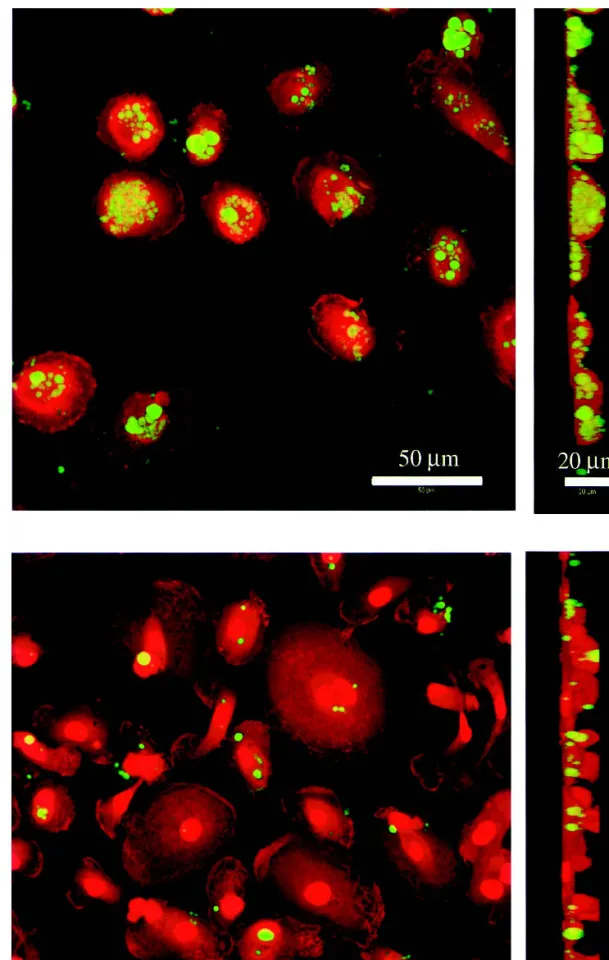
Phagocytosis of noncoated and PLL-g-PEG-coated PLGA microspheres was further investigated by CLSM to verify the intracellular localization of the microspheres in contrast to those attached to the outer membrane. The shape of the phagocytes was visualized by staining their actin cyto-skeleton with rhodamine-phalloidine (red), while the micro-spheres were labeled with 6-coumarin (green; Fig. 5). The result is a clear difference between the phagocytosis of non-coated vs. PLL-g-PEG-non-coated PLGA microspheres. Quanti-fication of uptake by counting the number of microspheres in the cells resulted in numbers, which were in reasonable agree-ment with the data obtained by fluorescence microscopy (data not shown).
A preliminary estimation of the cellular toxicity of the coatings was also performed. In control experiments, less than 5% of the MOs or DCs were found to be necrotic, irrespective of whether noncoated or PLL-g-PEG-coated microspheres were used. This corresponds to previous investigations on toxicity (14), immunogenicity, immunomodulatory potential, pyrogenicity and biodegradation (47–49) of PLL-g-PEG and suggests good biocompatibility of the coatings.
Previous attempts to inhibit phagocytosis of PLGA and
poly(L-lactide) (PLA) micro- and nanospheres by surface modification focused on coatings with poloxamer (11), high molecular weight PEG (50), or on the use of di-block and tri-block co-polymers of PLA/PLGA and PEG to prepare bulk modified micro- or nanospheres (8,9,51). For instance, the circulation time of PLA-PEG nanospheres was extended up to 6 h and they were found in increasing amounts in the heart, in blood vessels and in the lung (52). Inhibition of phagocytosis in the presence of serum or plasma was down to
Fig. 2. Phagocytosis of polystyrene particles by macrophages (a–d) and dendritic cells (e–h) upon the addition of noncoated particles (a, e), and particles coated with PLL-g-PEG (b, f), PLL-g-PEG-RGD (c, g), and PLL-g-PEG-RDG (d, h). Photographs were taken after 4 h of incubation at 37°C.
20–55%, revealing a comparable or even superior effect of the PLL-g-PEG-coated microspheres used in this study. Moreover, at both 4 and 37°C the repellent character of the PLL-g-PEG coatings on PLGA microspheres was maintained for up to 4 days (data not shown). In contrast, when in contact with water, the coatings of nonionic surfactants are subject to enhanced desorption followed by water uptake and subse-quent swelling, especially with microspheres prepared from fast degrading low molecular weight PLGA polymers (13). From a fabrication point of view, the spontaneous adsorption of PLL-g-PEG polymers to the carriers appears to be a su-perior technology to the covalent PEGylation of the polymer bulk. The combination of two separate platforms, i.e., the established PLGA microsphere technology and the PLL-g-PEG based surface modification, may allow separate devel-opment and optimization of carrier and coating. Conversely, the use of PEG-modified bulk polymers is expected to affect the physicochemical properties and the bioerosion of the
polymer matrix, and requires the composition and process parameters for microencapsulation to be optimized on a case-to-case basis.
In addition to the inhibition of nonspecific phagocytosis and in correspondence with the data from polystyrene par-ticles, we found that coatings of the PLGA microspheres with PLL-g-PEG-RGD resulted in ligand-specific phagocytosis by MO and DC (Fig. 4). As compared with noncoated micro-spheres the extent of phagocytosis of PLL-g-PEG-RGD-coated microspheres was decreased, which may be explained by some degree of microsphere agglomeration observed in the preparation. To our knowledge, this is the first dem-onstration of a technology that enables the targeting of biodegradable PLGA microspheres to specific cell surface re-ceptors while simultaneously preventing nonspecific phago-cytosis.
CONCLUSIONS
In this study, we demonstrated a simple and reproducible approach for the surface modification of biodegradable PLGA microspheres for targeting purposes. Coating with positively charged PLL-g-PEG by electrostatic interaction with negatively charged microspheres drastically reduced the nonspecific uptake by professional phagocytes such as MO and DC. Conjugation of a specific ligand to PLL-g-PEG en-ables receptor-specific targeting while maintaining the repel-lent character of PLL-g-PEG coatings. This should preclude rapid physiologic clearance of the microparticles by the MPS. The biospecificity of the interaction was proven by using the inactive nonspecific RDG-conjugated polymer as a control. Overall, our findings provide a potential technology to dimin-ish nonspecific phagocytosis of micro- and nanoparticles and
Fig. 3. Phagocytosis by macrophages (MOs) and dendritic cells (DCs). Typical distribution (a) showing the uptake of noncoated and PLL-g-PEG-coated polystyrene particles in MOs. Box plots for the phagocytosis by MOs (b) and DC (c) of noncoated particles and particles coated with either g-PEG, g-PEG-RGD, or PLL-g-PEG-RDG. Significance testing revealed differences between non-coated and PLL-g-PEG-non-coated particles (p ⱕ 0.05) and between PLL-g-PEG-RGD- and PLL-g-PEG-RDG-coated particles (p ⱕ 0.05) for both MOs and DCs.
Fig. 4. Phagocytosis of noncoated PLGA microspheres and micro-spheres coated with either g-PEG, g-PEG-RGD, or PLL-g-PEG-RDG by macrophages (a) and dendritic cells (b) presented as box plots. Significance testing revealed differences between non-coated and PLL-g-PEG-non-coated particles (p ⱕ 0.05) and between PLL-g-PEG-RGD- and PLL-g-PEG-RDG-coated particles (p ⱕ 0.05) for both macrophages and dendritic cells.
Fig. 5. Phagocytosis of noncoated (a) and PLL-g-PEG-coated (b) PLGA microspheres in macrophages as analyzed by confocal laser scanning microscopy. The outline of the individual cells was visualized by staining the actin cytoskeleton with rhodamine-phalloidin (red). Microspheres were labeled with 6-coumarin (green). Optical sections (xy, left panel) and yz-projections (right panel) allow to distinguish between extra-cellular and internalized microspheres.
at the same time target them to specific receptors. As a proof-of-concept this study was restricted to a model ligand, RGD. For the future we expect larger benefit of this novel technol-ogy to result from the use of more selective ligands, e.g., for the specific targeting of DC rather than MO (53). Moreover, in case suitable ligands can be provided, the technology may be also of use to target particulate drug carriers to other tissues and cells. This needs yet to be demonstrated by ex-periment.
ACKNOWLEDGMENTS
We would like to thank Michael Ulrich and Samantha Jilek for their support in isolating primary macrophages and dendritic cells and Stephanie Vandevondele and Samuele To-satti for their help with the peptide modified PLL-g-PEG. Pentti Tengvall at Linko¨ping Univeristy, Sweden, is acknowl-edged for valuable discussions regarding PLL-PEG immobi-lization and interpretation of results.
REFERENCES
1. C. Thomasin, G. Corradin, Y. Men, H. P. Merkle, and B. Gander. Tetanus toxoid and synthetic malaria antigen containing poly(lac-tide)/poly(lactide-co-glycolide) microspheres: Importance of polymer degradation and antigen release for immune response. J. Control. Release 41:131–145 (1996).
2. M. S. Shive and J. M. Anderson. Biodegradation and biocompat-ibility of PLA and PLGA microspheres. Adv. Drug Deliv. Rev. 28:5–24 (1997).
3. F. Ahsan, I. P. Rivas, M. A. Khan, and A. I. Torres Suarez. Targeting to macrophages: role of physicochemical properties of particulate carriers—liposomes and microspheres—on the phagocytosis by macrophages. J. Control. Release 79:29–40 (2002).
4. D. R. Absolom. Opsonins and dysopsonins: an overview. Meth-ods Enzymol. 132:281–318 (1986).
5. M. E. Norman, P. Williams, and L. Illum. Influence of block copolymers on the adsorption of plasma proteins to micro-spheres. Biomaterials 14:193–202 (1993).
6. G. L. Kenausis, J. Voros, D. L. Elbert, N. P. Huang, R. Hofer, L. Ruiz-Taylor, M. Textor, J. A. Hubbell, and N. D. Spencer. Poly(L-lysine)-g-poly(ethylene glycol) layers on metal oxide sur-faces: Attachment mechanism and effects of polymer architecture on resistance to protein adsorption. J. Phys. Chem. B. 104:3298– 3309 (2000).
7. G. R. Harper, M. C. Davies, S. S. Davis, T. F. Tadros, D. C. Taylor, M. P. Irving, and J. A. Waters. Steric stabilization of microspheres with grafted polyethylene oxide reduces phagocy-tosis by rat Kupffer cells in vitro. Biomaterials 12:695–700 (1991). 8. R. Gref, M. Luck, P. Quellec, M. Marchand, E. Dellacherie, S. Harnisch, T. Blunk, and R. H. Muller. ‘Stealth’ corona-core nanoparticles surface modified by polyethylene glycol (PEG): in-fluences of the corona (PEG chain length and surface density) and of the core composition on phagocytic uptake and plasma protein adsorption. Colloids Surfaces B: Biointerfaces. 18:301–313 (2000).
9. F. X. Lacasse, M. C. Filion, N. C. Philipps, E. Escher, J. N. Mc-Mullen, and P. Hildgen. Influence of surface properties at biode-gradable microsphere surfaces: effects on plasma protein adsorp-tion and phagocytosis. Pharm. Res. 15:312–317 (1998).
10. L. Illum, L. O. Jacobsen, R. H. Muller, E. Mak, and S. S. Davis. Surface characteristics and the interaction of colloidal particles with mouse peritoneal macrophages. Biomaterials 8:113–117 (1987).
11. S. E. Dunn, G. A. Coombes, M. C. Garnett, S. S. Davis, M. C. Davies, and L. Illum. In vitro cell interaction and in vivo biodis-tribution of poly(lactide-co-glycolide) nanospheres surface modi-fied by poloxamer and poloxamine copolymers. J. Control. Re-lease 44:65–76 (1997).
12. M. Luck, K. F. Pistel, Y. X. Li, T. Blunk, R. H. Muller, and T.
Kissel. Plasma protein adsorption on biodegradable microspheres consisting of poly(D,L-lactide-co-glycolide), poly(L-lactide) or ABA triblock copolymers containing poly(oxyethylene) - Influ-ence of production method and polymer composition. J. Control. Release 55:107–120 (1998).
13. M. A. Tracy, K. L. Ward, L. Firouzabadian, Y. Wang, N. Dong, R. Qian, and Y. Zhang. Factors affecting the degradation rate of poly(lactide-co-glycolide) microspheres in vivo and in vitro. Bio-materials 20:1057–1062 (1999).
14. D. L. Elbert and J. A. Hubbell. Self-assembly and steric stabili-zation at heterogeneous, biological surfaces using adsorbing block copolymers. Chem. Biol. 5:177–183 (1998).
15. E. Walter, D. Dreher, M. Kok, L. Thiele, S. G. Kiama, P. Gehr, and H. P. Merkle. Hydrophilic poly(DL-lactide-co-glycolide) mi-crospheres for the delivery of DNA to human-derived macro-phages and dendritic cells. J. Control. Release 76:149–168 (2001). 16. E. Walter and H. P. Merkle. Microparticle-mediated transfection of non-phagocytic cells in vitro. J. Drug Target. 10:11–21 (2002). 17. S. Vandevondele, J. Vo¨ro¨s, M. Textor, and J. A. Hubbell. RGD-grafted poly-L-lysine-graft-(polyethylene glycol) copolymers block nonspecific protein adsorption while promoting cell adhe-sion. Biotechnol Bioeng. in press (2002)
18. N. P. Huang, R. Michel, J. Voros, M. Textor, R. Hofer, A. Rossi, D. L. Elbert, J. A. Hubbell, and N. D. Spencer. Poly(L-lysine)-g-poly(ethylene glycol) layers on metal oxide surfaces: Surface-analytical characterization and resistance to serum and fibrino-gen adsorption. Langmuir 17:489–498 (2001).
19. E. Walter, K. Moelling, J. Pavlovic, and H. P. Merkle. Microen-capsulation of DNA using poly(DL-lactide-co-glycolide): Stabil-ity issues and release characteristics. J. Control. Release 61:361– 374 (1999).
20. F. Sallusto, M. Cella, C. Danieli, and A. Lanzavecchia. Dendritic cells use macropinocytosis and the mannose receptor to concen-trate macromolecules in the major histocompatibility complex class II compartment: downregulation by cytokines and bacterial products. J. Exp. Med. 182:389–400 (1995).
21. L. Thiele, B. Rothen-Rutishauser, S. Jilek, H. Wunderli-Allenspach, H. P. Merkle, and E. Walter. Evaluation of particle uptake in human blood monocyte-derived cells in vitro. Does phagocytosis activity of dendritic cells measure up with macro-phages? J. Control. Release 76:59–71 (2001).
22. L. Cochand, P. Isler, F. Songeon, and L. P. Nicod. Human lung dendritic cells have an immature phenotype with efficient man-nose receptors. Am. J. Respir. Cell Mol. Biol. 21:547–554 (1999). 23. M. L. Albert, B. Sauter, and N. Bhardwaj. Dendritic cells acquire antigen from apoptotic cells and induce class I-restricted CTLs. Nature 392:86–89 (1998).
24. S. C. Finnemann and E. Rodriguez-Boulan. Macrophage and retinal pigment epithelium phagocytosis: apoptotic cells and pho-toreceptors compete for alphavbeta3 and alphavbeta5 integrins, and protein kinase C regulates alphavbeta5 binding and cytoskel-etal linkage. J. Exp. Med. 190:861–874 (1999).
25. A. Rubartelli, A. Poggi, and M. R. Zocchi. The selective engulf-ment of apoptotic bodies by dendritic cells is mediated by the alpha(v)beta3 integrin and requires intracellular and extracellu-lar calcium. Eur. J. Immunol. 27:1893–1900 (1997).
26. S. M. Moghimi, C. J. H. Porter, I. S. Muir, L. Illum, and S. S. Davis. Non-Phagocytic Uptake of Intravenously Injected Micro-spheres in Rat Spleen - Influence of Particle-Size and Hydro-philic Coating. Biochem. Biophys. Res. Commun. 177:861–866 (1991).
27. R. Alyautdin, D. Goethier, V. Petrov, and D. Kharkevich. and J. Kreuter. Analgesic activity of the hexapeptide dlargin adsorbed on the surface of polysorbate 80-coated poly(butyl cyanoacry-late) nanoparticles. Eur. J. Pharm. Biopharm. 41:44–48 (1995). 28. R. H. Muller, S. Maassen, H. Weyhers, and W. Mehnert.
Phago-cytic uptake and cytotoxicity of solid lipid nanoparticles (SLN) sterically stabilized with poloxamine 908 and poloxamer 407. J. Drug Target. 4:161–170 (1996).
29. J. C. Neal, S. Stolnik, M. C. Garnett, S. S. Davis, and L. Illum. Modification of the copolymers poloxamer 407 and poloxamine 908 can affect the physical and biological properties of surface modified nanospheres. Pharm. Res. 15:318–324 (1998). 30. L. Araujo and R. Lobenberg. and J. Kreuter. Influence of the
surfactant concentration on the body distribution of nanopar-ticles. J. Drug Target. 6:373–385 (1999).
31. H. Takeuchi, H. Kojima, T. Toyoda, H. Yamamoto, T. Hino, and Y. Kawashima. Prolonged circulation time of doxorubicin-loaded liposomes coated with a modified polyvinyl alcohol after intra-venous injection in rats. Eur. J. Pharm. Biopharm. 48:123–129 (1999).
32. A. L. Klibanov. Targeted delivery of gas-filled microspheres, con-trast agents for ultrasound imaging. Adv. Drug Deliv. Rev. 37: 139–157 (1999).
33. K. Avgoustakis, A. Beletsi, Z. Panagi, P. Klepetsanis, A. G. Kary-das, and D. S. Ithakissios. PLGA-mPEG nanoparticles of cisplat-in: in vitro nanoparticle degradation, in vitro drug release and in vivo drug residence in blood properties. J. Control. Release 79: 123–135 (2002).
34. C. S. Cho, K. Y. Cho, I. K. Park, S. H. Kim, T. Sasagawa, M. Uchiyama, and T. Akaike. Receptor-mediated delivery of all trans-retinoic acid to hepatocyte using poly(L-lactic acid) nano-particles coated with galactose-carrying polystyrene. J. Control. Release 77:7–15 (2001).
35. J. E. Blackwell, N. M. Dagia, J. B. Dickerson, E. L. Berg, and D. J. Goetz. Ligand coated nanosphere adhesion to E- and P-selectin under static and flow conditions. Ann. Biomed. Eng. 29: 523–533 (2001).
36. S. Rudge, C. Peterson, C. Vessely, J. Koda, S. Stevens, and L. Catterall. Adsorption and desorption of chemotherapeutic drugs from a magnetically targeted carrier (MTC). J. Control. Release 74:335–340 (2001).
37. Y. Maitani, K. Kawano, K. Yamada, T. Nagai, and K. Takayama. Efficiency of liposomes surface-modified with soybean-derived sterylglucoside as a liver targeting carrier in HepG2 cells. J. Con-trol. Release 75:381–389 (2001).
38. A. Gessner, C. Olbrich, W. Schroder, O. Kayser, and R. H. Mull-er. The role of plasma proteins in brain targeting: species depen-dent protein adsorption patterns on brain-specific lipid drug con-jugate (LDC) nanoparticles. Int. J. Pharm. 214:87–91 (2001). 39. R. Igarashi, M. Takenaga, J. Takeuchi, A. Kitagawa, K.
Matsu-moto, and Y. Mizushima. Marked hypotensive and blood flow-increasing effects of a new lipo-PGE(1) (lipo-AS013) due to vas-cular wall targeting. J. Control. Release 71:157–164 (2001). 40. C. S. Cho, A. Kobayashi, R. Takei, T. Ishihara, A. Maruyama,
and T. Akaike. Receptor-mediated cell modulator delivery to hepatocyte using nanoparticles coated with carbohydrate-carrying polymers. Biomaterials 22:45–51 (2001).
41. M. A. Chattergoon, J. J. Kim, J. S. Yang, T. M. Robinson, D. J. Lee, T. Dentchev, D. M. Wilson, V. Ayyavoo, and D. B. Weiner. Targeted antigen delivery to antigen-presenting cells including dendritic cells by engineered Fas-mediated apoptosis. Nat. Bio-techol. 18:974–979 (2000).
42. P. Jeannin, T. Renno, L. Goetsch, I. Miconnet, J. P. Aubry, Y. Delneste, N. Herbault, T. Baussant, G. Magistrelli, C. Soulas, P. Romero, J. C. Cerottini, and J. Y. Bonnefoy. OmpA targets den-dritic cells, induces their maturation and delivers antigen into the
MHC class I presentation pathway. Nat. Immunol. 1:502–509 (2000).
43. Y. Shibata, W. J. Metzger, and Q. N. Myrvik. Chitin particle-induced cell-mediated immunity is inhibited by soluble mannan: mannose receptor-mediated phagocytosis initiates IL-12 produc-tion. J. Immunol. 159:2462–2467 (1997).
44. A. Regnault, D. Lankar, V. Lacabanne, A. Rodriguez, C. Thery, M. Rescigno, T. Saito, S. Verbeek, C. Bonnerot, P. Ricciardi-Castagnoli, and S. Amigorena. Fcgamma receptor-mediated in-duction of dendritic cell maturation and major histocompatibility complex class I-restricted antigen presentation after immune complex internalization. J. Exp. Med. 189:371–380 (1999). 45. P. Johansen, Y. Men, H. P. Merkle, and B. Gander. Revisiting
PLA/PLGA microspheres: an analysis of their potential in par-enteral vaccination. European Eur. J. Pharm. Biopharm. 50:129– 146 (2000).
46. M. Mu¨ller, J. Vo¨ro¨s, G. Csucs, E. Walter, G. Danuser, M. Textor, H. P. Merkle, and N. D. Spencer. Surface modification of PLGA microspheres. J. Biomed. Mat. Res. In press.
47. A. A. Bogdanov, R. Weissleder, H. W. Frank, A. V. Bogdanova, N. Nossif, B. K. Schaffer, E. Tsai, M. I. Papisov, and T. J. Brady. A new macromolecule as a contrast agent for MR-angiography— preparation, properties, and animal studies. Radiology 187:701– 706 (1993).
48. A. A. Bogdanov, C. Martin, A. V. Bogdanova, T. J. Brady, and R. Weissleder. An adduct of cis-diamminedichloroplatinum(II) and poly(ethylene glycol)poly(L-lysine)-succinate: synthesis and cy-totoxic properties. Bioconjugate Chem. 7:144–149 (1996). 49. H. J. P. Ryser, R. Mandel, A. Hacobian, and W. C. Shen.
Metho-trexate-poly(lysine) as a selective agent for mutants of chinese-hamster ovary cells defective in endocytosis. J. Cell. Physiol. 135: 277–284 (1988).
50. J. C. Leroux, F. De Jaeghere, B. Anner, E. Doelker, and R. Gurny. An investigation on the role of plasma and serum op-sonins on the internalization of biodegradable poly(D,L-lactic acid) nanoparticles by human monocytes. Life Sci. 57:695–703 (1995).
51. F. De Jaeghere, E. Allemann, J. Feijen, T. Kissel, E. Doelker, and R. Gurny. Cellular uptake of PEO surface-modified nanopar-ticles: evaluation of nanoparticles made of PLA:PEO diblock and triblock copolymers. J. Drug Target. 8:143–153 (2000).
52. T. Verrecchia, G. Spenlehauer, D. V. Bazile, A. Murry-Brelier, Y. Archimbaud, and M. Veillard. Non-stealth (poly(lactic acid/ albumin)) and stealth (poly(lactic acid-polyethylene glycol)) nanoparticles as injectable drug carriers. J. Control. Release 36: 49–61 (1995).
53. T. B. Geijtenbeek, D. S. Kwon, R. Torensma, S. J. van Vliet, G. C. van Duijnhoven, J. Middel, I. L. Cornelissen, H. S. Nottet, V. N. KewalRamani, D. R. Littman, C. G. Figdor, and Y. van Kooyk. DC-SIGN, a dendritic cell-specific HIV-1-binding pro-tein that enhances trans-infection of T cells. Cell 100:587–597 (2000).