HYPERICUM PERFORATUM IMPROVE MEMORY AND LEARNING IN ALZHEIMER'S MODEL:
(EXPERIMENTAL STUDY IN MICE)
Original Article
KHAYRA ZERROUKI
a,b*, NOUREDDINE DJEBLI
a, ESRA EROGLU OZKAN
c, NURTEN OZSOY
d, OZHAN GUL
e,
AFIFE MAT
caLaboratory of Pharmacognosy and Api-Phytotherapy Department of Biology FSNV-Mostaganem University, Mostaganem, Algeria, bDepartment of Biology-Faculty of Naturaal and Life Sciences Chlef University, cDepartment of Pharmacognosy Faculty of
Pharmacy-Istanbul Pharmacy-Istanbul, dDepartment of Biochemistry, Faculty of Pharmacy, e
Received: 05 Jul 2015 Revised and Accepted: 20 May 2016
Department of Pharmaceutical Toxicology Faculty of Pharmacy, Istanbul University, Istanbul, Turkey
Email: soumaia9@gmail.com ABSTRACT
Objective: The aim of this study, we based on protective and antioxidant efficiency of Hypericum perforatum that shows a wide range of beneficial effect in vitro and in vivo.
Methods: The in vitro antioxidant activity of the extract was assessed by using several antioxidant tests. The cytotoxic activity of Hypericum perforatum was also determined by using 3-(4, 5-dimethylthiazol-2-yl)-2, 5-diphenyltetrazolium bromide viability assay on ordinary used cell lines. In vivo experiments in Swiss mice were determined by performing behavioral, memory tests and histological study. According to tests results, H. perforatum may be relevant to the treatment of cognitive disorders.
Results: The results of chemical analysis showed a hight level of hyperforin and quercitin that had an important antioxidant activity proved in vitro with the 2, 2-diphenyl-1-picrylhydrazyl, Anti-lactoperoxidase and superoxide dismutases; this antioxidant activity was confirmed in vivo after the non-toxic results by means of improvement in behavioral and memory than the reducing shrunken in pyramidal cells of mice brains.
Conclusion: The present study suggests that Hypericum perforatum modulate the oxidative stress and be involved in the protective effect against oxidative damage and neurodegenerative diseases in mice.
Keywords: Neurotoxicity, Alzheimer’s disease, Phytotherapy, Hypericum perforatum, Neuroprotective, Mice
© 2016 The Authors. Published by Innovare Academic Sciences Pvt Ltd. This is an open access article under the CC BY license (http://creativecommons.org/licenses/by/4.0/)
INTRODUCTION
Alzheimer's disease (AD) is a multifactorial disease, complex, and progressive affecting the older population the most observed dementia cases is in persons over 65 y of age [1, 2].
Histological and pathological studies of AD have revealed that multiple cellular pathways are involved in AD progression [3]. The pathological features identified in the central nervous system (CNS) in AD are amyloid plaques, neurofibrillary tangles, inflammatory processes and disturbance of neurotransmitters [4, 5]. The dysfunction and degeneration of synapses in AD may involve Aβ-induced oxidative stress compromises the mitochondrial function by an oxidative-stress-mediated mechanism [6].
Reactive oxygen species (ROS) are produced by many redox processes that normally occur in the metabolism of aerobic cells. These species are very reactive and harmful to the cells. If not eliminated, ROS can damage vital molecules, such as proteins, DNA, and lipids. Cells express several defense mechanisms, including antioxidant enzymes and non-enzymatic compounds that help prevent the damaging effects of ROS [7].
Oxidative stress can also play an important role in the development of neurodegenerative disorders, such as Alzheimer’s and Parkinson’s diseases [8, 9]. There is an increasing interest in natural antioxidants, namely the phenols, present in medicinal and dietary plants, that might help prevent oxidative damage. [10-14]. In situations of increased free radical generation, the reinforcement of endogenous antioxidants via intake of dietary antioxidants may be of particular importance in attenuating the cumulative effects of oxidatively damaged molecules. In recent years, the consumption of Hypericum perforatum (St. John’s wort) derived products have increased dramatically, and presently it is one of the most consumed medicinal plants over the world [15]. The commercially available H.
perforatum derived products include sophisticated phytopharmaceuticals and nutraceuticals, teas, tinctures, juices and oil macerates [16]. H. perforatum has a wide range of medicinal applications, including skin wounds, eczema, burns, diseases of the alimentary tract and psychological disorders. Nowadays, its use in the treatment of mild to moderate depression has become prominent [17-19]. Numerous papers have been published concerning these aspects and several recent reviews can be pointed out [19-21]. In spite of this intense research activity, the antioxidant potential of H. perforatum extracts has been somewhat neglected. Current studies suggested that H. perforatum possessed protective effects against H2O2
The chemical composition of the Hypericum species is composed of naphthodianthrones (especially hypericin and pseudo hypericin), acylphloroglucinol derivatives (especially hyperforin and adhyperforin), flavonoids (especially quercetin, quercitrin, hyperoside and biapigenin), tannins, n-alkanes, xanthones and essential oils. [18, 23, 24]
induced apoptosis of PC12 cells than might be useful in the treatment of oxidative stress related to neurodegenerative diseases such as Alzheimer disease [22].
The aim of this study was to identify the potential of H. perforatum as a protective and therapeutic agent against neurodegenerative disorders and Alzheimer's disease.
The chemical composition of ethanolic extract of H. perforatum was analyzed by using HPLC-DAD. In order to determine the antioxidant potential of H. perforatum, was evaluated by employing several antioxidant tests. The cytotoxic activity of the extract was also determined by using MTT cell cytotoxicity screening assay on HeLa and NRK-52E cell lines. The in vivo studies on Swiss male mice were determined by utilizing behavioral, memory tests and histological analysis.
This represents the first report on the chemical composition, antioxidant, cytotoxic and anti-Alzheimer activity of H. perforatum that has been tested in this study.
MATERIALS AND METHODS Plant and animals
Aerial parts of H. perforatum were collected in Malatya (Turkey) in June 2010. The plant material was identified by Şükran Kültür and a voucher specimen (ISTE 93192), was deposited in the Herbarium of the University Of Istanbul Faculty Of Pharmacy, Istanbul, Turkey. The ethanol extracts were obtained by maceration of the plant material with ethanol for 3 d at room temperature and this procedure currently was repeated twice. The respective extracts were filtered and dried under reduced pressure at a temperature below 218.15 K. The crude methanol extract was lyophilized and stored at 153.15 K.
The in vivo studies were performed on Swiss mice (3-month-old), 22-26g, purchased from Pasteur Institute of Algiers. Mice were housed in the Laboratory animal care of Mostaganem University in 12-hour light-dark cycles at 196.15 K with free access to food and water.
Chemical agents
Hypericin, chlorogenic acid, rutin, hyperoside, isoquercitrin, quercitrin, kaempferol, quercetin, amentoflavone, hyperforin, AlCl3
Nitrobluetetrazolium (NBT), β-nicotinamide adenine dinucleotide reduced (NADH), soybean L-α-phosphatidylcholine Type IV-S, butylated hydroxytoluene (BHT), 3-(2-pyridyl)-5,6-bis (4-phenyl-sulfonic acid)-1,2,4-triazine (ferrozine), rutin and catechin were purchased from Fluka Chemical Co. (Buchs, Switzerland). Phenazinemethosulphate (PMS), 2,2-diphenyl-1-picryl-hydrazyl (DPPH•), ethylene diamine tetraacetic acid (EDTA), Gallic acid and ascorbic acid were obtained from Sigma Chemical Co. (St. Louis, MO, USA). Potassium ferric cyanide, trichloroacetic acid (TCA), thiobarbituric acid (TBA), ferrous and ferric chloride were obtained from Merck. All other reagents were of analytical grade.
and D-Galactose were obtained from Sigma-Aldrich (Taufkirchen, Germany). Pseudohypericin was trained from PhytoPlan (Heidelberg, Germany). Milli-Q ultrapure water was obtained from Millipore (Billerica, MA), HPLC grade acetonitrile, methanol, ethyl acetate and sodium dihydrogen phosphate dihydrate were obtained from Merck (Darmstadt, Germany) and orthophosphoric acid 85% was obtained from Fluka (Steinheim, Switzerland).
The 3-(4, 5-dimethylthiazol-2-yl)-2,5-diphenyl tetrazolium bromide (MTT) reagent were purchased from Sigma (St. Louis, USA). Dimethylsulfoxide (DMSO), trypsin (Biomatik, Canada), absolute alcohol, ethylene diamine tetraacetic acid (EDTA), sodium hydroxide (Merck, Germany). Fetal bovine serum (FBS), Dulbecco’s Modified Eagle Medium (DMEM), penicillin-streptomycin, phosphate buffer saline (PBS) were purchased from Multicell-Wisent Inc. (Quebec, Canada). Cytotoxicity detection kit (LDH) which containing catalyst, dye solution and stop solution were purchased from Roche (Mannheim. Germany). All other reagents were of analytical grade.
Experiment 1
The dried flowering aerial parts of H. perforatum were macerated in ethanol for 3 d at room temperature and the resulting extract was filtered through Whatman No.1. The residue from the filtration was extracted again twice having the same procedure. The filtrates obtained were combined and then evaporated to dryness under reduced pressure at a temperature below 318.15 K. The crude methanol extract was lyophilized and stored at 253.15 K [25, 26]. Extracts prepared with this procedure were used in the HPLC analysis and biological activity studies.
The extract has been tested by HPLC-DAD. The HPLC system consisted of a Shimadzu 10A model (Shimadzu Analytical and Measuring Instruments, Kyoto, Japan), a pump (LC-10AD), a diode-array detector (DAD) (SPD-M10A) and an autosampler (SIL-10AD).
Separation was accomplished with an ACE C18 column, 250 x 4.6 mm i.d., 5 µm (Advanced Chromatography Technologies, Alberdeen, Scotland). The elution conditions were as follows: flow rate: 1 cm3/60s; column temperature: 313.15 K; injection volume: 0.01 cm3
The solvent system used was a gradient of solvent A (0.3% formic acid in water (v/v)) and solvent B (0.3% formic acid in aceto-nitril (v/v)) to identify and quantify phenolic compounds and hyperforin (European Pharmacopoeia, 2008). The following gradient was applied: 0–1080 s, 82% A; 480–1080 s, 82–47% A; 1080–1086 s, 47– 3% A; 1086–1740 s, 3% A; 1740–2400 s, 3–82% A. The solvent system used was an isocratic of solvent A (Ethyl acetate: 15.6 g/dm
; detection: 590 nm for pseudo-hypericin and hypericin, 360 nm for phenolic compounds and 275 nm for hyperforin.
3
The control of the system and the data analysis procedure was performed with Shimadzu LC Solutions software.
sodium dihydrogen phosphate adjusted to pH: 2 with phosphoric acid: methanol/39:41:160/v/v/v) to identify and quantify pseudo-hypericin and pseudo-hypericin (European Pharmacopoeia, 2008). All solvents were filtered through 0.45 µm filters prior to use and degassed in an ultrasonic bath.
The calibration curves were prepared with external standards solution within the different concentration range in methanol. The experiment was performed for three times providing the same conditions. The calibration curves were constructed by using the average of peak areas.
The crude ethanol extract was dissolved in a mixture of methanol/ water (8:2, v/v). All samples were filtered through 0.45 µm filters into a vial for HPLC analysis. Each sample was prepared and injected for three times.
Experiment 2
In vitro antioxidant activity
Total soluble phenolic in the methanolic extracts was determined with Folin-Ciocalteu reagent according to the method of Slinkard & Singleton with some modifications [27].
The amount of total phenolic compounds was calculated from the calibration curve of Gallic acid standard solution (covering the concentration range between 0.05 and 0.4 mg/ml) and expressed as mg Gallic acid equivalents (GAE)/g dry weight (DW) of the plant material.
Total flavonoid content was established by using a method described by Sakanaka [28]. Total flavonoid contents were calculated based on the calibration curve prepared with standard catechin solution and expressed mg of (+)-catechin equivalents (CE) per g of DW of the plant material.
The DPPH radical scavenging activity of the methanolic extracts was measured according to the procedure described by Brand-Williams [28]. Two controls were utilized for this test, a negative control (containing all reagents except the test sample) and positive controls (using the reference antioxidants). The ability to scavenge DPPH radical was calculated by the following equation:
AA (DPPH radical scavenging activity %) = �(1 −Abs sample )517nm(Abs control )517nm � Χ 100
Lipid peroxidation (LPO) assay was prepared on the basis of the method described by Duh et al. and Brand-Williams et al. [39-30]. Formation of LPO products was assayed by the measurement of malondialdehyde (MDA) levels on the basis of MDA reacted with TBA at 532 nm according to Buege & Aust. The percentage inhibition of LPO was calculated by comparing the results of the sample with those of controls not treated with the extract using the following equation:
Inhibition effect (%) = �1 − Abs sample532nmAbs control (532nm)� Χ 100
The effect of the ethanolic extract on the generation of superoxide radicals was determined by the nitro blue tetrazolium (NBT) reduction method of Nishikimi [29]. Abilities to scavenge the superoxide radical were calculated by comparing the results of the
sample with those of controls not treated with the extract using the following equation:
SOD scavenging (%) = [1
− Abs sample (560nm)/Abs control (560nm)] Χ 100
FRAP assay was carried out in accordance with the procedure of Benzie & Strain [31-33]. The standard curve was constructed using iron sulphate heptahydrate solution (125-2000 µM), and the results were reported in mm Fe2+
In vitro cytotoxic activity
equivalents.
Cytotoxicity of the extract at various concentrations was determined on human cervix adenocarcinoma (HeLa, ATCC CCL-2) and
normal rat kidney epithelial (NRK-52E, ATCC
The results were generated from three independent experiments; each experiment was performed in triplicate.
CRL-6509) cell lines by the MTT assay, which is widely used for the measurement of cell viability. Briefly, the cells were seeded in 96-well plates at a density of 104 cells/well in 100 μl culture medium. Following 24-h incubation and attachment, the cells were treated with different concentrations of plant extracts and controls for 24 h. Dry methanolic extracts were dissolved in dimethyl sulphoxide (DMSO) as a solvent to obtain appropriate stock solutions of the extracts. Dilution of stock extracts of solutions was made in serum-free medium yielding final extracts concentrations from 0.125 to 2 mg/ml. DMSO and 5-fluorouracil (5-FU) were used solvent and positive controls, respectively The concentration range used for 5-FU was 50 to 1000 μm. The yellow MTT dye was reduced by succinic dehydrogenase in the mitochondria of viable cells to purple formazan crystals. The absorbance was measured by a microplate reader (BioTek, USA) at 570 nm with a reference wavelength of 670 nm. The reduction of absorbance was evaluated the inhibition of enzyme activity observed in cells compared to untreated (negative control) cells. Then, the half maximal inhibitory concentration (IC50) was expressed as the concentration of the sample caused an inhibition of 50% in enzyme activities in cells as flows [34].
IC % = 100 − [(mean absorbance of extract x100) /mean absorbance of solvent control]
Experiment 3
In vivo experimental design included three groups of mice Control group: Mice were administered freshwater orally and served as normal control. Alzheimer’s model group: Mice were treated with AlCl3 (100 mg/kg/day) concomitant with IP D-Gal in order of 120
mg/kg, respectively (1.5*10-6 cm3
Treated Alzheimer’s model group: Received concomitant with the Alzheimer’s dose, 0.1 cm
/s).
3
Both H. perforatum and chemical (ALCl
IP of H. perforatum in order of 200 mg/kg/day. The experiment lasted 3 mo.
3
Behavioral and memory tests
& D-Gal) were dissolved in distilled water in order to be administered for the needed dose. The different groups were monitored for the onset of postural abnormalities, general behavioral changes memory and learning. Locomotors activity refers to the movement from one location to another. In rodents, one of the most important components of exploration, a prominent activity of the rat’s repertoire of impulsive activity, is locomotion. We detected spontaneous motor activity over a four periods of 1200 s (300 s each) in an activity cage. Measure of motor exercise endurance was evaluated using a divided box apparatus counting the number of visited cases.
The anxiety test was established by means of dark/white connected rooms. The light/dark test is built on the innate aversion of rodents to brightly illuminated areas and on the spontaneous exploratory behavior of rodents in response to mild stressors, that is, novel environment and light. The test apparatus consists of a small obscure safe compartment (half box) and a large floodlit aversive compartment. Mice are placed in one of the rooms and the time
spent in each room was assessed. These tests were realized during 1200 s divides to four phases, 300 s each.
The hole board test was used to assess the behavior of mice confronted with a new environment. The test is to determine the effects of psychotropic drugs on exploratory behavior exhibited by the animal. It only takes a few minutes and does not require learning or conditioning of animals from them. Exploration holes movements are counted as a score of exploration.
The study of memory incorporates research methodologies from neuropsychology, human development and animal testing using a wide range of species.
Understanding memory has benefited greatly from animal research. Animal’s brains can be selectively lesions using neurotoxic methods (AlCl3
Non-spatial memory was used in neurology in order to assess memory disorder depends upon the hippocampus. For this aim of the experiment, only two arms of the maze were utilized. A baited lighted arm and the other arm were unlit. The subject is well placed on the central platform with two closed arms, and the two arms are straight successively for it to adapt to its new environment. The spending time in the lighted arm was assessed.
in this case) and be assessed comparatively to the treated mice. The assessment of animal memory utilizing different types of mazes has been largely used in neurosciences. Several models have been proposed recently, mainly trying to evaluate the accuracy of choice between the alternatives presented in the same day of the session, instead of looking for the accumulated learning through successive days of training.
Working memory relates to a short-term memory system used to complete a task, such as a relationship between objects and locations. To demonstrate that mice were matched for memory ability after the treatment period, working memory was evaluated in averaging the reentries and error entries, respectively, for the successive days of experimentation.
Histological study
Histological, analyses were performed on formalin-fixed and paraffin-embedded brain tissues from the experiment’s treatment groups. Briefly, whole formalin-fixed brains were cut in 2 nm thick slices along the sagittal plane and embedded in formalin. 2 mm thick serial sections were stained with Haematoxylin-Eosin, periodic acid Schiff, Luxol fast blue and Perls’ stain.
Statistical analysis
About in vivo studies, the data are expressed as means with SEM The statistical significance of differences between groups was assessed with an analysis of variance followed by Student Newman-Keuls. A P value of 0.05 or less was considered as a criterion for a statistically significant difference.
RESULTS Experiment 1
The LC chromatograms of the ethanolic extract of H. perforatum were showed in fıgure 1.
Fig. 1B
Fig. 1C
Fig. 1: It shows the LC chromatogram of ethanolic extract of H.
perforatum A: Pseudohypericin (1), hypericin (2) in the extract
at 590 nm (fig. 1A) B: Chlorogenic acid (1), rutin (2), hyperoside (3), isoquercitrin (4), quercitrin (5), kaempferol (6), quercetin
(7), amentoflavone (8) in the extract at 360 nm (fig. 1B) C: Hyperforin (1) in the extract at 275 nm (fig. 1C) Experiment 2
In vitro antioxidant activity In table 2 and table 3.
In vitro cytotoxic activity
Ethanolic extract of H. perforatum was evaluated for in vitro cytotoxic activity using the colorimetric MTT assay in HeLa and NRK-52E cell lines. The IC50 value (mg/cm3
Experiment 3: In vivo study
) is given in the table. The extract did not demonstrate significant cytotoxic activity against HeLa and NRK-52E cell lines.
Behavioral tests
The performance of mice in the step-through passive avoidance training and testing shows a remarkable difference between Alzheimer treated, and the Alzheimer’s model groups; locomotors activity test showed a hyperactivity in Alzheimer’s model group comparatively to the treated Alzheimer’s and control groups.
Anxiety tests presented in this study with the dark and light rooms, showed significant results for the treated Alzheimer’s comparatively to the Alzheimer’s model group.
The test of maze plus was assessed by the time spending in a covered arm squares noted as score per time of 1200 s, 300 s each phases showed high activity for the Alzheimer’s treated comparatively with the Alzheimer’s model group. fig. 2.
Memory tests
Non-spatial memory test preferably conditional results obtained during the five experimental tests show that Alzheimer’s model mice spent shortly time in the arm lit unlike treated Alzheimer’s and control mice, that put a very short time to get informed on the arm.
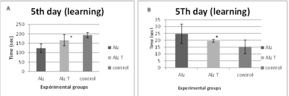
For the work spatial memory, the results of the treated Alzheimer’s group was very significant and very close to the control group results in the last experiment day comparatively to the Alzheimer’s model group. fig. 3.
Table 1: it shows the chemical compounds of the extract found in HPLC chromatogram
Compounds Retention time (min) Calibration equation values Linear regression (r2) H. perforatum (yield, %)
Pseudohypericin 4.86 y= 2.582269e+007x+1741.874 0.9998 0.075±0.0100 Hypericin 13.93 y= 6.03411e+007x+297.2292 0.9999 0.067±0.0400 Chlorogenic acid 4.33 y= 5110294x+1490.398 0.9999 0.008±0.0010 Rutin 8.89 y= 1.383368e+007+5188.182 0.9999 1.015±0.0400 Hyperoside 10.19 y= 2.849917e+007x+526.7023 0.9999 0.491±0.0900 Isoquercitrin 10.75 y= 1.671137e+007x–3712.788 0.9999 0.389±0.0600 Quercitrin 14.41 y= 1.205178e+007–3518.974 0.9999 0.135±0.0100 Kaempferol 17.09 y= 5.183916e+007x+4373.856 0.9999 0.002±0.0001 Quercetin 17.84 y= 3.688175e+007+18905.43 0.9999 0.125±0.0050 Amentoflavon 20.27 y= 2.207879e+007+772.0972 0.9996 0.006±0.0002 Hyperforin 27.75 y= 6212343x 0.9997 3.375±0.5000
Table 2: it shows the total extractable compounds (EC), total phenolic compounds (PC) (As Gallic acid equivalents) and total flavonoids (as catechin equivalents) in the extract
Extracts EC (mg/g DWa) PC (mg/g DWa) Flavonoid (mg/g DWa)
H. perfoatum 267.8 34.7 ± 0.9 31.8 ± 0.9 DW: Dried Weight
Table 3: it shows the antioxidant activity (EC50
Extracts
values) Average of EC50 (mg/ml)
Anti-LPO DPPH Superoxide Reducing power H. perforatum 2.74 ± 0.19 0.27 ± 0.12 1.38 ± 0.09 0.29 ± 0.02 Quercetin 0.059 ± 0.001 0.064 ± 0.001 0.461 ± 0.014 0.045 ± 0.008 *Values were the means of three replicates ± standard deviation.
Table 4: It shows the cytotoxic potential of the extract (IC50 value)
IC50 values (mg/ml)
Extracts HeLa NRK-52E H. perforatum 0.541 0.619
*Positive Control: (5-FU): 48.012 µM for HeLa, 12.645 µM for NRK-52E
Fig. 2: Shows the result of behavioral test; A: Locomotors activity of Alzheimer’s model mice (Alz) intoxicated by AlClз
(100 mg/kg Oral+0,1 cmP3P IP of D-Galactose 120 mg/kg),
Alzheimer’s treated (Alz. T) by Hypericum Perforatum (200 mg/kg) 0,1 cmP3P IP, and the control (Control) without any
treatment; for 90 d. B: Anxiety/curiosity test of Alzheimer’s model mice (Alz), Alzheimer’s treated (Alz. T), and the control (Control); for 90 d. C: curiosity test of Alzheimer’s model mice (Alz), Alzheimer’s treated (Alz. T), and the control (Control) for
90 d. (*) Significant result for Alz. T group compared to the control in the three behavior tests: P<0.05 Histological studies
H&E staining shows that there are typical neuropathological changes in the cerebral cortex and hippocampus of Alzheimer’s model. In the control groups, the neurons were full and arranged tightly; the nuclei were lit stained. By comparison in the model group mice the cytoplasm of neuron were shrunken, the nuclei were side moved and
dark stained, neurofibrillary degeneration and neurone loss were observed in the hippocampus. Hypericum perforatum administration showed moderated neuropathological changes. The treatment decreased the shape modifications of the neurons, with prolonged neurofibrillary reactions. fig. 4.
DISCUSSION
A complex mixture of bioactive secondary metabolites in several Hypericum species makes them valuable as herbal drugs [35-37]. H. perforatum (Common St. John’s wort), certainly the best known and worldwide the most abundant representative, is today the most investigated species of the genus [38]. Since the early 90’s, H. perforatum has been clinically studied from the perspective both of its chemical constituency and of its biological activity [39]. Around 2500 studies on Hypericum have been published to date (thereof c. 950 without H. perforatum; S. Crockett, pers. com.), including several reviews focused on the phytochemistry of H. perforatum [38,40-41], its pharmacology [42,44] or both aspects [38,45-47].
Phytochemical analysis of H. perforatum was achieved for the first time in this work; it was investigated the medicinal value of H. perforatum. According to European Pharmacopoeia, H. perforatum is a traditional medicinal plant with antidepressive and antioxidant properties. It contains a large number of constituents with documented biological activity including phenolic, a broad range of flavonoids and bioflavonoids, hypericin, adhyperforin, amentoflavone, hyperoside and isoquercitrin [47]. The chemical compounds of H. perforatum are provided in table 1. A total of 11 components were defined in the extract. Hyperforin constituted the major fraction (3.375%) this is due to the fact that hyperforin is photosensitive and its highest concentrations appear just before the blossom period [48].
It has been determined that hyperforin, a major constituent of H. perforatum, inhibits the proliferation of epithelial cells and human peripheral blood mononuclear cells, even when stimulated by phyto-hemagglutinin. Furthermore, another study [49], also justified the anticancer activity of extracts of H. perforatum by the presence of hyperforin and hypericin of the plant. Previous work [50] De Freitas Silva et al. demonstrated that compounds other than hypericin are highly relevant for both radical scavenging and inhibition of lipid peroxidation activities.
Rutin constituted the second major fraction, accounting for 1.015% of the extract. Umek et al. found that the content of rutin in H. perforatum was in a strong positive correlation with the altitude of the growing site, opposite to the quercitrin content [51].
Chlorogenic acid has been reported to inhibit lipid peroxidation by scavenging peroxyl radicals, thereby preventing the initiation of chain lipid peroxidation [50]. Also, chlorogenic acid had been reported to be an effective scavenger against peroxyl radicals [50]. According to the previous research [52], Chlorogenic acid has an effective antioxidant activity. These data are in accordance with the recently published chemical characterization of H. perforatum of Macedonia [48].
In the present study, using various in vitro experiments, we found that H. perforatum markedly inhibited XO activity, suggesting that besides direct scavenging [53,54]; inhibition of the XO enzyme may contribute to the scavenging action of the OR2RP-Pradical by H.
perforatum.
Considering antioxidant activity of the marine sponge species screened herein; the literature survey indicated that there has been only one former study describing the antioxidant potential of the compounds; 2-octaprenyl-1,4-hydroquinone and 2-(24-hydroxy)-octaprenyl-1,4-hydroquinone isolated from I. spinosula and its eight synthetic derivatives [55]. Their antioxidant activity was tested in vitro by scavenging of free radical DPPH and inhibition of the lipid peroxidation induced by the FeP++P/ascorbate system. Two natural
hydroquinone derivatives were found to display high antioxidant effect in both tests, which was in accordance with our results. In our DPPH radical scavenging assay, the highest scavenging effect (65.92%) was observed in I. variability of Antalya-I collection (table 3) and linear terpenes including.
C
C
Fig. 3: Shows the result of memory test A: Non-spatial memory test preferably conditional of Alzheimer’s model mice (Alz) intoxicated by AlClз (100 mg/kg Oral+0,1 cm3 IP of D-Galactose 120 mg/kg), Alzheimer’s treated (Alz. T.) by Hypericum Perforatum (200 mg/kg) 0,1
cm3IP, and the control (Control) without any treatment; for 90 d, B: work spatial memory test of Alzheimer’s model mice (Alz),
Alzheimer’s treated (Alz. T.), and the control (Control); for 90 d. (*) Significant result for Alz. T. group compared to the control in memory tests: (P<0.05)
Fig. 4: It shows the microscopic study of Cerebral cortex and Hippocampus performed by staining (HE) in model Alzheimer’s mice (Alz) induced by AlCl3 orally (100MG/KG)+0,1 cm3 IP of
D-Galactose (120 mg/Kg), treated Alzheimer’s model mice (Alz. T) with Hypericum perforatum for three months with 0,1 cm3
IP(200 mg/Kg) (T) compared to cerebral cortex & hippocampus of control mice (G × 400). (Alz) cerebral cortex characterized by
a decrease in cell density and neuronal degenerative and vacuolization (G × 400), (Alz. T) shows a decrease of edema and
vacuolization in treated Alzheimer’s (G × 400) (■)Low cell density; (◄ ) Vacuolization
The extract showed the DPPH radical scavenging activity in a dose-dependent manner. The results show that at 2.5 mg/cm3 the extract
(31.80±0.9%), did not differ in their DPPH radical scavenging activities (p > 0.05), which were comparable to that of quercetin (96.89 ± 0.72 %) at 0.31 mg/cm3. Our results were in agreement
with previous research [56] that reported the similar DPPH radical scavenging activity of 91.3% and 82.6% for H. scabroides and H. triquetrifolium, respectively. It was reported that H. perforatum extract showed relatively high DPPH radical scavenging effect with an average IC50 value of 10.63 µg/cm3
The inhibition of Fe (III) and ascorbic acid-induced lipid peroxidation assay results revealed that the extract inhibited TBARS formation and destruction of phospholipid in a
concentration-dependent manner. The highest antioxidant activity of the extract was observed in high concentration (5 mg/cm
[57]. It was evident that H. perforatum, H. triquetrifolium and H. scabroides extracts had stronger effects on the DPPH radicals compared to HSM used in this study. It can be explained with the different phytochemical composition. It is known that only flavonoids of a certain molecular structure, particularly those with a certain hydroxyl position will determine the antioxidant properties [58].
3). The antioxidant
activity of the extract was determined lower than quercetin activity. Based on the EC50 values, the antioxidant activity of HPM was
significantly lower (p<0.05) with an EC50 value of 2.74 ± 0.219
mg/cm3in comparison with quercetin (0.059 ± 0.001 mg/cm3).
Although the inhibitory effect of the extract on TBARS formation was less than that of quercetin, the extract may also be supposed to protect against damage to cell membranes, because it also reduces the level of peroxides. However, the extract has the capacity to remove free radicals and inhibit lipid peroxidation. The results consistent with the other in vitro studies with Hypericum species. Couladis and Silva et al. have reported that the methanolic extract of H. perforatum shown to inhibit lipid peroxidation with an EC50 value
of 26 µg/cm3 [59,60]. It is reported that the methanolic extract of H.
perforatum shown to inhibit (70%) lipid peroxidation [57]. It was also reported by Kızıl et al. that the extracts obtained from H. triquetrifolium and H. scabroides showed high potential to inhibit lipid peroxidation [56]
The extract was found to be an efficient scavenger of superoxide radical generated in PMS/NADH system, and its superior activity was comparable (p>0.05) to that of quercetin. Both extract and quercetin exhibited dose-dependent inhibition of superoxide radical.
.
The reducing power of the extract was found to be correlated with increasing absorbance (at 700 nm) as compared with quercetin. The extract has shown the potential of reducing ferric (III) form of iron to ferrous (II). These results compared with previous studies, similar results were reported for H. triquetrifolium and H. scabroides [56]. Antioxidant activity of the extract was examined by using four different methods. Table 3 shows EC50
There was a great correlation between total phenolic contents and reductive potential and lipid peroxidation inhibitory activity. As a result, this extract has antioxidant activity due to the phenolic compounds have the potential to act as antioxidants by scavenging free radicals, reducing power and blocking the formation of peroxide radicals and reducing power.
values of the extract in antioxidant properties. The differences of the results explained diverse mechanisms of oxidative stress.
Since the cytotoxic activity potential of the extracts having antioxidant activity is an important parameter, the cytotoxic activity of the HeLa and NRK-52E cell lines is investigated. The results reveal that the extracts have no cytotoxic activity.
Antidepressant activity of H. perforatum is certainly the main reason for the general public’s enthusiasm for this herbal medication [43]. The classical use of (dried alcoholic) extracts of H. perforatum for the treatment of mild to moderate depression has been demonstrated to be effective in several trials and meta-analyses, in the same citation the antidepressant activity of H. perforatum-based formulations can be attributed to several classes of secondary metabolites, which
exhibit additive, synergetic and partly antagonistic effects [61,42]. According to the actual state of scientific knowledge, the total extracts must be considered as the active principle [41].
The effects of H. perforatum on the central nervous system were investigated using various behavioral and memory models, including locomotors activity, dark/lighted rooms and hole-board for the behavior assessment, non-spatial memory and worked spatial memory for the memory experiments. According to the results, it was found that extracts prepared from H. perforatum were effective as depressant drug. This conclusion suggested that the anti-depressant effects of H. perforatum were remarkable in behavioral tests and may be used for therapeutic purposes in depression [62]. The species (rodents) in which aluminum-induced neurobehavioral effects (e. g., changes in locomotors activity, learning, and memory) have been observed fail to develop significant cytoskeletal pathology, but exhibit a number of neurochemical alterations following in vivo or in vitro exposure [63, 64]. In this case, the locomotors activity decreased in treated Alzheimer’s comparatively to the Alzheimer’s group that confirms that hyperactivity observed for the Alzheimer’s group could be the result of stress conditions [65].
Anxiety-related behavior is measured as a preference for the closed arms [66-68]. The percentage of open arm entries also indicates anxiety levels, especially in mice, which tend to be more impulsive and spend less time per entry in any arm. In this study, the time spent in opens area and close to the control group. The hole-board test provides a simple method for measuring the response of an animal to an unfamiliar environment and is widely used to assess emotionality, anxiety and/or responses to stress [69-71].
After the treatment administration of both Aluminum and Hypericum the head dip counts decreased without changing in locomotion. These results indicate that H perforatum has satisfactory in anxiolytic effect in this paradigm [72]. After “training” an experimental animal, such as a mouse, the only way to be sure that a “memory” was formed is by evoking it back, i.e., by recalling it in a “test” session: this “memory” is expressed by a behavior that differs from that one emitted in the training session. Training sessions consist of repeating a number of trials, several days in a row (4-8 trials a day, for 2 to 5 d-or more, when training to a criterion) [73]. Non-spatial memory conditional preference showed an improvement from the third day as same as the work spatial memory that improved from the 4Th day, that result confirm the damage reduction in the brain in treated Alzheimer’s comparatively to the Alzheimer group.
More specifically, the entire brain of patients with Alzheimer’s disease (AD) was shown to be subjected to an oxidative challenge [74]. In addition, overall peroxidation activity in brains of AD patients was significantly elevated compared to normal subjects [75]. In the reverse phenomena, H. perforatum extract contains flavonoids such as rutin, quercetin, and quercitrin, which demonstrated a free radical scavenging activity in a model of auto-oxidation of rat cerebral membranes [76].
In this investigation, the effect of H. perforatum lead to a reduction of neurotoxicity and Alzheimer's disease appeared as shrunken decreased in pyramidal cells, reduced effect of the decreasing number of the pyramidal cells. These brain moderation changes inducing by H. perforatum Alzheimer’s were due to reducing oxidative damage and edema which contribute to disease pathogenesis and were in accordance with some researchers research [77].
CONCLUSION
Operative treatments and medications are still absent to seize Alzheimer’s diseases. AD is a progressive neurodegenerative disorder that affects the elderly population particularly. Since pathogenesis of AD has not been clarified totally, yet, it is only a symptomatic treatment available. The most prescribed drug class against AD is cholinesterase inhibitors, which increase the level of acetylcholine/butyryl-choline in the brain.
Another factor contributing to the pathology of neurodegenerative diseases is oxidative stress, which leads to neuronal death. Inflammation is also linked to neurodegeneration by diverse
mechanisms such as accretion of proteins with abnormal conformations or via signals emanating from injured neurons. Considering the herbal treatment, we initiated an intensive research on the neuroprotective activity of the H. perforatum through the enzyme inhibition linked to neurodegeneration and antioxidant activity methods, which have so far let us to realize the in vitro and in vivo studies.
Antioxidant activity of the samples has been tested in a number of tests such as 2,2-diphenyl-1-picrylhydrazyl (DPPH), N,N-dimethylp-phenylenediamine (DMPD), superoxide, nitric oxide, hydrogen peroxide104 radical scavenging, ferric-reducing antioxidant power (FRAP), and phospho-molybdenum reducing antioxidant power (PRAP) assays and confirmed by in vivo study showing a decrease in cell degeneration.
ACKNOWLEDGEMENT
I would like to gratefully and sincerely to thank Dr. BOUAKLINE Houssam for his support, understanding, patience, and also the service of anatomy-pathology of Military hospital (HMRUO) of Oran that support me during my training.
ABBREVIATION
AD: Alzheimer disease; ROS, Reactive oxygen species; HPLC-DAD, High performance liquid chromatography-diode array detector); MTT, 3-(4,5-dimethylthiazol-2-yl)-2,5-diphenyltetrazolium bromide); NRK: Rattus norvegicus kidney; NBT, Nitro-bluetetra-zolium; NADH, Nicotinamide adenine dehydrogenase; BHT, Butylated-hydroxytoluene; PMS, Phenazinemethosulphate; EDTA, Ethylenediamine extra acetic acid; TCA, Trichloroacetic acid; DMSO, Dimethylsulfoxide; EDTA, Ethylenediamine tetraacetic acid; FBS, Fetal bovine serum; DMEM, Dulbecco’s Modified Eagle Medium; PBS, phosphate buffer saline; LDH, Lactate deshydrogenase; GAE, Galic acid equivalent.
CONFLICT OF INTERESTS Declared none
REFERENCES
1. Francis PT, Palmer AM, Snape M, Wilcock GK. The cholinergic hypothesis pharmacology, biochemistry and behavior of Alzheimer’s disease: a review of progress. J Neurol Neurosurg Psychiatry 1999;66:137–47.
2. Adams M, Gmunder F, Hamburger M. Plants traditionally used in age related brain disorders a survey of the ethno-botanical literature. J Ethnopharm 2007;113:363–81.
3. Reddy PH, McWeeney S. Mapping cellular transcriptosomes in autopsied Alzheimer’s disease subjects and relevant mouse models. Neurobiol Aging 2005;27:1060-77.
4. Selkoe DJ. Alzheimer’s disease: genes, proteins, and therapy. Physiol Rev 2001;81:741–66.
5. Bossy-Wetzel E, Schwarzenbacher R, Lipton SA. Molecular pathways to neurodegeneration. Nat Med 2004;10:2–9. 6. Mattson MP. Pathways towards and away from Alzheimer's
disease. Nature 2004;430:631–9.
7. Ben-Yoseph O, Boxer PA, Ross BD. Assessment of the role of the glutathione and pentose phosphate pathways in the protection of primary cerebrocortical cultures from oxidative stress. J Neurochem 1996;66:2329–37.
8. Behl C, Mosmann B. Antioxidant neuroprotection in Alzheimer’s disease as preventive and therapeutic approach. Free Radical Biol Med 2002;33:182–91.
9. Foy CJ, Passmore AP, Vahidassr MD, Young IS, Lawson JT. Plasma chain-breaking antioxidants in Alzheimer’s disease, vascular dementia, and Parkinson’s disease QJM. J Assoc Physicians India 1999;92:39–45.
10. Gardner PT, White TAC, McPhail DB, Duthie GG. The relative contributions of vitamin C, carotenoids and phenolics to the antioxidant potential of fruit juices. Food Chem 2000;68:471–4. 11. Halliwell B. Oxidative stress, nutrition and health. Experimental strategies for optimization of nutritional antioxidant intake in humans. Free Radical Res 1996;25:57–74.
12. Halliwell B. Establishing the significance and optimal intake of dietary antioxidants: the biomarker concept. Nutr Res 1999;57:104–13.
13. Youdim KA, Spencer JP, Schroeter H, Rice-Evans C. Dietary flavonoids as potential neuroprotectants. Biol Chem 2002;383:503–19.
14. Zheng W, Wang SY. Antioxidant activity and phenolic compounds in selected herbs. J Agric Food Chem 2001;49:5165–70.
15. Wills RBH, Bone K, Morgan M. Herbal products: active constituents, modes of action and quality control. Nutr Res Rev 2000;13:47–77.
16. Gaedcke F. St John’s wort herb extracts: manufacturing, standardization and characterization. Hypericum, medicinal and aromatic plants. In: E Ernst. Ed. London: Taylor and Francis Group; 2003;31:49–64.
17. Butterweck V. Mechanism of action of St. John’s wort in depression: what is known? CNS Drugs 2003;17:539–62. 18. Di Carlo G, Borrelli F, Ernst E, Izzo AA, St John’s. Wort: prozac
from the plant kingdom. Trends Pharmacol Sci 2001;22:292–7. 19. Di Carlo A, Baldereschi M, Amaducci L. Incidence of dementia,
Alzheimer's disease and vascular dementia in Italy. The ILSA Study. J Am Geriatr Soc 2002;50:41-8.
20. Agostinis P, Vantieghem A, Merlevede W, de Witte PAM. Hypericin in cancer treatment: more light on the way. Int J Biochem Cell Biol 2002;34:221-41.
21. Prince AM, Pascual D, Meruelo D, Liebes L, Mazur Y, Dubovi, et al. Influence of prenylated and nonprenylated flavonoids on liver microsomal lipid peroxidation and oxidative injury in rat hepatocytes. Food Chem Toxicol 2001;39:437–45.
22. Zou WQ, Puoti G, Xiao X, Yuan J, Qing L, Cali I, et al. Variably protease-sensitive prionopathy: a new sporadic disease of the prion protein. Ann Neurol 2010;68:162-72.
23. Bombardelli E, Morazzoni P. Hypericum perforatum. Fitoterapia LXVI 1995;1:43-68.
24. Hostettmann K, Marston A, Wolfender JL. Strategy in the search for new lead compounds and drugs from plants. Chimia 2005;59:291-4.
25. Trovato A, E Raneri, M Kouladis, O Tzakou, M Taviano, E Galati. Anti-inflammatory and analgesic activity of hypericum empetrifolium willd. (Guttiferae). Farmaco 2001;56:455–7. 26. Rabanal R, C Bonkanka, M Hernández-Pérez, C Sánchez-Mateo.
Analgesic and topical anti-inflammatory activity of Hypericum canariense L. and Hypericum glandulosum. J Ethnopharmacol 2005;96:591-6.
27. Slinkard K, Singleton VL. Total phenol analyses: automation and comparison with manual methods. Am J Enol Vitic 1977;28:49-55.
28. Sakanaka S, Tachibana Y, Okada Y. Preparation and antioxidant properties of extracts of Japanese persimmon leaf tea (kakinoha-cha). J Food Chem 2005;89:569–75.
29. Duh PD, Tu YY, Yen GC. Antioxidants activity of aqueous extract of Harnjyur (Chrysanthemum morifolium Ramat). Lebensmwiss Technol 1999;32:269-77.
30. Brand-Williams W, Cuvelier ME, Berset C. Use of free radical method to evaluate antioxidant activity. Lebensm Wiss Technol 1995;28:25-30.
31. Buege JA, Aust SD. Lipid peroxidation. Methods Enzymol 1978;51:302-10.
32. Nishikimi M, Rao NA, Yagi K. The occurrence of superoxide anion in the reaction of reduced phenazine methosulphate and molecular oxygen. Biochem Biophys Res Commun 1972;46: 849-54. 33. Oyaizu M. Studies on products of browning reactions:
antioxidative activities of products of browning reaction prepared from glucosamine. Jpn J Nutr 1986;103:413-9. 34. Alley MC, Scudiere DA, Monks A, Hursey ML, Czerwinski MJ,
Fine DL, et al. Feasibility of drug screening with panels of human tumor cell lines. Cancer Res 1988;48:4827-33.
35. Crockett SL, Schaneberg B, Khan I. Phytochemical profiling of new and old world hypericum (St. John’s wort) species. Phytochem Anal 2005;16:479–85.
36. Crockett SL, Eberhardt M, Kunert O, Schuhly W. Hypericum species in the paramos of central and south america: a special focus upon H. irazuense Kuntze. Phytochem Rev 2010;9:255–69.
37. Mártonfi P, Repcák M, Zanvit P. Secondary metabolites variation in Hypericum maculatum and its relatives. Biochem Syst Ecol 2006;34:56–9.
38. Nahrstedt A, Butterweck V. Lessons learned from herbal medicinal products: the example of St. John’s wort. J Nat Prod 2010;73:1015–21.
39. Röder C, Schaefer M, Leucht S. Meta-analyse zu wirksamkeit und verträglichkeit der behandlung der leichten und mittelschweren depression mit johanniskraut. Fortschr Neurol Psychiatr 2004;72:330–43.
40. Hölzl J, Petersen M. Chemical constituents of hypericum ssp. hypericum–the genus hypericum. In: E Ernst. Ed. London, Taylor and Francis New York; 2003.
41. Beerhues L. Biosynthesis of the active Hypericum perforatum constituents. Med Aromatic Plant Sci Biotechnol 2011;5:70–7. 42. Butterweck V, Schmidt M. St. John’s wort: role of active
compounds for its mechanism of action and efficacy. Wien Med Wochenschr 2007;157:356–61.
43. Linde K, Berner MM, Kriston L. St. John’s wort for major depression. Cochrane Database Systematic Rev 2008;4:1–143. 44. Linde K. St John’s wort–an overview. Forsch Komp Klas Nat
2009;16:146–55.
45. Roth L. Hypericum, Hypericin: Botanik, Inhaltsstoffe, Wirkung. Landsberg/lech: ecomed; 1990.
46. Avato P. A survey on the hypericum genus: secondary metabolites and bioactivity. Stud Nat Prod Chem 2005;30:603–34.
47. Müller WE. St John’s wort and its active principles in depression and anxiety Basel. J Med Chem 2006;49:5026–7. 48. Kristina Leuner, Victor Kazanski, Margarethe Müller, Kirill
Essin, Bettina Henke, Maik Gollasch, et al. Müller hyperforin-a key constituent of St. John’s wort specifically activates TRPC6 channels. FASEB J 2007;21:4101-11.
49. Schempp C, Müller K, Winghofer B, Schöpf E, Simon J. Johanniskraut (Hypericum perforatum L.). Eine Pflanze mit Relevanz für die Dermatologie. Der Hautarzt 2002;53:316–21. 50. De Freitas Silva DM, Ferraz VP, Ribeiro AM. Improved
high-performance liquid chromatographic method for GABA and glutamate determination in regions of the rodent brain. J Neurosci Methods 2009;177:289–93.
51. Umek A, Kreft S, Kartnig T, Heydel B. Quantitative phytochemical analyses of six Hypericum species growing in slovenia. Planta Med 1999;65:388–90.
52. Exarchou V, Nenadis N, Tsimidou M, Gerothanassis IP, Troganis A, Boskou D. Antioxidant activities and phenolic composition of extracts from greek oregano, greek sage, and summer savory. J Agric Food Chem 2002;50:5294–9.
53. Conforti F, Statti GA, Tundis R, Menichini F, Houghton P. Antioxidant activity of methanolic extract of Hypericum triquetrifolium turra aerial part. Fitoterapia 2002;73:479–83. 54. Tripathi YB, Pandey E, Dubey GP. Antioxidant property of
Hypericum perforatum (L.) of Indian origin and its comparison with established Medhya rasayanas of Ayurvedic medicine. Curr Sci 1999;76:27–9.
55. Ilkay Erdogan Orhan, Berrin Ozcelik, Belma Konuklugil, Annika Putz, Ulku Gokcen Kaban, Peter Proksch. Bioactivity screening of the selected turkish marine sponges and three compounds from agelas oroides. Rec Nat Prod 2012;6:356-67.
56. Kızıl G, Kızıl M, Yavuz M, Emen S, Hakimoğlu S. Antioxidant activities of ethanol extracts of Hypericumtriquetrifolium and Hypericumscabroides. Pharm Biol 2008;46:231-42.
57. Zou Y, Lu Y, Wei D. Antioxidant activity of a flavonoid-rich extract of Hypericumperforatum L. in vitro. J Agric Food Chem 2004;52:5032-9.
58. Rice-Evans CA, Miller NJ, Paganga G. Antioxidant properties of phenolic compounds. Trends Plant Sci 1997;2:152-9.
59. Couladis M, Badisa RB, Baziou P, Chaudhuri SK, Pilarinou E, Verykokidou. Antioxidant and cytotoxic activities of Hypericum sp. On brine shrimps and human cancer cell lines. Phytother Res 2002;16:719-22.
60. Silva BA, Ferreres F, Malva JO, Dias ACP. Phytochemical and antioxidant characterization of Hypericumperforatum alcoholic extracts. Food Chem 2005;90:157-67.
61. Kasper S, Caraci F, Forti B, Drago F, Aguglia E. Efficacy and tolerability of Hypericum extract for the treatment of mild to
moderate depression. Eur Neuropsychopharmacol 2010; 20:747–65.
62. Ozturk Y, Aydin S, Beis B, Baser KHC, Berberoglu H. Effects of H. perforatum and H. calycinum extracts on central nervous system in mice. Phytomedicine 1996;3:139-46.
63. Erasmus RT, Savory J, Wills MR. Aluminum neurotoxicity in experimental animals. Ther Drug Monit 1993;15:588-92. 64. Strong MJ, Garruto RM, Joshi JG. Can the mechanisms of
aluminum neurotoxicity be integrated into a unified scheme? J Toxicol Environ Health 1996;48:599-613.
65. Ouafa Rebai, Nour Eddine Djebli. Chronic exposure to aluminum chloride in mice: exploratory behaviors and spatial learning. Adv Biol Res 2008;2:26-33
66. Pellow S. Validation of open: closed arm entries in an elevated plus-maze as a measure of anxiety in the rat. J Neurosci Methods 1985;14:149-67.
67. Rodgers RJ. Ethopharmacological analysis of the effects of putative 'anxiogenic' agents in the mouse elevated plus-maze. Pharmacol Biochem Behav 1995;52:805-13.
68. Rodgers RJ, NJ Johnson. Factor analysis of spatiotemporal and ethological measures in the murine elevated plus-maze test of anxiety. Pharmacol Biochem Behav 1995;52:297-303.
69. Moraira EG, Nascimento N, Rogero JR. Gaba-ergicbenzodiazepine system is involved in the crotoxin-induced anxiogenic effect. Pharmacol Biochem Behav 2000;65:7-13.
70. Rodriguez Echandia EL, Broitman ST, Foscolo MR. Effect of the chronic ingestion of chlorimipramine and desipramine on the
hole board response to acute stresses in male rats. Pharmacol Biochem Behav 1987;26:207-10.
71. Takeda H, Tsuji M, Matsumiya T. Changes in head-dipping behavior in the hole-board test reflect the anxiogenic and/or anxiolytic state in mice. Eur J Pharmacol 1998;350:21-9. 72. Xiuyan Wei, Jingyu Yang, Chunfu Wu. Anxiolytic effect of
baicalin in mice. Asian J Tradit Med 2006;1:3-4.
73. Jorge A. Quillfeldt; behavioral methods to study learning and memory in rats. Library of congress. Springer link; 2006. 74. Balazs L, Leon M. Evidence of an oxidative challenge in the
Alzheimer brain. Neurochem Res 1994;19:1131-7.
75. Marcus DL, Thomas C, Rodriguez C, Simberkoff K, Tsai JS, Strafaci JA. Increased peroxidation and reduced antioxidant enzyme activity in Alzheimer’s disease. Exp Neurol 1998;150:40–4.
76. Saija A, Scalese M, Lanza M, Marzullo D, Bonina F, Castelli F. Flavonoids as antioxidant agents: importance of their interaction with biomembranes. Free Radical Biol Med 1995;19:481–6.
77. Bhakta Prasad Gaire, Hocheol Kim. Neuroprotective effects of fructus chebulae extracts on experimental models of cerebral ischemia. J Tradit Chin Med 2014;34:69-75.
How to cite this article
• Khayra Zerrouki, Noureddine Djebli, Esra Eroglu Ozkan, Nurten Ozsoy, Ozhan Gul, Afife Mat. Hypericum perforatum improve memory and learning in alzheimer's model: (experimental study in mice). Int J Pharm Pharm Sci 2016;8(8):49-57