HAL Id: inserm-00147527
https://www.hal.inserm.fr/inserm-00147527
Submitted on 27 Jun 2007
HAL is a multi-disciplinary open access
archive for the deposit and dissemination of sci-entific research documents, whether they are pub-lished or not. The documents may come from teaching and research institutions in France or abroad, or from public or private research centers.
L’archive ouverte pluridisciplinaire HAL, est destinée au dépôt et à la diffusion de documents scientifiques de niveau recherche, publiés ou non, émanant des établissements d’enseignement et de recherche français ou étrangers, des laboratoires publics ou privés.
Knowledge modeling in image guided neurosurgery:
application in understanding intra-operative brain shift
Perrine Paul, Xavier Morandi, Pierre Jannin, Julien Cohen-Adad
To cite this version:
Perrine Paul, Xavier Morandi, Pierre Jannin, Julien Cohen-Adad. Knowledge modeling in image guided neurosurgery: application in understanding intra-operative brain shift. SPIE Medical Imaging 2006: Visualization, Image-Guided Procedures and Display, Mar 2006, San Diego, United States. �10.1117/12.655752�. �inserm-00147527�
SPIE Medical Imaging 2006 San Diego
Knowledge modeling in image-guided neurosurgery: application in
understanding intraoperative brain shift
Julien Cohen-Adad
a, Perrine Paul
a, Xavier Morandi
a,b, Pierre Jannin
*aa
Projet/Unité Visages-U746, INSERM-INRIA-CNRS, Faculté de Médecine, Université de Rennes 1
2 avenue du Pr. Léon Bernard, CS 34317, 35043 Rennes Cedex France;
b
Service de neurochirurgie, CHRU Pontchaillou, Rennes, France
ABSTRACT
During an image-guided neurosurgery procedure, the neuronavigation system is subject to inaccuracy because of anatomical deformations which induce a gap between the preoperative images and their anatomical reality. Thus, the objective of many research teams is to succeed in quantifying these deformations in order to update preoperative images. Anatomical intraoperative deformations correspond to a complex spatio-temporal phenomenon. Our objective is to identify the parameters implicated in these deformations and to use these parameters as constrains for systems dedicated to updating preoperative images. In order to identify these parameters of deformation we followed the iterative methodology used for cognitive system conception: identification, conceptualization, formalization, implementation and validation. A state of the art about cortical deformations has been established in order to identify relevant parameters probably involved in the deformations. As a first step, 30 parameters have been identified and described following an ontological approach. They were formalized into a Unified Modeling Language (UML) class diagram. We implemented that model into a web-based application in order to fill a database. Two surgical cases have been studied at this moment. After having entered enough surgical cases for data mining purposes, we expect to identify the most relevant and influential parameters and to gain a better ability to understand the deformation phenomenon. This original approach is part of a global system aiming at quantifying and correcting anatomical deformations.
Keywords: Image-guided surgery, brain shift, knowledge modeling, formalization, data mining
1. INTRODUCTION
During an image-guided neurosurgery procedure, the first main deformation (also called brain shift) occurs while opening the dura matter and the arachnoid. Following deformations are related to the surgical event. These deformations are complex because they depend on many inter-dependent factors such as gravity, loss of cerebro-spinal fluid (CSF), lesion size, etc. Another aspect of this phenomenon’s complexity is its spatio-temporal aspect. Deformations are different according to the timing of the surgical procedure and to the surgical action being performed. Deformations are also different according to the anatomical structures and their spatial location. As a consequence, the intraoperative cerebral anatomy diverges from the preoperative anatomy and therefore from its corresponding preoperative images. This can be problematic as surgeons partly base their action on functional and anatomical preoperative images. Thus, the objective of many research teams is to succeed in registering preoperative images regarding these deformations. This can be achieved
by using intraoperative imaging1 and/or biomechanical models2. Nevertheless, current research cannot provide an
image-only based complete working system for solving this complex problem because of several technical difficulties such as the limited intraoperative MRI spatial resolution and the poor temporal resolution of registration algorithms. For optimizing current systems we think that a better understanding of the deformation phenomenon would bring relevant constrains to the existing registration algorithms.
In this paper, we propose an approach based on numerical and symbolical information about the deformation phenomenon. This information can be either preoperative (e.g., patient positioning, lesion histology) or intraoperative
(e.g., gravity direction, arterial partial pressure of CO2, loss of CSF). One objective is to identify such parameters in
order to understand this deformation phenomenon more precisely. With our approach we want to isolate and to weight parameters involved in the anatomical deformations.
*
[email protected]; phone +33 2 23 23 45 88; fax +33 2 23 23 45 86 Copyright 2006 Society of Photo-Optical Instrumentation Engineers.
This paper was published in Proceedings of SPIE and is made available as an electronic reprint with permission of SPIE. One print or electronic copy may be made for personal use only. Systematic or multiple reproduction, distribution to multiple locations via electronic or other means, duplication of any material in this paper for a fee or for commercial purposes, or modification of the content of the paper are prohibited.
HAL author manuscript inserm-00147527, version 1
HAL author manuscript
2. MATERIAL AND METHODS
In order to identify causes related to the deformations we followed the iterative methodology used for cognitive system conception, i.e., identification, conceptualization, formalization, implementation and validation. Introduced by A. Gomez-Perez et al. for an industrial use, this iterative process tends to an optimal solution thanks to a progressive
refinement3,4. To apply this method to our context we studied the literature related to brain shift. Then, parameters
probably involved in this phenomenon were considered. Software was developed to save into an SQL database parameters and cortical shift measurements for each surgical case. By now we have achieved the first four phases of the iterative schema which are described as follows.
The objective of the identification phase is to provide an exhaustive list of parameters probably involved in the deformations. We based our identification on the experts’ consensus, which can be found in the literature. In this manuscript we only quote the most relevant articles that helped the identification phase. The iterative process will then confirm or infirm the relevance of each parameter. Some research groups have already partially started to identify
parameters implicated in the deformations1,5,6,7.
The conceptualization phase provides an ontology for organizing all parameters. Some logical concepts were extracted from the list of parameters in order to describe them more precisely (e.g., patient-linked parameters, physiological-linked parameters). To validate this phase, we presented our ontology to two neurosurgeons of the University Hospital Center in Rennes. They evaluated the relevance of our ontology by pointing out the redundancy of some parameters and by confirming the quasi-independence of each category of parameters. One main difficulty of this work was that each parameter may be related to one or more other parameters. Inter-dependences between parameters have been considered during this phase.
The formalization established an interface for all identified concepts to be understood. It provided a model of parameters
based on a universal language. We chose the UML* representation. This language has mostly been chosen in order to
match an existing UML class diagram that models surgical procedure8.
During the implementation phase, we filled an SQL database with surgical cases. For each surgical case two kinds of data have been entered:
- Qualitative or quantitative measurements of parameters related to the deformations,
- Intraoperative cortical shift measurements.
Methods for measurement of parameters are described in table 1. Intraoperative cortical shift measurements were achieved with a surgical tool, which was localized by the infrared camera of the neuronavigation system. We measured the mean displacement of the cortical surface by taking some landmarks on the cortical surface and by comparing them
with the preoperative MRI. This method has already been used for brain shift quantification1.
3. RESULTS
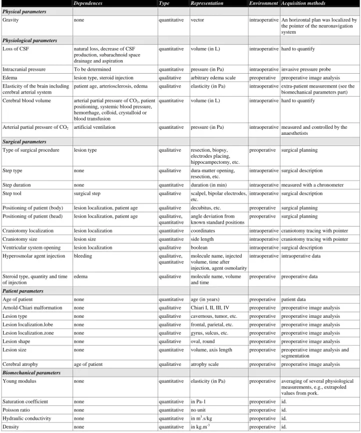
As a first step, 30 parameters were identified as being involved in the anatomical deformations. All of them were described by a model following an ontological approach. They are listed on table 1. We also included inter-parameter dependences although these were not described at a numerical level. Indeed the difficulty was to stay at a general level of description for a better understanding of this notion of inter-dependence. We also specified on table 1 the manner of acquiring and representing some of the parameters. Subsection 3.1 presents the results of the identification and conceptualization phases and subsection 3.2 shows the UML model of parameters.
3.1. Identification process
Physical parameters. Gravity is the only parameter that was put under the physical category. Its influence on the main
direction of matter displacement was often reported in the literature5,7,9,10,11,12,13,14,15. While opening the arachnoid, the
loss of CSF creates an unoccupied space within the intracranial cavity. As a consequence the brain collapses following
*
http://www.uml.org
the direction of gravity. Furthermore, during surgical resection the residual matter also tends to collapse. Gravity has no inter-dependence with other listed parameters.
Physiological parameters. Loss of CSF is also often quoted2,5,6,7,9,10,11,12,13,15,16,17,18. As described previously the brain occupies CSF space during the intervention. This fluid flows through the cranial window but it can also be drained. In some cases, reduction of CSF production was noticed. Intracranial pressure is said to have direct influence on the
deformations7,9,10. Nevertheless one must be aware that the brain shift phenomenon is the direct consequence of an
intracranial hyper- or hypo-tension while opening the arachnoid. We are here at the border between the cause and the description of the phenomenon. Anyway it may be of interest to consider this parameter as it can physically be quantified
for our database. Edema presence and size influence the intracranial pressure and thus the brain shift6,7,11,12,13. In the
literature, tissue swelling was also mentioned as a parameter, but we chose not to distinguishing between tissue swelling and edema. Edema presence depends on steroid injection. The elasticity of the brain, including the arterial system, determines its potentiality of being deformed and thus its capability of replacing the CSF lost. It depends on patient age, on arteriosclerosis and on edema width. Although one can easily determine an average of the brain and arterial system’s elasticity through extra-patient measurement, it is difficult to quantify these parameters for one patient. Cerebral blood
volume has been reported as influencing the deformations7,13,14,18. It depends on many factors such as CO2 partial
pressure, patient positioning, magnitude of bleeding and potential transfusion of colloid, crystalloid or blood. Arterial
partial pressure of CO2 is measured and controlled by anaesthetists. It determines vessel dilatation and thus the perfusion,
which influences the deformations1,5,13.
Surgical-related parameters. Type of surgical procedure may predict the magnitude of deformation5,7,9,11. For instance, T. Hartkens et al. showed differences in amplitudes of deformation on 24 patients by comparing three kinds of
intervention: resection, biopsy and functional interventions9. They underlined that in the case of a resection,
deformations occurred at a higher degree. The notion of surgical step was considered as well7,10,12,13,15,16,17,18. Indeed, we
intuitively and obviously understand that hair shaving induces less deformation than dura matter opening and that the kind of deformation is different between the access to the lesional area (matter displacement) and the removal of the lesional area (matter removal). We can refer to a model of surgical procedures proposed by P. Jannin et al. that helps to
clarify this notion of surgical step8. Step duration was also considered. For instance, D.L.G. Hill et al. underlined the
implication of this parameter by quantifying cortical surface at a one-hour interval before resection1. Furthermore, the
type of surgical tool must be noticed as they do not all have the same influence on matter. The positioning of the patient
may also have an influence on the brain shift phenomenon1,6,7,12,15. For instance, a ventral decubitus position induces an
increase of the intracranial pressure; consequently when opening the dura matter the brain swells instead of collapsing. Patient positioning may depend on the gravity, the craniotomy localization and the age of patient because some positionings are too risky for elderly people (i.e., extreme angles between body’s and head’s axis). Craniotomy size and
craniotomy localization are also involved in the deformations1,5,6,9,17,19. These parameters naturally depend on the lesion
localization and the positioning of the patient. Ventricular system opening may induce anatomical deformations that
occur at a deeper level7,6,15. Hyperosmolar agent injection has an influence on bleeding. This could have an impact on the
deformations1,6,7,10,11,13,15,16,17,19. To better understand these inter-dependences we can look at the Monroe-Kelly law,
which correlates volumes of blood, CSF and tissue20. As noted before, steroid injection has an influence on edema and
thus on intracranial pressure, which is directly correlated to brain shift19.
Patient-related parameters. These a priori known parameters include age of patient1, Arnold-Chiari malformation (related to intracranial pressure and CSF flowing) and lesion-related information. We distinguished the lesion size6,7,15,16,19 (related to the loss of matter after resection), the lesion localization1,6,7,9,14,15,16,19 (related to the positioning of the patient), the lesion shape and the lesion type (related to edema). Cerebral atrophy, which is related to patient age, was
sometimes quoted as being involved in the deformations14,19. It determines the proportion of CSF within the intracranial
cavity related to the brain volume. Its influences may be described as follows: when the neurosurgeon opens the dura matter, a higher quantity of CSF than normal flows out from the cranial window, the brain shift phenomenon may then be more substantial.
Biomechanical parameters. Biomechanical parameters are also related to the deformations, as they describe the brain’s behaviour. We preferred to define a single biomechanical category rather than to include them in the physiological category, since their values are a priori known. These parameters are: the Young modulus (ratio of stress to strain on the loading plane along the loading direction), the saturation coefficient (describes the quantity of water within the brain),
the Poisson ratio (ratio of lateral strain to axial strain), the hydraulic conductivity (capacity of a soil to conduct water through it) and the density of tissues.
3.2. Working with the parameters
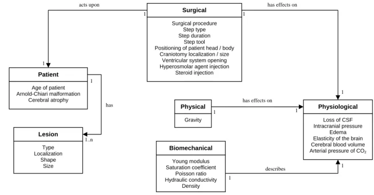
We proposed a UML model of parameters, which provides a convenient overview of all parameters implicated in the deformation (see figure 1). Each category of parameters was represented by a class. Each parameter of deformation was represented by a class attribute. We designated a ‘Physical’ class which contained the physical parameters, a class ‘Physiological’ which contained the physiological-related parameters, a ‘Surgical’ class which contained the parameters describing the surgical procedure, a ‘Patient’ class which contained the patient-related parameters, a ‘Lesion’ class which contained the lesion-related parameters and a ‘Biomechanical’ class which contained the parameters used in biomechanical brain modeling.
This model of parameters has been implemented into a web-based software in order to fill the database with surgical cases. Surgeons have also used this interface to plan surgical procedures, i.e., to enter information related to patient, pathology, positioning, and predictive surgical steps. The data of two surgical cases has already been entered. Both patients for whom the database has been filled underwent surgery for a tumor and a cavernous removal respectively.
Surgical
Surgical procedure Step type Step duration
Step tool
Positioning of patient head / body Craniotomy localization / size
Ventricular system opening Hyperosmolar agent injection
Steroid injection
Physiological
Loss of CSF Intracranial pressure
Edema Elasticity of the brain Cerebral blood volume Arterial pressure of CO2 Physical Gravity Patient Age of patient Arnold-Chiari malformation Cerebral atrophy Lesion Type Localization Shape Size Biomechanical Young modulus Saturation coefficient Poisson ratio Hydraulic conductivity Density has effects on acts upon has effects on has describes 1 1 1 1 1 1 1 1 1 1..n
Figure 1. UML class diagram showing parameters involved in brain deformations
Dependences Type Representation Environment Acquisition methods
Physical parameters
Gravity none quantitative vector intraoperative An horizontal plan was localized by the pointer of the neuronavigation system
Physiological parameters
Loss of CSF natural loss, decrease of CSF production, subarachnoid space drainage and aspiration
quantitative volume (in L) intraoperative hard to quantify
Intracranial pressure To be determined quantitative pressure (in Pa) intraoperative invasive pressure probe Edema lesion type, steroid injection qualitative arbitrary edema scale preoperative preoperative image analysis Elasticity of the brain including
cerebral arterial system
patient age, arteriosclerosis, edema qualitative elasticity (in Pa) intraoperative extra-patient measurement (see the biomechanical parameters part) Cerebral blood volume arterial partial pressure of CO2, patient
positioning, systemic blood pressure, hemorrhage, colloid, crystalloid or blood transfusion
quantitative volume (in L) intraoperative hard to quantify
Arterial partial pressure of CO2 artificial ventilation quantitative pressure (in Pa) intraoperative measured and controlled by the
anaesthetists
Surgical parameters
Type of surgical procedure lesion type qualitative resection, biopsy, electrodes placing, hippocampectomy, etc.
preoperative surgical planning
Step type none qualitative dura-matter opening,
resection, etc.
intraoperative surgical description Step duration none quantitative duration (in min) intraoperative measured with a chronometer Step tool surgical step qualitative scalpel, bipolar electrodes,
etc.
intraoperative surgical description Positioning of patient (body) lesion localization, patient age qualitative decubitus, etc. preoperative surgical planning Positioning of patient (head) lesion localization, patient age qualitative,
quantitative
angle deviation from known standard positions
preoperative surgical planning
Craniotomy localization lesion localization quantitative coordinates intraoperative craniotomy tracing with pointer Craniotomy size lesion size quantitative side length intraoperative craniotomy tracing with pointer Ventricular system opening lesion localization qualitative boolean intraoperative surgical description
Hyperosmolar agent injection bleeding qualitative, quantitative
molecule name, injected volume, time after injection, agent osmolarity
intraoperative intraoperative data
Steroid type, quantity and time of injection
edema qualitative molecule name, volume and time
preoperative preoperative data
Patient parameters
Age of patient none quantitative age (in years) preoperative patient data
Arnold-Chiari malformation none qualitative Chiari I, II, III, IV preoperative preoperative image analysis Lesion type none qualitative cavernous, tumor, etc. preoperative preoperative image analysis Lesion localization.lobe none qualitative frontal, parietal, etc. preoperative preoperative image analysis Lesion localization.zone none qualitative gyrus, sulcus, etc. preoperative preoperative image analysis Lesion shape none qualitative oval, round preoperative preoperative image analysis Lesion size none quantitative volume, axis length preoperative preoperative image analysis and
segmentation
Cerebral atrophy age of patient qualitative atrophy scale preoperative preoperative image analysis
Biomechanical parameters
Young modulus none quantitative elasticity (in Pa) preoperative averaging of several physiological measurements, e.g., extrapoled values from pork.
Saturation coefficient none quantitative in Pa-1 preoperative id.
Poisson ratio none quantitative no unit preoperative id.
Hydraulic conductivity none quantitative in m3.s/kg preoperative id.
Density none quantitative in kg.m-3 preoperative id.
Table 1. List of parameters involved in anatomical intraoperative brain deformations. L stands for Liter, Pa for Pascal, m for meter, s for second, kg for kilogram and min for minute.
4. DISCUSSION
Numerical and symbolical parameters involved in brain deformations were identified, organized under certain criteria and formalized into a UML class diagram. That model was implemented into a web-based software in order to fill a database. This database contains all parameters and cortical shift measurements related to each surgical case. After having entered enough surgical cases for data mining purposes, we expect to gain a better understanding of the deformation phenomenon and its most relevant parameters. Present literature only partially quotes probable parameters involved in the deformations. Our work enables a better understanding of these parameters by following an ontological approach. It also presents a new direction for finding constrains for algorithms dedicated to registering intraoperative with preoperative images.
4.1. Data acquisition
Intraoperative acquisition of some parameters is a difficult task. We proposed methods to acquire some parameters, e.g., measurement of the direction of gravity could be performed thanks to a neuronavigation system. Some other parameters are more difficult to quantify. For example, the volume of the intracranial cavity, which is often quoted in the literature, can be quantified by segmentation of the preoperative MRI, but the main difficulty would be to propose a standard edge for the region of interest. Furthermore, the shape of the intracranial cavity should also be considered because the brain does not behave the same way for different cranial shapes at equivalent intracranial volumes. We did however acquire some parameters that are not mentioned in the literature, such as the gender of the patient and its laterality.
4.2. Limits of the method
Although our aim was to reach an exhaustive identification, we are aware that this literature-based study is not fully objective. It was based primarily on the experts’ consensus. Some questions remain concerning the comprehensiveness of our approach:
- How to estimate the relevance of each of the parameters quoted in the literature?
- Are there other parameters involved in the deformations that were not considered?
We chose an iterative method in order to validate the implication and weight of each parameter. Furthermore, this work is at the first step of that iterative process, which aims at identifying in a most relevant way all parameters involved in anatomical deformations. They will be of use either for understanding the deformation phenomenon or as complementary quantified data as input for preoperative images registration systems. The aim therefore is not to provide a methodology for modeling the brain behaviour like the ones used for biomechanical models. This is, rather, an approach of knowledge modeling applied to the problem of anatomical deformations.
The conceptualization phase was mainly based on intuition. Indeed, we could have chosen other criteria for the categorization and for the instancing of the parameters. This raises two other questions:
- Are the categories proposed to describe the parameters the most relevant?
- Are the dependences between parameters exhaustive?
Concerning the first point, the chosen categories were considered as help for future investigations rather than as a standard basis for categorization of parameters. The conceptualization phase had no influence on the parameters’ intrinsic descriptions, but did have influence on their inter-dependences. These inter-dependences were designed with the help of a neurosurgeon, and were unequally hard to identify. On the one hand, some parameters have clearly identified dependences such as edema presence which is conditioned by the lesion type (e.g., a tumor will often generate edema and cavernous will not). On the other hand, parameters such as the cerebral blood volume or the intracranial pressure have poorly identified dependences. The difficulty is to set the granularity level we need for such an analysis. We have tried here to initiate a study of these inter-dependences that should be pursued through a collaboration between several expert groups such as physiologists, neurosurgeons, physicians and mathematicians. For instance, a more exhaustive description of the encountered inter-dependences could be achieved by integrating existing physical and physiological models for each of the parameters. The formalization provided in this paper brings a convenient overview of the parameters and could thus help such a collaboration.
4.3. Perspectives
The proposed UML class diagram does not show the weight of each parameter implicated in the deformations, e.g., we think that some parameters, such as gravity or loss of CSF, will have particular influences on the deformations. Thus, a graduation of each parameter towards their influence (amplitude of the deformations) and relevance (interest for the neurosurgeon) is conceivable. The notion of relevance corresponds to the interest for the neurosurgeon of being aware about certain deformations. For instance, if a deformation is detected far from the surgical region of interest, the neurosurgeon would be informed about both this deformation and its poor relevance.
The database of qualified and quantified parameters could provide several possibilities of investigation. Firstly, a data mining process could be of interest in order to extract the most relevant parameters related to anatomical deformations. This investigation could give us a new perspective on brain shift understanding. Secondly, a statistic-based algorithm could give for each surgical case a mean and a maximal deformation for a priori known parameters (e.g., patient positioning, lesion size, lesion location, gravity direction, etc.). These mean and maximum statistical values could then be used as input for registration processes based on intraoperative imaging or biomechanical models.
Moreover, one of the main difficulties of our work was the non-existence of standards for characterization of some physiological parameters. For instance, there is no consensus around edema qualification. An edema is said to be more or less prominent depending on the physician who qualifies it. We think therefore that the standardization of some parameters would increase the accuracy of some parameter qualifications.
This approach is the first step of an investigation for better understanding brain deformations during surgery. We believe that results from further studies will confirm some surgeons’ intuition about brain behaviour and will provide additional information for the quantification of these deformations.
REFERENCES
1. D.L.G. Hill, C.R. Maurer, R.J Maciunas, J.A Barwise, J.M Fitzpatrick W.Y Wang, “Measurement of intraoperative brain surface deformation under a craniotomy”, Neurosurgery, 43(3), 514-526, 1998.
2. M.I. Miga, D.W. Roberts, F.E. Kennedy, L.A. Platenik, A. Hartov, K.E. Lunn and D.K. Paulsen, “Modeling of retraction and resection for intraoperative updating of images”, Neurosurgery, 49(1), 75-85, 2001.
3. A. Gomez-Perez, M. Fernandez and A.J. De Vicente, “Towards a Method to Conceptualize Domain Ontologies”, ECAI-96 Workshop on Ontological Engineering, Budapest, 1996.
4. C. Warren and A.D. Whitefield, “The role of task characterisation in transferring models of users: the example of engineering design”, Human-Computer Interaction – Interact'87, 237-243, 1987.
5. D.W. Roberts, A. Hartov, F.E. Kennedy, M.I. Miga and K.D. Paulsen, “Intraoperative brain shift and deformation: a quantitative analysis of cortical displacement in 28 cases”, Neurosurgery, 43(4), 749-758, 1998.
6. C. Nimsky, O. Ganslandt, S. Cerny, P. Hastreiter, G. Greiner and R. Fahlbusch, “Quantification of, visualization of, and compensation for brain shift using intraoperative magnetic resonance imaging”, Neurosurgery, 47(5), 1070-1079, 2000.
7. N.L. Dorward, O. Alberti, B. Velani, F.A. Gerritsen, W.F. Harkness, N.D. Kitchen and D.G. Thomas, “Postimaging brain distortion: magnitude, correlates, and impact on neuronavigation”, Journal of Neurosurgery, 88(4), 656-662, 1998
8. P. Jannin, M. Raimbault, X. Morandi, L. Riffaud and B. Gibaud, “Model of surgical procedures for multimodal image-guided neurosurgery”, Computer Aided Surgery, 8(2), 98-106, 2003.
9. T. Hartkens, D.L. Hill, A.D. Castellano-Smith, D.J. Hawkes, C.R. Maurer, A.J. Martin, W.A. Hall, H. Liu and C.L. Truwit, “Measurement and analysis of brain deformation during neurosurgery”, IEEE Transaction on Medical Imaging, 22(1), 82-92, 2003.
10. O. Skrinjar, A. Nabavi and J. Duncan, “Model-driven brain shift compensation”, Medical Image Analysis, 6(4), 361-373, 2002.
11. R.D. Bucholz, D.D. Yeh, J. Trobaugh, L.L. McDurmont, C.D. Strum and C. Baumann, “The correction of stereotactic inaccuracy caused by brain shift using an intraoperative ultrasound device”, Medicine and Medical Robotics and Computer-assisted Surgery, Springer-Verlag, Berlin, 459-466 1997.
12. P. Hastreiter, C. Rezk-Salama, G. Soza, M. Bauer, G. Greiner, R. Fahlbusch, O. Ganslandt and C. Nimsky, “Strategies for brain shift evaluation”, Medical Image Analysis, 8(4), 447-464, 2004.
13. M.H. Reinges, H.H. Nguyen, T. Krings, B.O. Hutter, V. Rohde and J.M. Gilsbach, “Course of brain shift during microsurgical resection of supratentorial cerebral lesions: limits of conventional neuronavigation”, Acta Neurochirurgica, 146(4), 369-377, 2004.
14. M.I. Miga, J.M. Fitzpatrick, R.L. Galloway and K.D. Paulsen, “Incorporation of surface-based deformations for updating images intraoperatively”, SPIE Medical Imaging 2001: Image Processing, 4319, 169-178, 2001.
15. A. Nabavi, P.Mc.L. Black, D.T. Gering, C.-F. Westin, V. Metha, R.S. Pergolizzi, Jr.M. Ferrant, S.K. Warfield, N. Hata, R.B. Schwarts, W.M. Wells, R. Kikinis and F.A. Jolesz, “Serial intraoperative MR imaging of brain shift”, Neurosurgery, 48, 787-798, 2001.
16. G.E. Keles, K.R. Lamborn and M.S. Berger, “Coregistration accuracy and detection of brain shift using intraoperative sononavigation during resection of hemispheric tumors”, Neurosurgery, 53(3), 556-62, 2003.
17. X. Pennec, P. Cachier and N. Ayache, “Tracking brain deformations in time sequences of 3D US images”, Pattern Recognition Letters - Special Issue on Ultrasonic Image Processing and Analysis, 24(4-5), 801-813, 2003.
18. S.K. Warfield, S.J. Haker, I.-F. Talos, C.A. Kemper, N. Weisenfeld, A.U.J. Mewes, D. Goldberg-Zimring, K.H. Zou, C.-F. Westin, W.M. Wells, C.M.C. Tempany, A. Golby, P.M. Black, F.A. Jolesz and R. Kikinis, “Capturing intraoperative deformations: research experience at Brigham and Women’s hospital”, Medical Image Analysis, 9(2), 145-162, 2005.
19. CR.Jr. Maurer, D.L.G. Hill, A.J. Martin, H. Liu, M. McCue, D. Rueckert, D. Lloret, W.A. Hall, R.E. Maxwell, D.J. Hawkes and C.L. Truwit, “Investigation of intraoperative brain deformation using a 1.5-T interventional MR system: preliminary results”, IEEE Transaction on Medical Imaging, 17(5), 817-825, 1998.
20. B. Mokri, “The Monro-Kellie hypothesis: applications in CSF depletion”, Neurology, 56(12), 1746-1748, 2001.