Candidate genes and functional noncoding variants identified
in a canine model of obsessive-compulsive disorder
The MIT Faculty has made this article openly available.
Please share
how this access benefits you. Your story matters.
Citation
Tang, Ruqi, Hyun Noh, Dongqing Wang, Snaevar Sigurdsson, Ross
Swofford, Michele Perloski, Margaret Duxbury, et al. “Candidate
Genes and Functional Noncoding Variants Identified in a Canine
Model of Obsessive-Compulsive Disorder.” Genome Biology 15, no. 3
(2014): R25.
As Published
http://dx.doi.org/10.1186/gb-2014-15-3-r25
Publisher
BioMed Central Ltd
Version
Final published version
Citable link
http://hdl.handle.net/1721.1/86097
Terms of Use
Creative Commons Attribution
variants identified in a canine model of
obsessive-compulsive disorder
Tang et al.
Tang et al. Genome Biology 2014, 15:R25 http://genomebiology.com/2014/15/3/R25
R E S E A R C H
Open Access
Candidate genes and functional noncoding
variants identified in a canine model of
obsessive-compulsive disorder
Ruqi Tang
1,2,3†, Hyun Ji Noh
1†, Dongqing Wang
2, Snaevar Sigurdsson
1, Ross Swofford
1, Michele Perloski
1,
Margaret Duxbury
4, Edward E Patterson
4, Julie Albright
5, Marta Castelhano
5, Adam Auton
6, Adam R Boyko
7,
Guoping Feng
1,2, Kerstin Lindblad-Toh
1,8*and Elinor K Karlsson
1,9*Abstract
Background: Obsessive-compulsive disorder (OCD), a severe mental disease manifested in time-consuming repetition of behaviors, affects 1 to 3% of the human population. While highly heritable, complex genetics has hampered attempts to elucidate OCD etiology. Dogs suffer from naturally occurring compulsive disorders that closely model human OCD, manifested as an excessive repetition of normal canine behaviors that only partially responds to drug therapy. The limited diversity within dog breeds makes identifying underlying genetic factors easier.
Results: We use genome-wide association of 87 Doberman Pinscher cases and 63 controls to identify genomic loci associated with OCD and sequence these regions in 8 affected dogs from high-risk breeds and 8 breed-matched controls. We find 119 variants in evolutionarily conserved sites that are specific to dogs with OCD. These case-only variants are significantly more common in high OCD risk breeds compared to breeds with no known psychiatric problems. Four genes, all with synaptic function, have the most case-only variation: neuronal cadherin (CDH2), catenin alpha2 (CTNNA2), ataxin-1 (ATXN1), and plasma glutamate carboxypeptidase (PGCP). In the 2 Mb gene desert between the cadherin genes CDH2 and DSC3, we find two different variants found only in dogs with OCD that disrupt the same highly conserved regulatory element. These variants cause significant changes in gene expression in a human neuroblastoma cell line, likely due to disrupted transcription factor binding.
Conclusions: The limited genetic diversity of dog breeds facilitates identification of genes, functional variants and regulatory pathways underlying complex psychiatric disorders that are mechanistically similar in dogs and humans. Background
Obsessive compulsive disorder (OCD) is a common (1 to 3% of the population) and debilitating neuropsychiatric dis-order characterized by persistent intrusive thoughts and time-consuming repetitive behaviors [1]. Twin studies show OCD is very heritable (approximately 45 to 65% genetic in-fluences for early onset OCD), but the underlying genetics is complex [2,3]. More than 80 candidate gene studies of OCD and a recent genome-wide association study (GWAS) yielded no significant, replicable associations [4]. The most
strongly associated genes in the OCD GWAS implicate dis-rupted glutamatergic neurotransmission and signaling in disease pathogenesis [4].
Artificial mouse models have proven more effective for elucidating the neural pathways underlying OCD-like be-haviors. Mice lacking Sapap3, which encodes a postsynaptic scaffolding protein found at glutamatergic synapses, exhib-ited excessive grooming and increased anxiety, symptoms alleviated by treatment with selective serotonin reuptake inhibitors, the same drug frequently used to treat OCD patients [5]. Optogenetic stimulation of the orbitofrontal cortex region affected by the Sapap3 mutation reversed de-fective neural activity and suppressed compulsive behavior [6]. Resequencing of exons of DLGAP3 (the human SAPAP3 gene) revealed excessive rare non-synonymous var-iants in human OCD and trichotillomania individuals [7].
* Correspondence:[email protected];[email protected]
†Equal contributors 1
Broad Institute of MIT and Harvard, 7 Cambridge Center, Cambridge, MA 02142, USA
9
Center for Systems Biology, Department of Organismic and Evolutionary Biology, Harvard University, Cambridge, MA 02138, USA
Full list of author information is available at the end of the article
© 2014 Tang et al.; licensee BioMed Central Ltd. This is an Open Access article distributed under the terms of the Creative Commons Attribution License (http://creativecommons.org/licenses/by/2.0), which permits unrestricted use, distribution, and reproduction in any medium, provided the original work is properly cited. The Creative Commons Public Domain Dedication waiver (http://creativecommons.org/publicdomain/zero/1.0/) applies to the data made available in this article, unless otherwise stated.
Tang et al. Genome Biology 2014, 15:R25 http://genomebiology.com/2014/15/3/R25
Canine OCD is a naturally occurring model for human OCD that is genetically more complex than induced ani-mal models [8]. Phenotypically, canine and human OCD are remarkably similar. Canine compulsive disorder mani-fests as repetition of normal canine behaviors such as grooming (lick dermatitis), predatory behavior (tail chas-ing) and suckling (flank and blanket suckchas-ing). Just as in human patients, approximately 50% of dogs respond to the treatment with selective serotonin reuptake inhibitors or clomipramine [9]. Particular dog breeds (genetically isolated populations) have exceptionally high rates of OCD, including Doberman Pinschers (DPs), bull terriers and German shepherds [10-12]. The high disease rates and rather limited genetic diversity of dog breeds suggests that OCD in these populations, while multi-genic, may be less complex than in humans, facilitating genetic mapping and functional testing of associated variants [13,14].
In an earlier GWAS of canine OCD, we associated CDH2, a neural cadherin gene involved in synaptic plas-ticity, with OCD in DPs [14]. Here, we use a more powerful algorithm, MAGIC [15], to reanalyze the data from this study and identify new OCD-associated re-gions. These regions are enriched for genes involved in synapse formation and function, as are regions with patterns of reduced variation consistent with artificial selection. We sequence the top candidate regions, 5.8 Mb in total, and find that four genes, all with synaptic function, are enriched for case-specific variants: neuronal-cadherin (CDH2), catenin alpha2 (CTNNA2), ataxin-1 (ATXN1), and plasma glutamate carboxypeptidase (PGCP). Furthermore, two intergenic mutations between the cad-herin genes CDH2 and desmocollin 3 (DSC3) disrupt a non-coding regulatory element and alter gene expression in a human neuroblastoma cell line. Our results implicate abnormal synapse formation and plasticity in OCD, and point to disrupted expression of neural cadherin genes as one possible cause.
Results
Genome-wide association studies and homozygosity mapping
Using the raw data (included in Gene Expression Omnibus accession numbers GSE53488 and GSE53577) from the GWAS by Dr Dodman and collaborators [14], which in-cluded 92 DP cases and 68 DP controls extensively pheno-typed for canine OCD, we reanalyzed the Affymetrix genotype intensity data with a new calling algorithm, MAGIC [15]. MAGIC relaxes certain assumptions used in other callers, such as Hardy-Weinberg equilibrium in genotype clusters, to dramatically improve the accuracy of genotypes called from Affymetix v2 Canine GeneChip data. This yielded a 2.4-fold denser SNP map for association mapping (55,651 SNPs; 35,941 SNPs with minor allele frequency (MAF) >0.05) but a slightly
smaller sample size, with 87 cases and 63 controls passing MAGIC quality filters (compared to our original dataset of 14,700 SNPs with MAF > 0.05 in 92 cases and 68 controls; Figure 1a,b). The increased density allowed us to identify 13 new candidate OCD-associated regions (P < 0.0001) in addition to the original chromo-some 7 locus in CDH2 (Table S1 in Additional file 1). We estimate that this dataset explains 0.56 ± 0.18 of pheno-type variance [16].
We tested all Gene Ontology (GO) gene sets with 5 to 1,000 genes (5,206 sets) for enrichment in the new GWAS regions using INRICH, a permutation based software that rigorously controls for region size, SNP density, and gene size and gene number [17]. Overall, we observe an excess of sets with P < 0.01 (25 sets, P = 0.03; Figure 1e). The top set ‘GO:0045295 Gamma catenin binding’ is significant even after stringent correction for the number of gene sets tested (P = 5.9 × 10⁻⁵, Pcorrected= 0.05) and includes genes
under each of three peaks of association spanning ap-proximately 3 Mb on chromosome 7: CDH2, DSC3 and DSG1 (Figure 1d,e; Table S2 in Additional file 1). The GWAS regions also include two of 13 genes in ‘GO:0048814 Regulation of dendrite morphogenesis’ (P = 0.002): the calcium binding synaptogenesis gene SDC2 and the postsynaptic density protein gene TNIK, which encodes a serine-threonine kinase involved in AMPA re-ceptor trafficking and synaptic function [18,19].
The DP breed, like all dog breeds, was created through population bottlenecks and artificial selection for mor-phological and behavioral traits, potentially driving some OCD risk alleles to very high frequency and thus un-detectable by GWAS. Consistent with this hypothesis, we find functional connections between associated genes and genes in the 13 largest autosomal regions of fixation in the DP breed (25.7 Mb in total; Table S3 in Additional file 1). For example, the tyrosine kinase FER mediates cross-talk between CDH2 and integrins [20], and deple-tion of presynaptic FER inhibits synaptic formadeple-tion and transmission [21]. CTNNA2 interacts with CDH2 to regulate the stability of synaptic cell junctions [22]. While most fixed regions contain many genes, making it difficult to identify top candidates, several contain just one gene, including the neuronal protein gene LINGO2 and the synaptic-2 like glycoprotein gene TECRL.
We also identified 128 regions of unusually low vari-ability in the DP breed compared to 24 other dog breeds (23.73 Mb; Table S4 in Additional file 1) [23]. When we test these regions of reduced variability (RRVs) for gene set enrichment in the entire GO catalog, as described above, 10 GO terms are more enriched in DP RRVs than any other breed (Figure 1f ). Half of these have clear relevance to brain function, including regulation of neurotransmitters, neural projection, and dendrite morphogenesis. We also see enrichment for mannose
c
f
e
d
a
b
Figure 1 (See legend on next page.)
Tang et al. Genome Biology 2014, 15:R25 Page 3 of 14
binding-related genes, echoing the strong enrichment in GWAS regions for alpha-mannosidase activity. Mannose structures are concentrated at excitatory synapses, in-cluding glutamate receptors [24,25].
Targeted sequencing
We designed a sequencing array (Tables S5 and S6 in Additional file 1) that targeted nine of the top GWAS regions, including the CDH2 locus (3.9 Mb; Figure S1 in Additional file 1) as well as genes and conserved ele-ments within the five largest DP fixed regions (Table S3 in Additional file 1). We focused on fixed regions (total-ing 1.8 Mb) that were also fixed in two other OCD af-fected breeds, German shepherds (LUPA reference panel [26]) and bull terriers (20 dogs) (Figure 1c). We se-quenced eight cases and eight matched controls from breeds at high risk for OCD, including eight DP, four German shepherds, two Shetland sheepdogs and two Jack Russell terriers (Figure 2a). We selected DPs based on their genotype for the CDH2 risk haplotype [14] (two homozygous cases, two heterozygous cases, and four controls without the risk haplotype). We captured 92% of the target regions at >20× coverage, with 76× mean read depth coverage per sample (Table S7 in Additional file 1). In total, we detected 24,930 high-quality SNPs, 7,645 short INDELs, and 173 deletions, with high con-cordance to the SNP array data (median 99.5% for ap-proximately 390 SNPs tested per sample; Table S8 in Additional file 1).
Case-only variant discovery from sequence data
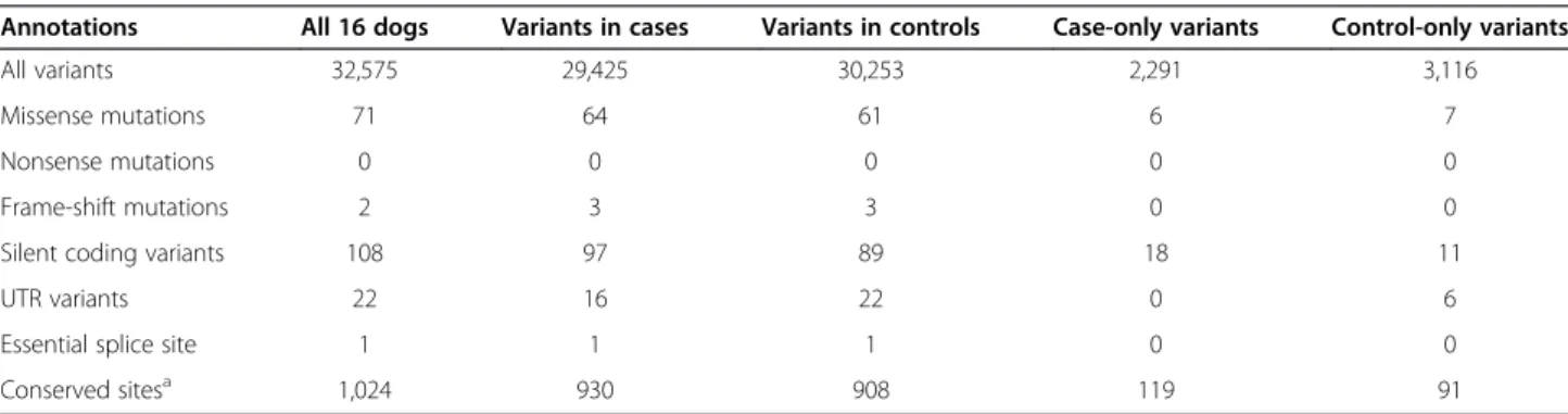
With our small sample size (eight cases and eight con-trols from four different breeds), we did not expect to have sufficient power to detect statistically significant al-lelic associations with OCD. Instead, we focused on vari-ants seen only in OCD cases (‘case-only varivari-ants’) as the strongest causal candidates. Of 32,575 variants, 2,291 variants are case-only (2,002 SNPs and 289 INDELs; 80 to 966 per dog), while 3,116 variants are specific to con-trol dogs (‘concon-trol-only variants’; 2,698 SNPs and 418 INDELs; 156 to 1,476 per dog) (Table 1; Table S9 in Additional file 1). While there is no significant difference between the total number of case- and control-only vari-ants (Wilcoxon test P = 0.63; Figure 2b), case dogs have
a significantly greater number in evolutionarily con-strained elements (median 15 versus 4, Wilcoxon test P = 0.02; Materials and methods; Figure 2b; Table S9 in Additional file 1). Excluding coding variants increases the difference further (median 15 versus 3, Wilcoxon test P = 0.01), suggesting that the excess of case-only functional variants may be due largely to noncoding variation.
Genotyping case-only variants in independent samples
We genotyped 114 case-only, evolutionarily constrained variants in an independent set of dogs from breeds with high rates of OCD (‘OCD-risk breeds’; 69 dogs) and breeds with normal rates of OCD and other psychiatric disorders (‘control breeds’; 19 dogs). Except for 14 cases from OCD-risk breeds, we have no individual OCD phenotype information for these dogs (Figure 2a). We find that the case-only variants identified in the sequence data are significantly more common in OCD-risk breeds, with median frequency (FOCD) of 0.17, than in control breeds,
where the median frequency (Fcontrol) is 0.05 (Wilcoxon
test P = 0.045; Table S10 in Additional file 1). The median frequency increases to 0.20 when only phenotyped cases are considered (Wilcoxon test P = 0.015, comparison with Fcontrol). We also observe an inverse correlation
be-tween the frequency difference bebe-tween OCD-risk and control breeds and the frequency across all genotyped dogs (Pearson’s R = −0.63, P = 8.4 × 10⁻¹⁴
; Figure 2c). Thus, the variants most enriched in OCD-risk breeds are other-wise rare, potentially due to either positive selective pres-sure in OCD-risk breeds or negative selection in the control breeds. While this suggests an association with OCD, we note that other traits may also systematically dif-fer between the two breed groups.
Gene-based analysis
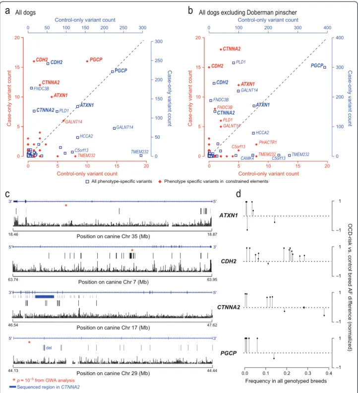
We identified genes enriched with case-only variants using a gene-based analysis method that accounts for multiple independent variants within a gene and greatly increases power for identifying disease-associated genes [28]. Four genes have an excess of case-only variation in evolutionarily constrained elements, even after cor-recting for gene size: ATXN1, CDH2, CTNNA2, and PGCP (10, 16, 12, and 16 case-only variants, respectively;
(See figure on previous page.)
Figure 1 Associated and fixed regions in Doberman Pinschers are enriched for brain-related pathways. (a) The original GWAS dataset showed a single peak of association at CDH2 [14]. (b) Recalling with MAGIC yielded a 2.4-fold denser SNP dataset and allowed us to define 17 distinct regions of association with P < 0.0001 using linkage disequilibrium (LD) clumping (Figure S1 in Additional file 1), a subset of which were targeted for sequencing (red dots, genes labeled above peak). (c) In four breeds with high rates of OCD, we identified regions of fixation (black boxes), a subset of which we targeted for sequencing (red boxes). Sequenced regions were selected because they were large and overlapping between breeds (Table S1 in Additional file 1). (d) LD clumping identified three distinct regions of association on chromosome 7 (boxes, with targeted regions in red). (e) The top Gene Ontology gene sets enriched in the GWAS regions. (f) GO gene sets enriched in Doberman Pinscher regions of reduced variability (RRVs) but not in 24 other breeds (grey circles, most at 0).
Figure 2 (See legend on next page.)
Tang et al. Genome Biology 2014, 15:R25 Page 5 of 14
Figure 3a; Text S1 and Table S11 in Additional file 1). Because the sequenced DPs were selected based on their haplotype at CDH2, we confirmed that the case-only enrichment at CDH2 persists even when DPs are excluded (Figure 3b). RNA-Seq data show all four genes are expressed in the dog brain (KL-T, unpublished observations).
Three of the four candidate genes, CDH2, PGCP and ATXN1, are associated with OCD in the DP GWAS study (chr7:63867472, P = 2.1 × 10⁻⁵; chr29:44152594, P = 1.5 × 10⁻⁵; chr35:18565131, P = 1.6 × 10⁻⁵, respect-ively), while the fourth, CTNNA2, falls in a large region of fixation (900 kb) in the DP breed (Figure 3c). In our genotyping dataset, the case-only variants in these four genes are more common in OCD-risk breeds (FOCD=
0.08 versus Fcontrol= 0.026, Wilcoxon test P = 2.95 × 10⁻⁴;
Figure 3d), particularly in CDH2 (FOCD= 0.23 versus
Fcontrol= 0.027, P = 0.001) and in PGCP (FOCD= 0.014
versus Fcontrol= 0.0, P = 0.047). We see a similar, though
weaker, pattern in ATXN1 (FOCD= 0.022 versus Fcontrol=
0.0, P = 0.3) and CTNNA2 (FOCD= 0.185 versus Fcontrol=
0.026, P = 0.13). In CTNNA2, the difference is clearer (P = 0.054) if only variants with frequency <0.20 are considered.
Of the 40 variants genotyped in these four genes, seven overlap chromatin marks, potentially indicating regulatory function. Four variants in CDH2 overlap H3K27Ac histone marks and/or DNase1 hypersensitivity
clusters. Three of these (chr7:63845160, chr7:63852056, and chr7:63832008) are observed in OCD-risk breeds, at frequencies of 0.435, 0.050, and 0.022, respectively, and never seen in control breeds. The fourth variant (chr7:63806661) is four-fold more common in OCD-risk breeds (frequency = 0.11 versus 0.026 in control breeds). Three variants in ATXN1 alter regions transcribed in the dog brain (KL-T, unpublished RNA-Seq data), including a putative enhancer variant not seen in the control breeds (chr35:18850625, OCD-risk breed frequency = 0.014). These variants, which lie in genes enriched for case-only variants, are overrepresented in cases, and alter putative regulatory elements, are strong candidates for further functional elucidation.
Single variant analysis
We next sought to identify the top candidate functional variants in the sequencing data. We first looked for coding variants found exclusively in cases. Most were missense mutations disrupting genes with little known relevance to brain functions (Table S12 and Text S2 in Additional file 1). More intriguing, one of our two Jack Russell terrier cases has a 1.2 kb deletion (chr29:44178339–44179516; Figure S2 in Additional file 1) overlapping exon 2 of the gene PGCP, causing a frameshift and loss of 70 amino acids from the protein. PGCP is one of the four genes enriched for case-only variants, and, while none of the DP cases has this particular deletion, a nearby SNP is
(See figure on previous page.)
Figure 2 Targeted sequencing identifies case-only variants that alter constrained elements and are more common in high OCD risk breeds. (a) We performed targeted sequencing of a small number of cases and controls from four breeds (row 1) and subsequently genotyped the top candidate variants in a larger panel of dogs from those four breeds as well as two more high‘OCD-risk’ breeds and two low risk ‘control breeds’ (row 2). (b) Across all variants identified in the sequencing data, the number of case-only (orange box) and control-only (purple box) variants is similar, but constrained elements are enriched for case-only variants. Boxes mark the 25th to 75th percentile across dogs, with the median shown as a thick line, and whiskers extending to values within 1.5 times the difference between the 25th to 75th percentiles. Outliers are marked with circles. (c) Case-only variants have higher frequency in OCD-risk breeds and lower frequency across all genotyped breeds. The x-axis represents allele frequencies across all genotyped dogs. The y-axis represents normalized allele frequency (AF) differences between OCD-risk and control breeds ([AFOCD-risk- AFcontrol]/[AFOCD-risk+ AFcontrol]). The straight downward line represents the linear model for the data points. The blue shade shows the 95% confidence interval for this model. Area under the curve (shaded in grey) is notably larger in AFOCD-risk> AFcontrolthan in AFOCD-risk< AFcontrol, showing that case-only variants are more common in OCD-risk breeds than in control breeds.
Table 1 Sequence variants identified by targeted resequencing of 5.8 Mb in eight cases and eight controls
Annotations All 16 dogs Variants in cases Variants in controls Case-only variants Control-only variants
All variants 32,575 29,425 30,253 2,291 3,116
Missense mutations 71 64 61 6 7
Nonsense mutations 0 0 0 0 0
Frame-shift mutations 2 3 3 0 0
Silent coding variants 108 97 89 18 11
UTR variants 22 16 22 0 6
Essential splice site 1 1 1 0 0
Conserved sitesa 1,024 930 908 119 91
a
Figure 3 Gene-based analysis identifies four top genes enriched for case-only variants in constrained regions. (a) Genes are plotted according to the number of case-only (y-axis) and control-only (x-axis) variants within a gene and its 5 kb flanking regions from our sequence data. Blue squares denote all the variants and the corresponding axes are shown in blue; red diamonds denote variants in constrained elements only and the corresponding axes are shown in red. Genes that are plotted above the identity line harbor more case-only than control-only variants. (b) A similar analysis excluding DP, the breed used to identify genes for sequencing, shows that the enrichment pattern persists for several genes. (c) The case-only variants in constrained elements, when plotted with gene structure and evolutionary conservation, show clustering in ATXN1 (5’ end). Dimmed bars represent canine variants that failed to lift over onto hg19. The conservation track shows a measure of evolutionary conservation in dog, human, mouse and rat [29]. (d) In each gene, SNPs with the greatest risk allele frequency (AF) difference between OCD-risk and control breeds (y-axis) tend to have lower frequency across all genotyped breeds (y-axis). SNPs are shown as solid circles with vertical lines.
Tang et al. Genome Biology 2014, 15:R25 Page 7 of 14
among the most strongly associated in the GWAS (chr29:44152594, P = 1.5 × 10⁻⁵; Figure 1; Figure S3 in Additional file 1). Using quantitative PCR (qPCR), we validated the deletion in the Jack Russell terrier cases and tested 74 dogs from OCD-risk breeds (including 10 unphenotyped Jack Russell terriers and 14 dogs from several breeds diagnosed with OCD) and 20 dogs from control breeds. We found the deletion in three Jack Russell terriers and in one Welsh terrier with OCD, and in none of the control breed dogs, suggest-ing it is associated with increased risk of OCD in mul-tiple breeds.
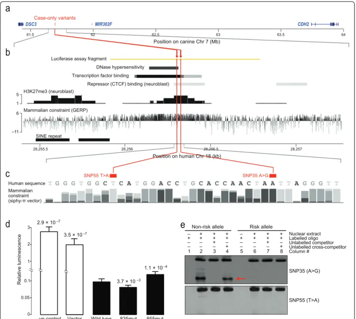
We next looked for non-coding variants seen only in cases, focusing on 15 seen in more than one DP case. All but two are near the GWAS peak in intron 2 of CDH2 (chr7:63867472, P = 2.1 × 10⁻⁵), reflecting the selection of DP dogs for sequencing based on their genotype at this locus. None of the 13 is obviously func-tional based on evolutionary constraint and histone marks. The other two variants are more interesting, changing a conserved region approximately 172 kb away from an associated GWAS SNP (chr7:61865715, P = 1.6 × 10⁻⁵), in the gene desert between the cadherin genes CDH2 and DSC3 (Figure 4a). The first SNP (chr7:61693835, T changed to C; SNP35 T > C) is exclu-sively found in three of four sequenced DP cases and showed the overall DP breed frequency of approximately 0.30 in our genotyping data set. The second SNP, a pri-vate variant in the fourth DP case (chr7:61693855; SNP55 A > T), is just 20 bases away and alters the same highly conserved region (Figure 4b,c).
Functional assessment of candidate variants
Because the region altered by SNP35 and SNP55 showed evidence of regulatory function (Figure 4b), we tested whether the risk alleles disrupt gene expression using a luciferase reporter assay. Including the wild-type region in the reporter construct lowers expression 14-to 20-fold in human neuroblas14-toma SK-N-BE(2) cells (vector versus wild type, t-test Bonferroni corrected P = 3.5 × 10⁻⁷; negative control versus wild type, t-test Bonferroni corrected P = 2.9 × 10⁻⁷; Figure 4d). Adding the SNP55 risk variant to the construct, however, significantly increases expression relative to the wild-type version, suggesting the regulatory element no lon-ger functions normally (1.6-fold change, paired t-test Bonferroni corrected P = 1.1 × 10⁻⁴; Figure 4d). Curiously, the SNP35 risk allele has the opposite effect, repressing expression even further (0.9-fold change, paired t-test Bonferroni corrected P = 3.7 × 10⁻³; Figure 4d).
Using an electrophoretic mobility shift assay (EMSA) to examine DNA protein binding in the region, we see that, while the SNP55 risk allele causes no apparent change relative to wild-type, the SNP35 risk allele shows
markedly reduced binding (Figure 4e). Three transcrip-tion factors are predicted by TRANSFAC [32] to bind the wild-type sequence but not the SNP35 variant (PRRX2, Oct-1 and Nobox; Figure S4 in Additional file 1). How-ever, we saw no evidence that these three proteins bind the region in a supershift assay (Figure S5 in Additional file 1), suggesting other factors are critical. More than 90 transcription factors are predicted to bind the wild-type sequence using various discovery tools [33]. Thus, both SNP35 and SNP55 significantly change the silencing activ-ity of the regulatory element, but in opposite directions and possibly through different mechanisms.
Discussion
Through a small GWAS (fewer than 90 cases and 70 controls) we identified OCD-associated loci, which, par-ticularly when analyzed together with regions of low variability, implicate specific cellular pathways in disease etiology. We sequenced nine of the top regions of asso-ciation and five regions of fixation in eight OCD cases and eight breed-matched controls. We found a notable excess of case-only variation in evolutionarily conserved regions, particularly in non-coding elements with poten-tial regulatory function. This suggests noncoding vari-ation is a major factor in canine OCD similar to human neuropsychiatric diseases, and unlike most artificially in-duced mouse models. While the dog population is com-posed of >400 genetically isolated breed populations, just a small number of breeds are highly enriched for OCD, suggesting that OCD risk variants are more preva-lent in these breeds. We show that the case-only variants found in the sequence data are in fact significantly more common in OCD-risk breeds compared to breeds with no increased risk of psychiatric disorders.
By comparing the sequence data using gene-based tests, we confirmed one gene (CDH2) and identified three novel ones (CTNNA2, ATXN1, and PGCP) strongly implicated for involvement in disease.
CDH2, a neural cadherin, encodes a calcium dependent cell-cell adhesion glycoprotein important for synapse as-sembly, where it mediates presynaptic to postsynaptic ad-hesions [34]. Disrupting expression of CDH2 in cultured mouse neurons causes synapse dysfunction, synapse elim-ination and axon retraction [35].
CTNNA2 encodes a neuronal-specific catenin protein that links cadherins to the cytoskeleton [34,36] and is associated with bipolar disorder [37], schizophrenia [38], attention deficit hyperactivity disorder [38] and excitement-seeking [39]. Mice with a deletion of CTNNA2 showed disrupted brain morphology and impaired startle modulation [40]. Cadherin-catenin complexes play a pivotal role in synapse formation and synaptic plasticity and therefore may be involved in the process of learning and memory [41].
ATXN1 encodes a chromatin binding protein that reg-ulates the Notch pathway [42], a developmental pathway also active in the adult brain, where it mediates neuronal migration, morphology and synaptic plasticity [43]. Mice with a deletion of ATXN1 showed pronounced deficits in learning and memory [44].
CDH2, CTNNA2 and ATXN1 have similar spatial expression patterns in the brain and are important during brain development and for synaptic plasticity. CDH2 and CTNNA2 are highly expressed in the pre-frontal cortex, amygdala, thalamus and fetal brain [34,45]. ATXN1 is highly expressed in the prefrontal
Figure 4 Two intergenic case-only variants disrupt a repressor element and change gene expressionin vitro. (a) The two case-only variants are just 20 bases apart in a 2.5 Mb gene desert between DSC3 and CDH2 on canine chromosome 7. (b) The syntenic region of human chromosome 18 shows markers of DNase hypersensitivity, transcription factor binding, repressor binding, histone methylation [30], and mammalian constraint [31]. (c) Both variants, SNP55 and SNP35, alter bases that are highly constrained across mammals, SNP55 to an A and SNP35 to a T. Compositions of the four bases for each position in the 29 mammals comparison [27] are shown with different grey scale and height of bars (the higher the more conserved in multiple species). (d) The wild-type regulatory sequence represses luciferase reporter expression in SK-N-BE(2) neuroblastoma cells. Both variants significantly change the extent of repression relative to wild type, with SNP35-G more repressive and SNP55-A less repressive. The firefly luciferase expression in the test plasmids were normalized against the co-transfected Renilla luciferase expression in pGL4.73. The P-value of the significance of the change relative to wild type is shown above each bar, with vertical lines showing standard error of the mean. (e) An electrophoretic mobility shift assay testing the wild-type alleles (lanes 1 to 4) and the OCD-risk alleles (lanes 5 to 8) of SNP35 (top gel) and SNP55 (bottom gel) show that nuclear protein binding (red arrow) to the SNP35 locus is disrupted by the risk allele. Nuclear extract was derived from SK-N-BE(2) cells. We used a 200-fold molar excess of competitor where appropriate.
Tang et al. Genome Biology 2014, 15:R25 Page 9 of 14
cortex, basal ganglia, cerebellum and fetal brain [45,46].
Intriguingly, the three genes appear to have functional connections to the top SNPs (association P < 10-5) in a recent human OCD GWAS, which found no single asso-ciations reaching genome-wide significance, but impli-cated glutamatergic signaling pathways [4] (Figure S6 in Additional file 1). Most notably, one of the top associ-ated genes in human patients, GRIK2, encodes a glutam-ate receptor recruited to the synaptic membrane by CDH2/catenin complexes [47] and another top candi-date, PKP2, mediates CDH2 cell adhesion and desmo-somal junctions [48]. In addition, several genes whose expression levels correlate with the top human OCD-associated SNPs interact with the genes we identify in dogs: LRSAM1 (cerebellum) and NARS (frontal lobe) interact with ATXN1; SPAG9 (cerebellum) acts in develop-mental pathways with CDH2 and CTNNA2 [49].
The fourth gene, PGCP, encodes a poorly characterized plasma glutamate carboxypeptidase. It may help hydrolyze N-acetylaspartylglutamate (NAAG), the third most abun-dant neurotransmitter in the brain, to glutamate and N-acetylaspartate (NAA) [34], suggesting a potential role in glutamatergic synapse dysfunction. PGCP is associated with migraine [50], which is frequently co-morbid with OCD [51].
We hypothesize that CDH2, CTNNA2, ATXN1, and PGCP may work in concert to regulate glutamatergic synapse formation and function in the cortico-striatal-thalamo-cortical (CSTC) brain circuit previously impli-cated in the pathogenesis of OCD [52-56].
Single variant analysis corroborates our hypothesis of dysregulated synapse formation in OCD. All four se-quenced DP cases had one of two mutations (SNP35 and SNP55) in a regulatory region, between DSC3 and CDH2, that we show acts as a strong silencer. The OCD-risk allele of SNP55 significantly increased the re-porter gene expression while the OCD-risk allele of SNP35 had the opposite effect. While surprising, other studies have shown that either deletion or reciprocal duplication of loci such as 17p11.2 and 15q13.3 can cause neuropsychiatric disorders [57]. For SNP35, we confirmed using EMSA that the OCD-risk allele changes DNA binding. We saw no change at SNP55, although in vitro assays may not capture all relevant in vivo reac-tions. The regulatory element is between CDH2 (2.2 Mb away) and DSC3 (0.3 Mb away), both cadherin genes involved in gamma-catenin binding (Figure 1d,e), sug-gesting disrupted gamma-catenin binding may be an important risk factor for OCD. Additional sequence data from DSC3 (not included in the current targeted sequencing design) and more functional analysis are needed to understand the two SNPs’ effects on CDH2 and DSC3.
Conclusions
Modeling neuropsychiatric disorders in animals is compli-cated by both limited understanding of the underlying neurobiology and subjective diagnostic criteria [58]. Natu-rally occurring canine compulsive disorder is a remarkably good model for human disease, as it is equivalent by most clinical metrics, including age of onset, symptoms, and pharmacological response. The work we present here sug-gests similar genetic etiology as well. We harness the lim-ited genetic diversity of dog breeds, and high rates of OCD in particular breeds, to identify genes, pathways and non-coding candidate functional variants. Dogs suffer from a wide range of psychiatric disorders and have been strongly selected for a variety of behavioral traits, making them a uniquely powerful natural model organism for investigating inherited psychiatric diseases in humans.
Materials and methods
GWAS and sequencing region selection
The GWAS using the sample set and phenotypes pub-lished previously [14] was rerun using genotypes called with the new MAGIC algorithm [15]. Briefly, MAGIC (Multidimensional Analysis for Genotype Intensity Clus-tering) does not use prior information to make genotype calls (that is, cluster locations Hardy-Weinberg equilib-rium, or complex normalization of probe intensities). In-stead, it performs quantile normalization of the data for each chip independently followed by a principal compo-nent analysis of all chips on a SNP-by-SNP basis, neatly summarizing the raw data.
The processed data are then clustered into genotype calls through expectation maximization using a t-distribution mixture model. Association was calculated with a standard chi-squared test in PLINK [59] (SNP genotype rate >90%, individual genotype rate >25%, MAF >5%) and regions were defined with linkage disequi-librium -based clumping around SNPs with P < 0.0001 (that is, SNPs within 1 Mb with r2> 0.8 and P < 0.01) (Table S1 and Figure S1 in Additional file 1). We identified regions of fixation as regions of >1,000 kb with more than five SNPs and >95% SNPs with MAF <0.05 and selected a subset found in breeds prone to OCD for targeted sequen-cing. From the associated and fixed regions we designed a 5.8 Mb targeted sequencing array that optimized inclusion of potential genes of interest within the design limitations (Tables S5 and S6 in Additional file 1).
Gene set enrichment analysis
We expanded the GWAS regions to include all genes within 500 kb of the original region start or end (Table S1 in Additional file 1). We defined RRVs by comparing the DP breed to 24 other dog breeds from a published reference dataset and identifying the 1% least variable 150 kb regions in DP [23]. We ran INRICH with
1,000,000 permutations to test regions for enrichment in any gene sets from the GO catalog. We tested all gene sets with between 5 and 1,000 genes (downloaded from the Gene Ontology website on 18 May 2013) [17]. We used a map file of 16,433 genes lifted over to canFam2.0 from the hg19 RefSeq Gene catalog (UCSC Genome Brower, single match using default parameters) [60]. To identify gene sets with unusually high enrichment in the DP RRVs, we calculated, for all sets with P < 0.05 and at least 2 RRV genes in DP, the average difference in en-richment P-values between DP and 24 other breeds [26] (Figure 1f ).
Sequenced samples
The targeted sequencing experiment comprised a total of eight cases and eight controls from multiple breeds: DP (four cases + four controls), German shepherd (two cases + two controls), Jack Russell terrier (one case + one control) and Shetland sheepdog (one case + one control) (Figure 2a). The four DP cases supplied by Dr. Meurs showed flank sucking behavior, while the German Shepherd, Jack Russell terrier and Shetland sheepdog cases were tail-chasers.
Targeted sequencing and variant calling
The 16 samples were individually barcoded and the targeted regions were captured by a NimbleGen Sequence Capture 385 K Array according to the man-ufacturer’s protocol. The captured samples were then pooled and sequenced on an Illumina Genome Analyzer II. Paired-end 76-bp reads were aligned to canFam2.0 and PCR duplicates were removed using Picard [61], and realignment and recalibration were processed through Genome Analysis Toolkit (GATK) [62,63]. SNPs and small INDELs were identified using GATK. We only considered the variants that pass the GATK standard filters. Larger structural variants were detected by GenomeSTRiP [64]. We manually checked the alignments of all discovered deletion sites for aberrant read pairs and read depth using Integrative Genomics Viewer [65] to ensure the reliability of the calls. We excluded a Shetland sheepdog pair where the control had lower SNP accuracy, when comparing case-and control-only variant counts (Figure 2b case-and Table S9 in Additional file 1).
Genotyping candidate sequence variants
We first selected case-specific variants that were within evolutionarily constrained elements determined by a 29 mammals sequence dataset [27]. We then selected a subset of the variants meeting one of the following cri-teria: (i) only variants within DP breed; (ii) case-only variants within CDH2, PGCP, CTNNA2 and ATXN1 that are identified by gene-based analysis; (iii) case-only
variants across at least two breeds; (iv) potential func-tional variants annotated as nonsense, splicing or mis-sense (predicted to be ‘probably’ or ‘possibly damaging’ by Polyphen-2 [66]) and case-only variants in at least one breed; (v) variants within CDH2 risk haplotype; and (vi) top associated variants from GWAS analysis. Of 140 variants that met one of the criteria, 127 variants passed Sequenom design standards, and were geno-typed using the Sequenom iPlex system. We employed an independent set of 94 dogs that consisted of 10 dogs without obvious health problems for each of 6 OCD-risk breeds (that is, 4 sequenced breeds and West Highland white terrier (Westie) and bull terrier (bull terrier)) and two control breeds without known psychiatric problems (greyhound and Leonberger), and 14 additional OCD cases from various breeds (2 bull terrier, 2 DP, 1 German shepherd, 1 Westie, 1 Golden re-triever, 1 Irish wolfhound, 1 pug, 1 Shiba, 1 Shepherd mix, 1 standard poodle, 1 Shih Tzu, 1 Welsh terrier). Geno-type data were cleaned by removing samples with missing genotype rates >10% and excluding SNPs with call rates <90%. After the quality control, 114 SNPs and 88 dogs (19 (10 Leonbergers + 9 Greyhounds) from control breeds and 69 (14 cases + 5 DP + 10 bull terrier + 10 Westie + 10 German shepherd + 10 Shetland sheepdogs + 10 Jack Russell terriers) from OCD-risk breeds or cases) were retained in our analysis (Figure 2a; Table S10 in Additional file 1).
Gene-based analysis
Each gene region was defined using the coordinates from RefSeq hg19 lifted over to canFam2.0 plus 5 kb flanking sequence on each side. We counted the number of case-and control-only variants case-and compared the counts for each gene. Genes that have excessive case-only variants relative to control-only variants were considered as poten-tial risk genes for OCD. The same analysis was applied to the variants within constrained elements. To correct for gene size, we calculated the ratio of the number of case-only variants and the number of control-case-only variants for each gene additionally.
Electrophoretic mobility shift assay
For each allele of the tested SNPs in a regulatory region between CDH2 and DSC3, pairs of 5’-biotinylated oligo-nucleotides were obtained from IDT Inc. (Coralville, IA, USA; Table S13 in Additional file 1). Equal volumes of forward and reverse oligos (100 μM) were mixed and heated at 95°C for 5 minutes and then cooled to room temperature. Fifty femtomoles of annealed probes were incubated at room temperature for 30 minutes with 10 mg SK-N-BE(2) nuclear extract (Active Motif Carlsbad, CA, USA). The remaining steps followed the LightShift Chemiluminescent EMSA Kit protocol (Thermo Scientific).
Tang et al. Genome Biology 2014, 15:R25 Page 11 of 14
Luciferase reporter assay
The activity of a putative regulatory element and the effect of SNP35 and SNP55 on gene expression were examined by luciferase reporter assay. We PCR ampli-fied an 879 bp-long orthologous sequence spanning SNP35 and SNP55 from human DNA samples (Table S14 in Additional file 1). The risk alleles were intro-duced using a site-directed mutagenesis kit. The wild-type and mutant DNA fragments were cloned into a firefly luciferase reporter plasmid (pGL4.23, Promega Madison, WI, USA). The test constructs were transiently co-transfected with a Renilla luciferase reporter plasmid (pGL4.73, Promega) as an internal control into neuro-blastoma SK-N-BE(2) cells. All constructs were tested in triplicates and repeated three times in a double-blinded manner.
Cell cultures
Human SK-N-BE(2) cells were purchased from ATCC. The cells were maintained at 37°C and 5% CO2 in 1:1
mixture of ATCC-formulated Eagle’s Minimum Essential Medium (EMEM) and F-12 K medium supplemented with 10% fetal bovine serum, 100 units/ml penicillin and 100μg/ml streptomycin.
Real-time qPCR
Real-time qPCR was performed using Quantifast SYBR Green PCR kit (QIAGEN, Hilden, Germany) on a Light-cycler 480 system (Roche Applied Science, Indianapolis, IN, USA). The reaction volumes were adjusted to 10 μl with 3μl of DNA (10 ng), 1 μl of both primers (10 μM) and 5 μl of Master Mix. The qPCR program was as fol-lows: pre-incubation at 95°C for 5 minutes, followed by 40 cycles of two-step amplification (10 s at 95°C, 1 mi-nute at 60°C). All the experiments were carried out in triplicates and include negative control without DNA. The primer sets used to detect PGCP deletion is shown in Table S15 in Additional file 1.
Data availability
The data presented in this publication are available through the NCBI Sequence Read Archive (SRP033723) and Gene Expression Omnibus (accession numbers GSE53488 and GSE53577). Datasets analyzed in the paper are also available at [67].
Additional file
Additional file 1: Texts S1 and S2, Tables S1 to S15, and Figures S1 to S6.
Abbreviations
bp:base pair; DP: Doberman Pinscher; EMSA: electrophoretic mobility shift assay; GATK: Genome Analysis Toolkit; GO: Gene Ontology; GWAS: genome-wide association study; MAF: minor allele frequency;
OCD: obsessive compulsive disorder; qPCR: quantitative polymerase chain reaction; RRV: regions of reduced variability; SNP: single-nucleotide polymorphism.
Competing interests
The authors declare that they have no competing interests.
Authors’ contributions
KLT, EKK and GF conceived and oversaw the study. EEP, JA, MD, MC and MP coordinated, collected, prepared samples and characterized samples for the study. RT, SS, MP and RS designed and performed the resequencing experiment. HJN, RT, EKK and KLT analyzed and interpreted the data. AB and AA performed genotype calling for GWAS. RT and DW performed the functional assays. HJN, RT, EKK, and KLT wrote the paper with input from the other authors. All authors read and approved the final manuscript.
Acknowledgements
We thank all the dog owners, breeders, and breed clubs, as well as the veterinarians in the US and Sweden for providing us with blood samples. This work was supported by a SPARC grant from the Broad Institute, Uppsala University, Medical Research Council, and a grant from NIMH/NIH
(R01MH081201), and by funds from the Poitras Center for Affective Disorders Research to GF. KLT is the recipient of a EURYI from the European Science Foundation. EKK is supported by fellowships from the American Cancer Society and the Charles A King Trust. We thank Andy Reynolds for help with genotype calling, Evan Mauceli for help with genotyping concordance analysis, Jeremy Johnson for project management assistance, Tarjei Mikkelsen and Xiaolan Zhang for supplying reagents for the luciferase assays, and Leslie Gaffney for help with illustration. We thank Kate Meurs and Åke Hedhammar for their work on sample collection. Our deepest appreciation goes to Nicholas Dodman, veterinary behaviorist and professor at Tufts University, and animal behaviorist Alice Moon-Fanelli, who performed extensive and careful phenotyping of OCD in the Doberman pinscher GWAS sample set using methodologies developed by Dr. Dodman and his group. This careful phenotyping, described in the earlier published GWAS study [14], provided a foundation for the work described herein. We also acknowledge the valuable material and intellectual contributions of Drs. Edward Ginns and Marzena Galdzicka to this study.
Author details
1Broad Institute of MIT and Harvard, 7 Cambridge Center, Cambridge, MA
02142, USA.2McGovern Institute for Brain Research and Department of Brain
and Cognitive Sciences, Massachusetts Institute of Technology, Cambridge, MA 02139, USA.3Renji Hospital, School of Medicine, Shanghai Jiao Tong
University, Shanghai 200127, China.4College of Veterinary Medicine,
University of Minnesota, St. Paul, MN 55108, USA.5Department of Clinical
Sciences, College of Veterinary Medicine, Cornell University, Ithaca, NY 14853, USA.6Department of Genetics, Albert Einstein College of Medicine, 1301
Morris Park Avenue, Van Etten B06, Bronx, NY 10461, USA.7Department of
Biomedical Sciences, Cornell University, Ithaca, NY 14853, USA.8Science for
Life Laboratory, Department of Medical Biochemistry and Microbiology, Uppsala University, 75237 Uppsala, Sweden.9Center for Systems Biology,
Department of Organismic and Evolutionary Biology, Harvard University, Cambridge, MA 02138, USA.
Received: 5 November 2013 Accepted: 14 March 2014 Published: 14 March 2014
References
1. Laks J, Fontenelle LF, Chalita A, Mendlowicz MV: Absence of dementia in late-onset schizophrenia: a one year follow-up of a Brazilian case series. Arquivos de neuro-psiquiatria 2006, 64:946–949.
2. van Grootheest DS, Cath DC, Beekman AT, Boomsma DI: Twin studies on obsessive-compulsive disorder: a review. Twin Res Hum Genet 2005, 8:450–458. 3. Taylor S: Molecular genetics of obsessive-compulsive disorder: a
comprehensive meta-analysis of genetic association studies. Mol Psychiatry 2013, 18:799–805.
4. Stewart SE, Yu D, Scharf JM, Neale BM, Fagerness JA, Mathews CA, Arnold PD, Evans PD, Gamazon ER, Osiecki L, McGrath L, Haddad S, Crane J, Hezel D, Illman C, Mayerfeld C, Konkashbaev A, Liu C, Pluzhnikov A, Tikhomirov A, Edlund CK, Rauch SL, Moessner R, Falkai P, Maier W, Ruhrmann S, Grabe HJ,
Lennertz L, Wagner M, Bellodi L, et al: Genome-wide association study of obsessive-compulsive disorder. Mol Psychiatry 2012, 18:788–798.
5. Welch JM, Lu J, Rodriguiz RM, Trotta NC, Peca J, Ding JD, Feliciano C, Chen M, Adams JP, Luo J, Dudek SM, Weinberg RJ, Calakos N, Wetsel WC, Feng G: Cortico-striatal synaptic defects and OCD-like behaviours in Sapap3-mutant mice. Nature 2007, 448:894–900.
6. Burguiere E, Monteiro P, Feng G, Graybiel AM: Optogenetic stimulation of lateral orbitofronto-striatal pathway suppresses compulsive behaviors. Science 2013, 340:1243–1246.
7. Zuchner S, Wendland JR, Ashley-Koch AE, Collins AL, Tran-Viet KN, Quinn K, Timpano KC, Cuccaro ML, Pericak-Vance MA, Steffens DC, Krishnan KR, Feng G, Murphy DL: Multiple rare SAPAP3 missense variants in trichotillo-mania and OCD. Mol Psychiatry 2009, 14:6–9.
8. Overall KL: Natural animal models of human psychiatric conditions: assessment of mechanism and validity. Prog Neuropsychopharmacol Biol Psychiatry 2000, 24:727–776.
9. Overall KL, Dunham AE: Clinical features and outcome in dogs and cats with obsessive-compulsive disorder: 126 cases (1989–2000). J Am Vet Med Assoc 2002, 221:1445–1452.
10. Moon-Fanelli AA, Dodman NH, Cottam N: Blanket and flank sucking in Doberman Pinschers. J Am Vet Med Assoc 2007, 231:907–912. 11. Moon-Fanelli AA, Dodman NH, Famula TR, Cottam N: Characteristics of
compulsive tail chasing and associated risk factors in Bull Terriers. J Am Vet Med Assoc 2011, 238:883–889.
12. Luescher AU: Diagnosis and management of compulsive disorders in dogs and cats. Clin Tech Small Anim Pract 2004, 19:233–239.
13. Karlsson EK, Sigurdsson S, Ivansson E, Thomas R, Elvers I, Wright J, Howald C, Tonomura N, Perloski M, Swofford R: Genome-wide analyses implicate 33 loci in heritable dog osteosarcoma, including regulatory variants near CDKN2A/B. Genome Biol 2013, 14:R132.
14. Dodman NH, Karlsson EK, Moon-Fanelli A, Galdzicka M, Perloski M, Shuster L, Lindblad-Toh K, Ginns EI: A canine chromosome 7 locus confers compulsive disorder susceptibility. Mol Psychiatry 2010, 15:8–10. 15. Boyko AR, Quignon P, Li L, Schoenebeck JJ, Degenhardt JD, Lohmueller KE,
Zhao K, Brisbin A, Parker HG, vonHoldt BM, Cargill M, Auton A, Reynolds A, Elkahloun AG, Castelhano M, Mosher DS, Sutter NB, Johnson GS, Novembre J, Hubisz MJ, Siepel A, Wayne RK, Bustamante CD, Ostrander EA: A simple genetic architecture underlies morphological variation in dogs. PLoS Biol 2010, 8:e1000451.
16. Yang J, Lee SH, Goddard ME, Visscher PM: GCTA: a tool for genome-wide complex trait analysis. Am J Hum Genet 2011, 88:76–82.
17. Lee PH, O’Dushlaine C, Thomas B, Purcell SM: INRICH: interval-based enrichment analysis for genome-wide association studies. Bioinformatics 2012, 28:1797–1799.
18. Ethell IM, Hagihara K, Miura Y, Irie F, Yamaguchi Y: Synbindin, A novel syndecan-2-binding protein in neuronal dendritic spines. J Cell Biol 2000, 151:53–68.
19. Coba MP, Komiyama NH, Nithianantharajah J, Kopanitsa MV, Indersmitten T, Skene NG, Tuck EJ, Fricker DG, Elsegood KA, Stanford LE, Afinowi NO, Saksida LM, Bussey TJ, O'Dell TJ, Grant SG: TNiK is required for postsynaptic and nuclear signaling pathways and cognitive function. J Neurosci 2012, 32:13987–13999.
20. Arregui C, Pathre P, Lilien J, Balsamo J: The nonreceptor tyrosine kinase fer mediates cross-talk between N-cadherin and beta1-integrins. J Cell Biol 2000, 149:1263–1274.
21. Lee SH, Peng IF, Ng YG, Yanagisawa M, Bamji SX, Elia LP, Balsamo J, Lilien J, Anastasiadis PZ, Ullian EM, Reichardt LF: Synapses are regulated by the cytoplasmic tyrosine kinase Fer in a pathway mediated by p120catenin, Fer, SHP-2, and beta-catenin. J Cell Biol 2008, 183:893–908.
22. Takeichi M, Abe K: Synaptic contact dynamics controlled by cadherin and catenins. Trends Cell Biol 2005, 15:216–221.
23. Vaysse A, Ratnakumar A, Derrien T, Axelsson E, Rosengren Pielberg G, Sigurdsson S, Fall T, Seppala EH, Hansen MS, Lawley CT, Karlsson EK, Bannasch D, Vila C, Lohi H, Galibert F, Fredholm M, Haggstrom J, Hedhammar A, Andre C, Lindblad-Toh K, Hitte C, Webster MT: Identification of genomic regions associated with phenotypic variation between dog breeds using selection mapping. PLoS Genet 2011, 7:e1002316. 24. Churchill L, Cotman C, Banker G, Kelly P, Shannon L: Carbohydrate
composition of central nervous system synapses, Analysis of isolated synaptic junctional complexes and postsynaptic densities. Biochim Biophys Acta 1976, 448:57–72.
25. Clark RA, Gurd JW, Bissoon N, Tricaud N, Molnar E, Zamze SE, Dwek RA, McIlhinney RA, Wing DR: Identification of lectin-purified neural glycoproteins, GPs 180, 116, and 110, with NMDA and AMPA receptor subunits: conservation of glycosylation at the synapse. J Neurochem 1998, 70:2594–2605.
26. Hedges DJ, Burges D, Powell E, Almonte C, Huang J, Young S, Boese B, Schmidt M, Pericak-Vance MA, Martin E, Zhang X, Harkins TT, Zuchner S: Exome sequencing of a multigenerational human pedigree. PloS ONE 2009, 4:e8232.
27. Lindblad-Toh K, Garber M, Zuk O, Lin MF, Parker BJ, Washietl S, Kheradpour P, Ernst J, Jordan G, Mauceli E, Ward LD, Lowe CB, Holloway AK, Clamp M, Gnerre S, Alfoldi J, Beal K, Chang J, Clawson H, Cuff J, Di Palma F, Fitzgerald S, Flicek P, Guttman M, Hubisz MJ, Jaffe DB, Jungreis I, Kent WJ, Kostka D, Lara M, et al: A high-resolution map of human evolutionary constraint using 29 mammals. Nature 2011, 478:476–482. 28. Huang H, Chanda P, Alonso A, Bader JS, Arking DE: Gene-based tests of
association. PLoS Genet 2011, 7:e1002177.
29. Siepel A, Bejerano G, Pedersen JS, Hinrichs AS, Hou M, Rosenbloom K, Clawson H, Spieth J, Hillier LW, Richards S, Weinstock GM, Wilson RK, Gibbs RA, Kent WJ, Miller W, Haussler D: Evolutionarily conserved elements in vertebrate, insect, worm, and yeast genomes. Genome Res 2005, 15:1034–1050.
30. Dunham I, Kundaje A, Aldred SF, Collins PJ, Davis CA, Doyle F, Epstein CB, Frietze S, Harrow J, Kaul R, Khatun J, Lajoie BR, Landt SG, Lee BK, Pauli F, Rosenbloom KR, Sabo P, Safi A, Sanyal A, Shoresh N, Simon JM, Song L, Trinklein ND, Altshuler RC, Birney E, Brown JB, Cheng C, Djebali S, Dong X, Ernst J, et al: An integrated encyclopedia of DNA elements in the human genome. Nature 2012, 489:57–74.
31. Cooper GM, Stone EA, Asimenos G, Green ED, Batzoglou S, Sidow A: Distribution and intensity of constraint in mammalian genomic sequence. Genome Res 2005, 15:901–913.
32. Wingender E, Dietze P, Karas H, Knuppel R: TRANSFAC: a database on transcription factors and their DNA binding sites. Nucleic Acids Res 1996, 24:238–241.
33. Calakos N, Patel VD, Gottron M, Wang G, Tran-Viet KN, Brewington D, Beyer JL, Steffens DC, Krishnan RR, Zuchner S: Functional evidence implicating a novel TOR1A mutation in idiopathic, late-onset focal dystonia. J Med Genet 2010, 47:646–650.
34. Pruitt KD, Tatusova T, Brown GR, Maglott DR: NCBI Reference Sequences (RefSeq): current status, new features and genome annotation policy. Nucleic Acids Res 2012, 40:D130–D135.
35. Pielarski KN, van Stegen B, Andreyeva A, Nieweg K, Jungling K, Redies C, Gottmann K: Asymmetric N-cadherin expression results in synapse dysfunction, synapse elimination, and axon retraction in cultured mouse neurons. PloS ONE 2013, 8:e54105.
36. Abe K, Chisaka O, Van Roy F, Takeichi M: Stability of dendritic spines and synaptic contacts is controlled by alpha N-catenin. Nat Neurosci 2004, 7:357–363.
37. Scott LJ, Muglia P, Kong XQ, Guan W, Flickinger M, Upmanyu R, Tozzi F, Li JZ, Burmeister M, Absher D, Thompson RC, Francks C, Meng F, Antoniades A, Southwick AM, Schatzberg AF, Bunney WE, Barchas JD, Jones EG, Day R, Matthews K, McGuffin P, Strauss JS, Kennedy JL, Middleton L, Roses AD, Watson SJ, Vincent JB, Myers RM, Farmer AE, et al: Genome-wide association and meta-analysis of bipolar disorder in individuals of European ancestry. Proc Natl Acad Sci U S A 2009, 106:7501–7506. 38. Chu TT, Liu Y: An integrated genomic analysis of gene-function correlation
on schizophrenia susceptibility genes. J Hum Genet 2010, 55:285–292. 39. Terracciano A, Esko T, Sutin AR, de Moor MH, Meirelles O, Zhu G, Tanaka T,
Giegling I, Nutile T, Realo A, Allik J, Hansell NK, Wright MJ, Montgomery GW, Willemsen G, Hottenga JJ, Friedl M, Ruggiero D, Sorice R, Sanna S, Cannas A, Raikkonen K, Widen E, Palotie A, Eriksson JG, Cucca F, Krueger RF, Lahti J, Luciano M, Smoller JW, et al: Meta-analysis of genome-wide association studies identifies common variants in CTNNA2 associated with excitement-seeking. Transl Psychiatry 2011, 1:e49.
40. Park C, Falls W, Finger JH, Longo-Guess CM, Ackerman SL: Deletion in Catna2, encoding alpha N-catenin, causes cerebellar and hippocampal lamination defects and impaired startle modulation. Nat Genet 2002, 31:279–284. 41. Arikkath J, Reichardt LF: Cadherins and catenins at synapses: roles in
synaptogenesis and synaptic plasticity. Trends Neurosci 2008, 31:487–494. 42. Tong X, Gui H, Jin F, Heck BW, Lin P, Ma J, Fondell JD, Tsai CC: Ataxin-1 and
Brother of ataxin-1 are components of the Notch signalling pathway. EMBO Rep 2011, 12:428–435.
Tang et al. Genome Biology 2014, 15:R25 Page 13 of 14
43. Ables JL, Breunig JJ, Eisch AJ, Rakic P: Not(ch) just development: Notch signalling in the adult brain. Nat Rev Neurosci 2011, 12:269–283. 44. Matilla A, Roberson ED, Banfi S, Morales J, Armstrong DL, Burright EN, Orr
HT, Sweatt JD, Zoghbi HY, Matzuk MM: Mice lacking ataxin-1 display learn-ing deficits and decreased hippocampal paired-pulse facilitation. J Neurosci 1998, 18:5508–5516.
45. Wu C, Orozco C, Boyer J, Leglise M, Goodale J, Batalov S, Hodge CL, Haase J, Janes J, Huss JW 3rd, Su AI: BioGPS: an extensible and customizable portal for querying and organizing gene annotation resources. Genome Biol 2009, 10:R130.
46. Servadio A, Koshy B, Armstrong D, Antalffy B, Orr HT, Zoghbi HY: Expression analysis of the ataxin-1 protein in tissues from normal and spinocerebellar ataxia type 1 individuals. Nat Genet 1995, 10:94–98.
47. Coussen F, Normand E, Marchal C, Costet P, Choquet D, Lambert M, Mege RM, Mulle C: Recruitment of the kainate receptor subunit glutamate receptor 6 by cadherin/catenin complexes. J Neurosci 2002, 22:6426–6436. 48. Barth M, Rickelt S, Noffz E, Winter-Simanowski S, Niemann H, Akhyari P,
Lichtenberg A, Franke WW: The adhering junctions of valvular interstitial cells: molecular composition in fetal and adult hearts and the comings and goings of plakophilin-2 in situ, in cell culture and upon re-association with scaffolds. Cell Tissue Res 2012, 348:295–307. 49. Matthews L, Gopinath G, Gillespie M, Caudy M, Croft D, de Bono B,
Garapati P, Hemish J, Hermjakob H, Jassal B, Kanapin A, Lewis S, Mahajan S, May B, Schmidt E, Vastrik I, Wu G, Birney E, Stein L, D'Eustachio P: Reactome knowledgebase of human biological pathways and processes. Nucleic Acids Res 2009, 37:D619–D622.
50. Anttila V, Stefansson H, Kallela M, Todt U, Terwindt GM, Calafato MS, Nyholt DR, Dimas AS, Freilinger T, Muller-Myhsok B, Artto V, Inouye M, Alakurtti K, Kaunisto MA, Hamalainen E, de Vries B, Stam AH, Weller CM, Heinze A, Heinze-Kuhn K, Goebel I, Borck G, Gobel H, Steinberg S, Wolf C, Bjornsson A, Gudmundsson G, Kirchmann M, Hauge A, Werge T, et al: Genome-wide association study of migraine implicates a common susceptibility variant on 8q22.1. Nat Genet 2010, 42:869–873.
51. Vasconcelos LP, Silva MC, Costa EA, da Silva Junior AA, Gomez RS, Teixeira AL: Obsessive compulsive disorder and migraine: case report, diagnosis and therapeutic approach. J Headache Pain 2008, 9:397–400.
52. Ting JT, Feng G: Glutamatergic synaptic dysfunction and obsessive-compulsive disorder. Curr Chem Genomics 2008, 2:62–75.
53. Ahmari SE, Spellman T, Douglass NL, Kheirbek MA, Simpson HB, Deisseroth K, Gordon JA, Hen R: Repeated cortico-striatal stimulation generates persistent OCD-like behavior. Science 2013, 340:1234–1239. 54. Marsh R, Maia TV, Peterson BS: Functional disturbances within
frontostriatal circuits across multiple childhood psychopathologies. Am J Psychiatry 2009, 166:664–674.
55. Milad MR, Rauch SL: Obsessive-compulsive disorder: beyond segregated cortico-striatal pathways. Trends Cogn Sci 2012, 16:43–51.
56. Pittenger C, Bloch MH, Williams K: Glutamate abnormalities in obsessive compulsive disorder: neurobiology, pathophysiology, and treatment. Pharmacol Ther 2011, 132:314–332.
57. Girirajan S, Campbell CD, Eichler EE: Human copy number variation and complex genetic disease. Annu Rev Genet 2011, 45:203–226.
58. Nestler EJ, Hyman SE: Animal models of neuropsychiatric disorders. Nat Neurosci 2010, 13:1161–1169.
59. Purcell S, Neale B, Todd-Brown K, Thomas L, Ferreira MA, Bender D, Maller J, Sklar P, de Bakker PI, Daly MJ, Sham PC: PLINK: a tool set for whole-genome association and population-based linkage analyses. Am J Hum Genet 2007, 81:559–575.
60. Karolchik D, Barber GP, Casper J, Clawson H, Cline MS, Diekhans M, Dreszer TR, Fujita PA, Guruvadoo L, Haeussler M, Harte RA, Heitner S, Hinrichs AS, Learned K, Lee BT, Li CH, Raney BJ, Rhead B, Rosenbloom KR, Sloan CA, Speir ML, Zweig AS, Haussler D, Kuhn RM, Kent WJ: The UCSC Genome Browser database: 2014 update. Nucleic Acids Res 2014, 42:D764–D770. 61. Picard. [http://picard.sourceforge.net/]
62. McKenna A, Hanna M, Banks E, Sivachenko A, Cibulskis K, Kernytsky A, Garimella K, Altshuler D, Gabriel S, Daly M, DePristo MA: The Genome Analysis Toolkit: a MapReduce framework for analyzing next-generation DNA sequencing data. Genome Res 2010, 20:1297–1303.
63. DePristo MA, Banks E, Poplin R, Garimella KV, Maguire JR, Hartl C, Philippakis AA, del Angel G, Rivas MA, Hanna M, McKenna A, Fennell TJ, Kernytsky AM, Sivachenko AY, Cibulskis K, Gabriel SB, Altshuler D, Daly MJ: A framework
for variation discovery and genotyping using next-generation DNA sequencing data. Nat Genet 2011, 43:491–498.
64. Handsaker RE, Korn JM, Nemesh J, McCarroll SA: Discovery and genotyping of genome structural polymorphism by sequencing on a population scale. Nat Genet 2011, 43:269–276.
65. Robinson JT, Thorvaldsdottir H, Winckler W, Guttman M, Lander ES, Getz G, Mesirov JP: Integrative genomics viewer. Nat Biotechnol 2011, 29:24–26. 66. Adzhubei IA, Schmidt S, Peshkin L, Ramensky VE, Gerasimova A, Bork P,
Kondrashov AS, Sunyaev SR: A method and server for predicting damaging missense mutations. Nat Methods 2010, 7:248–249. 67. FTP site for data files used in the study. [ftp://ftp.broadinstitute.org/pub/
vgb/dog/OCD_GenomeBiology2014paper/]
doi:10.1186/gb-2014-15-3-r25
Cite this article as: Tang et al.: Candidate genes and functional noncoding variants identified in a canine model of obsessive-compulsive disorder. Genome Biology 2014 15:R25.
Submit your next manuscript to BioMed Central and take full advantage of:
• Convenient online submission
• Thorough peer review
• No space constraints or color figure charges
• Immediate publication on acceptance
• Inclusion in PubMed, CAS, Scopus and Google Scholar
• Research which is freely available for redistribution
Submit your manuscript at www.biomedcentral.com/submit