Publisher’s version / Version de l'éditeur:
Journal of the American Chemical Society, 132, 43, pp. 15136-15139, 2010-10-11
READ THESE TERMS AND CONDITIONS CAREFULLY BEFORE USING THIS WEBSITE. https://nrc-publications.canada.ca/eng/copyright
Vous avez des questions? Nous pouvons vous aider. Pour communiquer directement avec un auteur, consultez la première page de la revue dans laquelle son article a été publié afin de trouver ses coordonnées. Si vous n’arrivez pas à les repérer, communiquez avec nous à [email protected].
Questions? Contact the NRC Publications Archive team at
[email protected]. If you wish to email the authors directly, please see the first page of the publication for their contact information.
NRC Publications Archive
Archives des publications du CNRC
This publication could be one of several versions: author’s original, accepted manuscript or the publisher’s version. / La version de cette publication peut être l’une des suivantes : la version prépublication de l’auteur, la version acceptée du manuscrit ou la version de l’éditeur.
For the publisher’s version, please access the DOI link below./ Pour consulter la version de l’éditeur, utilisez le lien DOI ci-dessous.
https://doi.org/10.1021/ja105028w
Access and use of this website and the material on it are subject to the Terms and Conditions set forth at
Water-soluble J-type rosette nanotubes with giant molar ellipticity
Borzsonyi, Gabor; Beingessner, Rachel L.; Yamazaki, Takeshi; Cho,
Jae-Young; Myles, Andrew J.; Malac, Marek; Egerton, Ray; Kawasaki, Masahiro;
Ishizuka, Kazuo; Kovalenko, Andriy; Fenniri, Hicham
https://publications-cnrc.canada.ca/fra/droits
L’accès à ce site Web et l’utilisation de son contenu sont assujettis aux conditions présentées dans le site LISEZ CES CONDITIONS ATTENTIVEMENT AVANT D’UTILISER CE SITE WEB.
NRC Publications Record / Notice d'Archives des publications de CNRC:
https://nrc-publications.canada.ca/eng/view/object/?id=3ecae9fa-2a93-413f-a86f-627e443da5c6
https://publications-cnrc.canada.ca/fra/voir/objet/?id=3ecae9fa-2a93-413f-a86f-627e443da5c6
Water-Soluble J-Type Rosette Nanotubes with Giant Molar Ellipticity
Gabor Borzsonyi,†,‡Rachel L. Beingessner,†Takeshi Yamazaki,†Jae-Young Cho,†
Andrew J. Myles,†Marek Malac,†,§Ray Egerton,†,§Masahiro Kawasaki,⊥
Kazuo Ishizuka,#
Andriy Kovalenko,†,|and Hicham Fenniri*,†,|
National Institute for Nanotechnology, Departments of Chemistry, Physics, and Mechanical Engineering, UniVersity of Alberta, 11421 Saskatchewan DriVe, Edmonton, Alberta, T6G 2M9, Canada., JEOL USA, Inc., 11 Dearborn Road,
Peabody, Massachusetts 01960, and HREM Research, 14-48 Matsukazedai, Higashimastuyama, 355-0055, Japan
Received June 9, 2010; E-mail: [email protected]
Abstract: A new self-assembling tricyclic module (×K1) featuring
the Watson-Crick H-bonding arrays of guanine and cytosine fused to an internal pyridine ring was synthesized. When dis-solved in water at room temperature, this module rapidly self-assembles into hexameric rosettes, which then stack to form J-type rosette nanotubes (RNTs) with increased inner/outer diameters and the largest molar ellipticity ever reported (4 × 10⁶
deg · M-1·m-1). Using a combination of imaging and spectroscopic
techniques we established the structure of ×K1-RNT and have shown that the extended π system of the self-assembling module resulted in a new family of J-type RNTs with enhanced inter-modular electronic communication.
Since their discovery in 1936 by Scheibe1
and Jelley,2
J-aggregates3
have seen a broad range of applications because they display coherent, cooperative phenomena such as superradiance and giant oscillator strength as a result of the long-range delocalization of their electronic excitation.3a,4
J-aggregates have for example been used as organic photoconductors,5
optical switches,6
NLO devices,7
critically coupled resonators,8
LEDs,9
sensitizers,10
photovoltaics,11
nanowires,12
and sensors.13
J-aggregates are usually dye crystallites in which the transition dipoles of the constituent molecules strongly couple to form a collective narrow line width optical transition possessing oscillator strength derived from the aggregated monomers.14
In this report we use self-assembly and self-organization strategies15
to build water-soluble J-type nanotubular architecture with unprecedented control over dimensions, supramolecular organization, and physical properties relative to earlier reports on J-aggregates. This approach offers opportunities for further tunability of electronic properties through subtle changes in the core structure and functionalization of the self-assembling molecular modules.
The G∧C motif16
is a self-complementary hybrid molecule of the DNA bases guanine and cytosine. This motif and its deriva-tives17
(2, Figure 1) were studied extensively in water and organic solvents and were shown to undergo hierarchical self-assembly into hexameric rosettes which further organize into a linear stack called a rosette nanotube (RNT).16c,18
Here we present a water-soluble tricyclicvariant 1, termed ×K1. This molecule features the same H-bonding arrays, but separated by an internally fused pyridine ring. Building upon the design criteria formulated and successfully
established in the case of its bicyclic congener, ×K1 was specifically designed to (a) self-assemble in water to form RNTs with increased inner/outer diameters relative to K1 (2) and (b) engender an extendedπ system that would enhance electronic transport along the RNT’s main axis.
Following is the synthesis, self-assembly, and characterization of ×K1 using circular dichroism (CD), UV-vis and fluorescence spectroscopy, scanning electron microscopy (SEM), transmission electron microscopy (TEM), tapping mode atomic force microscopy (TM-AFM), energy filtering TEM (EFTEM), off-axis electron holography (EH), and quantitative phase technology (QPt). We also report remarkable optical properties suggesting enhanced electronic communication for this new class of RNTs.
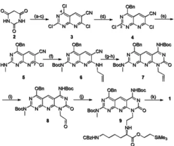
The synthesis of ×K1 (Scheme 1) began by transforming commercially available barbituric acid in three steps to bicycle 3. Three consecutive regioselective SNAr reactions at positions 4, 2,
and 7 with benzyl alcohol, methylamine, and allylamine, respec-tively, provided the functionalized compound 6. However, for the third SNAr reaction to proceed, it was necessary to electronically
†National Institute for Nanotechnology, University of Alberta. ‡Department of Chemistry, University of Alberta.
§
Department of Physics, University of Alberta.
|
Department of Mechanical Engineering, University of Alberta.
⊥
JEOL USA, Inc.
#
HREM Research.
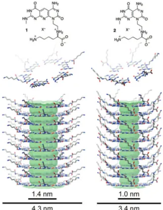
Figure 1. ×K1 (1) and K1 (2) modules, corresponding hexameric rosettes (middle), and RNTs (lower) (X ) CF3CO2-).
deactivate the ring by protecting the methylamine in compound 5 with an electron-withdrawing group. Ring closure of 6 using trichlorocarbonyl isocyanate/NH3, followed by protection of the
amine, generated tricycle 7. Oxidative cleavage of alkene 7 and
subsequent reductive amination of aldehyde 8 with protected
L-lysine provided compound 9. Finally, deprotection of 9 with TFA/ thioanisole furnished the target ×K1 motif (1) as a TFA salt.
Characterization of ×K1 by SEM revealed that this tricyclic compound rapidly self-assembles into RNTs when dissolved in water (pHfinal)2.8) (Figure 2C) at room temperature. This is an
advantage over the bicyclic G∧C base 2 which requires heat to drive the entropically driven self-assembly process as a result of its reduced hydrophobic character.18b
TM-AFM (Figure 2A) and TEM (Figure 2B) of uranyl acetate-stained samples established the RNTs’ outer diameter of 4.2 ( 0.2 and 4.4 ( 0.2 nm, respectively, in excellent agreement with the theoretical value 4.3 nm. These measurements were further verified using three direct TEM imaging methods (without staining agent), from which the diameter of a single RNT was measured to be 4.5 ( 1 nm (EFTEM), 4.4 ( 0.75 nm (EH), and 4.9 ( 0.8 nm (QPt).19
In contrast with EFTEM, EH and QPt use elastic scattering of the fast electrons in TEM instead of the inelastically scattered electrons to obtain quantitative measurements. Based on these results, the inner and outer diameters of ×K1-RNTs relative to K1-RNTs increased by 0.4 and 0.9 nm, respectively.
CD and UV-vis spectroscopic techniques were used to monitor the self-assembly process in solution. The CD spectra of ×K1 (2.1 × 10⁻⁵M) feature two couplets centered at 257 and 380 nm with positive maxima at 264 and 388 nm (Figures 3B). The CD signal intensity increased ca. 40× over 7 days reaching a giant molar ellipticity, with an unprecedented value of 4 × 10⁶deg · M-1·m-1
for a chiral helical stack.20
Briefly heating a dilute solution of ×K1 (1.9 × 10⁻⁵M) to 100 °C (10 s) or acidification with TFA (0.1%
v/v) leads to the complete disappearance of the CD profile, thus suggesting that the induced CD is the result of the supramolecular chirality of the RNTs. In agreement with these studies, variable temperature CD (VT-CD) established the reversibility of the CD profile.19
The intensity of the CD couplet is proportional to the fourth power of the dipole strength and the relative orientation of the chromophores and is inversely proportional to the square of the interchromophoric distance.21
Since the latter is probably the same for RNTs assembled from K1 and ×K1, the very large molar ellipticity recorded is therefore an indication of the much larger transition dipole strength and an optimal relative orientation of the corresponding dipole moments.
UV-vis, CD, and fluorescence spectroscopy unveiled the remarkable optical properties of the RNTs assembled from ×K1 (×K1-RNT). First, the UV-vis spectra of K1 versus ×K1 show a significant shift of all the maxima toward the red in the latter case. K1 showed maxima at 236 and 285 nm, whereas ×K1 showed maxima at 246 and 363 nm in addition to a unique, sharp, and lower energy band at 388 nm (Figure 3A). This band is unique to Scheme 1.Synthesis of ×K1a
a(a) POCl
3, DMF, reflux, 15 h, 90%; (b) malonitrile,β-alanine, CH3CN, 30 h, 50%; (c) 180 °C, 48 h, 75%; (d) BnOH, Et3N, CH2Cl2, -40 °C, 2 h, 70%; (e) CH3NH2(2 M in THF), DIPEA, CH2Cl2, 0 °C, 2 h, 60%; (f) Boc2O, DIPEA, DMAP, THF, rt (room temperature), 12 h then allylamine, DIPEA, CH2Cl2, rt, 2 h, 50%; (g) Cl3CONCO, CH2Cl2, rt, 5 h then 7 N NH3in MeOH, rt, 12h, 74%; (h) Boc2O, DIPEA, DMAP, THF, rt, 12 h, 96%; (i) OsO4in t-BuOH, 50% NMO in H2O, THF, rt, 6 h, then NaIO4in H2O, rt, 48 h, 85%; (j) ProtectedL-Lysine, DIPEA, 1,2-DCE, rt, 10 min, then Na(OAc)3BH, rt, 48 h, 82%; (k) 94/6 (v/v) TFA/thioanisole, rt, 96 h, 87%.
Figure 2. Imaging of ×K1 RNTs (0.1 mg/mL in water) by (A) TM-AFM (5µm scan, height scale ) 0-10 nm), (B) TEM, and (C) SEM. White arrows point to individual RNTs; scale bars in nm.
Figure 3. (A) UV-vis, (B) circular dichroism, and (C) fluorescence (λexc)388 nm) spectra of ×K1-RNTs (ca. 2 × 10⁻⁵M in water).
J. AM. CHEM. SOC.9VOL. 132, NO. 43, 2010 15137 C O M M U N I C A T I O N S
×K1-RNT as it has never been observed for bicyclic G∧C derivatives such as K116c,18
or nonassembled ×K1. Because this band is narrow and red-shifted and grew over time and because it decreased with increasing temperature (Figures S3, S4), dilution (Figures S5, S6),19
or titration with TFA (data not shown), we propose that ×K1 modules assumed a J-type arrangement1-3
within the RNTs.
Second, the growth of the J-band is associated with the growth of a couplet centered on the same λmax(388 nm) (Figures 3B,
S2). Variable temperature UV-vis and CD showed that this couplet is associated with the supramolecular organization of ×K1-RNT, as it disappears with heating and grows upon cooling (Figure S4).
Third, a subtle hypochromic effect (ca. 8%) was observed over 7 days (Figure 3A) whereas a pronounced hyperchromic effect (up to ca. 50%) was recorded upon thermally induced disassembly (Figure S5).19
While this result suggests that RNT formation has already substantially progressed prior to the initial measurements (i.e., within minutes), the UV-vis and CD profiles, notably the appearance and growth of the red-shifted band along with the CD couplet, suggest that the formation of J-type RNTs proceeds in at least two stages: the first step (within minutes) leading to rapid formation of RNTs and the second (within days) during which ×K1 modules adopt a particularly favorable supramolecular arrangement for exciton coupling within the RNT construct. We refer to these states as confor-mational states I and II (CS-I, CS-II). As evidenced by time-dependent SEM,19
the shape, dimension, and hierarchy/ aggregation states of ×K1-RNTs were the same in CS-I and CS-II. However, the transition from CS-I to CS-II clearly has a dramatic effect on the relative orientation of the transition dipoles of the self-assembling modules and their exciton delocalization.3
To further establish the J-type nature of ×K1-RNTs, steady-state fluorescence spectroscopy was carried out. Three excitation wavelengths were used to observe the changes in emission upon transition from CS-I to CS-II, namely theλmaxof CS-I (356 nm),
the isosbestic point (369 nm), and the λmaxof the J-band (388
nm) for CS-II. The time-dependent emission spectra resulting from excitation at 356 and 369 nm showed a decrease in fluorescence intensity and a change in band shape due to the growth of the narrow J-band fluorescence peak at 393 nm (Figures S7, S8). Excitation of the J-band at 388 nm minimizes the absorption of CS-I and shows growth of the J-band fluorescence emission at 393 nm (Figure 3C). The absorption and emission spectra are nearly mirror image and the Stokes shift for the J-band is only 5 nm, consistent with the formation of J-type aggregates.22
In addition, the emission of CS-II (relative to CS-I) is more intense possibly as a result of an increased quantum yield of the J-aggregate.
As is the case for J-type aggregates, the remarkably strong CD couplet observed for ×K1 coincides with the newly formed red-shifted J-band and is likely due to coupling of stronger electric dipole transitions between adjacent molecules in the RNTs.21
This may stem from the higher polarizability, largerπ electron system, and supramolecular arrangement of the dipole moments of ×K1 relative to its parent module 2, whose effect is 3-fold: (a) it increases the hydrophobic/amphiphilic character of ×K1, which results in stronger assemblies in water and polar solvents; (b) it promotes stronger and largerπ-π interactions, which are favorable to establishing optimal interchromophoric distances and geometries for exciton coupling; and (c) it increases electronic delocalization.
In summary, here we describe the design, synthesis, and characterization of a new class of water-soluble RNTs from a tricyclicself-assembling module (×K1). UV-vis and CD experi-ments revealed interesting optical properties. In particular, the formation of highly ordered J-type RNTs suggests long-range intermodular electronic communication relative to the parent RNTs (derived from 2). Detailed photophysical studies to determine the size of the coherent domain of the J-aggregates within the ×K1-RNTs are underway. Our results suggest also that by further extending the ring system of the G∧C motif, we should be able to realize an electronically conducting RNT with tremendous practical and fundamental potential.4-13
Acknowledgment. We thank NSERC, NRC, and the University
of Alberta for supporting this program.
Supporting Information Available: Synthetic procedures for ×K1;
UV-vis, CD and fluorescence studies; SEM, TEM, AFM, EH, QPt and EFTEM procedures and studies; and computational methods. This material is available free of charge via the Internet at http://pubs.acs.org.
References
(1) Scheibe, G. Angew. Chem. 1936, 49, 563. (2) Jelley, E. E. Nature 1936, 138, 1009.
(3) (a) Mo¨bius, D. AdV. Mater. 1995, 7, 437. (b) Kuhn, H.; Kuhn, C. In
J-Aggregates; Kobayashi, T., Ed.; World Scientific: Singapore, 1996; Chapter 1. (c) Mishra, A.; Behera, R. K.; Behera, P. K.; Mishra, B. K.; Behera, G. B. Chem. ReV. 2000, 100, 1973.
(4) Kobayashi, T., Ed. J-Aggregates; World Scientific: Singapore, 1996. (5) (a) Lin, H.; Camacho, R.; Tian, Y.; Kaiser, T. E.; Wu¨rthner, F.; Scheblykin,
I. G. Nano Lett. 2010, 10, 620. (b) Jin, W.; Yamamoto, Y.; Fukushima, T.; Ishii, N.; Kim, J.; Kato, K.; Takata, M.; Aida, T. J. Am. Chem. Soc.
2008, 130, 9434. (c) Law, K. -Y. Chem. ReV. 1993, 93, 449. (d) Eisele,
D. M.; Knoester, J.; Kirstein, S.; Rabe, J. P.; Vanden Bout, D. A. Nat.
Nano. 2009, 4, 658. (e) Borsenberger, P. M.; Chowdry, A.; Hoesterey, D. C.; Mey, W. J. Appl. Phys. 1978, 44, 5555.
(6) (a) Zheng, J.; Qiao, W.; Wan, X.; Gao, J. P.; Wang, Z. Y. Chem. Mater.
2008, 20, 6163. (b) Sasaki, F.; Kobayashi, S. Appl. Phys. Lett. 1993, 63,
2887. (c) Tian, M.; Tatsuura, S.; Furuki, M.; Sato, Y.; Iwasa, I.; Pu, L. S.
J. Am. Chem. Soc. 2003, 125, 348. (d) Adhikari, R. M.; Shah, B. K.; Palayangoda, S. S.; Neckers, D. C. Langmuir 2009, 25, 2402. (7) (a) Wang, Y. Chem. Phys. Lett. 1986, 126, 209. (b) Wang, Y. J. Opt. Soc.
Am. B 1991, 8, 981. (c) Kobayashi, S. Mol. Cryst. Liq. Cryst. 1992, 217, 77.
(8) Tischler, J. R.; Bradley, M. S.; Bulovic, V. Opt. Lett. 2006, 31, 2045. (9) (a) Tischler, J. R.; Bradley, M. S.; Zhang, Q.; Atay, T.; Nurmikko, A.;
Bulovic, V. Org. Electron. 2007, 8, 94. (b) Walker, B. J.; Nair, G. P.; Marshall, L. F.; Bulovic, V.; Bawendi, M. G. J. Am. Chem. Soc. 2009,
131, 9624.
(10) James, T. H., Ed. The Theory of the Photographic Process; Macmillan: New York, 1977.
(11) (a) Sayama, K.; Tsukagoshi, S.; Mori, T.; Hara, K.; Ohga, Y.; Shinpou, A.; Abe, Y.; Suga, S.; Arakawa, H. Sol. Energy Mater. Sol. Cells 2003,
80, 47. (b) Kawasaki, M.; Aoyama, S. Chem. Commun. 2004, 988. (c) Tameev, A. R.; Vannikov, A. V.; Schoo, H. F. M. Thin Solid Films 2004,
451/452, 109. (d) Meng, F.; Chen, K.; Tian, H.; Zuppiroli, L.; Nuesch, F.
Appl. Phys. Lett. 2003, 82, 3788. (e) Steiger, R.; Pugin, R.; Heier, J. Colloids
Surf., B 2009, 74, 484. (f) Zhang, Q.; Atay, T.; Tischler, J. R.; Bradley, M. S.; Bulovic, V.; Nurmikko, A. V. Nat. Nano. 2007, 2, 555. (g) Wang, X.-F.; Kitao, O.; Zhou, H.; Tamiaki, H.; Sasaki, S. J. Phys. Chem. C 2009,
113, 7954. (h) Spitler, M. T.; Parkinson, B. A. Acc. Chem. Res. 2009, 42, 2017.
(12) Lagoudakis, P. G.; de Souza, M. M.; Schindler, F.; Lupton, J. M.; Feldmann, J. Phys. ReV. Lett. 2004, 93, 257401–1.
(13) Whitten, D. G.; Achyuthan, K. E.; Lopez, G. P.; Kim, O.-K. Pure Appl.
Chem. 2006, 78, 2313.
(14) Vanburgel, M.; Wiersma, D. A.; Duppen, K. J. Chem. Phys. 1995, 102, 20.
(15) (a) Lehn, J.-M. Science 2002, 295, 2400. (b) Whitesides, G. M.; Simanek, E. E.; Mathias, J. P.; Seto, C. T.; Chin, D. N.; Mammen, M.; Gordon, D. M. Acc. Chem. Res. 1995, 28, 37. (c) Cornelissen, J. J. L. M.; Rowan, A. E.; Nolte, R. J. M.; Sommerdijk, N. A. J. M. Chem. ReV. 2001, 101, 4039. (d) Lawrence, D. S.; Jiang, T.; Levett, M. Chem. ReV. 1995, 95, 2229. (e) Prins, L J.; Reinhoudt, D. N.; Timmerman, P. Angew. Chem.,
Int. Ed. 2001, 40, 2382. (f) Shimizu, T.; Masuda, M.; Minamikawa, H.
Chem. ReV. 2005, 105, 1401.
(16) (a) Marsh, A.; Silvestri, M.; Lehn, J.-M. Chem. Commun. 1996, 1527. (b) Mascal, M.; Hext, N. M.; Warmuth, R.; Moore, M. H.; Turkenburg, J. P.
Angew. Chem., Int. Ed. Engl. 1996, 35, 2204. (c) Fenniri, H.; Mathivanan, P.; Vidale, K. L.; Sherman, D. M.; Hallenga, K.; Wood, K. V.; Stowell, J. G. J. Am. Chem. Soc. 2001, 123, 3854.
(17) (a) Beingessner, R.; Deng, B.-L.; Fanwick, P. E.; Fenniri, H. J. Org. Chem.
A.; Weibing, L.; Heirtzler, F. R.; Kwok, D. Y.; Fenniri, H. J. Org. Chem.
2008, 73, 4248.
(18) (a) Fenniri, H.; Deng, B.-L.; Ribbe, A. E. J. Am. Chem. Soc. 2002, 124, 11064. (b) Fenniri, H.; Deng, B.-L.; Ribbe, A. E.; Hallenga, K.; Jacob, J.; Thiyagarajan, P. Proc. Natl. Acad. Sci. U.S.A. 2002, 99, 6487. (c) Moralez, J. G.; Raez, J.; Yamazaki, T.; Motkuri, R. K.; Kovalenko, A.; Fenniri, H.
J. Am. Chem. Soc. 2005, 127, 8307. (d) Johnson, R. S.; Yamazaki, T.; Kovalenko, A.; Fenniri, H. J. Am. Chem. Soc. 2007, 129, 5735. (e) Moralez, J. G.; Raez, J.; Fenniri, H. J. Am. Chem. Soc. 2004, 126, 16298. (f) Yamazaki, T.; Fenniri, H.; Kovalenko, A. ChemPhysChem 2010, 11, 361.
(19) See Supporting Information.
(20) (a) Kim, O.-K.; Je, J.; Jernigan, G.; Buckley, L.; Whitten, D. J. Am. Chem.
Soc. 2006, 128, 510. (b) Nuckolls, C.; Katz, T. J.; Castellanos, L. J. Am.
Chem. Soc. 1996, 118, 3767. (c) von Berlepsch, H.; Kirstein, S.; Bo¨ttcher, C. J. Phys. Chem. B 2003, 107, 9646.
(21) Berova, N.; Di Bari, L.; Pescitelli, G. Chem. Soc. ReV. 2007, 36, 914. (22) Chan, J. M. W.; Tischler, J. R.; Kooi, S. E.; Bulovic, V.; Swager, T. M.
J. Am. Chem. Soc. 2009, 131, 5659.
JA105028W
J. AM. CHEM. SOC.9VOL. 132, NO. 43, 2010 15139 C O M M U N I C A T I O N S