Publisher’s version / Version de l'éditeur:
Journal of Natural Products, 72, 7, pp. 1237-1240, 2009-07-24
READ THESE TERMS AND CONDITIONS CAREFULLY BEFORE USING THIS WEBSITE. https://nrc-publications.canada.ca/eng/copyright
Vous avez des questions? Nous pouvons vous aider. Pour communiquer directement avec un auteur, consultez la première page de la revue dans laquelle son article a été publié afin de trouver ses coordonnées. Si vous n’arrivez pas à les repérer, communiquez avec nous à PublicationsArchive-ArchivesPublications@nrc-cnrc.gc.ca.
Questions? Contact the NRC Publications Archive team at
PublicationsArchive-ArchivesPublications@nrc-cnrc.gc.ca. If you wish to email the authors directly, please see the first page of the publication for their contact information.
NRC Publications Archive
Archives des publications du CNRC
This publication could be one of several versions: author’s original, accepted manuscript or the publisher’s version. / La version de cette publication peut être l’une des suivantes : la version prépublication de l’auteur, la version acceptée du manuscrit ou la version de l’éditeur.
For the publisher’s version, please access the DOI link below./ Pour consulter la version de l’éditeur, utilisez le lien DOI ci-dessous.
https://doi.org/10.1021/np800795q
Access and use of this website and the material on it are subject to the Terms and Conditions set forth at
Characterization of a Dispiroketal Spirolide Subclass from Alexandrium
ostenfeldii
Roach, Joy S.; LeBlanc, Patricia; Lewis, Nancy I.; Munday, Rex; Quilliam,
Michael A.; MacKinnon, Shawna L.
https://publications-cnrc.canada.ca/fra/droits
L’accès à ce site Web et l’utilisation de son contenu sont assujettis aux conditions présentées dans le site LISEZ CES CONDITIONS ATTENTIVEMENT AVANT D’UTILISER CE SITE WEB.
NRC Publications Record / Notice d'Archives des publications de CNRC:
https://nrc-publications.canada.ca/eng/view/object/?id=4ecc5f97-20a3-4c44-9102-a97c6b2fb80b
https://publications-cnrc.canada.ca/fra/voir/objet/?id=4ecc5f97-20a3-4c44-9102-a97c6b2fb80b
Characterization of a Dispiroketal Spirolide Subclass from Alexandrium ostenfeldii
Joy S. Roach,†Patricia LeBlanc,†Nancy I. Lewis,†Rex Munday,‡Michael A. Quilliam,†and Shawna L. MacKinnon*,†
Institute for Marine Biosciences, National Research Council of Canada, 1411 Oxford Street, Halifax, NoVa Scotia, Canada B3H 3Z1, and Ruakura Agricultural Research Centre, AgResearch, PriVate Bag 3123, East Street, Hamilton, New Zealand
ReceiVed December 15, 2008
A new subclass of spirolide marine toxins, represented by spirolides H (1) and I (2), were isolated from the marine dinoflagellate Alexandrium ostenfeldii. Spirolides H and I are structurally distinct from other spirolides in that they contain a 5:6 dispiroketal ring system rather than the trispiroketal ring system characteristic of previously isolated spirolides. The structures were assigned using a combination of spectrometric and spectroscopic techniques. Previously isolated spirolides containing a cyclic imine moiety showed toxicity in the mouse bioassay. Spirolide H contains this cyclic imine moiety but does not show toxicity in the mouse assay, suggesting that the presence of the cyclic imine moiety is not the only structural requirement for toxicity.
Spirolides are a class of macrocycles structurally characterized by a spiro-linked cyclic iminium or keto amine functionality and a polycyclic ether moiety.1-5Toxicity in the mouse bioassay is the
only presently known biological activity of spirolides. Intraperito-neal injection of spirolides containing a cyclic imine moiety results in neurological symptoms, including convulsions, followed by rapid death.2Studies on their mode of action suggest that in mammalian
systems spirolides are muscarinic acetylcholine receptor antagonists and weak L-type transmembrane calcium channel activators.6
Though toxic to mice, human illness has never been directly correlated to spirolide ingestion.7 A cyclic imine moiety is the
common structural feature of spirolides showing toxicity in the mouse assay, suggesting that this is the pharmacophore.2Spirolides
were first identified in extracts of the digestive glands of mussels and scallops from the Atlantic coast of Nova Scotia.1,2Later, the
marine dinoflagellate Alexandrium ostenfeldii was discovered to be their source.8Chemical investigations of various shellfish extracts
and cultured dinoflagellate isolates led to the isolation and structural elucidation of spirolides A-G and several derivatives.1-5Cultures
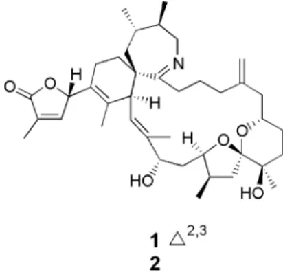
of a single-cell isolate of A. ostenfeldii (AOSH1) from Ship Harbour, Nova Scotia, yielded spirolides A, C, and 13-desmethyl C.3Using LC/MS methodology, spirolides H (1) and I (2) (Figure
1) were isolated from methanol extracts of wet cells of another Ship Harbour isolate of A. ostenfeldii, AOSH2. Unlike previously isolated spirolides, which contain a 5:5:6 or a 5:6:6 trispiroketal ring moiety,1-51and 2 contain a 5:6 dispiroketal ring system. Here
we report the structural elucidation of 1 and 2, a new subclass of spirolide marine toxins, and discuss the toxicity of 1 to mice.
Results and Discussion
The spirolide profile of AOSH2 was investigated using LC/MS analysis. A methanolic extract of cultured dinoflagellate cells was partitioned between CH2Cl2and H2O. Separation of the components
in the CH2Cl2extract on a C18chromatography column followed
by LC/MS analysis of the fractions resulted in the identification of a spirolide-containing fraction. Examination of the LC/MS profile revealed the presence of two components with m/z 650 and 652, which had not been previously isolated and identified. Isolation of these components was undertaken using a modified isolation procedure that varied from previous spirolide isolation procedures in that an LH-20 column chromatography step was not employed. This change significantly decreased the time needed to isolate
spirolide components. Characterization of 1 and 2 was achieved using a combination of spectrometric and 1D and 2D NMR spectroscopic techniques.
High-resolution mass spectrometry experiments determined the molecular formulas of 1 and 2 to be C40H60NO6([M + H]
+
m/z 650.4407) and C40H62NO6([M + H]+m/z 652.4570), respectively.
The MS/MS spectra of both components contained a fragment ion at m/z 164, which indicated the presence of a cyclic imine ring containing vicinal dimethyl groups, as seen in previously isolated spirolides.1-5Comparison of the1H and13C NMR spectral data
showed that the structures of 1 and 2 were similar except that the latter compound lacked the downfield proton atδ 7.13 and a carbon atδ 149.4, but possessed two proton signals at δ 1.65 and 2.56 and a carbon signal atδ 36.2.
The1H,13C, DEPT 135, and HSQC NMR data showed that the
carbons of 1 were distributed as 10 quaternary, 10 methine, 13 methylene, and 7 methyl carbons (Table 1). Inspection of DEPT-135 and HSQC NMR data of 2 revealed that it contained an additional methylene carbon and one less quaternary carbon, which explained the difference in molecular formula for 1 and 2. The assignments of the γ-lactone ring at C-1 to C-4, a vinyl double bond at C-21, and an imine at C-25 in the structures of 1 and 2 were confirmed by COSY and TOCSY data (Figure 2), HMBCs, and comparison with the NMR spectral data of previously isolated spirolides.1-5 Further analysis of COSY and TOCSY spectra
established four1H-1H spin systems corresponding to the partial
structures (a-d) for both 1 and 2 (Figure 2). The most significant difference between the two compounds was observed in partial structure a, which included theγ-lactone ring system, C-1 through C-4. For compound 1, the presence of unsaturation between C-2 and C-3 was indicated by the following: COSY and TOSCY * To whom correspondence should be addressed. Tel: 902-426-6351.
Fax: 902-426-9413. E-mail: shawna.mackinnon@nrc-cnrc.gc.ca.
†National Research Council of Canada. ‡AgResearch.
Figure 1. Structures of spirolides H (1) and I (2) and relative configuration of 1.
10.1021/np800795q CCC: $40.75 Published 2009 by the American Chemical Society Published on Web 07/02/2009
correlations of H-3 with H-4 and H-33 and of H-33 with H-4 (Figure 2), and HMBC correlations of H-33/C-1, C-2, C-3 and H-3/C-1, C-4. The additional methylene carbon in theγ-lactone ring of 2 was evidenced by COSY and TOCSY correlations of H-33 with H-2, H-3, H-4 and of H-2 with H-3 (Figure 2) and supported by HMBCs of H-33/C-3 and H-3b/C-5, C-6.
Chemical shifts of 1 (Table 1) were similar to those reported in the spirolide literature, except that one carbon resonance was observed atδ 112.9, characteristic of a 5:6 spiroketal carbon and corresponding to C-15. The absence of other downfield carbon signals in the range δ 112-119 indicated that there were no additional 5:6, 5:5, or 6:6 linkages. Furthermore, there were no carbon signals corresponding to the methylene carbons found at positions C-16 and C-17 in previously isolated spirolides.1-5These
observations suggested the presence of a dispiroketal ring system, which was confirmed by HMBC connectivities of H-13/C-15 and H-37/C-13, C-15, C-16, C-17 in 1.
The relative configuration of 1 (Figure 1) derived from ROESY data (Table 1) was in agreement with the relative configuration reported for spirolide D, in the area of their common structure, namely, C-7, -10, -12, and -13, in addition to C-16, -26, -28, and -29, which correspond to C-19, -29, -31, and -32, respectively, in spirolide D (Figure 2).9Further, ROESY correlations from H-4 to
H-3 and H3-34 and not to the methylene protons at C-32, and from
H-3 to H-4 and H3-34 only, favor an S* configuration at C-4, relative
to the rest of the spirolide structure in 1.
One of the major fragmentation pathways for [M + H]+
spirolide ions is the intramolecular retro-Diels-Alder reaction of the
six-Table 1. 1H and13C Data for Spirolides H (1) and I (2) in CD 3OHa
1 2
position δC, mult. δH(J in Hz) HMBCb ROESYc δC, mult. δH(J in Hz)
1 176.5, qC 182.0, qC 2 130.9, qC 36.5, CH 2.80, m 3 149.4, CH 7.13, dq (1.7, 1.7) 1, 4 4, 34 36.2, CH2 2.56, m 1.65, m 4 82.0, CH 5.98, m 35 3, 34 79.0, CH 5.41, dd (10.9, 5.8) 5 126.2, qC 129.4, qC 6 133.2, qC 130.7, qC 7 47.7, CH 3.78, bd (10.6) 24a, 24b, 34, 35 47.5, CH 3.76, d (10.6) 8 120.9, CH 5.07, bd (10.6) 10, 27a, 27b, 28, 40 121.2, CH 5.06, d (10.6) 9 149.4, qC 149.3, qC 10 75.5, CH 4.30, dd (7.3, 4.6) 8, 12 8, 11a, 11b, 36 75.4, CH 4.29, dd (6.6, 4.7) 11a 41.7, CH2 1.82, m 10, 11b, 12 41.6, CH2 1.84, m 11b 1.74, m 10, 12 10, 11a, 12 1.74, m 12 79.3, CH 4.27, q (6.5) 10, 14, 15 11a, 11b, 13, 14a 79.1, CH 4.27, q (6.5) 13 38.5, CH 2.40, m 15 12, 14b, 36 38.5, CH 2.40, m 14a 36.9, CH2 2.48, dd (12.7, 7.9) 12, 13, 36 12, 14b 36.8, CH2 2.48, m 14b 1.60, dd (12.7, 7.1) 12, 13, 15, 36 13, 14a, 19, 36 1.60, m 15 112.9, qC 112.8, qC 16 71.9, qC 72.0, qC 17 36.2, CH2 〈1.62〉dm 18b, 37 36.1, CH2 〈1.62〉dm 18a 29.9, CH2 1.52, m 18b, 37 30.1, CH2 1.52, m 18b 1.43, m 17, 18a, 19 1.44, m 19 74.7, CH 3.63, m 14b, 18b, 20a 74.5, CH 3.63, m 20a 43.7, CH2 2.42, m 44.0, CH2 2.44, m 20b 2.18, dd (14.4, 6.7,) 19, 21, 38 19 2.18, m 21 147.6, qC 147.6, qC 22a 35.4, CH2 2.42, m 35.3, CH2 2.44, m 22b 2.10, m 2.10, m 23a 22.7, CH2 1.90, m 22.7, CH2 1.92, m 23b 1.78, m 1.78, m 24a 36.9, CH2 3.25, m 7, 24b 36.8, CH2 3.25, m 24b 2.74, dt (20.9, 6.7) 25 7, 24a, 35 2.74, m 25 202.9, qC 176.5, qC 26 52.0, qC 52.0, qC 27a 36.5, CH2 2.01, m 36.7, CH2 2.03, m 27b 1.78, m 1.78, m 28 37.2, CH 1.10, m 8 37.2, CH 1.10, m 29 38.5, CH 1.66, m 30a, 30b, 39 38.5, CH 1.66, m 30 51.9, CH2 4.17, dd (12.8, 4.6) 29, 30b, 31a 52.0, CH2 4.20, m 3.58, m 25, 28, 29, 39 29, 30a, 39 3.58, m
31a 31.7, CH2 1.98, m 30a, 32a 32.1, CH2 1.98, m
31b 1.70, m 1.70, m 32a 20.0, CH2 2.27, m 31a 20.5, CH2 2.27, m 32b 1.70, m 1.70, m 33 10.4, CH3 1.90, t (1.5) 1, 2, 3 14.9, CH3 1.23, d (7.0) 34 16.7, CH3 1.76, s 5, 6, 7, 26 3, 4, 7 16.5, CH3 1.65, s 35 12.7, CH3 1.94, s 8, 9, 10 7, 24b 12.6, CH3 1.93, s 36 15.7, CH3 1.00, d (6.9) 12, 13, 14 10, 13, 14b 15.7, CH3 1.00, d (7.0) 37 21.7, CH3 1.27, s 15, 16, 17 17, 18a 21.7, CH3 1.27, s 38a 113.5, CH2 4.90, m 20, 22 113.4, CH2 4.89, m 38b 4.84, m 20, 22 4.86, m 39 18.5, CH3 1.05, d (6.9) 28, 29, 30 29, 30b 18.5, CH3 1.05, d (6.8) 40 20.0, CH3 1.06, d (6.9) 28, 29, 30 8 20.0, CH3 1.06, d (6.8)
aSpectra were recorded at 500.13 MHz (1H) and 125.77 MHz (13C). Chemical shiftsδ
HandδCwere referred to CHD2OH ) 3.30 ppm and13CD3OH
)49.0 ppm (13C), respectively.bHMBC correlations are from the proton stated to the indicated carbon.cUnambiguous ROESY correlations, recorded at 700 MHz, are from the proton stated to the indicated proton.dAverage value for an incompletely resolved methylene group.
membered monounsaturated ring followed by the six-centered concerted loss of H2O at C-10, cleaving the C-11 to C-12 bond.
This produces fragment ion masses in the range m/z 408-460 for known spirolides.7,10On inspection, spirolide H (1) was determined
to be most similar structurally to spirolide C.3The MS/MS spectrum
of spirolide C gave a fragment ion at m/z 458 for the species produced from the retro Diels-Alder reaction. In comparison, the MS/MS spectra of 1 and 2 contained a prominent ion at m/z 402 (Figure 3). The mass difference between spirolide C and 1 is in agreement with the absence of the five-membered cyclic ether moiety. Fragment ions at m/z 632 and 614, and at m/z 634 and 616 for 1 and 2, respectively, corresponded to the successive loss of H2O at C-10 and C-16. The proposed fragmentation pattern of 1 is
illustrated in Figure 4.
Toxicological investigations have shown that spirolides A, B, and C, 13-desmethylspirolide C, and 20-methylspirolide G are highly toxic to mice, with intraperitoneal LD50values between 6.9
and 99µg/kg.11In contrast, only transient hunching and lethargy
were observed in mice injected intraperitoneally with 1 at doses up to 2000µg/kg. Our results indicate that cyclic imine functionality is not the only requirement for toxicity of the spirolides, as suggested by earlier studies on the toxicity of these substances.
Structural studies are presently underway to investigate the con-formational features that are responsible for the difference in toxicity seen between spirolide H and previously isolated spirolides.
Experimental section
General Experimental Procedures.NMR spectra were measured on a Bruker DRX-500 spectrometer (Bruker Canada Ltd.) with the following conditions: frequency 500.13 MHz (1H), 125.7 MHz (13C);
solvents CD3OH (referenced to1H 3.30 and13C 49.0), 5 mm tubes,
temperature 20 °C; standard Bruker pulse sequences for1H single pulse,
double quantum filtered COSY, TOSCY (160 ms mixing time), HSQC, HMBC (60 and 90 ms mixing time),13C DEPT 135, and13C{1
H}-waltz decoupled experiments. A Bruker Avance III (700 MHz) (Bruker Canada Ltd.) [frequency 700.23 MHz (1H), 176.07 MHz (13C); solvents
CD3OH (referenced to1H 3.30 and13C 49.0), 5 mm tubes, temperature
20 °C] was used to acquire ROESY (400 ms mixing time) data on spirolide H. The initial identification of spirolide compounds in the culture was performed on an Agilent 1100 LC/MSD single quadrupole mass spectrometer equipped with a pneumatically assisted electrospray ionization source. Accurate mass spectra for measurements were performed on a Micromass Q-TOF Premiere (Micromass, Manchester, UK) time-of-flight mass spectrometer. Product ion MS/MS spectra were acquired on a hybrid 4000 QTrap mass spectrometer. Separations were performed on a 1.8µm Agilent Zorbax C18silica (4.6 × 50 mm) column
that was eluted isocratically with 74% A (0.1% TFA) and 26% B (0.1%
Figure 2.COSY and TOCSY correlations (curved arrows) in 1 and 2 and partial structures a-d (bold lines) representing1H-1H
spin systems.
Figure 3.Product ion mass spectra of the [M + H]+
ions of 1 (a),
m/z 650, and 2 (b), m/z 652. Conditions: electrospray ionization; collision energy of 55 V; enhanced product ion scan on an API 4000 Qtrap mass spectrometer.
TFA, CH3CN) at a flow rate of 1 mL/min. Analyses were conducted
in positive ion mode using selected ion monitoring of [M + H]+
ions. The column eluent was split, with 10% going to the mass spectrometer.
Biological Material. The A. ostenfeldii culture (AOSH2) was initiated from a single cell isolated from plankton samples collected at Ship Harbour, NS, in 2000. The clonal isolate was identified by Nomarski contrast interference microscopy and by epifluorescence microscopy after staining the thecal plates with calcofluor.12The culture
is maintained at the National Research Council’s Institute for Marine Biosciences.
Culturing of A. ostenfeldii Clonal Isolates.AOSH2 was initiated in L1 growth medium diluted 1:10 with sterile seawater in multiwell tissue culture plates. Cultures were scaled-up in full-strength L1 medium by serial transfer into 15 mL culture tubes and then into 2.8 L Fernbach flasks, total culture volume 80 L. Unialgal cultures were maintained at an ambient photon flux density of 90 mmol m-2s-1at 14 °C and a
14:10 h light/dark photocycle in a controlled growth chamber. Cells were harvested in late exponential growth phase by gravity filtration onto a 20 mm Nitex mesh sieve and concentrated by centrifugation (2750g) for 20 min.
Isolation of Spirolides from Cultured A. ostenfeldii.The wet cell pellets of AOSH2 (47.4 g) were extracted four times with CH3OH (250
mL) followed by sonication. After centrifugation, the methanolic supernatants were pooled and evaporated to dryness. The residue was dissolved in H2O (300 mL) and partitioned with CH2Cl2(3 × 300 mL).
The CH2Cl2extract (892 mg) was dissolved in 30% CH3OH/H2O and
subjected to a C18flash chromatography column, which was conditioned
and eluted with 50% CH3OH/H2O. Fractions containing spirolides were
combined, evaporated to dryness, and purified using an Agilent Zorbax SB C18 HPLC column, which was eluted isocratically with 26%
CH3CN/H2O (0.1% TFA) and monitored at 210 nm. The yields of 1
and 2 were determined by NMR quantitation to be 1.03 and 0.20 mg, respectively.
Spirolide H:white solid;1H and13C NMR (Table 1); HRMS ([M +
H]+
m/z 650.4407 (calcd for C40H60NO6, 650.4421).
Spirolide I:white solid;1H and13C NMR (Table 1); HRMS ([M +
H]+m
/z 652.4570 (calcd for C40H62NO6, 652.4577).
Acknowledgment.The authors thank the following from the NRC-Institute for Marine Biosciences: Mr. I. Burton for acquisition of NMR data, Dr. J. Melanson and Mr. W. Hardstaff for acquisition of mass spectrometry data, and Dr. J. Simmons-Boyce and Dr. R. Syvitski for their helpful discussions.
Supporting Information Available:1H and13C NMR spectra for
1and the1H NMR spectrum of 2. This material is available free of
charge via the Internet at http://pubs.acs.org.
References and Notes
(1) Hu, T.; Curtis, J. M.; Oshima, Y.; Quilliam, M. A.; Walter, J. A.; Watson-Wright, W. M.; Wright, J. L. C. J. Chem. Soc., Chem. Commun. 1995, 2159–2161.
(2) Hu, T.; Curtis, J. M.; Oshima, Y.; Walter, J. A.; Wright, J. L. C. Tetrahedron Lett. 1996, 37, 7671–7674.
(3) Hu, T.; Burton, I. W.; Cembella, A. D.; Curtis, J. M.; Quilliam, M. A.; Walter, J. A.; Wright, J. L. C. J. Nat. Prod. 2001, 64, 308–312. (4) MacKinnon, S. L.; Walter, J. A.; Quilliam, M. A.; Cembella, A. D.;
LeBlanc, P.; Burton, I. W. J. Nat. Prod. 2006, 69, 983–987. (5) Cimineiello, P.; Dell’Aversano, C.; Fattorusso, E.; Forino, M.; Grauso,
L.; Tartaglione, L.; Guerrini, F.; Pistocchi, R. J. Nat. Prod. 2007, 70, 1878–1883.
(6) Gill, S.; Murphy, M.; Clausen, J.; Richard, D.; Quilliam, M.; MacKinnon, S.; LeBlanc, P.; Mueller, R.; Pulido, O. Neurotoxicology
2003, 24, 593–604.
(7) Sleno, L.; Windhust, A. J.; Volmer, D. A. Anal. Bioanal. Chem. 2004, 378, 969–976.
(8) Cembella, A. D.; Bauder, A. G.; Lewis, N. I.; Quilliam, M. A. J. Plankton Res. 2001, 23, 1413–1419.
(9) Falk, M.; Burton, I. W.; Hu, T.; Walter, J. A.; Wright, J. L. C. Tetrahedron 2001, 57, 8659–8665.
(10) Sleno, L.; Chalmers, M. J.; Volmer, D. A. Anal. Bioanal. Chem. 2004, 378, 977–986.
(11) Quilliam, M.; Spirolides: DiscoVery, Toxicity and Pharmacological Potential; Presented at the 122nd AOAC Annual Meeting & Exposi-tion, Dallas, TX, Sept 2008.
(12) Fritz, L.; Triemer, R. E. In Toxic Dinoflagellates; Anderson, D. M., White, A. W., Baden, D. G., Eds.; Elsevier-North Holland: New York, 1985; pp 117-120.
NP800795Q
![Figure 3. Product ion mass spectra of the [M + H] + ions of 1 (a), m/z 650, and 2 (b), m/z 652](https://thumb-eu.123doks.com/thumbv2/123doknet/14155172.472405/4.883.504.777.64.730/figure-product-ion-mass-spectra-m-h-ions.webp)
