Publisher’s version / Version de l'éditeur:
Vous avez des questions? Nous pouvons vous aider. Pour communiquer directement avec un auteur, consultez la première page de la revue dans laquelle son article a été publié afin de trouver ses coordonnées. Si vous n’arrivez pas à les repérer, communiquez avec nous à PublicationsArchive-ArchivesPublications@nrc-cnrc.gc.ca.
Questions? Contact the NRC Publications Archive team at
PublicationsArchive-ArchivesPublications@nrc-cnrc.gc.ca. If you wish to email the authors directly, please see the first page of the publication for their contact information.
https://publications-cnrc.canada.ca/fra/droits
L’accès à ce site Web et l’utilisation de son contenu sont assujettis aux conditions présentées dans le site LISEZ CES CONDITIONS ATTENTIVEMENT AVANT D’UTILISER CE SITE WEB.
The Journal of Biological Chemistry, 286, 12, pp. 10735-10743, 2011-03-25
READ THESE TERMS AND CONDITIONS CAREFULLY BEFORE USING THIS WEBSITE.
https://nrc-publications.canada.ca/eng/copyright
NRC Publications Archive Record / Notice des Archives des publications du CNRC :
https://nrc-publications.canada.ca/eng/view/object/?id=2e1e9d12-78ff-4088-9e83-ae51d163ba33
https://publications-cnrc.canada.ca/fra/voir/objet/?id=2e1e9d12-78ff-4088-9e83-ae51d163ba33
NRC Publications Archive
Archives des publications du CNRC
This publication could be one of several versions: author’s original, accepted manuscript or the publisher’s version. / La version de cette publication peut être l’une des suivantes : la version prépublication de l’auteur, la version acceptée du manuscrit ou la version de l’éditeur.
For the publisher’s version, please access the DOI link below./ Pour consulter la version de l’éditeur, utilisez le lien DOI ci-dessous.
https://doi.org/10.1074/jbc.M110.194423
Access and use of this website and the material on it are subject to the Terms and Conditions set forth at
Structural and functional studies of the Escherichia coli
phenylacetyl-CoA monooxygenase complex
Grishin, Andrey M.; Ajamian, Eunice; Tao, Limei; Zhang, Linhua; Menard,
Robert; Cygler, Miroslaw
Structural and Functional Studies of the Escherichia coli
Phenylacetyl-CoA Monooxygenase Complex
*
□SReceived for publication, October 13, 2010, and in revised form, December 10, 2010Published, JBC Papers in Press, January 19, 2011, DOI 10.1074/jbc.M110.194423
Andrey M. Grishin‡, Eunice Ajamian‡, Limei Tao§, Linhua Zhang§, Robert Menard§, and Miroslaw Cygler‡§1 From the‡Department of Biochemistry, McGill University, Montreal, Quebec H3G 1Y6, Canada and the§Biotechnology Research
Institute, National Research Council, Montreal, Quebec H4P 2R2, Canada
The utilization of phenylacetic acid (PA) in Escherichia coli occurs through a hybrid pathway that shows features of both aerobic and anaerobic metabolism. Oxygenation of the aromatic ring is performed by a multisubunit phenylacetyl-coenzyme A oxygenase complex that shares remote homology of two sub-units to well studied bacterial multicomponent monooxyge-nases and was postulated to form a new bacterial multicompo-nent monooxygenase subfamily. We expressed the subunits PaaA, B, C, D, and E of the PA-CoA oxygenase and showed that PaaABC, PaaAC, and PaaBC form stable subcomplexes that can be purified. In vitro reconstitution of the oxygenase subunits showed that each of the PaaA, B, C, and E subunits are necessary for catalysis, whereas PaaD is not essential. We have determined the crystal structure of the PaaAC complex in a ligand-free form and with several CoA derivatives. We conclude that PaaAC forms a catalytic core with a monooxygenase fold with PaaA
being the catalytic␣subunit and PaaC, the structuralsubunit.
PaaAC forms heterotetramers that are organized very differ-ently from other known multisubunit monooxygenases and lacks their conservative network of hydrogen bonds between the di-iron center and protein surface, suggesting different associa-tion with the reductase and different mechanisms of electron transport. The PaaA structure shows adaptation of the common access route to the active site for binding a CoA-bound sub-strate. The enzyme-substrate complex shows the orientation of the aromatic ring, which is poised for oxygenation at the ortho-position, in accordance with the expected chemistry. The PA-CoA oxygenase complex serves as a paradigm for the new sub-family multicomponent monooxygenases comprising several hundred homologs.
Aromatic organic compounds represent a major class of environmental pollutants (1). Many microbes can grow using these molecules as an abundant source of nutrients and a car-bon source. Under anaerobic conditions, the metabolism of aromatic compounds proceeds through initial conversion to a CoA derivative, which is then reduced in an ATP-dependent
reaction (2). In the presence of oxygen, bacteria utilize a wide variety of oxygenases to activate the inert aromatic ring (3). In aerobic pathways that utilize multisubunit dioxygenases or non-heme monooxygenases, both the hydroxylation of the aro-matic ring and its cleavage are oxygen-dependent (1). Dioxyge-nases incorporate two adjacent hydroxyl groups into the aro-matic ring (4). For metabolism that relies on monooxygenases, the consecutive action of two such enzymes is required, with insertion of one hydroxyl group at each step, as is the case in Pseudomonas stutzeriOX1 with toluene/o-xylene monooxyge-nase and phenol hydroxylase performing these reactions (5).
An aerobic hybrid pathway combines the features of classic aerobic and anaerobic strategies (6). As in anaerobic metabo-lism, the aromatic compound is attached to CoA, but the sub-strate is oxygenated, whereas the ring opening proceeds with-out oxygen. This pathway is present in many bacteria and frequently serves as a core component of an extensive meta-bolic network involved in degradation of 2-phenylethylamine, styrene, phenylacetaldehyde, phenylacetyl amides, n-phenylal-kanoic acids with even numbers of carbon atoms, and phenyla-cetyl esters (7). Escherichia coli has only a subset of these enzymes that are required for the utilization of phenylacetic acid (PA)2 and of 2-phenylethylamine, encoded by the paa
operon (6) (see Fig. 1). The enzymatic steps along the PA deg-radation pathway and enzymes associated with each step have been unequivocally established only recently (8).
The key enzyme of the PA-CoA metabolic pathway is a ring oxygenase that binds the aromatic CoA thioester and performs a strictly aerobic oxygenation reaction. Phylogenetic analysis suggests that the enzyme belongs to the family of bacterial mul-ticomponent monooxygenases (BMMs) and forms the proto-type of a new branch (9). BMMs were previously divided into six major groups (10) and include well studied enzymes such as methane monooxygenase (MMO) (11, 12), phenol hydroxylase (5, 13), toluene/o-xylene monooxygenase (ToMO) (14, 15), and toluene 4-monooxygenase (16, 17).
The PA-CoA oxygenase complex was proposed to contain five subunits, PaaA, B, C, D, and E (9). The PaaA subunit is distantly related to the catalytic (␣) subunit of other well char-*This work was supported in part by the National Sciences and Engineering
Research Council of Canada, the National Research Council, the Canadian Institutes of Health Research, and the University of Saskatchewan. This work was supported by Grant GSP-48370 from Canadian Institutes of Health Research (to M. C.).
□S The on-line version of this article (available at http://www.jbc.org) contains
supplemental text, Tables S1–S4, and Figs. S1–S8.
1To whom correspondence should be addressed: Biotechnology Research
Institute, NRC, 6100 Royalmount Ave., Montreal, PQ H4P 2R2, Canada. Tel.: 514-496-6321; E-mail: mirek@bri.nrc.ca.
2The abbreviations used are: PA, phenylacetic acid (phenylacetate); BMM,
bacterial multicomponent monooxygenases; BMMH, hydroxylase compo-nent of BMM; MBP, maltose-binding protein; MMO, methane monooxyge-nase complex; MMOH, hydroxylase of MMO; 2-OH-PA, 2-hydroxy-phenyla-cetate; ToMO, toluene/o-xylene monooxygenase complex; ToMOH, hydroxylase of ToMO; PaaAC, PaaA–PaaC; Pipes, 1,4-piperazinediethane-sulfonic acid; ADA, [(carbamoylmethyl)imino]diacetic acid; SEC, size exclu-sion chromatography; Ni-NTA, nickel-nitrilotriacetic acid.
at CISTI - Natl Research Council, on March 30, 2011
www.jbc.org
acterized BMMs, sharing with them only 10 –15% sequence identity, with the iron-binding motifs being the easiest to rec-ognize. Of the other subunits, PaaC shows sequence similarity to PaaA, being analogous to the structural () subunit of BMMs. PaaB and PaaD have no detectable sequence similarity to subunits of other BMMs. The PaaACD complex was pro-posed to be analogous to ␣␥ hydroxylase complex of BMMs, whereas PaaB was suggested to be a regulatory subunit control-ling hydroxylase-reductase interactions (9). Based on amino acid sequence, PaaE was postulated to be a class IA reductase (classification of reductases according to Ref. 18). Interestingly, this is the first example of a monooxygenase complex contain-ing a class IA reductase. Usually, the monooxygenase-associ-ated reductases belong to either class IB (e.g. MMO) or class III (e.g. ToMO). Reductases class IA and IB differ in domain order and in cofactor specificity.
We have investigated protein-protein interactions between the putative components of the E. coli PA-CoA monooxyge-nase complex and identified several stable subcomplexes in vitro. By reconstituting the monooxygenase from its compo-nents, we show that PaaA, B, C, and E are necessary for activity, whereas PaaD, which was deemed to be important for in vivo function (9), had no effect on in vitro activity. We have deter-mined the crystal structure of the PaaAC subcomplex with and without bound PA-CoA substrate. This structure shows that PaaA is the catalytic subunit, whereas PaaC is the structural subunit. The PaaAC complex serves as the prototype for a new class of BMMs and provides the first structural example of a BMM oxygenase-substrate complex. Finally, we showed exper-imentally that PaaE is a reductase.
EXPERIMENTAL PROCEDURES
The detailed procedures of cloning and heterologous expres-sion of genes, protein purification and characterization, reac-tion measurement, crystallizareac-tion, and structure determinareac-tion are described in thesupplemental materials. Here we provide an abbreviated description.
Cloning, Expression, and Purification—Individual genes en-coding PaaA, B, C, D, and E from E. coli K-12 were cloned into a modified pGEX-4T1 (N-GST tag) (GE Healthcare), pET15b (N-His tag), pRDSFDuet-1 MCS2 (no tag), and pCDFDuet-1 MCS2 (no tag) vectors (Novagen). The paaE gene was also cloned into pMAL-c2x (N-MBP tag) vector (New England Bio-labs). Additionally the paaA-E segment of the entire paa
operon was cloned into the modified pET15b. E. coli expression strains BL21(DE3) or BL21 (Novagen) were used for protein production.
The purification of individual proteins and the coexpressed complexes was done as described previously (19). Briefly, the cells were lysed by sonication, cell debris was removed by cen-trifugation, and the supernatant was loaded on Ni-NTA (Qia-gen). The column was washed with buffer containing 50 mM
HEPES, pH 7.5, 1MNaCl, and 40 mMimidazole. The final
puri-fication step consisted of gel filtration chromatography on Superose 12 column (GE Healthcare). The purification of PaaB-His6C was accomplished following the same protocol, but with the omission of high salt wash. The growth of cells expressing PaaA-His6C in iron-enriched medium (20) or the addition of
0.1 mM ammonium ferrous sulfate to purified PaaA-His6C
resulted in aggregation of the protein.
GST-PaaB and GST-PaaD were purified by affinity chroma-tography using glutathione-Sepharose 4B (GE Healthcare) with the subsequent cleavage from the beads by TEV protease, fol-lowed by size exclusion chromatography on a Superose 12. The expression of MBP-PaaE was done in iron-enriched medium. The protein was purified using amylose resin (New England Biolabs) and used without further purification.
Protein-Protein Interaction Assays—Protein-protein interac-tions were evaluated using coexpression of different combina-tions of subunits with only one of the subunits having a His tag. The extracted proteins were loaded on a Ni-NTA-agarose col-umn, and the proteins retained on the column after washing were analyzed by SDS-PAGE (Table 1). The composition of buffers was varied from pH 6.5 to 8.5 with and without 0.4M
NaCl to study the effects of pH and ionic strength on complex formation. In addition, interactions of purified GST-PaaD with PaaABC as well as Paa-His6ABC with PaaD and MBP-PaaE
with PaaABC and PaaD were investigated by pull-down exper-iments using the appropriate columns (Table 2).
Enzyme Activity Assays—Mixtures of different Paa subunits, each at a final concentration of 2 M, were added to reaction
buffer containing 25 mMTris, pH 7.5, 500 MNADPH, 10 M
Fe(NH4)2(SO4)2, 10 MFAD, and were incubated for 15 min at
37 °C. The reaction was initiated by the addition of phenyla-cetyl-CoA to the final concentration of 300 M. After 20 h, the
reaction was stopped by the addition of HCl. The appearance of 2-hydroxy-phenylacetate (2-OH-PA) was quantitatively
moni-TABLE 1
Identified complexes between the Paa subunits by copurification
NA, not applicable.
Coexpression combinations High salt purification scheme Low salt wide pH range purification scheme
His6PaaC ⫹ PaaA His6PaaC ⫹ PaaA NA
His6PaaC ⫹ PaaB His6PaaC His6PaaC ⫹ PaaB
His6PaaC ⫹ PaaD His6PaaC His6PaaC
His6PaaC ⫹ PaaA ⫹ PaaB His6PaaC ⫹ PaaA ⫹ PaaB NA
His6PaaC ⫹ PaaA ⫹ PaaD His6PaaC ⫹ PaaA His6PaaC ⫹ PaaA
His6PaaC ⫹ PaaA ⫹ PaaE His6PaaC ⫹ PaaA His6PaaC ⫹ PaaA
His6PaaE ⫹ PaaB ⫹ PaaD Absence of specific protein NA
His6PaaA ⫹ PaaB Absence of specific protein NA
His6PaaA ⫹ PaaD Absence of specific protein NA
His6PaaA ⫹ PaaB ⫹ PaaC His6PaaA ⫹ PaaB ⫹ PaaC NA
His6PaaA ⫹ PaaB ⫹ PaaC ⫹ PaaD His6PaaA ⫹ PaaB ⫹ PaaC His6PaaA ⫹ PaaB ⫹ PaaC
His6PaaA ⫹ PaaB ⫹ PaaC ⫹ PaaD ⫹ PaaE His6PaaA ⫹ PaaB ⫹ PaaC His6PaaA ⫹ PaaB ⫹ PaaC
Phenylacetyl-CoA Monooxygenase
at CISTI - Natl Research Council, on March 30, 2011
www.jbc.org
tored by LC-MS/MS on an Agilent 1200 HPLC system coupled to a Agilent QQQ6410 mass spectrometer (Agilent Technolo-gies, Inc., Palo Alto, CA). Separations were performed on a SymmetryShield RP18 column (Waters).
Crystallization—Crystallization of PaaA-His6PaaC was
described previously (19). All of the crystals were obtained using hanging drop vapor diffusion with protein concentration of 8 mg/ml. Two conditions were optimized for complexes of PaaAC with CoA, 3-hydroxybutyril-CoA, benzoyl-CoA, and phenylacetyl-CoA. For the first condition, the reservoir solu-tion contained 100 mMPipes, pH 6.5, 15% (v/v) PEG 400MME
(Sigma), whereas the second condition was 100 mMPipes, pH
6.5, 5% (v/v) PEG 400MME, 5% (v/v) isopropanol. PaaAC with acetyl-CoA was crystallized in 0.1Msodium citrate buffer, pH
5.5, and 15% (w/v) PEG 6000 (Fluka). Crystals of ligand-free PaaAC were obtained in 100 mMADA, pH 5.5.
X-ray Data Collection, Structure Solution, and Refinement— The crystals were cryoprotected in 25% (v/v) glycerol or 25% (v/v) 2-methyl-2,4-pentanediol. The diffraction data were col-lected with a Mar300CCD detector at the CMCF-1 beamline at the Canadian Light Source. Data integration and scaling was performed with HKL2000 (21). The structure of PaaA-His6C
was solved by molecular replacement using the programs PHASER (22) and MOLREP (23) with the PaaC structure (Pro-tein Data Bank code 1OTK)3as a search model. Subsequent
model building and refinement was performed using COOT (24) and REFMAC5 (25). Pertinent details are presented in Table 3. Analysis of structure using MolProbity (26) showed models of good quality.
RESULTS
Interactions between Subunits of the PA-CoA Oxygenase Complex—The five proteins encoded by paaABCDE were pos-tulated to form a multicomponent oxygenase involved in the hydroxylation of phenylacetyl-CoA. To analyze protein-pro-tein interactions among these five subunits, we have coex-pressed and purified them in various combinations (Table 1). We were able to overexpress each subunit individually but only PaaB, C, and D were soluble, whereas PaaA and E were insolu-ble. However, when PaaA and PaaC were coexpressed, they formed a well behaved soluble complex. When the entire paaABCDEoperon was expressed, only PaaA, B, and C but not PaaD or PaaE could be detected. Other coexpression strategies are summarized in Table 1. An attempt to improve PaaE solu-bility by coexpression with various combinations of other
sub-in a soluble protesub-in.
Interactions between the subunits were established by affin-ity purification of various combinations of coexpressed PaaA, B, C, D, and E where only one subunit carried a His6tag. Protein association was tested at pH 6.5, 7.5, and 8.4, at low and high ionic strength (0 –1MNaCl). Under all of the tested conditions,
we observed PaaA-His6C, PaaAB-His6C, and Paa-His6ABC complexes, whereas in the absence of salt, we also detected the His6PaaC-PaaB complex (Table 1). No evidence for the
forma-tion of a stable complex between PaaD or PaaE and other pro-teins (or their combinations) was found. Pull-down experi-ments using GST-PaaD and MBP-PaaE as baits and purified PaaABC or individual subunits were also negative (Table 2).
The apparent molecular mass of the PaaA-His6C complex
(calculated molecular mass, 64.8 kDa) was determined to be ⬃180 kDa by size exclusion chromatography (SEC) and ⬃160 kDa by dynamic light scattering, consistent with either hetero-tetramers or heterohexamers. The molecular mass of PaaABC (molecular mass, 75.5 kDa) was ⬃90 –120 kDa (SEC) and 160 – 200 kDa (dynamic light scattering), making the prediction of stoichiometry uncertain. Finally, the molecular mass of PaaB-His6C (molecular mass, 40.0 kDa) was ⬃40 kDa (SEC) and 38 – 43 kDa (dynamic light scattering), indicating heterodimer. Similar SEC analysis of PaaB (molecular mass, 11.0 kDa) showed that it elutes as a peak with an apparent molecular mass of 33 kDa, corresponding to trimers in solution. The PaaD (molecular mass, 18.5 kDa) elutes from the SEC column with an apparent molecular mass of 21 kDa, indicating a monomer.
Paa Components Necessary for Enzymatic Activity—Because there is a discrepancy between the in vivo (9) and in vitro (8) data as to which subunits are necessary for activity, we first reconstituted all five components, PaaABCDE, in the presence of the required cofactors and 10 MFe2⫹, and after the addition
of PA-CoA, we followed the reaction by LC-MS (supplemental Fig. S1). The reaction product (III in Fig. 1) is expected to rap-idly decompose to 2-OH-PA (VI in Fig. 1) (9). We observed the disappearance of the substrate and concomitant appearance of VI. We also detected PA (compound I), a byproduct of sub-strate hydrolysis (supplemental Fig. S1). The measured activity of the reconstituted PA oxygenase was ⬃5 nmol䡠min⫺1䡠mg⫺1
(supplemental Table S1). The rather low activity of the recon-stituted complex may be due to partially active PaaE, which could only be expressed in a soluble form as a MBP-tagged protein.
To determine whether all five proteins are indispensable for the oxygenation of phenylacetyl-CoA, we analyzed the reaction mixture with different subsets of PaaABCDE proteins. The maximum reaction rate was observed when PaaA, B, C, and E subunits were present (Table 4). The presence or absence of PaaD had no effect on the reaction outcome, suggesting that this component is not essential for the reaction in vitro. Absence of PaaB leads to a more than 100-fold drop in the product concentration. PaaAC and PaaABC were able to generate small amounts of the product, suggesting a single turnover utilizing the reduced Fe2⫹present in the reaction
mixtures.
3R. Zhang, unpublished observations.
Pulldown assays to identify interactions of PaaD and PaaE with other subunits
Affinity resin Bait Prey Interact?
Glutathione-Sepharose GST-PaaD PaaC No Glutathione-Sepharose GST-PaaD PaaAC No Glutathione-Sepharose GST-PaaD PaaABC No
Ni-NTA PaaA-His6C PaaD No
Ni-NTA Paa-His6ABC PaaD No
Ni-NTA His6PaaB PaaD No
Ni-NTA His6PaaC PaaD No
Amylose resin MBP-PaaE PaaABC ⫹ PaaD No
at CISTI - Natl Research Council, on March 30, 2011
www.jbc.org
To establish whether the oxygen for the hydroxylation reac-tion is obtained from water or from gaseous O2, we analyzed the reaction products in the presence of H2O18. No incorporation
of O18 at position 2-OH of 2-OH-PA was observed by mass
spectrometry, indicating that the oxygen is derived from O2 (supplemental Table S2). In the same experiment, we observed, as expected, the incorporation of O18into the carboxyl of the
2-OH-PA and PA.
The Reductase PaaE—To identify the type of iron-sulfur cluster in PaaE, we have recorded the absorption spectrum of MBP-PaaE (supplemental Fig. S2). The spectrum with maxima at 325, 415, and 460 nm closely resembles that of spinach [2Fe-2S] ferredoxin (27).
We next investigated the cofactor preference of the PaaE, which requires nicotinamide adenine dinucleotide, NAD(P), and a riboflavin, FAD or FMN, cofactors and is expected to transfer electrons from NAD(P)H to the di-iron center of PaaA through the riboflavin cofactor and the iron-sulfur center. The measured activity was 8.5 times higher in the presence of NADPH than in the presence of NADH and 3 times higher in the presence of FAD than in the presence of FMN ( supplemen-tal Table S1). PaaE showed the same cofactor preference when cytochrome c was used as an artificial electron acceptor instead of PaaABC (supplemental Table S1).
Crystal Structure of PaaAC—Of the three stable complexes, PaaAC, PaaABC, and PaaBC, we obtained crystals only of the PaaA-His6PaaC subcomplex. PaaA and PaaC share only 17% FIGURE 1. Aerobic hybrid phenylacetate catabolic pathway as updated recently (8). The product of the PA-CoA monooxygenase is the 1,2-epoxide derivative of PA-CoA (compound III). The previously proposed dihydrodiol derivative of PA-CoA (compound VII) (35) is shown in parentheses.
TABLE 3
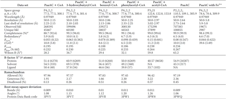
X-ray data collection and refinement statistics
Data set PaaAC ⴙ CoA 3-hydroxybutyryl CoAPaaAC ⴙ benzoyl-CoAPaaAC ⴙ phenylacetyl-CoAPaaAC ⴙ acetyl-CoAPaaAC ⴙ PaaAC PaaAC with Fe2ⴙ
Space group P41212 P41212 P41212 P41212 P3221 P212121 P41212
a, b, c (Å) 77.5, 77.5, 300.1 77.4, 77.4, 301.4 77.6, 77.6, 300.7 77.6, 77.6, 300.4 122.8, 122.8, 153.8 110.3, 109.1, 305.9 78.4, 78.4, 309.9
Wavelength (Å) 0.97949 0.97949 0.97949 0.97949 0.97949 0.97949 0.97949
Resolution (Å) 50.0-2.15 50.0-2.03 50.0-2.06 50.0-2.25 50.0-2.97 50.0-2.64 50.0-5.8 Last shell of resolution (Å) 2.23-2.15 2.10-2.03 2.13-2.06 2.33-2.25 3.02-2.97 2.73-2.64 5.9-5.8
Observed hkl 268595 599496 339272 300130 175298 707383 19671 Unique hkl 45467 60180 58037 44543 27899 108855 2987 Completeness (%)a 88.7 (92.6) 99.3 (94.8) 99.5 (96.6) 99.1 (94.4) 99.0 (99.6) 99.9 (99.9) 98.4 (99.3) Redundancya 5.9 (6.0) 10.0 (4.1) 5.8 (4.2) 6.7 (5.9) 6.3 (6.3) 6.5 (4.0) 6.6 (7.0) Rsyma 0.055 (0.22) 0.063 (0.382) 0.078 (0.389) 0.098 (0.459) 0.15 (0.681) 0.08 (0.547) 0.044 (0.422) I/(I)a 10.8 (3.8) 11.8 (2.1) 8.6 (2.1) 6.5 (2.0) 11.2 (2.0) 10.0 (2.0) 39.6 (3.49) Rwork 0.195 0.195 0.188 0.186 0.238 0.234 Rfree(% hkl) 0.232 0.230 0.225 0.233 0.264 0.267 Wilson B (Å2) 28.2 34.7 29.4 32.1 59.8 65.2 B-factor Å2(# atoms) Protein 51.4 (6278) 60.9 (6269) 51.0 (6268) 50.0 (6269) 40.57 (8658) 54.9 (24587) Solvent 54.5 (333) 69.5 (378) 56.4 (457) 49.2 (348) NA 43.3 (217) Ligand 50.4 (48) 57.8 (54) 46.6 (56) 39.8 (57) 33.7 (102) NA Ramachandran Allowed (%) 97.96 97.57 97.83 97.45 96.42 97.19 Generous (%) 1.91 2.17 1.66 2.30 3.22 2.36 Disallowed (%) 0.13 0.26 0.51 0.26 0.37 0.45
Root mean square deviation
Bonds (Å) 0.009 0.010 0.01 0.011 0.011 0.009
Angles (°) 1.08 1.15 1.12 1.20 1.26 1.06
Protein Data Bank code 3PVY 3PVT 3PVR 3PW1 3PW8 3PWQ
aThe information for the last shell of resolution is given in parentheses.
TABLE 4
Activity of reconstituted Paa subunits
Paa subunits 2-OH-PA PA
M M ABCDE 86.3 22.7 ABC关5D兴Ea 87.9 40.15 ABCE 80.1 15.1 ACE 0.34 112.8 ACB 1.1 101.9 AC 0.97 96.9 BE 0.13b 56.6
a关5D兴 indicates 5-fold higher concentration of PaaD than the concentration of all
other compounds.
bThe value is at the edge of the detection limit, which is 0.1 M.
Phenylacetyl-CoA Monooxygenase
at CISTI - Natl Research Council, on March 30, 2011
www.jbc.org
mean square deviation of 1.7 Å for 191 C␣ atoms (of 302 for PaaA and 248 for PaaC). Their overall fold is characteristic of the hydroxylase components of monooxygenases (28). The core of the molecules is composed of six long ␣-helices: B, C, E, F, G, and H (named according to Ref. 11; Fig. 2a andsupplemental Table S3) and is similar to other monooxygenases. In PaaA the ␣-helices B, C, E, and F form a four-helix bundle, adopting a ferritin-like fold and encompassing a long tunnel. The di-iron-binding site of PaaA is unoccupied. PaaC lacks the N- and C-terminal ␣-helical extensions observed in PaaA (Fig. 2a) and differs in conformation of segments between ␣-helices G, G⬘, H, and J. The space between the helices of the ferritin bundle in PaaC is filled with side chains eliminating the tunnel ( supple-mental Fig. S3).
The PaaAC complex crystallized in three crystal forms. The stoichiometry of PaaA:PaaC was 1:2 in two forms and 1:1 in the third form. Comparison of molecular arrangements in different crystal packing environments allowed us to identify the (PaaAC)2heterotetramer (dimer of heterodimers) as the com-mon structural unit. PaaAC heterodimer interface is formed by helices B and C from each molecule, together forming an antiparallel four-helix bundle, with additional contacts be-tween helix K of PaaA and the C terminus of PaaC (Fig. 2b). The interface has a significantly hydrophobic character, with a par-ticularly large hydrophobic surface on PaaA (supplemental Fig. S4). The presence of such an extensive hydrophobic patch on the surface of PaaA is likely responsible for its high propensity for aggregation when expressed alone. Three salt bridges anchor the PaaA and PaaC subunits at the edges of the interface (Glu-33PaaC…Arg-90PaaA, Asp-35PaaC…Lys-282PaaA, and
ter-minal COOHPaaC…Arg-61PaaA). The two PaaAC heterodimers
are arranged in an antiparallel fashion to form a heterotetramer (Fig. 2c). This interface has a predominantly hydrophilic char-acter. The interactions of unpaired PaaC subunit (“free PaaC”) are described in thesupplemental materials.
Substrate Binding—To date no hydroxylase component of a BMM oxygenase was crystallized with its substrate in the active site. We have determined the structure of apo PaaAC as well as its complexes with CoA, acetyl-CoA, 3-hydroxybutytyl-CoA, benzoyl-CoA, and the substrate phenylacetyl-CoA. The ligands bind only to the PaaA subunit, accompanied by ordering of the loop between ␣-helices G and G⬘ (residues 199 –205) that now forms part of the ligand-binding site. The difference map unequivocally shows the substrate position in the active site (supplemental Fig. S5). The ligand/substrate binds within the tunnel formed by helices B, C, E, and F and the tips of helices G and I. This tunnel extends ⬃20 Å from the protein surface toward the center of PaaA and ends near the di-iron center (Fig. 2d andsupplemental Fig. S3a). The CoA moiety of the substrate forms interactions with ⬃30 residues (Fig. 2d). The ligands are accommodated in the tunnel with the adenine in an anti-con-formation, folded over the diphosphate moiety at the top of the tunnel and the pantothenate moiety extending toward the bot-tom of the tunnel (Fig. 2d). The adenine base is anchored by hydrogen bonds between the N6 amine group and OGSer-106
and COMet-193and by van der Waals contacts with Ile-268 and
Phe-264. The phosphate groups form multiple hydrogen
37, and Lys-103; the ␣-phosphate bonds with Ser-202, Asn-204, and Asn-218; whereas the -phosphate bonds with Arg-33 and Lys-214. Pantothenate and -mercaptoethylamine moieties make numerous van der Waals contacts along the tunnel with Gln-34, His-38, Ser-41, Ser-105, Asn-132, Gln-133, Leu-136, Tyr-144, Met-194, and Phe-195. The bottom of the binding site is formed by Phe-108 and three acidic residues (Glu-49, Glu-76, and Asp126) hydrogen-bonded to the NZLys68(Figs. 2d and 3a).
Phe-108 forms an “edge-to-face” contact with the aromatic ring. Only two other hydrophobic side chains, Ile-121 and Val-125, are in the proximity of the aromatic ring of the substrate. The relatively few contacts made by the phenylacetate are com-pensated by the many interactions with CoA, assuring adequate binding of the substrate.
To obtain iron-loaded PaaAC, we supplemented the media with iron, resulting in protein aggregation. The presence of as little as a 1:1 molar ratio of Fe(NH4)2SO4or CoCl2to PaaAC
arrested crystal growth, whereas with a 4-fold lower concentra-tion of Fe(NH4)2SO4(25 M), no iron was found in the
struc-ture. We therefore resorted to soaking PaaAC crystals with iron. Even very brief soaks led to drastically reduced diffraction resolution. Nevertheless, we were able to collect diffraction data to 5.8 Å resolution from these crystals. The anomalous map showed a large peak at the expected position of the iron center in PaaA. At this resolution, the peak could represent the expected di-iron center.
DISCUSSION
Role of Individual Components of the PA-CoA Oxygenase Complex—PaaAC displays very low sequence identity to the hydroxylase subunits of other BMMs (BMMH), and its struc-ture shows several feastruc-tures unique to this hydroxylase (see below), thus supporting the phylogenetic assignment of PA-CoA oxygenase as a prototype of a new subfamily of BMMs. Indeed, BLAST search identified several hundred bacterial homologs of PaaA and PaaC with sequence identity exceeding 30%. We hypothesize that like PaaC, the  subunits in other BMMs may be essential for structural integrity of the ␣ subunit. The PaaE subunit is a reductase with a preference for NADPH and FAD, capable of reducing cytochrome c.
We identified stable subcomplexes formed by PaaABC, PaaAC, and PaaBC. Furthermore, our in vitro reconstitution experiments showed that only PaaA, B, C, and E subunits of the E. coli monooxygenase are necessary for in vitro enzymatic activity and that the presence of PaaD is inconsequential. This supports a similar observation reported recently by investiga-tion of PaaABCDE proteins from Pseudomanas putida (8). In this light, the previous conclusion from in vivo studies of E. coli knock-out mutants for each of the paaABCDE genes, which indicated that PaaD is essential for the appearance of the prod-uct (9), may indicate that PaaD function is related to the matu-ration of the monooxygenase complex, rather than direct involvement in catalysis. Indeed, PaaD shows low sequence similarity to SufT from the suf operon, which may be involved in iron-sulfur cluster assembly. We hypothesize that in vivo, PaaD could assist either in maturation of the PaaE reductase
at CISTI - Natl Research Council, on March 30, 2011
www.jbc.org
Phenylacetyl-CoA Monooxygenase
at CISTI - Natl Research Council, on March 30, 2011
www.jbc.org
(insertion of an iron-sulfur cluster) or PaaA (insertion of di-iron center).
The function of the essential, small PaaB subunit is not yet clear. Although it has no sequence similarity to the ␥ or regu-latory subunits of BMMs, it may share their functions. PaaB forms a heterodimeric complex with PaaC that is sensitive to ionic strength, suggesting a predominantly hydrophilic interac-tion. A larger complex that includes PaaA (PaaACB) is more stable, suggesting possible additional interactions with PaaA. We hypothesize that the PaaCB heterodimer binds PaaA pre-dominantly through PaaC and that PaaB plays a regulatory rather than a structural role.
Comparison of PaaAC with Other BMMHs—Comparison of PaaA with other catalytic subunits of BMMHs shows that the ␣-helical core is structurally most conserved (see the multiple structure alignment insupplemental Fig. S6). Four of these hel-ices form the ferritin fold enclosing the active center. The struc-tures of the BMMHs show that the substrate-binding site, located in cavity 1 of the ␣-subunit, is connected to the surface through two consecutive cavities (cavities 2 and 3) formed within the helical core, as e.g. in MMOH (11) or through a continuous channel leading from the surface to the active cen-ter, as e.g. in ToMOH (14) or PaaA.
aromatic substrate binding in the reaction center of BMM hydroxylases and shows how the common monooxygenase fold has adapted to binding CoA derivatives. The elongated PA-CoA substrate molecule uses the same access route to the active center as in other BMMHs, extending along the channel and placing the PA moiety in the active center. The acyl moiety of the substrate is positioned in the region corresponding to cavity 1. The -mercaptoethylamine, pantothenate, and adenine moi-eties occupy the region equivalent to cavity 2, which in PaaA is substantially larger than in other BMMHs. The ribose and phosphates reside in cavity 3. Cavity 1 in BMMHs is usually lined with many hydrophobic residues (11, 14). Extensive mutagenesis studies as well as molecular modeling proved that residues lining cavity 1 are important for regiospecificity, for enantioselectivity, and for determining substrate specificity (29 –32). This cavity in PaaA is even more hydrophilic than in other BMMHs (supplemental Table S4), with the only strictly conserved residue being Phe-108, whose side chain makes “edge-to-face” interaction with the aromatic ring of the sub-strate (Figs. 2d and 3a). Phe-108 corresponds to Phe-188 in MMOH, which occludes the entrance to the reaction cavity and together with Leu-110 forms the so-called “leucine gate.”
An important difference between PaaAC and other BMMHs is in their oligomerization. Although PaaAC heterodimers interact in an antiparallel fashion to form heterotetramers (Fig. 2c), the BMMH ␣␥ heterotrimers pack parallel to each other (Fig. 2e) (14), with different parts of the surfaces involved in these interactions.
The structure of PaaA with PA-CoA provides new insight into the precise organization of the Michaelis complex. The interaction with Phe-108 helps to orient the PA substrate such that the C1–C2 bond of the aromatic ring is located exactly above the putative di-iron center, and the acetyl group is point-ing toward the entrance of the active site. This orientation is compatible with hydroxylation at the ortho-position. Our crys-tal structure provides a template for substrate binding in other BMMHs and will aid in protein engineering attempts that were previously made without the knowing the substrate positioning (29, 31, 32). For example, a recent attempt at modeling the toluene substrate in the active site of ToMOH (30) placed the aromatic ring in a different orientation than in our structure.
Differences in the Iron Center—The anomalous difference map based on crystals soaked with iron indicated that the iron binds at the expected site within PaaA. Aggregation of PaaAC in the presence of iron and significant reduction of resolution upon soaking crystals with iron suggest either a low affinity of PaaA for iron, possibly associated with a conformational rear-rangement, or a more complex mechanism of iron insertion that requires accessory protein(s). The positions of the di-iron ligands in PaaA are similar to those in other BMMHs, with the exception of one of the four iron-binding glutamates, which is
FIGURE 2. Structure of PaaAC. a, the stereo view of the superimposed PaaA (wheat) and PaaC (cyan). Core ␣-helices B, C, E, F, G, and H are marked. The PA-CoA substrate molecule is shown in sphere mode. b, the PaaAC heterodimer with ␣-helices participating in heterodimerization are shown in darker tones. c, the PaaAC heterotetramer showing head-to-tail association; d, stereo view of the PaaA substrate-binding site with bound PA-CoA. e, the ToMOH (␣␥)2
homodimer associated side-by-side. Subunits are colored the same as their homologs in PaaAC. The Fe2⫹ions and toluene molecules are shown as spheres.
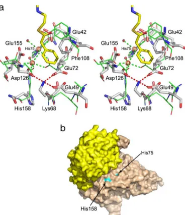
FIGURE 3. Comparison of substrate-binding sites in MMBs. a, superposi-tion of iron binding centers in PaaA (shown as sticks and labeled) and ToMOH (lines). Glu-72, Glu-155, and His-158 in PaaA in the absence of Fe2⫹are rotated
away. The “lysine bridge,” Lys-68 salt-bridged to Glu-49, Glu-72, and Asp-126, is also shown. b, surface representation of PaaAC showing that His-75 and His-158 near the di-iron center are partially solvent-exposed.
at CISTI - Natl Research Council, on March 30, 2011
www.jbc.org
replaced here by an aspartate (Fig. 3a). However, a local adjust-ment of the backbone brings the carboxylic group close to its position in the glutamate of other BMMHs. Therefore, we expect a similar di-iron center in the PaaA subunit, with one Fe2⫹ion coordinated by Glu-42, Glu-72, and His-75 and the
second Fe2⫹ion coordinated by Asp-126, Glu-155, and His-158
(Fig. 3a). In the absence of iron, the side chains of 72, Glu-155, and His-158 assume different conformations than their counterparts in MMOH and ToMOH.
PaaA contains a unique structural feature in close proximity to the iron-binding center, which we designate the “lysine bridge” (Fig. 3a). At its center is Lys-68, which forms salt bridges with three acidic residues, Glu-49, Glu-72, and Asp-126; the latter two are expected to coordinate Fe2⫹ions. The
residues of the lysine bridge belong to three ␣-helices of the ferritin-like bundle (B, C, and E). The lysine and the acidic res-idues are together conserved in all of the BMMHs homologous to PaaA with ⬎30% sequence identity. This lysine bridge may account for stabilization of the helices that form the ferritin core, which in PaaA are well ordered and have similar B-factors to other parts of the structure. MMOH, not having this feature, shows an increased flexibility of two ␣-helices of the ferritin-like four-helix bundle (E and F) in the absence of iron (33). Another possibility follows from the urease, where a lysine serves as an iron-binding ligand after carbamoylation (34).
PaaA also lacks the hydrogen bond network from the sur-face to the active center highly conserved in other BMMHs and formed by residues belonging to ␣-helices A (missing in PaaA), C, and F and two histidines from the metal-binding center (11, 14). This network was proposed to provide elec-tron transport path from the reductase to the iron-binding center of the hydroxylase. Additionally, two residues, Thr-213 and Asn-214 (MMOH numbers) from the helix E, that are conserved in BMMs and affect catalysis (10) are replaced in PaaA by Ala-129 and Ile-130, suggesting some differences in the reaction cycle. These residues are located next to an enlarged turn (-turn) of the ␣-helix E, the feature retained in PaaA.
Interactions with the Reductase—The hydroxylase compo-nent of BMMs are usually hetero-oligomers of the type (␣␥)2. However, the (PaaAC)2complex is organized differently from other BMMHs (Fig. 2, c and e). The ␣-helix A from the ␣-sub-unit and the homologous ␣-helix from the -sub␣-sub-unit, which play pivotal roles in ␣␥ dimerization, are absent in PaaA and PaaC.
This different oligomerization of (PaaAC)2brings the two
histidines of the iron-binding center very close to the protein surface (Fig. 3b). His-158 is separated from solvent by only one layer of side chains, whereas the side chain of His-75 is partially solvent-exposed in the absence of iron. Thus the di-iron center is accessible for direct electron transfer from the PaaE reduc-tase, suggesting interaction of PaaE with helices C and F of PaaA. The absence of the hydrogen bond network conserved in other BMMs and implicated in electron transfer supports this hypothesis. We hypothesize that the PaaB subunit, found to be essential for catalysis, may be directly involved in electron transport.
PA-CoA Oxygenase Is a Monooxygenase—Until recently, the product of the PA-CoA oxygenase complex was thought to be a nonaromatic dihydrodiol (VII; Fig. 1), suggesting that the PA-CoA oxygenase might be a dioxygenase (35). However, the PaaAC complex shows a typical monooxygenase structure. The formation of the dihydrodiol VII could, however, also be explained by a monooxygenase mechanism. Such a mechanism for PaaAC should account for the formation of two hydroxyl groups and the reduction of one of the double bonds of the benzene ring analo-gous to the known mechanism of complex of hydroxylase of tolu-ene 4-monooxygenase (17). Reiner and Hegeman (36) considered this mechanism in investigation of the metabolism of benzoic acid by Alcaligenes eutrophus. According to this mechanism, the PA-CoA oxygenase would operate through the 1,2-epoxide interme-diate (Fig. 1, III), which hydrolysis would give the previously expected product VII (Fig. 1, reaction H). If this was indeed the case, then VII would convert to the observed VI with the 2-OH group derived from water. We performed the reaction in H2O18
and unequivocally showed that this oxygen does not come from water but must come from O2, ruling out this proposed mecha-nism (III 3 VII 3 VI). That would suggest that observed product VI occurs by rearrangement of III. Indeed, the presence of III was recently observed directly as the product of the PA-CoA oxyge-nase (8), requiring only a monooxygeoxyge-nase activity. This is consis-tent with our data and reconciles the expected chemistry with the structure of PaaA.
Acknowledgment—We thank Allan Matte for helpful suggestions and comments on the manuscript.
REFERENCES
1. Cao, B., Nagarajan, K., and Loh, K. C. (2009) Appl. Microbiol. Biotechnol.
85, 207–228
2. Fuchs, G. (2008) Ann. N.Y. Acad. Sci. 1125, 82–99
3. Ullrich, R., and Hofrichter, M. (2007) Cell Mol. Life Sci. 64, 271–293 4. Gibson, D. T., and Parales, R. E. (2000) Curr. Opin. Biotechnol. 11,
236 –243
5. Cafaro, V., Izzo, V., Scognamiglio, R., Notomista, E., Capasso, P., Casbarra, A., Pucci, P., and Di Donato, A. (2004) Appl. Environ. Microbiol. 70, 2211–2219
6. Ferra´ndez, A., Min˜ambres, B., García, B., Olivera, E. R., Luengo, J. M., García, J. L., and Díaz, E. (1998) J. Biol. Chem. 273, 25974 –25986 7. Luengo, J. M., García, J. L., and Olivera, E. R. (2001) Mol. Microbiol. 39,
1434 –1442
8. Teufel, R., Mascaraque, V., Ismail, W., Voss, M., Perera, J., Eisenreich, W., Haehnel, W., and Fuchs, G. (2010) Proc. Natl. Acad. Sci. U.S.A. 107, 14390 –14395
9. Ferna´ndez, C., Ferra´ndez, A., Min˜ambres, B., Díaz, E., and García, J. L. (2006) Appl. Environ. Microbiol. 72, 7422–7426
10. Notomista, E., Lahm, A., Di Donato, A., and Tramontano, A. (2003) J. Mol. Evol. 56,435– 445
11. Rosenzweig, A. C., Frederick, C. A., Lippard, S. J., and Nordlund, P. (1993) Nature 366,537–543
12. Rosenzweig, A. C., Brandstetter, H., Whittington, D. A., Nordlund, P., Lippard, S. J., and Frederick, C. A. (1997) Proteins 29, 141–152 13. Sazinsky, M. H., Dunten, P. W., McCormick, M. S., DiDonato, A., and
Lippard, S. J. (2006) Biochemistry 45, 15392–15404
14. Sazinsky, M. H., Bard, J., Di Donato, A., and Lippard, S. J. (2004) J. Biol. Chem. 279,30600 –30610
15. Cafaro, V., Scognamiglio, R., Viggiani, A., Izzo, V., Passaro, I., Notomista, E., Piaz, F. D., Amoresano, A., Casbarra, A., Pucci, P., and Di Donato, A. (2002) Eur. J. Biochem. 269, 5689 –5699
Phenylacetyl-CoA Monooxygenase
at CISTI - Natl Research Council, on March 30, 2011
www.jbc.org
Natl. Acad. Sci. U.S.A. 105,19194 –19198
17. Mitchell, K. H., Rogge, C. E., Gierahn, T., and Fox, B. G. (2003) Proc. Natl. Acad. Sci. U.S.A. 100,3784 –3789
18. Batie, C. J., Ballou, D. P., and Correll, C. C. (1992) in Chemistry and Bio-chemistry of Flavoenzymes(Muller, F., ed) Vol. 3, pp. 543–556, CRC Press, Boca Raton, FL
19. Grishin, A. M., Ajamian, E., Zhang, L., and Cygler, M. (2010) Acta Crys-tallogr. Sect. F Struct. Biol. Cryst. Commun. 66,1045–1049
20. Jaganaman, S., Pinto, A., Tarasev, M., and Ballou, D. P. (2007) Protein Expression Purif. 52,273–279
21. Otwinowski, Z., and Minor, W. (1997) Methods Enzymol. 276, 307–326 22. McCoy, A. J., Grosse-Kunstleve, R. W., Adams, P. D., Winn, M. D.,
Storoni, L. C., and Read, R. J. (2007) J. Appl. Crystallogr. 40, 658 – 674 23. Vagin, A., and Teplyakov, A. (1997) J. Appl. Crystallogr. 30, 1022–1025 24. Emsley, P., and Cowtan, K. (2004) Acta Crystallogr. D60, 2126 –2132 25. Murshudov, G. N., Vagin, A. A., Lebedev, A., Wilson, K. S., and Dodson,
E. J. (1999) Acta Crystallogr. D Biol. Crystallogr. 55, 247–255
26. Chen, V. B., Arendall, W. B., 3rd, Headd, J. J., Keedy, D. A., Immormino, R. M., Kapral, G. J., Murray, L. W., Richardson, J. S., and Richardson, D. C.
27. Tagawa, K., and Arnon, D. I. (1962) Nature 195, 537–543
28. Sazinsky, M. H., and Lippard, S. J. (2006) Acc. Chem. Res. 39, 558 –566
29. Borodina, E., Nichol, T., Dumont, M. G., Smith, T. J., and Murrell, J. C. (2007) Appl. Environ. Microbiol. 73, 6460 – 6467
30. Notomista, E., Cafaro, V., Bozza, G., and Di Donato, A. (2009) Appl. En-viron. Microbiol. 75,823– 836
31. Pikus, J. D., Studts, J. M., McClay, K., Steffan, R. J., and Fox, B. G. (1997) Biochemistry 36,9283–9289
32. Leungsakul, T., Johnson, G. R., and Wood, T. K. (2006) Appl. Environ. Microbiol. 72,3933–3939
33. Sazinsky, M. H., Merkx, M., Cadieux, E., Tang, S., and Lippard, S. J. (2004) Biochemistry 43,16263–16276
34. Jabri, E., Carr, M. B., Hausinger, R. P., and Karplus, P. A. (1995) Science
268, 998 –1004
35. Ismail, W., El-Said Mohamed, M., Wanner, B. L., Datsenko, K. A., Eisen-reich, W., Rohdich, F., Bacher, A., and Fuchs, G. (2003) Eur. J. Biochem.
270, 3047–3054
36. Reiner, A. M., and Hegeman, G. D. (1971) Biochemistry 10, 2530 –2536
at CISTI - Natl Research Council, on March 30, 2011
www.jbc.org