African Great Apes Are Naturally Infected with Polyomaviruses
Closely Related to Merkel Cell Polyomavirus
䌤
†
Fabian H. Leendertz,
1*‡ Nelly Scuda,
2‡ Kenneth N. Cameron,
3§ Tonny Kidega,
4Klaus Zuberbu
¨hler,
4,5Siv Aina J. Leendertz,
1,6Emmanuel Couacy-Hymann,
7Christophe Boesch,
8Se
´bastien Calvignac,
1and Bernhard Ehlers
2*
Research Group Emerging Zoonoses, Robert Koch Institute, Berlin, Germany
1; Division of Viral Infections, Robert Koch Institut,
Berlin, Germany
2; Mountain Gorilla Veterinary Project, Inc., Baltimore, Maryland
3; Budongo Conservation Field Station,
Masindi, Uganda
4; School of Psychology, University of St. Andrews, St. Andrews, Scotland, United Kingdom
5;
Norwegian School of Veterinary Science, Oslo, Norway
6; LANADA/Laboratoire Central de la
Pathologie Animale, Bingerville, Co
ˆte d’Ivoire
7; and Department of Primatology,
Max Planck Institute for Evolutionary Anthropology, Leipzig, Germany
8The oncogenic Merkel cell polyomavirus (MCPyV) infects humans worldwide, but little is known about the
occurrence of viruses related to MCPyV in the closest phylogenetic relatives of humans, great apes. We
analyzed samples from 30 wild chimpanzees and one captive gorilla and identified two new groups of
poly-omaviruses (PyVs). These new viruses are by far the closest relatives to MCPyV described to date, providing
the first evidence of the natural occurrence of PyVs related to MCPyV in wild great apes. Similar to MCPyV,
the prevalence of these viruses is relatively high (>30%). This, together with the fact that humans in West and
Central Africa frequently hunt and butcher primates, may point toward further MCPyV-like strains spreading
to, or already existing in, our species.
Polyomaviruses (PyVs) are known to infect a wide range of
birds and mammals (31). This includes humans, from which
eight PyVs have been identified to date, namely, BK virus
(BKV) (17), JC virus (JCV) (41), KIPyV (2), WUPyV (19),
Merkel cell polyomavirus (MCPyV) (15), human PyV 6
(HPyV6) and HPyV7 (47), and a Trichodysplasia
spinulosa-associated PyV (TSV) (52). Primary PyV infection usually
oc-curs in childhood and seems to result in lifelong persistence. In
healthy humans, PyVs have not been associated with severe
acute disease. However, PyV reactivation can cause severe
diseases in the case of immunodeficiency (23).
MCPyV has been associated to (and named after) Merkel
cell carcinoma (MCC), a rare but aggressive skin cancer (15).
Since the first report of MCC in 1972 (under the former
des-ignation “trabecular carcinoma”) (51), its incidence has been
increasing (46). MCC occurs worldwide, and its association
with MCPyV was observed in studies from several continents
(4, 18, 26, 56). Benign MCPyV infection seems to be both
common (
⬎40%) and geographically widespread (47).
MCPyV’s closest relatives known to date have been
identi-fied from two nonhuman primate species: B-lymphotropic
polyomavirus (LPV; from some African green monkeys
(Chlo-rocebus aethiops) (7) and chimpanzee polyomavirus (ChPyV)
from one chimpanzee (Pan troglodytes) (24). This raises the
possibility that MCPyV is actually part of a primate-specific
subgroup. However, little is known about the natural
occur-rence of viruses related to MCPyV in the closest relatives of
humans, the African great apes.
MATERIALS AND METHODS
Sample collection and DNA isolation.Necropsy samples (spleen, lymph nodes, bone marrow, thymus, lung, liver, intestine, muscle, heart, pancreas, blood, urine; n⫽ 88) were collected from 29 chimpanzees (Pan troglodytes verus) from the Taï National Park in Coˆte d’Ivoire. The chimpanzees had died of anthrax (34), respiratory diseases (30), or other causes (35) between 2001 and 2009. Necropsy samples were also collected from one chimpanzee (Pan troglodytes schwein-furthii), which was found dead in the Budongo Forest area, Uganda, and one gorilla (Gorilla gorilla gorilla), which had been confiscated and died in the Projet Protection des Gorilles in the Republic of Congo. For all samples originating from Coˆte d’Ivoire, the sample collectors wore fully closed body protection suits and masks due to a history of ebola and anthrax in these populations and to avoid any contamination of samples with human pathogens. For the two great apes sampled in the Republic of Congo and Uganda, the sample collectors wore at a minimum single-use gloves and surgical masks (35). Permission for sample col-lection from wild primates was obtained from the authorities of national parks of each country, and tissue samples were exported with the appropriate CITES permissions from Coˆte d’Ivoire, Uganda, the Republic of Congo, and Germany. Importations took place according to German veterinary regulations for import of organic materials. All samples from Coˆte d’Ivoire were preserved in liquid nitrogen upon arrival at the research camps and were later transferred to⫺80°C at the Robert Koch Institute. Other samples were stored in RNAlater (Qiagen, Hilden, Germany). DNA was isolated using a DNeasy tissue kit (Qiagen).
PyV PCR and sequencing. (i) Generic PCR.For identification of PyVs related to MCPyV, generic nested PCR targeting a short fragment of the VP1 gene (approximately 260 bp) was performed using two pairs of degenerate and de-oxyinosine-substituted primers (see Table S1 in the supplemental material). All primers were designed to bind sites within the VP1 gene identified as being conserved among a wide range of PyVs. They were derived from the sequence of
* Corresponding author. Mailing address for Fabian H. Leendertz
(primates and zoonotic risk questions): Research Group Emerging
Zoonoses, Robert Koch Institute, Berlin, Germany. Phone: 49 18754
2592. Fax: 49 18754 2181. E-mail: leendertzf@rki.de. Mailing address
for Bernhard Ehlers (polyomavirus detection questions): Division of
Viral Infections, Robert Koch Institute, Nordufer 20, Berlin 13353,
Germany. Phone: 49 1888754 2347. Fax: 49 1888754 2598. E-mail:
ehlersb@rki.de.
† Supplemental material for this article may be found at http://jvi
.asm.org/.
‡ These authors contributed equally to the work.
§ Present address: Global Health Program, Wildlife Conservation
Society, New York, NY.
MCPyV (GenBank accession no. FJ173815) in order to bias amplification toward sequences from PyVs related to MCPyV. PCR was performed in a total volume of 25l with 0.4 l AmpliTaq Gold (Applied Biosystems), 20 pmol of each primer, 200M deoxynucleoside triphosphates, 2 mM MgCl2, and 5% dimethyl
sulfoxide. A T-Gradient thermocycler from Biometra was used with the following cycling conditions: 95°C for 12 min and 45 cycles of 95°C for 30 s, 46°C (1st round) or 50°C (2nd round) for 30 s, and 72°C for 2 min, followed by a 15-min final extension step at 72°C.
(ii) Long-distance PCR.Nested specific primers were derived from the se-quences amplified with the generic PCR (see Table S1 in the supplemental material). They were designed tail to tail for the amplification and sequencing of the remaining parts of the genomes. Nested long-distance PCR was performed with the TaKaRa-Ex PCR system, according to the instructions of the manufac-turer (Takara Bio Inc., Japan).
(iii) Diagnostic PCR.For the differential detection of chimpanzee PyVs, three primer sets were selected. The first set (VP1-ptv1) detects P. troglodytes verus PyV1a (PtvPyV1a) and PtvPyV1b, the second set (VP1-ptv2a/b) detects PtvPyV2a and -2b, and the third one (VP1-ptv2c) detects PtvPyV2c (see Table S1 in the supplemental material). They were used under the PCR conditions de-scribed above, except that AmpliTaq Gold was used at 0.2l/25-l reaction volume. Cycling conditions were conducted as follows: 95°C for 12 min and 45 cycles of 95°C for 30 s; 55°C ptv1), 58°C ptv2a/b), or 60°C (VP1-ptv2c) for 30 s; and 72°C for 1 min, followed by a 10-min final extension step at 72°C.
(iv) Testing strategy.First, the chimpanzee samples and the gorilla sample were tested with the generic PCR. Then, a core set of samples from which
sufficient spleen and lymph node aliquots were available (21 samples from 15 P. t. verus chimpanzees) was tested with the generic PCR as well as all three diagnostic, PyV-specific PCRs (Table 1). A supplementary diagnostic PCR was also used for testing of nine additional P. t. verus chimpanzees and was conducted mainly with the VP1-ptv2c PCR only, since PtvPyV2c was the first virus discov-ered in the course of this study (43 samples from nine individuals) (data not shown). Additional testing of these samples with the other diagnostic PCRs was not possible because of sample limitations.
Sequencing.All PCR products were purified by using a PCR purification kit (Qiagen) and directly sequenced with a BigDye Terminator cycle sequencing kit (Applied Biosystems, Warrington, Great Britain) in a 377 DNA automated sequencer (Applied Biosystems).
Sequence analyses.Phylogenetic analyses were performed so as to determine the positions of the newly identified strains in the Polyomaviridae family tree and decipher the relationships between the strains belonging to the clade of MCPyV-related PyVs. For that purpose, two alignments were generated and analyzed: (i) an amino acid alignment of the three main coding sequences (VP1, VP2, and large T), gathering most available polyomaviral genomes (i.e., a comprehensive sample of the overall genomic diversity of this viral family), and (ii) a nucleotide alignment of all distinct VP1 sequences from MCPyV-related PyVs (i.e., MCPyVs and the sequences depicted in this article) identified to date.
VP1, VP2, and large T amino acid sequence data set preparation and phylo-genetic analysis.VP1, VP2, and large T sequences were recovered from the complete genomes of polyomaviruses standing for the entire extent of the genetic diversity of the family (i.e., 4 avian polyomaviruses and 21 mammalian polyoma-viruses, among which are the 8 known human polyomaviruses; see Table S2 in
TABLE 1. Generic and specific PCR detection of great ape PyVs
Sample identifier Organ
PCR result
Generic PCR
Diagnostic PCR with primer set:
VP1-ptv1 VP1-ptv2a/b VP1-ptv2c
P. t. verus 1 5740
Spleen
—
a—
PtvPyV2a
—
P. t. verus 2 6446
Lymph node
—
—
—
—
P. t. verus 3 2756
Spleen
PtvPyV1b
PtvPyV1b
—
PtvPyV2c
P. t. verus 4
2293
Lymph node
—
—
—
—
5744
Lymph node
—
—
—
PtvPyV2c
2221
Spleen
—
—
—
—
5743
Spleen
—
—
—
PtvPyV2c
P. t. verus 5 5749
Spleen
—
PtvPyV1a
—
—
P. t. verus 6
3147
Spleen
—
PtvPyV1a
—
PtvPyV2c
6444
Spleen
PtvPyV1a
PtvPyV1a
—
PtvPyV2c
P. t. verus 7 4699
Spleen
—
—
—
—
P. t. verus 8 4579
Spleen
PtvPyV2b
—
PtvPyV2b
—
P. t. verus 9
6499
Lymph node
PtvPyV2c
—
—
PtvPyV2c
6498
Spleen
—
—
—
—
P. t. verus 10 6501
Spleen
PtvPyV1a
PtvPyV1a
PtvPyV2a
—
P. t. verus 11 6506
Spleen
—
—
—
PtvPyV2c
P. t. verus 12 2757
Spleen
—
—
—
—
P. t. verus 13 6433
Lymph node
PtvPyV1b
—
—
—
P. t. verus 14
6435
Lymph node
—
PtvPyV1a
—
PtvPyV2c
5779
Spleen
—
—
—
—
P. t. verus 15 6452
Spleen
—
—
—
—
P. t. schweinfurthii 5760
Spleen
PtsPyV1
NT
bNT
—
G. g. gorilla 5766
Spleen
GggPyV1
NT
NT
NT
a
—, PCR negative.
b
the supplemental material). The sequences from four MCPyVs were included in all data sets so as to account for the known diversity of the species (Asian and non-Asian MCPyV sequences were included [47]). The VP1 data set was com-pleted with one sequence previously determined from a virus infecting a chim-panzee, but the complete genome of that virus had otherwise not been sequenced (24). Finally, all sequences obtained in the course of this study were added to the data sets, irrespective of their length.
Sequences were aligned in the SeaView program (version 4) (20) using Muscle software (13, 14) and on a web server using T-Coffee software (http://www .tcoffee.org/; 40). Both methods produced alignments of similar quality; however, the T-Coffee alignments were arbitrarily retained for further analyses. From each coding sequence alignment, a protein alignment of better quality was produced using the Gblocks server (http://molevol.cmima.csic.es/castresana/Gblocks _server.html) (8, 50). This resulted in removing blocks of the alignments where the hypothesis of homology was likely to be overoptimistic, a strategy that has been proved to lead to better estimation of phylogenetic trees (50). The auto-matically cured alignments were then manually edited in SeaView before being reduced to unique sequences using the FaBox program (55). The process re-sulted in three alignments: (i) VP1 with 38 unique sequences and 203 amino acids (aa); (ii) VP2 with 28 unique sequences and 63 aa; and (iii) large T with 32 unique sequences and 407 aa. From those data sets, a concatenation which ultimately was composed of an alignment of 673 positions, including 39 unique sequences, was assembled. Preliminary phylogenetic analyses of individual align-ments supported similar topologies, in particular when it comes to our clade of interest (data not shown; individual alignments and trees are available upon request). Therefore, the concatenated alignment was used for all following anal-yses.
Model selection was performed on the concatenated data set using the Prot-Test program (version 2.4) (1). Because we intended to use the software BEAST thereafter and this software currently implements only 6 substitution matrices (JTT, MtREV, Dayhoff, WAG, CpREV, and Blosum62), likelihoods were com-puted for models using these substitution matrixes. All model add-ons (⫹I, ⫹G, or⫹I⫹G for rate heterogeneity modeling and ⫹F for use of empirical equilib-rium frequencies of amino acids) were activated. Base trees for calculations were maximum likelihood (ML) optimized (i.e., for each model, a tree was recon-structed using PhyML) (21). Likelihoods were compared according to the Akaike information criterion (AIC), resulting in selecting WAG⫹I⫹G as a model of amino acid evolution.
Phylogenetic analyses were performed in both ML and Bayesian frameworks under that model of amino acid evolution. ML analysis was performed on the PhyML web server (http://www.atgc-montpellier.fr/phyml/) (21, 22). Equilibrium frequencies, topology, and branch lengths were optimized; the starting tree was determined using the BioNJ program; and both nearest-neighbor interchange (NNI) and subtree pruning and regrafting (SPR) algorithms of tree search were used (keeping the best outcome). Branch robustness was assessed by performing nonparametric bootstrapping (500 replicates). Bayesian analyses were performed using BEAST (version 1.5.3) (12). Besides allowing modeling of the amino acid substitution process, BEAST also allows modeling of evolutionary rate variation and tree shape. Analyses were run under the assumption of a relaxed, uncorre-lated lognormal clock and two speciation models (Yule process and birth-death model). Four independent runs totaling 8,000,000 generations were performed under each speciation model. Trees and numerical values taken by all parameters were sampled every 1,000 generations. The Tracer program (version 1.5) was used to check that individual runs had reached convergence, that independent runs converged on the same zones of parameter spaces, and that chain mixing was satisfactory (global effective sample size values,⬎100) (12). Tree samples were then gathered into a single file (after removal of a visually conservative 10% burn-in period) using the LogCombiner program (version 1.5.3; distributed with BEAST), and the information from approximately 7,200 trees was summarized onto the maximum clade credibility trees using the TreeAnnotator program (version 1.5.3; distributed with BEAST). Posterior probabilities were taken as a measure of branch robustness. Ninety-five percent highest posterior density (HPD) intervals for the maximum patristic distance observed for two clades of interest were also determined from the output of BEAST analyses.
VP1 nucleotide sequence data set preparation and phylogenetic analysis.All available MCPyV VP1 sequences as well as all sequences generated for this study were aligned using the Muscle program, as implemented in SeaView. Given the good quality of the alignment, only minor manual editing of the nucleotide alignment was necessary. Reduction to unique sequences with Fabox resulted in an alignment of 1,182 positions comprising 25 sequences.
The nucleotide substitution model to which the data were a better fit was determined using the jModeltest program (version 0.1.1) (21, 44). Three substi-tution schemes (Jukes and Cantor [JC], Hasegawa, Kishino, and Yano [HKY],
and global time reversible [GTR]) were examined along with rate variation (⫹I, ⫹G, ⫹I⫹G) and base frequency (⫹F) modeling. Base trees for calculations were maximum likelihood optimized (21). According to the AIC, comparisons of model likelihoods were more favorable to a GTR⫹G model.
Phylogenetic analyses were performed in both maximum likelihood and Bayes-ian frameworks under that model of nucleotide evolution. ML and BayesBayes-ian analyses were performed as described above, with the exceptions that in Bayesian analysis tree shape was modeled by a coalescent process (constant population size or exponential growth) and that two experiments were run according to each model for 4,000,000 generations total (accordingly, ca. 3,500 trees constituted the final samples).
For both data sets, ML and Bayesian methods globally supported congruent topologies with consistent branch supports (even though bootstrap [Bp] and posterior probability [pp] are not directly comparable [11]), as did different speciation and coalescent models. All .xml files (including sequence alignments) used for Bayesian analyses are available upon request. Figures summarizing phylogenetic analyses were drawn using the FigTree program (version 1.3.1; http://tree.bio.ed.ac.uk/software/figtree/).
Polyomaviral species delineation.So as to put the extent of the genetic diver-sity of the newly identified ape strains in an appropriate context, we compared it to the known genetic diversity prevailing in four human polyomaviruses, all of which are currently considered constituting viral species (MCPyV, BKV, JCV, and WUPyV). For this, we took the VP1-coding sequence as a proxy of the global genomic divergence of polyomaviruses.
In addition to the VP1 nucleotide alignment, which already encompassed the known genetic diversity of MCPyV, three new data sets were assembled: (i) BKV sequences spanning the entire BKV genetic diversity were taken from a study of Krumbholz et al. (32). Representatives of the seven clades identified in this study were included, resulting in an alignment of 1,089 positions comprising 26 se-quences. All sequences were unique. (ii) JCV sequences representing the overall genetic diversity of the species were taken from a study of Kitchen et al. (29). VP1 sequences from the 92 sequences chosen by these authors to compose four main regional groups (Asia, Europe, Japan, and the Americas) were gathered, resulting in an alignment of 1,065 positions. The latter was then reduced to the 64 unique sequences that it comprised. (iii) WUPyV sequences were drawn from a study of Bialasiewicz et al. (6). A total of 19 sequences representing the six clades described in this study were gathered, constituting an alignment of 1,110 positions. From those sequences, 10 could be identified to be unique.
For each data set, pairwise distances were first computed using SeaView. As pairwise distances are, in general, poor indicators of true evolutionary distances (36), we also computed patristic (tree) distances. For this, all data sets were analyzed along a common pipeline. The model of nucleotide substitution to which the data set was a best fit was first determined using jModeltest, as detailed above (for BKV, GTR⫹I; for JCV, GTR⫹I⫹G; for WUPyV, GTR⫹I). Then the maximum likelihood tree was determined using PhyML (as implemented in Seaview) under that model. Finally, patristic distances were computed from that tree using the software Patristic (16). From both pairwise and patristic distance matrices, maximum distances within known species were determined. Minimum patristic distances separating newly identified strains from their closest relatives were finally identified.
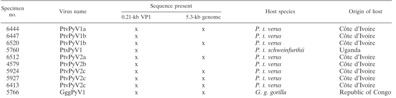
Provisional nomenclature and abbreviations of novel polyomaviruses.Names and abbreviations for newly detected PyVs were formed from host genus and species names and numbered arbitrarily following the results of the species delineation, e.g., G. g. gorilla PyV1 (GggPyV1). They are listed in Table 2. Published PyVs that were used in the analyses are listed in Table S2 in the supplemental material.
Nucleotide sequence accession numbers.PyV sequences determined in the course of this study have been deposited in GenBank under the following ac-cession numbers: for PtvPyV1a, HQ385746; for PtvPyV1b, HQ326775 and HQ385747; for PtsPyV1, HQ326774; for PtvPyV2a, HQ385748; for PtvPyV2b, HQ326776; for PtvPyV2c, HQ385749 to HQ385751; and for GggPyV1, HQ385752.
RESULTS
A generic nested PCR strategy aimed at amplifying a
frag-ment of the VP1 gene from MCPyV-related viral strains was
employed to test necropsy samples of 25 wild chimpanzees and
one gorilla. Fifteen chimpanzees (14/24 P. t. verus chimpanzees
and 1/1 P. t. schweinfurthii chimpanzee) and the gorilla were
PCR positive. In all cases, sequencing of PCR products
con-firmed their infection with PyVs. For 13 chimpanzees (12 P. t.
verus chimpanzees and 1 P. t. schweinfurthii chimpanzee) and
the gorilla, preliminary BLAST analyses gave MCPyV as a first
hit (3). These MCPyV-like viruses are the focus of the present
study (information on the other chimpanzee PyVs will be
pub-lished elsewhere).
On the basis of the MCPyV-like VP1 sequences obtained
using the first protocol, nested specific primers (see Table S1 in
the supplemental material) were used for long-distance PCR
amplification and sequencing of the remaining parts of the
genomes. Seven complete genome sequences, six from
chim-panzees (P. t. verus) and one from the gorilla (Table 2), could
be determined. All genomes exhibited the typical set of PyV
open reading frames (i.e., VP1, VP2, VP3, large T, and
small T) and lacked any agnoprotein open reading frame.
Genome analysis of great ape PyVs.
A high overall degree of
sequence similarity of the novel great ape PyVs to MCPyV,
including in regions otherwise less conserved among PyVs, was
observed (see Table S3 in the supplemental material). Analysis
of the noncoding control region (NCCR), the most variable
region of the PyV genome, revealed a high degree of similarity
of the great ape PyVs to MCPyV. An important motif of this
region is the DNA element GAGGC and its complement,
GCCTC. Repeats of these motifs are considered
large-T-anti-gen-binding sites (reviewed in reference 25). The highest
num-ber of these elements is found in the NCCRs of MCPyV
isolates (n
⫽ 7 to 8), which are to be compared to those of
simian virus 40 (SV40), BKV, JCV (n
⫽ 6), and LPV (n ⫽ 4).
In MCPyV, these elements are also present in one or two
overlapping, palindromic octamers which possibly affect
bind-ing of T-antigen hexamers and initiation of DNA replication
(33). Interestingly, the novel African great ape PyVs also
har-bor eight or nine elements and three palindromic octamers
(Fig. 1).
The large T proteins exhibit a high degree of conservation of
functional domains (CR1, J, RB-binding, ori-binding, and Zn
finger domains) (43). Additionally, a region of approximately
180 amino acids extending from the J domain to the
ori-bind-ing domain rich in serine, glutamine, and threonine and
pos-sibly affecting RB-binding function (25) is also highly
con-served among the novel PyVs and MCPyVs but is absent from
LPV, BKV, JCV, and SV40 (see Fig. S1 in the supplemental
material).
In addition to its function as a virion structural protein, the
major capsid protein VP1 plays an important role during the
initial interaction of PyVs with host cells through its outfacing
loops (BC, HI) (48, 49) and contains antigenic domains (39).
Modifications in PyV VP1 loops have been shown to alter host
specificity (38). Therefore, the striking similarity between the
great ape PyVs and MCPyV in VP1, even in the highly variable
loop regions, is particularly supportive of a close relationship
between great ape PyVs and MCPyV (data not shown).
Phylogenetic analysis.
Phylogenetic trees inferred from an
alignment of concatenated VP1, VP2, and large T sequences
(673 aa) comprising most known PyVs evidenced the close
relationship of wild great ape PyVs to MCPyV, which
alto-gether formed a highly supported monophyletic group (Bp and
pp values, 100 and 1, respectively) (Fig. 2). Within this group,
TABLE 2. Polyomaviruses detected in wild great apes
Specimen
no. Virus name
Sequence present
Host species Origin of host
0.21-kb VP1 5.3-kb genome
6444
PtvPyV1a
x
x
P. t. verus
Co
ˆte d’Ivoire
6447
PtvPyV1b
x
P. t. verus
Co
ˆte d’Ivoire
6520
PtvPyV1b
x
x
P. t. verus
Co
ˆte d’Ivoire
5760
PtsPyV1
x
P. t. schweinfurthii
Uganda
6512
PtvPyV2a
x
x
P. t. verus
Co
ˆte d’Ivoire
4579
PtvPyV2b
x
P. t. verus
Co
ˆte d’Ivoire
5924
PtvPyV2c
x
x
P. t. verus
Co
ˆte d’Ivoire
5927
PtvPyV2c
x
x
P. t. verus
Co
ˆte d’Ivoire
6413
PtvPyV2c
x
x
P. t. verus
Co
ˆte d’Ivoire
5766
GggPyV1
x
x
G. g. gorilla
Republic of Congo
FIG. 1. Noncoding control region of great ape PyVs. DNA sequences of the discovered great ape PyVs and a selection of published sequences
from MCPyVs and other PyVs were compared using the ClustalW program (as implemented in the MacVector program, version 10.6) with
additional manual adjustments. Base 1 corresponds to base 5316 of the MKL-1 isolate of MCPyV (GenBank accession no. FJ173815). Conserved
regions are outlined with solid lines. The part of the alignment comprising the great ape PyVs and the MCPyVs is boxed with a dotted line.
T-antigen-binding elements (GAGGC or the complement, GCCTC) are boxed with red (GAGGC) and blue (GCCTC) lines.
the maximum patristic distance was estimated to stand
be-tween 0.119 and 0.270 aa substitution per site (95% HPD
interval), while the maximum patristic distance to members of
the sister clade comprising primate polyomavirus sequences
was assessed as lying at between 0.539 and 0.910 aa substitution
per site (95% HPD interval) (Fig. 2). Of note, MCPyV was
firmly established as monophyletic (Bp, 91; pp, 1; Fig. 2).
Phylogenetic trees were also inferred from a nucleotide
alignment of 1,182 bp so as to refine the picture of MCPyV-like
virus relationships (all known unique MCPyV sequences were
included). This confirmed the MCPyV monophyly (Bp, 100;
pp, 1) and found support for the existence of at least two clades
of wild great ape PyVs (Fig. 3). The most basal one comprised
sequences from the gorilla and chimpanzees of the subspecies
P. t. verus only (GggPyV1 and PtvPyV2 clade). GggPyV1
ap-peared to have diverged first from all other sequences of this
clade (Bp, 82; pp, 1; Fig. 3). All other sequences (PtvPyV1 and
PtsPyV1 clade) identified from chimpanzees (P. t. verus and P.
t. schweinfurthii) belonged to another clade which was found to
be in sistership with the MCPyV clade (Fig. 3). Within this
clade, the sequence determined from a P. t. schweinfurthii
individual was always found to be the first to diverge, though
with moderate statistical support.
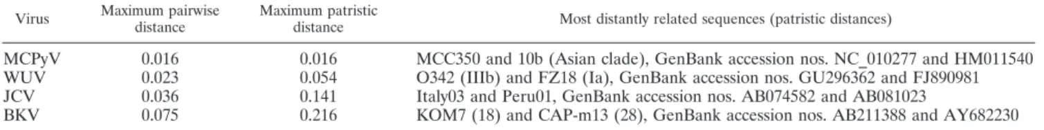
Maximum VP1 patristic distances within recognized human
polyomavirus species varied from 0.016 (MCPyV) to 0.216
(BKV) nucleotide substitution per site (Table 3). All strains
belonging to the clades PtvPyV1/PtsPyV1, PtvPyV2, and
Ggg-PyV1 exhibited minimum patristic distances from the other
clades greater than 0.216 (respective minima, 0.354, 0.272, and
0.272 nucleotide substitution per site, respectively; Table 4).
Within those species, subclades were defined that gather
se-quences whose patristic distances do not outscore the
maxi-FIG. 2. Bayesian chronogram deduced from the analysis of a 673-amino-acid concatenation of VP1, VP2, and large T sequences. PyVs
identified from human hosts are in red, PyVs from chimpanzees in blue, and PyV from a gorilla in green. Ninety-five percent HPD intervals of
maximum patristic distances are indicated in parentheses for two clades (lines are drawn to the corresponding nodes). The clade formed by
MCPyVs and the newly described ape PyVs is highlighted. Statistical support for branches is given where Bp values are
ⱖ70 and pp values are
ⱖ0.95. Bp values are shown below the branches, and pp values are shown above the branches (pp values are those obtained from analyses
performed under the Yule model of speciation). The scale axis is in number of amino acid substitution per site. This chronogram was rooted using
a relaxed clock. A maximum likelihood analysis of the same data set concluded a similar topology, and thus, the results are not shown here. It is
noted that when only VP1 sequences were considered, ChPyV was found to be the sister taxon of the MCPyV-related PyV clade.
mum patristic distance observed between MCPyVs (PtvPyV1a
and -b, PtsPyV1, PtvPyV2a to -c).
Prevalence of great ape PyVs.
Since diverse groups of PyVs
were identified in chimpanzees, three specific PCR tests
(di-rected against PtvPyV1a/b, PtvPyV2a/b, or PtvPyV2c; see
Ta-ble S1 in the supplemental material) were used to get insight
into their respective prevalence and to determine the extent of
coinfection. All PCR products were sequenced to check for
proper clade and subclade identification. In the core sample
set (21 samples from 15 individuals), PtvPyV1 was detected in
6/15 individuals, PtvPyV2a/b was detected in 3/15 individuals,
and PtvPyV2c was detected in 6/15 individuals, corresponding
to prevalence rates of approximately 40%, 20%, and 40%,
respectively. Four of 15 individuals were coinfected (2 with
PtvPyV1a/2c, 1 with PtvPyV1b/2c, and 1 with PtvPyV1a/2a;
Table 1). Furthermore, PtvPyV2c-specific PCR was performed
FIG. 3. Bayesian chronogram deduced from the analysis of a 1,182-nucleotide VP1 alignment. PyVs identified from human hosts are in red,
PyVs from chimpanzees in blue, and PyV from a gorilla in green. Statistical support for branches is given where Bp values are
ⱖ70 and pp values
are
ⱖ0.95. Bp values are shown below the branches, and pp values are shown above the branches (pp values are those obtained from analyses
performed under the constant-population-size model of coalescence). The scale axis is in number of nucleotide substitution per site. This
chronogram was rooted using a relaxed clock (midpoint rooting of the maximum likelihood tree identified the same root). A maximum likelihood
analysis of the same data set concluded a similar topology, and thus, the results are not shown here.
TABLE 3. Maximum pair-wise and patristic distances observed in four human polyomaviral species
Virus Maximum pairwise distance
Maximum patristic
distance Most distantly related sequences (patristic distances)
MCPyV
0.016
0.016
MCC350 and 10b (Asian clade), GenBank accession nos. NC_010277 and HM011540
WUV
0.023
0.054
O342 (IIIb) and FZ18 (Ia), GenBank accession nos. GU296362 and FJ890981
JCV
0.036
0.141
Italy03 and Peru01, GenBank accession nos. AB074582 and AB081023
on nine additional individuals, among which three were found
to be positive (not listed). Together with the data from the core
set (Table 1), PtvPyV2c was thus detected in 9/24 individuals
(38%).
DISCUSSION
Here PyVs infecting wild African great apes were discovered
and shown to be closely related to human MCPyV using
ge-nome analysis (Fig. 1; see Fig. S1 and Table S3 in the
supple-mental material) and phylogenetic analysis (Fig. 2 and 3).
These findings are particularly interesting since they
convinc-ingly demonstrate that MCPyV stems from a (so far)
primate-specific and even ape-primate-specific group of PyVs.
Though this might at first be seen as an argument in favor of
the cospeciation hypothesis (42), two diverging interpretations
of our phylogenetic trees can be made that end up with
con-trasting conclusions about the processes at play along
MCPyV-related PyV evolution. If three distinct MCPyV-MCPyV-related PyVs
whose present descendants would be MCPyV, PtvPyV2/GggPyV1,
and Ptv/PtsPyV1, respectively, are assumed to have been
in-fecting the last common ancestor of African great apes, then
the fact that along two of the corresponding evolutionary
lin-eages (those leading to PtvPyV2/GggPyV1 and Ptv/PtsPyV1)
species-specific patterns are not contradicted would indeed be
consistent with the codivergence hypothesis. On the contrary,
if the last common ancestor of all African great apes is
as-sumed to have been infected with only one MCPyV-related
PyV (the last common ancestor of all MCPyV-related PyVs),
then strict codivergence is an unsatisfactory explanation for the
observed phylogenetic pattern (i.e., a bad overlap of viral and
host phylogenies) and some degree of host switching has to be
assumed as well. Our data do not allow favoring one hypothesis
over the other. Therefore, the question of the modalities of
evolution of PyVs related to MCPyV remains largely open.
The finding of new viral strains that cannot be identified
directly as belonging to a recognized species raises the
ques-tion of their taxonomic status (i.e., are they likely to represent
new viral species?). Viral species delineation is often
contro-versial (54), including among the Polyomaviridae (31). In
par-ticular, when only sequences are available, it is impossible to
define any objective threshold beyond which genetic
diver-gence undoubtedly reflects the existence of separate viral
spe-cies (54). Here we find that three entities—two clades (Ptv/
PtsPyV1 and PtvPyV2) and a single sequence (GggPyV1)—
present minimum patristic distances to their closest outgroup
relatives greater than the maximum patristic distance observed
within BKV, the human polyomavirus with the highest degree
of genetic diversity known thus far. On the basis of this
obser-vation, we propose to provisionally consider those three
enti-ties as separate viral species (or at least as separate taxa of the
same rank as BKV). This should obviously not be taken as
implying that they have different biological properties, whose
characterization will be required to truly install those groups of
viruses as valid viral species (45, 54).
This being said, it is striking that PyVs related to MCPyV
circulating in chimpanzees are, from a genetic standpoint,
much more diverse than the human MCPyVs described so far
(and are actually even more diverse than any human PyV), all
the more so since nearly the entire extent of this diversity could
be found in a single P. t. verus chimpanzee community of the
Taï National Park, Co
ˆte d’Ivoire. This could be interpreted as
indicating that West Africa (and possibly Central Africa) is a
hot spot of MCPyV-related PyV diversity. These data may
point toward a higher degree of genetic diversity of MCPyV in
humans worldwide and specifically in West and Central Africa,
where conclusive studies with human populations are missing
(47).
Though our analyses do not provide direct evidence that
MCPyV was transmitted from apes to humans, the presence of
PyVs related to MCPyV in wild great apes is also significant
with respect to human health. SV40 and LPyV are much more
distantly related to MCPyV than PtvPyV1, PtvPyV2, or
Ggg-PyV1. However, they seem to infect humans, as suggested by
their identification in human samples (7, 10, 27, 37). If the
assumption is made that the closer that two species are the
higher that the likelihood of successful transmission of their
respective pathogens is (the preferential host switching
hy-pothesis) (9), then transmission of great ape PyVs to sympatric
humans must be considered. Besides, the practical
determi-nants of a successful transmission to human populations are
already in place. First, according to our data set, PtvPyV is
infecting chimpanzees (including coinfections with various
strains) at a high prevalence rate, at least in the Taï Forest,
Co
ˆte d’Ivoire. Second, nonhuman primates (including
chim-panzees and gorillas) still represent an important proportion of
the bush meat consumed in West and Central Africa (ca. 12%)
(5). Hunting and butchering of bush meat have been shown to
provide the major routes of pathogen transmission to humans
(e.g., human immunodeficiency viruses) (28, 53). This could
well apply to PyVs related to MCPyV.
The wide variety of MCPyV-related viruses in African great
apes presented here calls for larger studies to unravel the
diversity of PyVs related to MCPyV currently circulating in
nonhuman primates of West and Central Africa (and more
particularly in apes) as well as in local human populations. It is
reasonable to assume that such studies will reveal
unprece-dented levels of PyV diversity, which will provide a sound basis
TABLE 4. Minimum patristic distance involving the new
great ape PyVs
PyV Minimum patristic distance Closest relative(s) (patristic distance) Minimum patristic distance to other groups of sequences
6444_PtvPyV1a
0.043
a6520_PtvPyV1b
0.354
b6447_PtvPyV1b
0.010
6520_PtvPyV1b
6520_PtvPyV1b
0.010
6447_PtvPyV1b
5760_PtsPyV1c
0.078
6520_PtvPyV1b
6512_PtvPyV2a
0.028
4579_PtvPyV2b
0.272
4579_PtvPyV2b
0.028
6512_PtvPyV2a
5924_PtvPyV2c
0.002
5927_PtvPyV2c
5927_PtvPyV2c
0.002
5924_PtvPyV2c
6413_PtvPyV2c
0.006
5927_PtvPyV2c
5766_GggPyV1
0.272
6512_PtvPyV2a
0.272
aValues outscoring MCPyV maximum patristic distance are italicized.
b
Values outscoring the maximum patristic distance observed in a human PyV (i.e., 0.216 substitution per site for BKV) are in boldface.
for a better assessment of both PyV natural history and the risk
that nonhuman primate PyVs really pose.
ACKNOWLEDGMENTS
We thank the Ivorian authorities for long-term support, especially
the Ministry of the Environment and Forests, as well as the Ministry of
Research, the directorship of the Taï National Park, the Swiss
Re-search Center in Abidjan, the Uganda Wildlife Authority, and the
Uganda National Council for Science and Technology for granting us
permission to conduct this research. We also thank the CITES
author-ities of Co
ˆte d’Ivoire, Uganda, Republic of Congo, and Germany for
permission. We are grateful to S. Schenk, S. Metzger, and the field
assistants of the Taï chimpanzee project and T. King and C.
Cham-berlain of the Aspinall Foundation for sample collection; F.
Babwe-teera and R. Wittig for their support; and D. Wevers, S. Liebmann, C.
Walter, N. Yasmum, S. Ko
¨ndgen, A. Blasse, and C. Hedemann for
technical support in the laboratory.
The Budongo Conservation Field Station receives core funding from
the Royal Zoological Society of Scotland. This work was supported
by the Robert Koch Institute and the Max Planck Society.
REFERENCES
1. Abascal, F., R. Zardoya, and D. Posada. 2005. ProtTest: selection of best-fit models of protein evolution. Bioinformatics 21:2104–2105.
2. Allander, T., K. Andreasson, S. Gupta, A. Bjerkner, G. Bogdanovic, M. A. Persson, T. Dalianis, T. Ramqvist, and B. Andersson.2007. Identification of a third human polyomavirus. J. Virol. 81:4130–4136.
3. Altschul, S. F., W. Gish, W. Miller, E. W. Myers, and D. J. Lipman. 1990. Basic local alignment search tool. J. Mol. Biol. 215:403–410.
4. Becker, J. C., R. Houben, S. Ugurel, U. Trefzer, C. Pfohler, and D. Schrama. 2009. MC polyomavirus is frequently present in Merkel cell carcinoma of European patients. J. Investig. Dermatol. 129:248–250.
5. Bennett, E. L., E. Blencowe, K. Brandon, D. Brown, R. W. Burn, G. Cowl-ishaw, G. Davies, H. Dublin, J. E. Fa, E. J. Milner-Gulland, J. G. Robinson, J. M. Rowcliffe, F. M. Underwood, and D. S. Wilkie.2007. Hunting for consensus: reconciling bushmeat harvest, conservation, and development policy in West and Central Africa. Conserv. Biol. 21:884–887.
6. Bialasiewicz, S., R. Rockett, D. W. Whiley, Y. Abed, T. Allander, M. Binks, G. Boivin, A. C. Cheng, J. Y. Chung, P. E. Ferguson, N. M. Gilroy, A. J. Leach, C. Lindau, J. W. Rossen, T. C. Sorrell, M. D. Nissen, and T. P. Sloots. 2010. Whole-genome characterization and genotyping of global WU poly-omavirus strains. J. Virol. 84:6229–6234.
7. Brade, L., N. Muller-Lantzsch, and H. zur Hausen. 1981. B-lymphotropic papovavirus and possibility of infections in humans. J. Med. Virol. 6:301–308. 8. Castresana, J. 2000. Selection of conserved blocks from multiple alignments
for their use in phylogenetic analysis. Mol. Biol. Evol. 17:540–552. 9. Charleston, M. A., and D. L. Robertson. 2002. Preferential host switching by
primate lentiviruses can account for phylogenetic similarity with the primate phylogeny. Syst. Biol. 51:528–535.
10. Delbue, S., S. Tremolada, E. Branchetti, F. Elia, E. Gualco, E. Marchioni, R. Maserati, and P. Ferrante.2008. First identification and molecular charac-terization of lymphotropic polyomavirus in peripheral blood from patients with leukoencephalopathies. J. Clin. Microbiol. 46:2461–2462.
11. Douady, C. J., F. Delsuc, Y. Boucher, W. F. Doolittle, and E. J. Douzery. 2003. Comparison of Bayesian and maximum likelihood bootstrap measures of phylogenetic reliability. Mol. Biol. Evol. 20:248–254.
12. Drummond, A. J., and A. Rambaut. 2007. BEAST: Bayesian evolutionary analysis by sampling trees. BMC Evol. Biol. 7:214.
13. Edgar, R. C. 2004. MUSCLE: a multiple sequence alignment method with reduced time and space complexity. BMC Bioinform. 5:113.
14. Edgar, R. C. 2004. MUSCLE: multiple sequence alignment with high accu-racy and high throughput. Nucleic Acids Res. 32:1792–1797.
15. Feng, H., M. Shuda, Y. Chang, and P. S. Moore. 2008. Clonal integration of a polyomavirus in human Merkel cell carcinoma. Science 319:1096–1100. 16. Fourment, M., and M. J. Gibbs. 2006. PATRISTIC: a program for
calculat-ing patristic distances and graphically comparcalculat-ing the components of genetic change. BMC Evol. Biol. 6:1.
17. Gardner, S. D., A. M. Field, D. V. Coleman, and B. Hulme. 1971. New human papovavirus (B.K.) isolated from urine after renal transplantation. Lancet i:1253–1257.
18. Garneski, K. M., A. H. Warcola, Q. Feng, N. B. Kiviat, J. H. Leonard, and P. Nghiem.2009. Merkel cell polyomavirus is more frequently present in North American than Australian Merkel cell carcinoma tumors. J. Investig. Dermatol. 129:246–248.
19. Gaynor, A. M., M. D. Nissen, D. M. Whiley, I. M. Mackay, S. B. Lambert, G. Wu, D. C. Brennan, G. A. Storch, T. P. Sloots, and D. Wang.2007. Identi-fication of a novel polyomavirus from patients with acute respiratory tract infections. PLoS Pathog. 3:e64.
20. Gouy, M., S. Guindon, and O. Gascuel. 2010. SeaView version 4: a multi-platform graphical user interface for sequence alignment and phylogenetic tree building. Mol. Biol. Evol. 27:221–224.
21. Guindon, S., and O. Gascuel. 2003. A simple, fast, and accurate algorithm to estimate large phylogenies by maximum likelihood. Syst. Biol. 52:696–704. 22. Guindon, S., F. Lethiec, P. Duroux, and O. Gascuel. 2005. PHYML
on-line—a web server for fast maximum likelihood-based phylogenetic infer-ence. Nucleic Acids Res. 33:W557–W559.
23. Jiang, M., J. R. Abend, S. F. Johnson, and M. J. Imperiale. 2009. The role of polyomaviruses in human disease. Virology 384:266–273.
24. Johne, R., D. Enderlein, H. Nieper, and H. Muller. 2005. Novel polyomavirus detected in the feces of a chimpanzee by nested broad-spectrum PCR. J. Virol. 79:3883–3887.
25. Johnson, E. M. 2010. Structural evaluation of new human polyomaviruses provides clues to pathobiology. Trends Microbiol. 18:215–223.
26. Katano, H., H. Ito, Y. Suzuki, T. Nakamura, Y. Sato, T. Tsuji, K. Matsuo, H. Nakagawa, and T. Sata.2009. Detection of Merkel cell polyomavirus in Merkel cell carcinoma and Kaposi’s sarcoma. J. Med. Virol. 81:1951–1958. 27. Kean, J. M., S. Rao, M. Wang, and R. L. Garcea. 2009. Seroepidemiology of
human polyomaviruses. PLoS Pathog. 5:e1000363.
28. Keele, B. F., F. Van Heuverswyn, Y. Li, E. Bailes, J. Takehisa, M. L. San-tiago, F. Bibollet-Ruche, Y. Chen, L. V. Wain, F. Liegeois, S. Loul, E. M. Ngole, Y. Bienvenue, E. Delaporte, J. F. Brookfield, P. M. Sharp, G. M. Shaw, M. Peeters, and B. H. Hahn.2006. Chimpanzee reservoirs of pan-demic and nonpanpan-demic HIV-1. Science 313:523–526.
29. Kitchen, A., M. M. Miyamoto, and C. J. Mulligan. 2008. Utility of DNA viruses for studying human host history: case study of JC virus. Mol. Phylo-genet. Evol. 46:673–682.
30. Kondgen, S., H. Kuhl, P. K. NⴕGoran, P. D. Walsh, S. Schenk, N. Ernst, R. Biek, P. Formenty, K. Matz-Rensing, B. Schweiger, S. Junglen, H. Ellerbrok, A. Nitsche, T. Briese, W. I. Lipkin, G. Pauli, C. Boesch, and F. H. Leendertz. 2008. Pandemic human viruses cause decline of endangered great apes. Curr. Biol. 18:260–264.
31. Krumbholz, A., O. R. Bininda-Emonds, P. Wutzler, and R. Zell. 2009. Phy-logenetics, evolution, and medical importance of polyomaviruses. Infect. Genet. Evol. 9:784–799.
32. Krumbholz, A., P. Wutzler, and R. Zell. 2008. The non-coding region of BK subtype II viruses. Virus Genes 36:27–29.
33. Kwun, H. J., A. Guastafierro, M. Shuda, G. Meinke, A. Bohm, P. S. Moore, and Y. Chang.2009. The minimum replication origin of Merkel cell poly-omavirus has a unique large T-antigen loading architecture and requires small T-antigen expression for optimal replication. J. Virol. 83:12118–12128. 34. Leendertz, F. H., H. Ellerbrok, C. Boesch, E. Couacy-Hymann, K. Matz-Rensing, R. Hakenbeck, C. Bergmann, P. Abaza, S. Junglen, Y. Moebius, L. Vigilant, P. Formenty, and G. Pauli.2004. Anthrax kills wild chimpanzees in a tropical rainforest. Nature 430:451–452.
35. Leendertz, F. H., G. Pauli, K. Maetz-Rensing, W. Boardmann, C. Nunn, H. Ellerbrok, S. A. Jensen, S. Junglen, and C. Boesch.2006. Pathogens as drivers of population declines: the importance of systematic monitoring in great apes and other threatened mammals. Biol. Conserv. 131:325–337. 36. Lefebure, T., C. J. Douady, M. Gouy, and J. Gibert. 2006. Relationship
between morphological taxonomy and molecular divergence within Crusta-cea: proposal of a molecular threshold to help species delimitation. Mol. Phylogenet. Evol. 40:435–447.
37. Martini, F., A. Corallini, V. Balatti, S. Sabbioni, C. Pancaldi, and M. Tog-non.2007. Simian virus 40 in humans. Infect. Agents Cancer 2:13. 38. Mezes, B., and P. Amati. 1994. Mutations of polyomavirus VP1 allow in vitro
growth in undifferentiated cells and modify in vivo tissue replication speci-ficity. J. Virol. 68:1196–1199.
39. Murata, H., B. Teferedegne, L. Sheng, A. M. Lewis, Jr., and K. Peden. 2008. Identification of a neutralization epitope in the VP1 capsid protein of SV40. Virology 381:116–122.
40. Notredame, C., D. G. Higgins, and J. Heringa. 2000. T-Coffee: a novel method for fast and accurate multiple sequence alignment. J. Mol. Biol. 302:205–217.
41. Padgett, B. L., D. L. Walker, G. M. ZuRhein, R. J. Eckroade, and B. H. Dessel.1971. Cultivation of papova-like virus from human brain with pro-gressive multifocal leucoencephalopathy. Lancet i:1257–1260.
42. Perez-Losada, M., R. G. Christensen, D. A. McClellan, B. J. Adams, R. P. Viscidi, J. C. Demma, and K. A. Crandall.2006. Comparing phylogenetic codivergence between polyomaviruses and their hosts. J. Virol. 80:5663– 5669.
43. Pipas, J. M. 1992. Common and unique features of T antigens encoded by the polyomavirus group. J. Virol. 66:3979–3985.
44. Posada, D. 2008. jModelTest: phylogenetic model averaging. Mol. Biol. Evol. 25:1253–1256.
45. Pringle, C. R. 1991. The 20th Meeting of the Executive Committee of the ICTV. Virus species, higher taxa, a universal virus database and other mat-ters. Arch. Virol. 119:303–304.
46. Pulitzer, M. P., B. D. Amin, and K. J. Busam. 2009. Merkel cell carcinoma: review. Adv. Anat. Pathol. 16:135–144.
Buck.2010. Merkel cell polyomavirus and two previously unknown polyoma-viruses are chronically shed from human skin. Cell Host Microbe 7:509–515. 48. Stehle, T., and S. C. Harrison. 1997. High-resolution structure of a poly-omavirus VP1-oligosaccharide complex: implications for assembly and re-ceptor binding. EMBO J. 16:5139–5148.
49. Stehle, T., Y. Yan, T. L. Benjamin, and S. C. Harrison. 1994. Structure of murine polyomavirus complexed with an oligosaccharide receptor fragment. Nature 369:160–163.
50. Talavera, G., and J. Castresana. 2007. Improvement of phylogenies after removing divergent and ambiguously aligned blocks from protein sequence alignments. Syst. Biol. 56:564–577.
51. Toker, C. 1972. Trabecular carcinoma of the skin. Arch. Dermatol. 105:107– 110.
52. van der Meijden, E., R. W. Janssens, C. Lauber, J. N. Bouwes Bavinck, A. E. Gorbalenya, and M. C. Feltkamp.2010. Discovery of a new human
poly-omavirus associated with trichodysplasia spinulosa in an immunocompro-mised patient. PLoS Pathog. 6:e1001024.
53. Van Heuverswyn, F., Y. Li, C. Neel, E. Bailes, B. F. Keele, W. Liu, S. Loul, C. Butel, F. Liegeois, Y. Bienvenue, E. M. Ngolle, P. M. Sharp, G. M. Shaw, E. Delaporte, B. H. Hahn, and M. Peeters.2006. Human immunodeficiency viruses: SIV infection in wild gorillas. Nature 444:164.
54. Van Regenmortel, M. H. 2010. Logical puzzles and scientific controversies: the nature of species, viruses and living organisms. Syst. Appl. Microbiol. 33:1–6.
55. Villesen, P. 2007. FaBox: an oline tool for FASTA sequences. Mol. Ecol. Notes 7:965–968.
56. Wieland, U., C. Mauch, A. Kreuter, T. Krieg, and H. Pfister. 2009. Merkel cell polyomavirus DNA in persons without Merkel cell carcinoma. Emerg. Infect. Dis. 15:1496–1498.