HAL Id: hal-02627969
https://hal.inrae.fr/hal-02627969
Submitted on 26 May 2020
HAL is a multi-disciplinary open access
archive for the deposit and dissemination of
sci-entific research documents, whether they are
pub-lished or not. The documents may come from
teaching and research institutions in France or
abroad, or from public or private research centers.
L’archive ouverte pluridisciplinaire HAL, est
destinée au dépôt et à la diffusion de documents
scientifiques de niveau recherche, publiés ou non,
émanant des établissements d’enseignement et de
recherche français ou étrangers, des laboratoires
publics ou privés.
Distributed under a Creative Commons Attribution| 4.0 International License
Optimization and evaluation of a non-invasive tool for
peste des petits ruminants surveillance and control
Arnaud Bataille, Olivier Kwiatek, Salima Belfkhi, Lucile Mounier, Satya
Parida, Mana Mahapatra, Alexandre Caron, Chobi Clement Chubwa, Julius
Keyyu, Richard Kock, et al.
To cite this version:
Arnaud Bataille, Olivier Kwiatek, Salima Belfkhi, Lucile Mounier, Satya Parida, et al.. Optimization
and evaluation of a non-invasive tool for peste des petits ruminants surveillance and control. Scientific
Reports, Nature Publishing Group, 2019, 9, 8 p. �10.1038/s41598-019-41232-y�. �hal-02627969�
optimization and evaluation of
a non-invasive tool for peste des
petits ruminants surveillance and
control
Arnaud Bataille
1,2, Olivier Kwiatek
1,2, Salima Belfkhi
1,2, Lucile Mounier
2, Satya parida
3,
Mana Mahapatra
3, Alexandre Caron
2,4,5, Chobi Clement Chubwa
6, Julius Keyyu
7,
Richard Kock
8, Bryony A. Jones
8& Geneviève Libeau
1,2Peste des petits ruminants (PPR) is a highly contagious and devastating viral disease affecting mainly sheep and goats, but also a large number of wild species within the order Artiodactyla. A better understanding of PPR transmission dynamics in multi-host systems is necessary to efficiently control the disease, in particular where wildlife and livestock co-occur. Notably, the role of wildlife in PPR epidemiology is still not clearly understood. Non-invasive strategies to detect PPR infection without the need for animal handling could greatly facilitate research on PPR epidemiology and management of the disease in atypical hosts and in complex field situations. Here, we describe optimized methods for the direct detection of PPR virus genetic material and antigen in fecal samples. We use these methods to determine the detection window of PPR in fecal samples, and compare the sensitivity of these methods to standard invasive sampling and PPR diagnostic methods using field samples collected at a wildlife-livestock interface in Africa. Our results show that quantitative reverse transcription PCR (RT-QPCR) amplification of PPRV from fecal swabs has good sensitivity in comparison to ocular swabs. Animals infected by PPRV could be identified relatively early on and during the whole course of infection based on fecal samples using RT-QPCR. Partial gene sequences could also be retrieved in some cases, from both fecal and ocular samples, providing important information about virus origin and relatedness to other PPRV strains. Non-invasive strategies for PPRV surveillance could provide important data to fill major gaps in our knowledge of the multi-host PPR epidemiology.
Peste des petits ruminants (PPR) is a highly contagious and devastating disease caused by a virus of the genus Morbillivirus in the family Paramyxoviridae1. PPR virus (PPRV) affects mainly sheep and goats, but also a large
number of wild species within the order Artiodactyla1,2. PPR occurrence in livestock must be notified to the
World Organization for Animal Health (OIE) and the disease is now the target of a global eradication campaign3.
PPR is endemic in large parts of Africa, the Middle East and Asia, and is still spreading globally, with emergence notably reported in Georgia4, Mongolia5, and most recently within the European Union in Bulgaria6.
Better understanding of PPR transmission dynamics is necessary to efficiently control the disease. Notably, the role of wildlife in PPR epidemiology is still not clearly understood7,8. Importantly, the potential for spill-over
PPR infection between wildlife and livestock and the direction of these spill-overs are still poorly studied. Also, the capacity of PPR virus to be transmitted and maintained within wildlife populations without a domestic host source has still not yet been demonstrated. The role of wildlife is particularly important to explore in Africa and Central-South Asia including the Himalayas where multi-host systems composed of a large diversity and population of wild ungulate species and domestic populations co-occur. A range of wildlife-livestock interfaces
1CIRAD, UMR ASTRE, F-34398, Montpellier, France. 2ASTRE, Univ Montpellier, CIRAD, INRA, Montpellier,
France. 3The Pirbright Institute, Ash Road, Pirbright, Woking, Surrey, UK. 4CIRAD, UMR ASTRE, RP-PCP, Maputo,
Mozambique. 5Faculdade de Veterinaria, Universidade Eduardo Mondlane, Maputo, Mozambique. 6Ngorongoro
District Council, Arusha, Tanzania. 7Tanzania Wildlife Research Institute, Arusha, Tanzania. 8Royal Veterinary College,
Received: 21 November 2018 Accepted: 5 March 2019 Published: xx xx xxxx
www.nature.com/scientificreports
www.nature.com/scientificreports/
provides ample opportunities for virus sharing between wild and domestic hosts. So far, the knowledge about the role of wildlife in PPR epidemiology, recently reviewed8,9, is limited to outbreaks from ex-situ populations
in zoos and fenced enclosures, and to rare recent in situ epidemics affecting mountain goats10–13 and, on one
occasion, free-ranging antelope and wild caprines in Mongolia14. Transmission of PPR between domestic and
non-domestic species could hinder control of the disease in some areas. Importantly, recent mass mortality of saïga antelopes in Mongolia15 due to PPR infection showed that this disease also represents a serious threat to
some critically endangered wildlife populations14.
Despite the need for more data, wildlife disease surveys are rare because animal capture is very costly and requires complicated logistics in remote areas. Permits for protected animals are also difficult to obtain for inva-sive sampling and sample transportation across borders. Even for domestic animals, it may be hard to obtain samples in some regions if farmers are reluctant to have their animals handled and tested for PPR infection. Non-invasive strategies to detect PPR infection without the need for animal capture and handling could greatly facilitate research on PPR epidemiology in atypical hosts (e.g. wildlife) and in complex field situations.
Non-invasive samples, notably fecal samples, have been used to study wildlife ecology and health for more than two decades16. Fecal samples are now also being used to explore virus diversity in wildlife17,18. Several studies
have shown that PPR antigen and RNA can be detected in feces of infected goats for weeks after infection19–21.
However no study so far has looked into optimizing diagnostic methods for PPR detection in fecal material and compared the sensitivity of such methods with standard diagnosis from invasive sampling. Indeed, robust commercial serological and virological diagnostic kits are available to detect PPR infection, but they were mainly developed for domestic small ruminants (goat and sheep) and for high quality, invasive samples. These methods need to be adapted for PPR virus detection from fecal samples.
Here, we describe optimized methods for the detection of PPR virus genetic material and antigen in fecal samples. We use these methods to determine the detection window of PPR virus in fecal samples, and compare the sensitivity of these methods to standard invasive sampling and PPR diagnostic methods using field samples collected in Africa at the wildlife-livestock interface.
Results
Optimization of PPR antigen detection in fecal samples.
Preliminary tests performed with fecal samples spiked with serially diluted PPR vaccine showed that fecal samples contained inhibitors that lowered the sensitivity of reverse transcription Polymerase Chain Reaction (RT-PCR) and Enzyme-linked immunosorbent (ELISA) assays used for the diagnosis of PPR (see Supplementary Information). Viral RNA extraction with mag-netic beads increased the sensitivity of downstream quantitative RT-PCR assays (RT-QPCR), compared to a col-umn-based RNA extraction approach. Our preliminary tests also showed that some RT-PCR kits appeared to be less sensitive to inhibitors from fecal samples than others. Increasing incubation time for antigen capture ELISA (AgELISA) assay on fecal samples also increased the sensitivity of the assays without compromising specificity. Optimized protocols were developed to detect PPRV in fecal samples using RT-QPCR, RT-PCR, and AgELISA based on these preliminary results (see Supplementary Information).Detection window of PPR viral antigen in rectal fecal material.
We evaluated the time window of PPR excretion in rectal fecal material using rectal swabs collected daily from day 0 post infection (dpi) to 14 dpi during a previously described experimental challenge on Saanen goats22. We focused on samples collected fromfour individuals infected by an intranasal route with a highly virulent PPR strain (Morocco 2008) as this route of infection simulates natural infection. PPRV was detected in rectal swabs from 5 dpi until the end of the experi-ment (14 dpi) using our optimized RT-QPCR method (Fig. 1). PPRV was also detected in fecal swabs by antigen capture ELISA, but the first positive result was obtained at 6 dpi, with strong optical density values obtained from 7 dpi. Antigen capture ELISA results became negative at 12 dpi for one individual, while rectal swabs taken at 12 to 14 dpi from the other 3 animals were still positive, although close to the threshold (Fig. 1).
PPR viral antigen detection on field fecal samples.
As part of a larger study on the role of wild-life in PPR transmission, suspected cases of PPR were investigated in domestic sheep and goat flocks kept in close proximity to wild herbivores in the Ngorongoro district in the Serengeti ecosystem of northern Tanzania in 2015. Conjunctival swabs were collected from animals with clinical signs of PPR infection (see Table S1 in Supplementary Information), and tested in the field for presence of PPR antigen using the lateral flow device (LFD) Peste-Test Rapid Field Test for PPRV (BDSL Irvine, UK). Further conjunctival and nasal swabs were tested in the laboratory of the Pirbright Institute for the presence of PPRV genetic material using published RT-PCR23and RT-QPCR24 methods targeting a portion of the PPR N gene. If present, fecal samples were collected by gloved
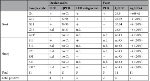
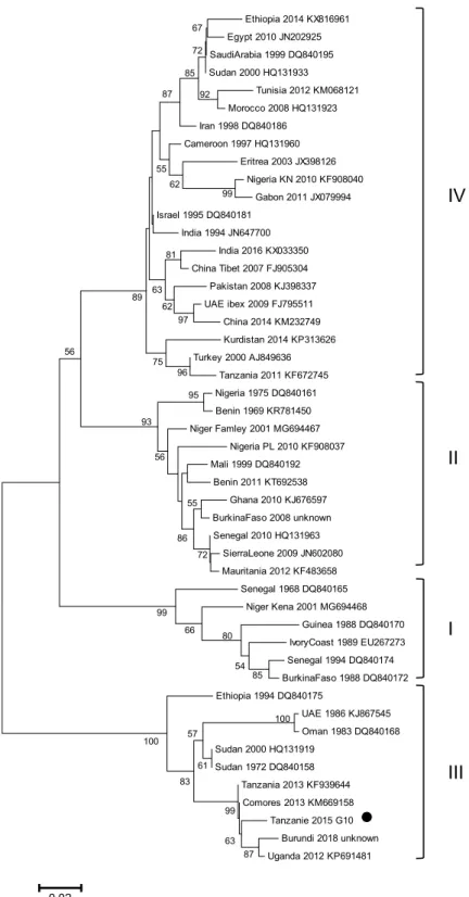
hand from the rectum of the same animals, and were tested for presence of PPRV using RT-QPCR, RT-PCR and AgELISA methods optimized during this study. We analysed fecal samples collected from five goats and six sheep. Viral genetic material could be detected in fecal samples by RT-QPCR in all goats tested positive by RT-PCR and RT-QPCR on ocular swabs (Table 1). Only one out of six sheep (animal S14) was found positive by PCR and LFD on ocular swabs whereas all sheep fecal samples remained negative. Only two fecal samples that tested positive by RT-QPCR were found positive by RT-PCR and AgELISA. These two samples had relatively high Ct values (Ct: 23–27) by RT-QPCR (Table 1). The PCR products obtained after RT-PCR were sequenced and aligned to publicly available partial N gene sequences to identify the genetic lineage using phylogenetic analysis. Sequences retrieved from ocular swabs and fecal samples were identical (Genbank accession number MK201803), and were positioned within the lineage III of PPRV in the phylogenetic tree (Fig. 2).
Discussion and Conclusion
Non-invasive samples, especially fecal samples, could represent a very useful tool to detect PPR in wildlife and, to a lesser extent, domestic ruminants from remote and complex wildlife-livestock interfaces. Easier and cheaper sampling in complex field situations would provide the means to fill in major gaps in our knowledge of PPR epidemiology, increasing our chances of successful eradication of this disease25,26. Our results based on infection
experiments and domestic animals at the wildlife-livestock interface suggest that fecal samples may offer a good alternative to standard ocular or nasal swabs, when such invasive sampling is difficult to obtain.
Results from infection experiments suggest that shedding of PPR virus in feces starts five days after infection. Ocular excretion of the virus starts three days earlier22, so fecal samples have some disadvantages in this regard.
However, PPRV genetic material could be detected in rectal swabs by RT-QPCR until the end of the experiment, confirming results obtained in previous studies19,20 and suggesting that animals infected by the virus could be
identified relatively early on and during the whole course of infection based on fecal samples.
We put in place optimized protocols to detect PPR genetic material and antigens in fecal samples despite the presence of inhibitors. These protocols may be implemented in any laboratory with standard equipment. Samples obtained from domestic animals at the wildlife-livestock interface in the Serengeti ecosystem in northern Tanzania gave us the opportunity to test our protocols in a field situation. RT-QPCR results from fecal samples were similar to results obtained from ocular swabs, confirming that high quantity of virus genetic material can be retrieved in the field from feces of infected animals. PPRV RNA could be detected in ocular swabs of one sheep
Figure 1. Kinetics of viral shedding in fecal material of PPRV infected goats. Viral shedding was monitored by (a) RT-QPCR (expressed in Ct, limit of detection = 40 Ct) and (b) antigen capture ELISA (expressed in Sample/
Positive Control % following the manufacturers’ instructions, limit of detection = 20%). Samples were collected from four Saanen goats infected by an intranasal route with the highly virulent Peste des Petits Ruminants strain Morocco 200822. Fecal material was tested for presence of PPRV genetic material from day 4 post infection (dpi)
www.nature.com/scientificreports
www.nature.com/scientificreports/
performed on this animal when virus was shed in ocular excretions but not yet in fecal excretions. The fecal sam-ple may also have been degraded during transport. These results may also suggest that the issue of PCR inhibition in fecal samples, although reduced with our optimized method, has not been completely resolved. In all cases, these potential issues have to be kept in mind when interpreting diagnostic results.
PPRV RNA and antigen could also be retrieved from fecal material using RT-PCR and antigen capture ELISA. However, sensitivity of these two methods is lower than RT-QPCR. Also, the window of detection for antigens appears to be shorter and more variable, probably due to production of antibodies by infected animals. Still, these methods may be a useful addition to the RT-QPCR method. RT-PCR is especially interesting, as our results show that partial gene sequence can be retrieved from fecal material after amplification, providing important informa-tion about virus origin and relatedness to other PPRV strains27. In this study, sequencing results obtained from
both ocular swabs and fecal sample confirm the circulation in 2015 of the lineage III of PPRV in the Northern Tanzania. This lineage has been present in the region for several years7,28. Most likely, full genome of PPRV could
also be retrieved from fecal samples of infected animals, although concentration and quality of viral RNA would need to be high29.
In our field study, fecal material was directly collected from the rectum, possibly increasing our chances to detect PPRV material from rectal epithelium. Capacity of PPRV detection from excreted fecal material is likely to be more variable, but this study shows that chances are high to obtain virus sample from feces of infected ani-mals without the need to catch them. However, fresh fecal samples would need to be collected to ensure a good quality of viral RNA. In addition, PPRV is quickly inactivated in the environment30, so fecal samples needs to be
retrieved within 1–2 hours of excretion if one hopes to detect PPRV genetic material from this source. Thus fecal sampling for PPRV detection still involves particular logistics to follow closely animals and ensure collection of fresh samples and to have the necessary means to store the samples under optimal cold storage conditions until shipment to a laboratory. This being said, carefully designed protocols on wildlife species implemented in col-laboration with staff trained in wildlife ecology and/or management could easily ensure fresh collection of fecal samples after visual observation of the deposition, together with characteristics of the sampled individuals such as gender, age, body condition and reproductive status, which could prove important to relate PPRV infection to host traits. The choice of individuals and herds to sample should also be based on observation of clinical signs or on epidemiological data suggesting circulation of PPR in the area (for example, outbreak in domestic animals).
This study offers the first research data on how fecal samples could be exploited for non-invasive PPR detec-tion. Past attempts by our laboratory to detect PPR antibodies in fecal samples have failed so far (data not shown), suggesting that non-invasive strategies for PPR serological surveys may be harder to develop. On the other hand, some trials suggests that penside tests can detect PPR antigen in feces of domestic small ruminants31, but also of
wildlife such as saiga antelopes (R. Kock, personal communication). Implementing such tests in a routine manner would permit fast non-invasive diagnosis of PPR infection in the field, and therefore quick response of veterinary and wildlife services. PPR has a major socio-economic impact across many regions of the globe32, and represents
a threat to some endangered wildlife2. We will need all the tools available to better understand and control the
disease. Non-invasive strategies to sample PPR represent an important addition to this toolbox. Sample code
Ocular swabs Feces
PCR QPCR LFD antigen test PCR QPCR AgELISA
Goat G4 + no Ct + + 26.9 +(68%) G10 + 22.96 + + 22.92 +(120%) G11 + 36.96 + − 35.64 −(<20%) G16 n.d. 26.37 n.d. − 26.9 −(<20%) G74§ − no Ct n.d. n.d. no Ct −(<20%) Sheep S14 + no Ct + n.d. no Ct −(<20%) S19 n.d. no Ct n.d. n.d. no Ct −(<20%) S20 n.d. no Ct n.d. n.d. no Ct −(<20%) S31 n.d. no Ct − − no Ct −(<20%) S33 − no Ct n.d. n.d. no Ct −(<20%) S37* n.d. no Ct n.d. n.d. no Ct −(<20%) Total 11 6 11 5 5 11 11 Total positive 4 3 4 2 4 2
Table 1. Summary of results obtained from multiple diagnostic methods for PPRV genetic material or antigen
with ocular swabs or fecal material. Results for reverse transcription Polymerase Chain Reaction (PCR) and lateral flow device (LFD) antigen test are indicated as positive (+) or negative (−) status. Results for quantitative reverse transcription Polymerase Chain Reaction (QPCR) are presented in Ct values, with lower Ct values indicating presence of higher amount of target PPR genetic material and “no Ct” indicating no detection of PPR genetic material. Results of the antigen capture ELISA (AgELISA) are presented as positive (+) or negative (−) status, with OD values converted to Sample/Positive Control % following the manufacturers’ instructions between brackets. Tissue from oral lesions (*) or post-mortem samples (§) were collected instead of ocular
Material and Methods
Fecal samples from suspected cases of PPR in the field.
Reports of suspected PPR cases in domestic mixed sheep and goat flocks in Ngorongoro District of northern Tanzania were investigated. Animals with clinical signs of pyrexia, lacrimation, nasal discharge and/or mouth lesions were selected for diagnostic testing. Sampling was designed in accordance to guidelines and regulations of the Royal Veterinary College Ethics Committee andEthiopia 2014 KX816961 Egypt 2010 JN202925 SaudiArabia 1999 DQ840195 Sudan 2000 HQ131933 Tunisia 2012 KM068121 Morocco 2008 HQ131923 Iran 1998 DQ840186 Cameroon 1997 HQ131960 Eritrea 2003 JX398126 Nigeria KN 2010 KF908040 Gabon 2011 JX079994 Israel 1995 DQ840181 India 1994 JN647700 India 2016 KX033350 China Tibet 2007 FJ905304 Pakistan 2008 KJ398337 UAE ibex 2009 FJ795511 China 2014 KM232749 Kurdistan 2014 KP313626 Turkey 2000 AJ849636 Tanzania 2011 KF672745 Nigeria 1975 DQ840161 Benin 1969 KR781450 Niger Famley 2001 MG694467 Nigeria PL 2010 KF908037 Mali 1999 DQ840192 Benin 2011 KT692538 Ghana 2010 KJ676597 BurkinaFaso 2008 unknown Senegal 2010 HQ131963 SierraLeone 2009 JN602080 Mauritania 2012 KF483658 Senegal 1968 DQ840165 Niger Kena 2001 MG694468 Guinea 1988 DQ840170 IvoryCoast 1989 EU267273 Senegal 1994 DQ840174 BurkinaFaso 1988 DQ840172 Ethiopia 1994 DQ840175 UAE 1986 KJ867545 Oman 1983 DQ840168 Sudan 2000 HQ131919 Sudan 1972 DQ840158 Tanzania 2013 KF939644 Comores 2013 KM669158 Tanzanie 2015 G10 Burundi 2018 unknown Uganda 2012 KP691481 0,02
IV
II
I
III
67 72 85 87 92 55 62 99 81 63 62 97 89 56 75 96 95 93 56 55 86 72 99 66 80 54 85 100 100 57 61 83 99 63 87Figure 2. PPRV N gene phylogenetic analysis. Neighbour-joining tree showing the relationship between the
partial N gene sequence obtained in this study (indicated by black dot) and sequences publically available in GenBank (indicated by the country and sample name, the year and the GenBank Accession Number). The numbers at the nodes are bootstrap values obtained from 1000 replicates. Only bootstrap values > 50 are shown.
www.nature.com/scientificreports
www.nature.com/scientificreports/
were collected from animals with pyrexia and tested by PPR rapid diagnostic test. Conjunctival and nasal swabs were collected for conventional23 and quantitative PCR24 assays at the Pirbright Institute. Fecal samples were
col-lected by rectal examination from animals which had feces present in the rectum. All samples were put into a cool box with ice packs for transportation to the field base where they were stored in a freezer at −20C, until further transportation by cool box to the TAWIRI laboratory in Arusha where they were stored at −80C. Samples were shipped to Pirbright and CIRAD in dry ice.
Optimization of PPRV diagnostic from fecal samples.
Details of the methods used to detect and reduce the effect of inhibitors and increase sensitivity of RT-PCR, RT-QPCR, and ELISA assay performed on rectal swabs and fecal samples to detect the presence of PPR virus are provided as Supplementary Information. The optimized protocol is described below.Fecal sample preparation.
For RT-PCR, fecal samples were ground in 3 ml of Minimum Essential Media (MEM, 10% W/V) with 0.2 μm glass beads, and then centrifuged 3 min at 1000 g to collect supernatant. RNA was extracted from 150 μl of supernatant using the ID gene MAG universal extraction kit (IDvet genetics, Grabels, France) and a KingFisher automated extractor (ThermoFisher, IDvet genetics, France), following the manufac-turers’ instructions. For the antigen capture ELISA, fecal sample were ground in 3 ml (10% W/V) of the buffer 13 of the IDscreen PPR antigen capture ELISA kit (IDvet, Grabels, France) with 0.2 μm glass beads and then centri-fuged 3 min at 1000 g to collect supernatant.Detection of PPR genetic material in fecal samples.
A one-step RT-QPCR method was used to amplify the partial end of the N protein gene33, with the qscript XLT kit one-step RT-qPCR ToughMix (Quantabio, VWR,Fontenay-sous-Bois, France). The amplification cycle consisted of a reverse transcription step of 45 °C for 10 min, an initial denaturation at 95 °C for 10 min, followed by 40 cycles of 95 °C for 15 s and 60 °C for 45 s. Serially diluted stand-ard controls were included in the RT-QPCR runs to validate the test and obtain an estimation of the number of copy of PPR N protein in the samples. The RT-QPCR runs were performed on a LightCycler instrument (Roche, IDvet genet-ics, Montpellier, France). A RT-PCR was performed on samples found positive by RT-QPCR analysis. The qScript XLT One-Step RT-PCR Kit (Quantabio, VWR, Fontenay-sous-Bois, France) was used to amplify a 351 base pair (bp) segment of the PPRV N gene with the NP3/NP4 (Forward NP3: 5′-GTC-TCG-GAA-ATC-GCC-TCA-CAG-ACT-3′ and Reverse NP4: 5′-CCT-CCT-CCT-GGT-CCT-CCA-GAA-TCT-5′-GTC-TCG-GAA-ATC-GCC-TCA-CAG-ACT-3′) diagnostic primers modified from23. The
amplification cycle consisted of a reverse transcription step of 48 °C for 20 min, an initial denaturation at 94 °C for 3 min, followed by 40 cycles of 94 °C for 15 s and 60 °C for 30 sec and a final extension step at 72 °C for 1 min. PCR products were resolved on a 1.5% agarose gel to reveal the expected band size. Positive and negative controls were included in the RT-PCR runs to validate the test.
Enzyme-linked immunosorbent assay (ELISA) on fecal samples.
Fecal samples were tested for pres-ence of viral particles using the IDscreen PPR antigen capture ELISA (IDvet, Grabels, France). The assays were performed and analyzed following the manufacturer’s instructions except for the incubation step that was done overnight at room temperature to maximize the sensitivity of the test (see Supplementary Methods). Optical density (OD) values at 450 nm were recorded with a Sunrise ELISA reader (Tecan, Lyon, France). OD values were converted to S/P % following the manufacturers’ instructions. According to the cut-off value of the test, test sam-ples with S/P values ≥ 20% were considered positive. Presence of PPR antibodies in fecal samsam-ples was tested using the IDscreen PPR competition ELISA (IDvet, Grabels, France). The assays were performed and analyzed follow-ing the manufacturer’s instructions except for the incubation step that was done overnight at room temperature to maximize the sensitivity of the test (see Supplementary Methods). OD values were converted to percent competi-tion (PC). According to the cut-off value of the test, test samples with PC values ≤ 50% were considered positive.Data Availability
All data generated or analysed during this study are included in this published article (and its Supplementary Information files).
References
1. Baron, M. D., Diallo, A., Lancelot, R. & Libeau, G. Peste des Petits Ruminants virus in Advances in Virus Research (Academic Press) Eds Kielian, M., Maramorosch, K. & Mettenleiter, T. C. Vol 95, 1–42 (2016).
2. Aziz-ul, R., Wensman, J. J., Abubakar, M., Shabbir, M. Z. & Rossiter, P. Peste des petits ruminants in wild ungulates. Trop Anim
Health Prod. 50, 1815–1819 (2018).
3. OIE & FAO Global control and eradication of PPR, http://www.oie.int/eng/PPR2015/doc/PPR-Advocacy-EN.pdf. Accessed on 05 Jan 2017 (2015).
4. Donduashvili, M. et al. Identification of peste des petits ruminants virus, Georgia, 2016. Emerg Infect Dis. 24, 1576–1578 (2018). 5. OIE. World Animal Health Information System, http://www.oie.int/wahis_2/public/wahid.php/Reviewreport/Review?page_
refer=MapFullEventReport&reportid=20934. Accessed on 18 Jan 2017 (2017).
6. OIE. World Animal Health Information System, http://www.oie.int/wahis_2/public/wahid.php/Reviewreport/Review/ viewsummary?fupser=&dothis=&reportid=27029. Accessed on 14 Aug 2018 (2018).
7. Mahapatra, M. et al. Spillover of Peste des Petits Ruminants virus from domestic to wild ruminants in the Serengeti ecosystem, Tanzania. Emerg Infect Dis. 21, 2230–2234 (2015).
8. Munir, M. Role of wild small ruminants in the epidemiology of peste des petits ruminants. Transbound Emerg Dis. 61, 411–424 (2014).
9. Parida, S. et al. Peste des petits ruminants. Vet Microbiol. 181, 90–106 (2015).
10. Abubakar, M., Rajput, Z. I., Arshed, M. J., Sarwar, G. & Ali, Q. Evidence of peste des petits ruminants virus (PPRV) infection in Sindh Ibex (Capra aegagrus blythi) in Pakistan as confirmed by detection of antigen and antibody. Trop Anim Health Prod. 43, 745–747 (2011).
11. Hoffmann, B. et al. Fatalities in wild goats in Kurdistan associated with peste des petits ruminants virus. Transbound Emerg Dis. 59, 173–176 (2012).
12. Li, J. et al. Diagnosis of peste des petits ruminants in wild and domestic animals in Xinjiang, China, 2013–2016. Transbound Emerg
Dis. 64, e43–e47 (2017).
13. Marashi, M. et al. Peste des petits ruminants virus in vulnerable wild small ruminants, Iran, 2014–2016. Emerg Infect Dis. 23, 704–706 (2017).
14. Aguilar, X. F. et al. PPR virus threatens wildlife conservation. Science. 362, 165–166 (2018).
15. Shatar, M. et al. First genetic characterization of peste des petits ruminants virus from Mongolia. Arch Virol. 162, 3157–3160 (2017). 16. Waits, L. P. & Paetkau, D. Noninvasive genetic sampling tools for wildlife biologists: a review of applications and recommendations
for accurate data collection. J Wildl Manag. 69, 1419–1433 (2005).
17. Vibin, J. et al. Metagenomics detection and characterisation of viruses in faecal samples from Australian wild birds. Sci Rep. 8, 8686 (2018).
18. Bodewes, R. et al. Viral metagenomic analysis of feces of wild small carnivores. Virol J. 11, 89 (2014).
19. Ezeibe, M. C. O. et al. Persistent detection of peste de petits ruminants antigen in the faeces of recovered goats. Trop Anim Health
Prod. 40, 517–519 (2008).
20. Wasee Ullah, R. et al. Detection of peste des petits ruminants viral RNA in fecal samples of goats after an outbreak in Punjab province of Pakistan: A longitudinal study. BioMed Res Int. 2016, 1486824 (2016).
21. Kumar, S. K. et al. Molecular characterisation of lineage IV peste des petits ruminants virus using multi gene sequence data. Vet
Microbiol. 174, 39–49 (2014).
22. Enchery, F. et al. Development of a PPRV challenge model in goats and its use to assess the efficacy of a PPR Vaccine. Vaccine 37, 1667–1673 (2019).
23. Couacy-Hymann, E. et al. Rapid and sensitive detection of peste des petits ruminants virus by a polymerase chain reaction assay. J
Virol Methods. 100, 17–25 (2002).
24. Batten, C. A. et al. A real time RT-PCR assay for the specific detection of peste des petits ruminants virus. J Virol Methods. 171, 401–404 (2011).
25. Albina, E. et al. Peste des petits ruminants, the next eradicated animal disease? Vet Microbiol. 165, 38–44 (2013).
26. Baron, M. D., Diop, B., Njeumi, F., Willett, B. J. & Bailey, D. Future research to underpin successful peste des petits ruminants virus (PPRV) eradication. J Gen Virol. 98, 2635–2644 (2017).
27. Libeau, G., Diallo, A. & Parida, S. Evolutionary genetics underlying the spread of peste des petits ruminants virus. Animal Front. 4, 14–20 (2014).
28. Kgotlele, T. et al. Partial genetic characterization of peste des petits ruminants virus from goats in northern and eastern Tanzania.
Transbound Emerg Dis. 61, 56–62 (2014).
29. Oude Munnink, B. B. et al. Characterization of Posa and Posa-like virus genomes in fecal samples from humans, pigs, rats, and bats collected from a single location in Vietnam. Virus Evol. 3, vex022 (2017).
30. Kumar, N. et al. Peste des petits ruminants virus infection of small ruminants: a comprehensive review. Viruses. 6, 2287–2327 (2014). 31. Baron, J. et al. Development and testing of a field diagnostic assay for peste des petits ruminants virus. Transbound Emerg Dis. 61,
390–396 (2014).
32. Jones, B. A. et al. The economic impact of eradicating peste des petits ruminants: A benefit-cost analysis. PLoS One. 11, e0149982 (2016).
33. Kwiatek, O. et al. Quantitative one-step real-time RT-PCR for the fast detection of the four genotypes of PPRV. J Virol Methods. 165, 168–77 (2010).
Acknowledgements
The research was supported by a grant from the European Commission Animal Health and Welfare European Research Area Network for the IUEPPR Project “Improved Understanding of Epidemiology of PPR” in the framework of ANIHWA 2013, and by a grant (SI2.756606) from the European Commission Directorate General for Health and Food Safety awarded to the European Union Reference Laboratory for Peste des Petits Ruminants (EURL-PPR). Co-authors from the Pirbright Institute and from Royal Veterinary College were funded by the Biotechnology and Biological Sciences Research Council (grants BB/L013657/1 and BB/L013592/1, respectively). The Ngorongo Conservation Area Authority (Tanzania), and especially Dr Kuya Sayalel provided their assistance in sampling collection and permission in the area. The CIRAD thanks its collaborators at Boeringher-Ingelheim for granting permission to use samples acquired during an infection experiment realized in the frame of a collaborative project between CIRAD and Boeringher-Ingelheim. The CIRAD also thank the Montpellier zoo (France), and especially Laurie Virolle, for providing fecal material from its animals.
Author Contributions
A.B. and G.L. supervised the study and wrote the manuscript. O.K., S.B., M.M. and L.M. performed laboratory diagnostic procedures at CIRAD. M.M. performed laboratory diagnostic procedures at the Pirbright Institute. C.C.C., J.K., A.C., B.J. and R.K. recovered the material for the study. S.P. is coordinator of the IUEPPR Project “Improved Understanding of Epidemiology of PPR”. All Authors read and approved the final version of the manuscript.
Additional Information
Supplementary information accompanies this paper at https://doi.org/10.1038/s41598-019-41232-y.
Competing Interests: The authors declare no competing interests.
Publisher’s note: Springer Nature remains neutral with regard to jurisdictional claims in published maps and
www.nature.com/scientificreports
www.nature.com/scientificreports/
Open Access This article is licensed under a Creative Commons Attribution 4.0 International
License, which permits use, sharing, adaptation, distribution and reproduction in any medium or format, as long as you give appropriate credit to the original author(s) and the source, provide a link to the Cre-ative Commons license, and indicate if changes were made. The images or other third party material in this article are included in the article’s Creative Commons license, unless indicated otherwise in a credit line to the material. If material is not included in the article’s Creative Commons license and your intended use is not per-mitted by statutory regulation or exceeds the perper-mitted use, you will need to obtain permission directly from the copyright holder. To view a copy of this license, visit http://creativecommons.org/licenses/by/4.0/.