Application of microtechnologies for the
vascularization of engineered tissues
The MIT Faculty has made this article openly available.
Please share
how this access benefits you. Your story matters.
Citation
Gauvin, Robert et al. “Application of Microtechnologies for the
Vascularization of Engineered Tissues.” Vascular Cell 3.1 (2011): 24.
As Published
http://dx.doi.org/10.1186/2045-824X-3-24
Publisher
BioMed Central Ltd
Version
Final published version
Citable link
http://hdl.handle.net/1721.1/69018
Terms of Use
Creative Commons Attribution
R E V I E W
Open Access
Application of microtechnologies for the
vascularization of engineered tissues
Robert Gauvin
1,2,3,4, Maxime Guillemette
1, Mehmet Dokmeci
1,2and Ali Khademhosseini
1,2,3*Abstract
Recent advances in medicine and healthcare allow people to live longer, increasing the need for the number of organ transplants. However, the number of organ donors has not been able to meet the demand, resulting in an organ shortage. The field of tissue engineering has emerged to produce organs to overcome this limitation. While tissue engineering of connective tissues such as skin and blood vessels have currently reached clinical studies, more complex organs are still far away from commercial availability due to pending challenges with in vitro engineering of 3D tissues. One of the major limitations of engineering large tissue structures is cell death resulting from the inability of nutrients to diffuse across large distances inside a scaffold. This task, carried out by the vasculature inside the body, has largely been described as one of the foremost important challenges in engineering 3D tissues since it remains one of the key steps for both in vitro production of tissue engineered construct and the in vivo integration of a transplanted tissue. This short review highlights the important challenges for vascularization and control of the microcirculatory system within engineered tissues, with particular emphasis on the use of microfabrication approaches.
Keywords: microfabrication, biomimetic approaches, modular assembly
Introduction
Progress in the development of large tissue-engineered organs has so far been limited by the inability to gener-ate sophisticgener-ated three dimensional (3D) structures com-prised of a functional vasculature. The vascular system is a dynamic environment comprised of a variety of cell types that constantly remodels itself under the influence of endothelial, immune, nervous and endocrine cells [1]. Vascular growth and remodeling are coupled with devel-opmental and wound healing processes as well as the progression of various pathologies such as inflammation, cardiovascular diseases and cancer. Most of these pro-cesses depend on endothelial cells, which line the inter-ior of blood vessels and form the endothelium. This interface between circulating blood and the surrounding tissues is responsible for proper solute transport and molecular exchange. It ensures the delivery of sufficient oxygen and nutrients to cells to maintain tissue homeos-tasis. Cells in vivo are found to be at most a few
hundred microns away from the nearest capillary or blood vessel. Beyond this distance, diffusion is not effec-tive and tends to reduce cell survival and function. Therefore, the inability to adequately vascularize engi-neered tissues results in inefficient transport of nutrients and metabolites and often leads to cell death and tissue necrosis. Moreover, vascularization of engineered tissues plays an important role in graft perfusion and is also crucial in facilitating the integration of the implanted material with the host vasculature [2,3].
The fabrication of tissue constructs often requires cell seeding of 3D scaffolds. These scaffolds are gener-ally made of gels, foams or fibrous meshes and usugener-ally have basic macroscale properties that enable cell adhe-sion, migration and proliferation [4]. Although these properties are often sufficient to allow the formation of functional connective tissues such as skin [5,6], bladder [7] and cornea [8,9] and 3D tubular structures like blood vessels [10,11] and urinary tract [12], most tissue engineering approaches still lack the capability to sustain the growth of thick engineered organs [13]. Skin and cartilage have been among the first engi-neered tissues ready for clinical applications since they
* Correspondence: alik@rics.bwh.harvard.edu
1
Harvard-MIT Division of Health Sciences and Technology, Massachusetts Institute of Technology, Cambridge, MA, USA
Full list of author information is available at the end of the article
© 2011 Gauvin et al; licensee BioMed Central Ltd. This is an Open Access article distributed under the terms of the Creative Commons Attribution License (http://creativecommons.org/licenses/by/2.0), which permits unrestricted use, distribution, and reproduction in any medium, provided the original work is properly cited.
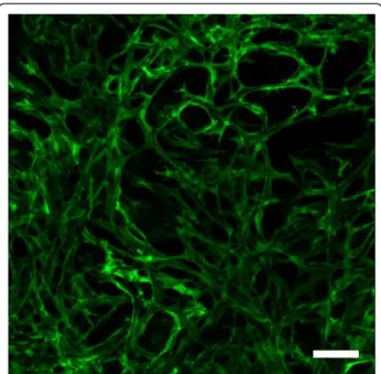
do not require extensive internal vasculature to survive in vivo. However, the fabrication of complex organs such as the heart or liver requires adequate vascular supply to ensure the survival of specialized cells within their 3D structure. To achieve this level of functional-ity, it is necessary to integrate a network of vessels ranging from a few microns to several millimeters within engineered tissues. The incorporation of this microcirculation into tissues and organs represents a considerable challenge which includes the engineering of vascular conduits having micron scale dimensions. It also requires a functional endothelium to improve vascular activity and avoid thrombosis, as well as other specialized cell types performing the physiological tis-sue function of interest. Various approaches have been proposed to design scaffolds comprised of a vascular network analogous to capillaries. These approaches rely on the release of growth factors [14,15] from the scaffolding material, the seeding of endothelial cells in the scaffold to promote angiogenesis [16] (Figure 1) or the use of microfabrication technologies to engineer branched microfluidic channels inside biocompatible materials [17]. Regardless of the application, these technologies all aim at improving mass and fluid trans-port as well as oxygen diffusion in engineered tissues producedin vitro.
Microvascularization of Engineered Tissues through Angiogenesis and Inosculation
The need for adequate solute transport in cell-seeded scaffolds is essential for tissue survival and function. A key approach in attempting to induce the growth of a vascular network within 3D engineered tissue has been the incorporation of growth factors into the scaffolding material. It was shown that a macroporous scaffold, obtained by particle leaching, freeze drying or other pore forming technologies and functionalized with growth factors such as vascular endothelial growth fac-tor (VEGF), basic fibroblast growth facfac-tor (bFGF) and platelet-derived growth factor (PDGF), can trigger the formation of vascular structures following in vivo implantation [18-22].
Both natural and synthetic scaffolding materials have been loaded with these pro-angiogenic molecules, lead-ing to the sproutlead-ing of capillary beds within the con-structs. However, the lack of directed growth of blood vessels to enable interconnectivity between the capillary networks still remains the biggest challenge of this technology.
Microvascular structures incorporated in tissue engi-neering scaffolds prior to their implantation can also be obtained by the co-culture of endothelial cells with the cell types of interest regarding tissue function. This cell-based approach uses the ability of endothelial cells to release growth factors and promotes the formation of capillariesin vitro. The resulting angiogenesis phenom-enon can be explained by the remodeling that occurs within the construct, which is driven by the endothelial network that activates the release of pro-angiogenic fac-tors in the construct. The remodeling of the extracellu-lar matrix (ECM) allowing the formation of vasculature is driven by matrix-metalloproteases (MMPs), regulating cell proliferation and migration within the tissue [2]. Using a skin model, it has been originally demonstrated that a capillary network can be successfully produced by culturing human umbilical vein endothelial cells (HUVECs) into the dermal part of the engineered sub-stitute leading to the formation of long-lasting and per-fused blood vessel networks following in vivo transplantation [3,16,23-25]. This inosculation enabled the perfusion of the tissue engineered capillary network by the vasculature of the host following implantation. The cell-based approaches have led to many interesting results including vascularized muscle [26], cardiac tissue [27], bone [28] and blood vessel [29], all of which have been implanted in animal models and have shown improved perfusion and inosculation with the host vas-culature. Similar results were obtained when HUVECs were cultured in a 3D collagen ECM in vivo, where a stable tree-like structure of a branched network was observed for an extended period of time [30]. Several
Figure 1 Co-culture of fibroblasts and endothelial cells for one month in a collagen-GAG porous construct, resulting in a 3D capillary system within the biomaterial. Confocal imaging of the full thickness of the scaffold showing green fluorescent protein (GFP)-labeled endothelial cells forming vascular channels throughout the 3D structure of the biomaterial. Gauvinet al. Vascular Cell 2011, 3:24
http://www.vascularcell.com/content/3/1/24
other recent studies also suggested that mesenchymal stem cells and endothelial cells have the ability to inter-act together to form a stable vascular network of capil-laries both underin vitro and in vivo conditions [31,32].
In vivo studies also demonstrated that the presence of vascular structures formedin vitro greatly accelerated inosculation of the implanted tissue with the host vascu-lature. Results from L’Heureux et al. have shown that the formation of a vasa vasorum in the wall of a tissue engineered blood vessel (TEBV) occurred 3 months fol-lowing implantationin vivo [33].
Similarly, Guillemetteet al. showed that a TEBV com-prised of a vasa vasorum engineered in vitro (TEBVwVV) allowed for complete tissue integration and functional vasa vasorum activity after only 2 weeksin vivo [3]. These studies have provided evidence for the need to incorporate a capillary network into engineered tissues prior to implantation, showing improved and accelerated tissue integration and capillary-like structure formation which enabled connections with the host vas-culaturein vivo. Therefore, rapid formation of a vascular network between the engineered tissue and the host at the transplanted site, similar to the process observed during wound healing, represents a promising solution to provide implanted cells with adequate supply of oxy-gen and nutrients. However, this strategy does not pro-vide a sustainable solution that can enable both perfusionin vitro and inosculation in vivo. In addition, these approaches did not result in the creation of organ scale constructsin vitro. In an attempt to engineer vas-culature into engineered tissues and organs, microscale technologies and microfluidics systems have emerged as efficient tools to create easily perfusable channels in bio-compatible and biodegradable 3D scaffolding materials.
The use of Microtechnologies to Engineer Vascularization in vitro
The application of microtechnologies to biomaterials can be used to reproduce the capillary network and allow the flow of culture medium through a construct duringin vitro studies. Unlike angiogenesis and inoscu-lation approaches, microfabricated devices with inte-grated microvasculature can be optimized to provide a uniform distribution of flow and mass transfer across the scaffolding material and thus provide the cells with an adequate supply of nutrients.
Soft lithographic and micromolding processes have been used to create microfluidic devices consisting of branched networks that can be connected to perfusion systems in vitro [17]. These technologies have been applied to polymers such as poly(dimethyl siloxane) (PDMS), lactic (co-glycolic acid) (PLGA) and poly-glycerol sebacate (PGS) in which channel networks can be perfused and seeded with vascular cells [34-37].
These techniques have been shown to be useful to regu-late the formation of vascular networks in a precise and efficient manner. The design of functional microvascular networks involves the integration of multiple parameters such as the geometry of branching and bifurcations, fluid mechanics, mass transport and structural rigidity.
These properties are of utmost importance since they greatly influence the stability, oxygen and nutrient distri-bution and therefore the functionality of engineered tis-sues. Microfluidic systems can effectively transport solutes in capillary channels ranging from a few milli-meters down to micromilli-meters [38]. The control of fluid mechanics and mass transport over this wide range of dimensions has been used to study bioactive molecules and therapeutics in cardiovascular research and tissue engineering [39]. Sophisticated devices have recently been fabricated to reproduce a lung assist apparatus allowing the blood being perfused within the micro-channels to be oxygenated by flowing through many parallel capillary-like channels analogous to the native lung architecture [40]. Although optimization of the gas transfer membrane and characterization of the blood flow in the device are still needed, this is a good exam-ple demonstrating the potential of microfabrication technologies to generate vascularized platforms for tis-sue engineering. Similarly, it was shown that microvas-cular cells could be seeded in the device to form a confluent endothelium on the walls of the vascular channels [41]. These studies demonstrated that micro-fabricated devices comprised of a fluidic network mod-eled on human vasculature can be successfully inosculatedin vivo [41].
Even though previous work has shown that microscale channels can be engineered in vitro, there is actually no available method to consecutively branch multi-dimen-sional channels inside a scaffold [42]. Top-down fabrica-tion processes are inherently planar in nature and therefore 3D structures mostly result from stacked 2D structures which are comprised of channels having rec-tangular cross sections [43]. Moreover, most microfabri-cation techniques have been developed for materials that are unable to sustain cell encapsulation, which represents a limitation of this approach to generate large vascularized tissues [36,44,45]. Few attempts were made to engineer perfused microfluidic devices in which two different cell types can be cultured [36,46], but a vascularized tissue with parenchymal cells has yet to be created and there are still no methods to build sustain-able large cell-laden structures with multi-dimensional branched networks. Other microscale fabrication approaches such as bioprinting and stereolithography are currently being investigated to create 3D branching vascular networks [47-50]. However, these methods not only require specialized facilities and expensive
equipment, but the fabrication processes involved are usually time consuming.
Biomimetic Approaches for Engineering Tissue Vascularization
Cells in the body are in contact with a complex 3D environment comprised of a combination of soluble fac-tors as well as the ECM and basement membrane pro-teins found in the tissue in which they reside. Most tissues consist of multiple cell types organized into hier-archical structures that allow them to regulate their function. Modular tissue engineering, or bottom-up approaches, have recently emerged as powerful fabrica-tion methods to generate 3D structures that mimic this organization and that recapitulate tissue structure and spatial resolution [51-54]. These techniques aim to gen-erate biomimetic mesoscale structures by engineering microscale components and by using them as building blocks to fabricate larger tissue structures [55]. This approach has been used to control the cell microenvir-onment and the macroscale properties of relatively large and complex engineered tissues [56,57].
Microgels are microscale hydrogels fabricated by mer-ging microscale fabrication and hydrogel chemistry. They exhibit properties similar to native ECM, can sus-tain cell encapsulation and have tunable geometrical, mechanical and biological properties which make them excellent candidates for tissue engineering applications. Based on these characteristics, cell-laden microgels were fabricated and then assembled into 3D tissue constructs to create precise
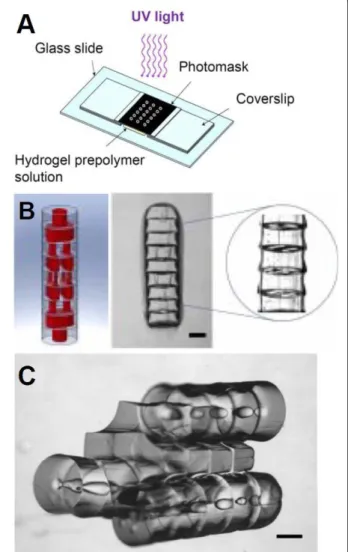
microarchitecture containing repeated functional units that mimic the in vivo structure of tissues and organs [52,54,58,59]. The directed assembly of microgels can be driven by simple thermodynamic processes in a two-phase oil-aqueous reactor [54,57]. Recent work has also shown the potential of modular assembly to produce vascularized tissuesin vitro [60,61]. Arrays of microgels with precisely defined structures and channels have been produced by photolithography and were assembled in a controlled manner resulting in 3D structures with multidimensional interconnected lumens resembling native vasculature (Figure 2) [61]. The validation of this biofabrication process was assessed by placing endothe-lial and smooth muscle cells inside the microgels follow-ing a precise and concentric fashion mimickfollow-ing the organization of blood vessels. The complete assembly was later strengthened by a secondary crosslinking step and it was shown that these assemblies could be per-fused with fluids [61].
One of the challenges of directed self-assembly tech-nology resides in the difficulty to scale-up the tissue produced. Larger structures have recently been built using lock-and-key principles and modular assembly
[56]. Ongoing work is currently focusing on long-term perfusion aiming at developing mature and functional vasculature in vitro. Maintaining physiological function-ality and blood flow in a high-density vascular network with optimized oxygen transfer conditions is critical to keep the tissue in an appropriate state of homeostasis. This biomimetic approach can also be utilized in the design of organ-on-a-chip technologies aiming at the fabrication of precise and reliable small scale models that can later be used for drug screening and physiologi-calin vitro studies [62].
Figure 2 Directed assembly of microgels using a modular approach. Schematic representation of a high-throughput photolithographic method (A). Design image of microgel arrays assembled into tubular structures embedded with 3D branching lumens and actual phase image of the microgel assembly after secondary crosslinking (B). Phase image of microgels following a sequential and directed assembly process (C). Scale bar: 500μm. (Adaptation from Du et al., Sequential assembly of cell-laden hydrogel constructs to engineer vascular-like microchannels, Biotechnol Bioeng, 2011, Copyright Wiley-VCH Verlag GmBH&Co. KGaA. Reproduced with permission.)
Gauvinet al. Vascular Cell 2011, 3:24 http://www.vascularcell.com/content/3/1/24
Conclusions
One of the major limiting factors in the field of tissue engineering is the difficulty to generate functional 3D tissues due to the inability to integrate vascular struc-tures into scaffolds. Building networks of vessels branched together into a complex interconnected struc-ture connecting across multiple length scales remain one of the greatest challenges in tissue engineering. Most cells in the human body are within a few hundred microns from a capillary, allowing the delivery of ade-quate nutrients and supplies to the tissues and organs. Since most tissue engineering scaffolds are unable to provide such proximity for continuous solutes and oxy-gen flow, the engineering of large tissues severely lacks from diffusion and transport properties. The methods currently investigated to generate vasculature in scaf-folds mainly involve the use of proangiogenic growth factors and cell-based approaches, which have shown promising results in vivo, but still cannot provide inlet and outlet vessels forin vitro perfusion. Despite all the advances in microfluidics, the use of microengineered 3D structures comprised of rationally designed and microfabricated channels offer limited functionality. These platforms do not provide a parenchymal space for cell types other than endothelial cells to grow within the constructs and present an integration problem with the host tissue. Modular and bottom-up approaches have recently emerged as promising biofabrication approaches in which functional microscale tissue building blocks can be assembled into 3D macroscale tissue constructs. These are relatively simple methods that allow the pro-duction of perfusable tissue, with precise control over the microscale features in a 3D construct. They are par-ticularly promising in the case of organ engineering, where tissue requires perfusion and needs to perform a specialized physiological function. The precise design of microscale components in a high-throughput fashion combined with the capability to link these components together to generate larger structures represents a pro-mising way to build vascularized 3D structures. There-fore, combining modular assembly methods with microfabrication technologies to engineer tissues and organs represent an effective method to control tissue architecture both at the micro and macroscale. This will be a major step forward in the field of tissue engineering that will not only result in the production of functional engineered tissues, but will also greatly help the transla-tion of the technology fromin vitro studies to in vivo applications.
Acknowledgements
This work was supported by the National Institutes of Health (EB008392; HL092836; HL099073; EB009196; DE019024), National Science Foundation (DMR0847287), the Institute for Soldier Nanotechnology, the Office of Naval
Research, and the US Army Corps of Engineers. RG holds a postdoctoral fellowship from FQRNT and a scientific fellowship from DRDC-NSERC.
Author details
1
Harvard-MIT Division of Health Sciences and Technology, Massachusetts Institute of Technology, Cambridge, MA, USA.2Center for Biomedical
Engineering, Brigham and Women’s Hospital, Harvard Medical School, Boston, MA, USA.3Wyss Institute for Biologically Inspired Engineering,
Harvard University, Boston, MA, USA.4Defence Research and Development Canada (DRDC), Valcartier, QC, Canada.
Authors’ contributions
All the authors have been involved in the writing process and have read and approved the final version of the manuscript.
Competing interests
The authors declare that they have no competing interests.
Received: 5 July 2011 Accepted: 31 October 2011 Published: 31 October 2011
References
1. Mikos AG, Herring SW, Ochareon P, Elisseeff J, Lu HH, Kandel R, Schoen FJ, Toner M, Mooney D, Atala A, Van Dyke ME, Kaplan D, Vunjak-Novakovic G: Engineering complex tissues. Tissue Eng 2006, 12:3307-3339.
2. Jain RK, Au P, Tam J, Duda DG, Fukumura D: Engineering vascularized tissue. Nat Biotechnol 2005, 23:821-823.
3. Guillemette MD, Gauvin R, Perron C, Labbe R, Germain L, Auger FA: Tissue engineered vascular adventitia with vasa vasorum improves graft integration and vascularization through inosculation. Tissue Eng Part A 2010, 16:2617-2626.
4. Freed LE, Engelmayr GC Jr, Borenstein JT, Moutos FT, Guilak F: Advanced material strategies for tissue engineering scaffolds. Adv Mater 2009, 21:3410-3418.
5. Yannas IV, Burke JF, Orgill DP, Skrabut EM: Wound tissue can utilize a polymeric template to synthesize a functional extension of skin. Science 1982, 215:174-176.
6. Bell E, Ehrlich HP, Buttle DJ, Nakatsuji T: Living tissue formed in vitro and accepted as skin-equivalent tissue of full thickness. Science 1981, 211:1052-1054.
7. Atala A, Bauer SB, Soker S, Yoo JJ, Retik AB: Tissue-engineered autologous bladders for patients needing cystoplasty. Lancet 2006, 367:1241-1246. 8. Carrier P, Deschambeault A, Audet C, Talbot M, Gauvin R, Giasson CJ,
Auger FA, Guerin SL, Germain L: Impact of cell source on human cornea reconstructed by tissue engineering. Invest Ophthalmol Vis Sci 2009, 50:2645-2652.
9. Vrana NE, Builles N, Justin V, Bednarz J, Pellegrini G, Ferrari B, Damour O, Hulmes DJ, Hasirci V: Development of a reconstructed cornea from collagen-chondroitin sulfate foams and human cell cultures. Invest Ophthalmol Vis Sci 2008, 49:5325-5331.
10. L’Heureux N, Paquet S, Labbe R, Germain L, Auger F: A completely biological tissue engineered human blood vessel. FASEB Journal 1998, 12:47-56.
11. L’Heureux N, McAllister TN, de la Fuente LM: Tissue-engineered blood vessel for adult arterial revascularization. N Engl J Med 2007, 357:1451-1453.
12. Magnan M, Levesque P, Gauvin R, Dube J, Barrieras D, El-Hakim A, Bolduc S: Tissue Engineering of a Genitourinary Tubular Tissue Graft Resistant to Suturing and High Internal Pressures. Tissue Eng 2008, 15:197-202. 13. Griffith LG, Naughton G: Tissue engineering–current challenges and
expanding opportunities. Science 2002, 295:1009-1014.
14. Ehrbar M, Djonov VG, Schnell C, Tschanz SA, Martiny-Baron G, Schenk U, Wood J, Burri PH, Hubbell JA, Zisch AH: Cell-demanded liberation of VEGF121 from fibrin implants induces local and controlled blood vessel growth. Circ Res 2004, 94:1124-1132.
15. Peters MC, Polverini PJ, Mooney DJ: Engineering vascular networks in porous polymer matrices. J Biomed Mater Res 2002, 60:668-678. 16. Black AF, Berthod F, L’Heureux N, Germain L, Auger FA: In vitro
reconstruction of a human capillary-like network in a tissue-engineered skin equivalent. Faseb J 1998, 12:1331-1340.
17. Borenstein JT, Terai H, King KR, Weinberg EJ, Kaazempur-Mofrad MR, Vacanti JP: Microfabrication Technology for Vascularized Tissue Engineering. Biomedical Microdevices 2002, 4:167-175.
18. Richardson TP, Peters MC, Ennett AB, Mooney DJ: Polymeric system for dual growth factor delivery. Nat Biotechnol 2001, 19:1029-1034. 19. Zisch AH, Lutolf MP, Ehrbar M, Raeber GP, Rizzi SC, Davies N, Schmokel H,
Bezuidenhout D, Djonov V, Zilla P, Hubbell JA: Cell-demanded release of VEGF from synthetic, biointeractive cell ingrowth matrices for vascularized tissue growth. Faseb J 2003, 17:2260-2262.
20. Zisch AH, Lutolf MP, Hubbell JA: Biopolymeric delivery matrices for angiogenic growth factors. Cardiovasc Pathol 2003, 12:295-310. 21. Perets A, Baruch Y, Weisbuch F, Shoshany G, Neufeld G, Cohen S:
Enhancing the vascularization of three-dimensional porous alginate scaffolds by incorporating controlled release basic fibroblast growth factor microspheres. J Biomed Mater Res A 2003, 65:489-497. 22. Lin H, Chen B, Sun W, Zhao W, Zhao Y, Dai J: The effect of
collagen-targeting platelet-derived growth factor on cellularization and vascularization of collagen scaffolds. Biomaterials 2006, 27:5708-5714. 23. Rochon MH, Fradette J, Fortin V, Tomasetig F, Roberge CJ, Baker K,
Berthod F, Auger FA, Germain L: Normal Human Epithelial Cells Regulate the Size and Morphology of Tissue-Engineered Capillaries. Tissue Eng Part A 2010, 16:1457-68.
24. Hudon V, Berthod F, Black AF, Damour O, Germain L, Auger FA: A tissue-engineered endothelialized dermis to study the modulation of angiogenic and angiostatic molecules on capillary-like tube formation in vitro. Br J Dermatol 2003, 148:1094-1104.
25. Tremblay PL, Hudon V, Berthod F, Germain L, Auger FA: Inosculation of tissue engineered capillaries with the host’s vasculature in a reconstructed skin transplanted on mice. Am J Transplant 2005, 5:1002-1010.
26. Levenberg S, Rouwkema J, Macdonald M, Garfein ES, Kohane DS, Darland DC, Marini R, van Blitterswijk CA, Mulligan RC, D’Amore PA, Langer R: Engineering vascularized skeletal muscle tissue. Nat Biotechnol 2005, 23:879-884.
27. Lesman A, Habib M, Caspi O, Gepstein A, Arbel G, Levenberg S, Gepstein L: Transplantation of a tissue-engineered human vascularized cardiac muscle. Tissue Eng Part A 2010, 16:115-125.
28. Tsigkou O, Pomerantseva I, Spencer JA, Redondo PA, Hart AR, O’Doherty E, Lin Y, Friedrich CC, Daheron L, Lin CP, Sundback CA, Vacanti JP, Neville C: Engineered vascularized bone grafts. Proc Natl Acad Sci USA 2010, 107:3311-3316.
29. Guillemette MD, Gauvin R, Perron C, Labbe R, Germain L, Auger FA: Tissue-Engineered Vascular Adventitia with Vasa Vasorum Improves Graft Integration and Vascularization Through Inosculation. Tissue Eng Part A 2010, 16:2617-26.
30. Koike N, Fukumura D, Gralla O, Au P, Schechner JS, Jain RK: Tissue engineering: creation of long-lasting blood vessels. Nature 2004, 428:138-139.
31. Sorrell JM, Baber MA, Caplan AI: Influence of adult mesenchymal stem cells on in vitro vascular formation. Tissue Eng Part A 2009, 15:1751-1761. 32. Ford MC, Bertram JP, Hynes SR, Michaud M, Li Q, Young M, Segal SS,
Madri JA, Lavik EB: A macroporous hydrogel for the coculture of neural progenitor and endothelial cells to form functional vascular networks in vivo. Proc Natl Acad Sci USA 2006, 103:2512-2517.
33. L’Heureux N, Dusserre N, Konig G, Victor B, Keire P, Wight TN, Chronos NA, Kyles AE, Gregory CR, Hoyt G, Robbins RC, McAllister TN: Human tissue-engineered blood vessels for adult arterial revascularization. Nat Med 2006, 12:361-365.
34. King KR, Wang CJ, Kaazempur-Mofrad MR, Vacanti JP, Borenstein JT: Biodegradable microfluidics. Advanced Materials 2004, 16:2007-2012. 35. Fidkowski C, Kaazempur-Mofrad MR, Borenstein J, Vacanti JP, Langer R,
Wang Y: Endothelialized microvasculature based on a biodegradable elastomer. Tissue Engineering 2005, 11:302-309.
36. Golden AP, Tien J: Fabrication of microfluidic hydrogels using molded gelatin as a sacrificial element. Lab Chip 2007, 7:720-725.
37. Choi NW, Cabodi M, Held B, Gleghorn JP, Bonassar LJ, Stroock AD: Microfluidic scaffolds for tissue engineering. Nat Mater 2007, 6:908-915. 38. Whitesides GM: The origins and the future of microfluidics. Nature 2006,
442:368-373.
39. Khademhosseini A, Langer R, Borenstein J, Vacanti JP: Microscale technologies for tissue engineering and biology. Proc Natl Acad Sci USA 2006, 103:2480-2487.
40. Hoganson DM, Anderson JL, Weinberg EF, Swart EJ, Orrick BK,
Borenstein JT, Vacanti JP: Branched vascular network architecture: a new approach to lung assist device technology. J Thorac Cardiovasc Surg 2010, 140:990-995.
41. Hsu WM, Carraro A, Kulig KM, Miller ML, Kaazempur-Mofrad M, Weinberg E, Entabi F, Albadawi H, Watkins MT, Borenstein JT, Vacanti JP, Neville C: Liver-assist device with a microfluidics-based vascular bed in an animal model. Ann Surg 2010, 252:351-357.
42. Chrobak KM, Potter DR, Tien J: Formation of perfused, functional microvascular tubes in vitro. Microvasc Res 2006, 71:185-196.
43. Borenstein JT, Tupper MM, Mack PJ, Weinberg EJ, Khalil AS, Hsiao J, Garcia-Cardena G: Functional endothelialized microvascular networks with circular cross-sections in a tissue culture substrate. Biomed Microdevices 2010, 12:71-79.
44. Bettinger CJ, Weinberg EJ, Kulig KM, Vacanti JP, Wang Y, Borenstein JT, Langer R: Three-Dimensional Microfluidic Tissue-Engineering Scaffolds Using a Flexible Biodegradable Polymer. Advanced Materials 2006, 18:165-169.
45. Bettinger CJ, Cyr KM, Matsumoto A, Langer R, Borenstein JT, Kaplan DL: Silk Fibroin Microfluidic Devices. Adv Mater 2007, 19:2847-2850.
46. Carraro A, Hsu WM, Kulig KM, Cheung WS, Miller ML, Weinberg EJ, Swart EF, Kaazempur-Mofrad M, Borenstein JT, Vacanti JP, Neville C: In vitro analysis of a hepatic device with intrinsic microvascular-based channels. Biomedical Microdevices 2008, 10:795-805.
47. Visconti RP, Kasyanov V, Gentile C, Zhang J, Markwald RR, Mironov V: Towards organ printing: engineering an intra-organ branched vascular tree. Expert Opin Biol Ther 2010, 10:409-420.
48. Huang J-H, Kim J, Agrawal N, Sudarsan AP, Maxim JE, Jayaraman A, Ugaz VM: Rapid Fabrication of Bio-inspired 3D Microfluidic Vascular Networks. Advanced Materials 2009, 21:3567-3571.
49. Guillotin B, Souquet A, Catros S, Duocastella M, Pippenger B, Bellance S, Bareille R, Remy M, Bordenave L, Amedee J, Guillemot F: Laser assisted bioprinting of engineered tissue with high cell density and microscale organization. Biomaterials 2010, 31:7250-7256.
50. Lu Y, Mapili G, Suhali G, Chen S, Roy K: A digital micro-mirror device-based system for the microfabrication of complex, spatially patterned tissue engineering scaffolds. J Biomed Mater Res A 2006, 77:396-405. 51. Nichol JW, Khademhosseini A: Modular Tissue Engineering: Engineering
Biological Tissues from the Bottom Up. Soft Matter 2009, 5:1312-1319. 52. McGuigan AP, Leung B, Sefton MV: Fabrication of cell-containing gel
modules to assemble modular tissue-engineered constructs. Nat Protoc 2006, 1:2963-2969.
53. McGuigan AP, Sefton MV: Design and fabrication of sub-mm-sized modules containing encapsulated cells for modular tissue engineering. Tissue Eng 2007, 13:1069-1078.
54. Du Y, Lo E, Ali S, Khademhosseini A: Directed assembly of cell-laden microgels for fabrication of 3D tissue constructs. Proc Natl Acad Sci USA 2008, 105:9522-9527.
55. Khademhosseini A, Langer R: Microengineered hydrogels for tissue engineering. Biomaterials 2007, 28:5087-5092.
56. Fernandez JG, Khademhosseini A: Micro-masonry: construction of 3D structures by microscale self-assembly. Adv Mater 2010, 22:2538-2541. 57. Du Y, Ghodousi M, Lo E, Vidula MK, Emiroglu O, Khademhosseini A:
Surfacedirected assembly of cell-laden microgels. Biotechnol Bioeng 2010, 105:655-662.
58. Yeh J, Ling Y, Karp JM, Gantz J, Chandawarkar A, Eng G, Blumling J, Langer R, Khademhosseini A: Micromolding of shape-controlled, harvestable cell-laden hydrogels. Biomaterials 2006, 27:5391-5398. 59. Chung SE, Park W, Shin S, Lee SA, Kwon S: Guided and fluidic
self-assembly of microstructures using railed microfluidic channels. Nat Mater 2008, 7:581-587.
60. McGuigan AP, Sefton MV: Vascularized organoid engineered by modular assembly enables blood perfusion. Proc Natl Acad Sci USA 2006, 103:11461-11466.
61. Du Y, Ghodousi M, Qi H, Haas N, Xiao W, Khademhosseini A: Sequential assembly of cell-laden hydrogel constructs to engineer vascular-like microchannels. Biotechnol Bioeng 2011, 108:1693-703.
Gauvinet al. Vascular Cell 2011, 3:24 http://www.vascularcell.com/content/3/1/24
62. Qi H, Du Y, Wang L, Kaji H, Bae H, Khademhosseini A: Patterned differentiation of individual embryoid bodies in spatially organized 3D hybrid microgels. Adv Mater 2010, 22:5276-5281.
doi:10.1186/2045-824X-3-24
Cite this article as: Gauvin et al.: Application of microtechnologies for the vascularization of engineered tissues. Vascular Cell 2011 3:24.
Submit your next manuscript to BioMed Central and take full advantage of:
• Convenient online submission
• Thorough peer review
• No space constraints or color figure charges
• Immediate publication on acceptance
• Inclusion in PubMed, CAS, Scopus and Google Scholar
• Research which is freely available for redistribution
Submit your manuscript at www.biomedcentral.com/submit

