HAL Id: hal-02446917
https://hal.archives-ouvertes.fr/hal-02446917
Submitted on 15 Dec 2020
HAL is a multi-disciplinary open access
archive for the deposit and dissemination of
sci-entific research documents, whether they are
pub-lished or not. The documents may come from
teaching and research institutions in France or
abroad, or from public or private research centers.
L’archive ouverte pluridisciplinaire HAL, est
destinée au dépôt et à la diffusion de documents
scientifiques de niveau recherche, publiés ou non,
émanant des établissements d’enseignement et de
recherche français ou étrangers, des laboratoires
publics ou privés.
Eu 2+ : A suitable substituent for Pb 2+ in CsPbX 3
perovskite nanocrystals?
Firoz Alam, K. David Wegner, Stéphanie Pouget, Lucia Amidani, Kristina
Kvashnina, Dmitry Aldakov, Peter Reiss
To cite this version:
Firoz Alam, K. David Wegner, Stéphanie Pouget, Lucia Amidani, Kristina Kvashnina, et al.. Eu 2+ :
A suitable substituent for Pb 2+ in CsPbX 3 perovskite nanocrystals?. Journal of Chemical Physics,
American Institute of Physics, 2019, 151 (23), pp.231101. �10.1063/1.5126473�. �hal-02446917�
CsPbX
3
perovskite nanocrystals?
Cite as: J. Chem. Phys. 151, 231101 (2019); https://doi.org/10.1063/1.5126473Submitted: 05 September 2019 . Accepted: 21 November 2019 . Published Online: 17 December 2019 Firoz Alam , K. David Wegner , Stephanie Pouget , Lucia Amidani , Kristina Kvashnina , Dmitry Aldakov , and Peter Reiss
COLLECTIONS
Paper published as part of the special topic on Colloidal Quantum Dots
Note: This paper is part of the JCP Special Topic on Colloidal Quantum Dots.
ARTICLES YOU MAY BE INTERESTED IN
Enhanced photoredox activity of CsPbBr3 nanocrystals by quantitative colloidal ligand
exchange
The Journal of Chemical Physics
151, 204305 (2019);
https://doi.org/10.1063/1.5129261
How surface-specific is 2nd-order non-linear spectroscopy?
The Journal of Chemical Physics
151, 230901 (2019);
https://doi.org/10.1063/1.5129108
Quasicubic model for metal halide perovskite nanocrystals
The Journal
of Chemical Physics
COMMUNICATION scitation.org/journal/jcpEu
2+
: A suitable substituent for Pb
2+
in CsPbX
3
perovskite nanocrystals?
Cite as: J. Chem. Phys. 151, 231101 (2019);doi: 10.1063/1.5126473
Submitted: 5 September 2019 • Accepted: 21 November 2019 • Published Online: 17 December 2019
Firoz Alam,1 K. David Wegner,1 Stephanie Pouget,2 Lucia Amidani,3,4 Kristina Kvashnina,3,4
Dmitry Aldakov,1 and Peter Reiss1,a)
AFFILIATIONS
1University Grenoble Alpes, CEA, CNRS, IRIG, SyMMES, STEP, 38000 Grenoble, France 2University Grenoble Alpes, CEA, IRIG, DEPHY, MEM, SGX, 38000 Grenoble, France
3The Rossendorf Beamline at ESRF—The European Synchrotron, CS40220, 38043 Grenoble Cedex 9, France
4Helmholtz Zentrum Dresden-Rossendorf (HZDR), Institute of Resource Ecology, P.O. Box 510119, 01314 Dresden, Germany
Note: This paper is part of the JCP Special Topic on Colloidal Quantum Dots. a)Author to whom correspondence should be addressed:peter.reiss@cea.fr
ABSTRACT
Eu2+is used to replace toxic Pb2+in metal halide perovskite nanocrystals (NCs). The synthesis implies injection of cesium oleate into a solution of europium (II) bromide at an experimentally determined optimum temperature of 130○C and a reaction time of 60 s. Structural
analysis indicates the formation of spherical CsEuBr3nanoparticles with a mean size of 43 ± 7 nm. Using EuI2instead of EuBr2leads to the formation of 18-nm CsI nanoparticles, while EuCl2does not show any reaction with cesium oleate forming 80-nm EuCl2nanoparticles. The obtained CsEuBr3NCs exhibit bright blue emission at 413 nm (FWHM 30 nm) with a room temperature photoluminescence quan-tum yield of 39%. The emission originates from the Laporte-allowed 4f7–4f65d1transition of Eu2+and shows a PL decay time of 263 ns. The long-term stability of the optical properties is observed, making inorganic lead-free CsEuBr3NCs promising deep blue emitters for optoelectronics.
Published under license by AIP Publishing.https://doi.org/10.1063/1.5126473., s
I. INTRODUCTION
Lead halide perovskites have not only become promising thin-film absorber materials in photovoltaics but also inspire intense research efforts in the form of colloidal semiconductor nanocrystals (NCs).1–3Initially, organic-inorganic hybrid perovskite NCs such
as methylammonium lead bromide (MAPbBr3) were developed, reaching up to unity photoluminescence quantum yield (PLQY) combined with narrow emission linewidths.4–8These features make hybrid perovskite NCs very appealing for light-emitting applica-tions; however, due to their high sensitivity to oxygen and moisture, efficient encapsulation strategies are required.9–11 Fully inorganic lead halide perovskites with the formula ABX3(A = Cs+, B = Pb2+, X = Cl−, Br−, or I−) have already been known since the end of the 19th century,12but their perovskite crystal structure and semiconducting nature were not reported until the 1950s.13In the form of colloidal
NCs, they show intrinsically higher stability than hybrid perovskites, albeit still lower than conventional II–VI, IV–VI, or III–V semi-conductor NCs due to their much stronger ionic characteristic.2,3,14 While it turned out challenging to stabilize small-sized CsPbX3NCs in the strong quantum confinement regime below approximately 5 nm,2 anion exchange has been shown to be an efficient way to fine tune their optical and electronic properties, with bandgap ener-gies covering the entire visible range.1,15–17 In the past few years, the high potential of CsPbX3NCs for use in diverse optoelectronic applications, such as light-emitting diodes,18–20 solar cells,21 and photodetectors,22–24has been demonstrated.
Despite these appealing features, the intrinsic toxicity of lead is a roadblock for real-life applications of perovskite NCs, which triggered research on its replacement by less toxic metals.25 Most of these works focused on elements neighboring lead in the peri-odic table of elements, namely, tin, bismuth, and antimony.26In the
J. Chem. Phys. 151, 231101 (2019); doi: 10.1063/1.5126473 151, 231101-1
case of tin, one has to consider the much higher oxidation sensitiv-ity of Sn2+as compared to Pb2+whose divalent state is stabilized by relativistic effects (6s2inert electron pair). Jellicoeet al. first reported the successful synthesis of CsSnBr3and CsSnI3NCs, however, with low PLQYs (0.14% and 0.06%).27Trivalent Bi3+is isoelectronic with Pb2+and can form 2D layered perovskite NCs of the formula A3B2X9 crystallizing in the trigonal space group P-3m1.28The same type of structure can be adopted by the lighter homolog Sb3+. The high-est reported PLQY for Cs3Bi2Br9QDs emitting at 410 nm (FWHM 48 nm) is 19.4%29 and for Cs3Sb2Br9 QDs emitting at the same wavelength (FWHM 41 nm) 46%.30
In contrast to these approaches, metal halide perovskite NCs involving rare earth (RE) ions for lead substitution have been essen-tially unexplored so far, although lanthanides have been used as dopants to modify the emission properties.31,32 Here, we report the first synthesis and main photophysical properties of colloidal CsEuBr3NCs. Europium has been chosen for its capacity of octa-hedral coordination in the divalent state and its almost identi-cal ionic radius with Pb2+ in this hexacoordinated configuration (117 pm/119 pm). The obtained NCs exhibit a PL peak centered at 413 nm, a narrow emission linewidth (≤30 nm FWHM), and a PLQY of 39%, which is the highest value reported for Pb-free ABX3NCs.
II. EXPERIMENTAL A. Materials
Chemicals:cesium carbonate (Cs2CO3, Aldrich, 99.9%), oleic acid (OA, Fisher Chemicals, 70%), 1-octadecene (ODE, Sigma-Aldrich, 90%), oleylamine (OLA, Acros Organics, 80%–90%), europium(II) bromide (EuBr2, Sigma-Aldrich, 99.99%), europium(II)
chloride (EuCl2, Sigma-Aldrich, 99.999%), europium(II) iodide
(EuI2, Sigma-Aldrich, 99.999%), anhydrous toluene, hexanes, and acetonitrile (all Sigma-Aldrich).
B. Methods
Synthesis of Cs-oleate: The synthesis was adapted from the method reported in Ref.14. 203.5 mg (0.62 mmol) of Cs2CO3, 10 ml of ODE, and 0.625 ml of dried OA (1.97 mmol) were loaded into a 50 ml three-neck flask equipped with a condenser. The reaction mix-ture was continuously stirred and degassed for 1 h at 120○C under primary vacuum using a Schlenk line for the removal of oxygen and moisture. Later, the system was switched to an argon atmosphere and the temperature was increased to 150○
C to get a clear solution of Cs-oleate. Cs-oleate solidifies at room temperature, and it must be heated to 100○C before injection.
Synthesis of CsEuBr3NCs:Colloidal CsEuBr3NCs were syn-thesized using a hot-injection method. Typically, within a glove-box 58.6 mg (0.188 mmol) of europium bromide and 5 ml of ODE were loaded into a 50 ml three-neck flask. Outside the glove-box, the flask was connected to a condenser and degassed at 120○C for 60 min using a Schlenk line. After backfilling with argon, dried OLA and OA (0.5 ml each) were injected into the reaction mixture. Within 15 min, a clear colorless solution was obtained and the reaction tem-perature was increased to 130○C. The Cs-oleate solution (0.4 ml of the 0.12 M stock solution, preheated to 100○
C) was swiftly injected, and 1 min later, the reaction was cooled down by immersion in an
ice/water bath. The CsEuBr3NCs were purified by adding 1.5 ml of hexanes followed by centrifugation at 1000 rpm for 5 min. The supernatant was discarded, and a second cycle of purification was carried out by adding 1.5 ml of anhydrous acetonitrile to the pre-cipitate followed by vortexing and centrifugation as before. Finally, the precipitated NCs were redispersed in a nonpolar solvent such as hexanes or toluene for further analysis.
Synthesis attempts for CsEuCl3 and CsEuI3 NCs: In the above-described reaction EuBr2 was replaced by EuCl2 or EuI2, respectively. In the former case, higher temperatures (170–180○C) and longer times (2 h) were required for the complexation of the europium salt prior to Cs-oleate injection.
C. Characterization
1. Powder X-ray diffraction
Powder X-ray diffraction was performed using a Panalyti-cal X’Pert powder diffractometer equipped with a copper anode (λKα1=1.5406 Å and λKα2=1.5444 Å) and an X’Celerator 1D detec-tor. It was configured in Bragg-Brentano geometry, with a variable divergence slit on the primary beam path and a set of antiscattering slits positioned before and after the sample. Axial divergence was limited by 0.02 rad Soller slits. The XRD samples were prepared in a glove box by drop-casting a concentrated NC dispersion in hexane on a disoriented silicon substrate and sealed with a double layer of airtight Kapton®foils within the sample holder.
2. XANES measurements on ID26 at ESRF
Eu L3-edge X-ray absorption near edge structure (XANES) in High-Energy Resolution Fluorescence Detection (HERFD) mode was acquired on the ID26 beamline of the ESRF.33 The incident energy was selected by a Si(311) double crystal monochromator. The footprint of the beam on the sample surface, oriented at 45○to the incident beam direction, was 600 μm horizontal times 150 μm ver-tical. The HERFD XANES at the Eu L3edge was collected using an X-ray emission spectrometer in Rowland geometry equipped with four spherically bent Ge(333) crystal analyzers. The spectrometer was moved to the energy of the maximum of the Eu Lα1 character-istic fluorescence line in order to collect only emitted photons in a 0.8 eV energy bandwidth around the maximum of the Eu Lα1. Col-lecting the emitted photons on a bandwidth smaller than the core-hole lifetime broadening results in a sharpening of the XANES fea-tures compared to the conventional fluorescence detected XANES, which integrates the full characteristic line.34The overall (incoming and emitted) energy resolution was 0.8 eV. All samples were mea-sured in a liquid He cryostat kept at 20 K in order to minimize the X-ray beam damage and the contact with air.
We carefully checked for X-ray beam damage on all samples by acquiring fast XANES of the edge region. On sensitive samples, we adapted the thickness of Al attenuators and the scan time per spectrum to acquire HERFD XANES with minimal X-ray damage and we measured single XANES on several sample spots to have the desired statistics.
3. SEM and EDX
A ZEISS Ultra 55+ scanning electron microscope equipped with an EDX probe (acceleration tension: 20 keV and distance
The Journal
of Chemical Physics
COMMUNICATION scitation.org/journal/jcpsample/electron source: 7 nm) was used to obtain the images of the NCs and to determine the elemental composition. For sample preparation, a concentrated colloidal solution of CsEuBr3NCs in chloroform is drop cast on a cleaned silicon substrate.
4. Transmission electron microscopy
Conventional transmission electron microscopy (TEM) images were acquired on a JEOL 3010 LaB6 microscope equipped with a thermionic gun at 300 kV accelerating voltage. The samples were prepared by drop-casting diluted NC solution onto 200-mesh carbon-coated copper grids. STEM-HAADF images were recorded on an aberration corrected FEI Titan Themis3microscope using an acceleration voltage of 200 kV.
5. UV-visible absorption spectroscopy
UV-vis spectroscopy was performed with a Hewlett Packard 8452A single beam spectrophotometer operating in the wavelength range of 190–820 nm and using a diode array for detection. The samples were prepared by diluting the NC dispersions in toluene or hexane in quartz cuvettes with a path length of 4 mm or 1 cm. The background was acquired using the pure solvent.
6. Steady-state and time-resolved photoluminescence
Photoluminescence spectra were recorded using a modular Flu-orolog FL3-22 system from Horiba-Jobin Yvon equipped with a double grating excitation monochromator and an iHR320 imag-ing spectrometer. A Hamamatsu R928P photomultiplier and quartz cuvettes with a path length of 1 cm were used for the measure-ment. For the PL measurements, the concentration of the sam-ples was adjusted to an absorbance around 0.1 at the excitation wavelength.
PL lifetimes of the NCs were obtained using a NanoLED pulsed source from Horiba (emission wavelength: 360 nm and repetition rate: 1 MHz). The output signal was controlled and analyzed with Data Station (v2.7) and Decay Analysis (v6.8) software from Horiba Scientist. The quantum yield measurements were performed at room temperature using an integration sphere, Hamamatsu Quantaurus-QY Absolute PL quantum yield spectrometer C11347-11.
III. RESULTS AND DISCUSSION
Alkali halide compounds of divalent RE ions are generally synthesized by high temperature reactions between stoichiometric amounts of the RE halide and the alkali halide.35 In the case of europium(II) and cesium, the formation of perovskite-type ternary
compounds of the formula CsEuX3 (X = Cl, Br) was observed, while ARE2X5 compounds are obtained for smaller alkaline ions (Rb, K). In attempts to develop synthetic methods for nanoparti-cles of CsEuX3, major challenges are related to the low solubility and high oxidation sensitivity of europium(II) halides used as
precur-sors. As a starting point, the well-established hot-injection method reported for CsPbBr3 NCs was applied,14 using EuBr2 instead of PbBr2. In brief, a cesium oleate solution was quickly injected into a hot mixture of EuBr2, oleic acid (OA), and oleylamine (OLA) in 1-octadecene (ODE). For an optimized reaction temperature of 130○
C and a reaction time of 60 s (vide infra), CsEuBr3NCs of approxi-mately spherical shape with a mean size of 43 ± 7 nm were obtained
as revealed by SEM and TEM analyses shown inFig. 1. Shorter reac-tion times (5–10 s) did not give access to smaller NCs but yielded ill-defined mixtures of larger particles and sheet-like structures (cf. Fig. S1). In the high-resolution TEM image [Fig. 1(b)], lattice planes can be identified throughout the entire particle, confirming the high crystallinity of the obtained NCs, which are sensitive to beam dam-age as visible by the darker areas in the imdam-age. The observed lattice spacing of 0.295 nm corresponds to (004) planes in CsEuBr3(ICDD card No. 04-014-8774)36and cannot be found in related secondary phases such as CsBr or EuBr2. EDX analyses resulted in a Cs:Eu ratio of 1:0.92, i.e., close to the expected 1:1 ratio in CsEuBr3, indi-cating that possible other phases such as Cs2EuBr4or Cs4EuBr6do not form or are minority phases.37We note, however, a slightly ele-vated Br ratio of 3.86, which we attribute to remaining bromide after purification and/or oleylammonium bromide passivating the surface.
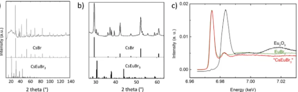
The structure of CsEuBr3single crystals has been reported to correspond to a distorted 3D perovskite structure, with ana−a− c+tilting scheme of the EuBr6octahedra.36It is isotypic to GdFeO3 and crystallizes in the orthorhombic space groupPbnm. The pow-der X-ray data of the synthesized CsEuBr3NCs [Figs. 2(a)and2(b)] reveal prominent peaks similar to the diffraction pattern of CsBr as well as a number of additional peaks characteristic of the forma-tion of the ternary structure. However, the diffracforma-tion pattern does not match the powder XRD data reported for ground CsEuBr3 sin-gle crystals.38Analysis of the XRD peak linewidth does not reveal the presence of different sets of peaks and therefore does not give any indication that several crystalline phases coexist. We empha-size that the obtained XRD data cannot be assigned to Eu2+-doped CsBr nanocrystals, which exhibit essentially the crystal structure of CsBr with few very low intensity additional peaks in the lower 2 theta angle range.32,38 The data were also compared with
sev-eral other related structures, namely, CsPbBr3nanocrystals as well as bulk EuBr2, Cs2PbBr4, and Cs4PbBr6, yet none of these phases fits the experimental data. Several reasons can be at the origin of the observed structural differences. First, structural modifications occurring during the measurement could be eventually expected in view of the high sensitivity against radiation damage observed in Eu2+-doped CsBr used in X-ray radiography based on photo-stimulated luminescence (PSL).39 However, by examining a series
of diffractograms recorded in successive scans, we can exclude this effect being responsible for the observed structural features. Sec-ond, AREX3compounds are well-known to form several hettotypes
FIG. 1. (a) SEM image of the obtained CsEuBr3NCs (mean size 43 ± 7 nm), (b)
high-resolution, and (c) low-resolution STEM-HAADF images.
J. Chem. Phys. 151, 231101 (2019); doi: 10.1063/1.5126473 151, 231101-3
FIG. 2. (a) Powder X-ray diffractogram of CsEuBr3NCs. The broad feature in the 10○–30○range originates from two Kapton foils used to protect the sample against
oxidation during the measurement (cf. Fig. S2). For comparison, the diffraction patterns of bulk CsBr (ICCD No. 00-005-0588) and CsEuBr3(ICCD No. 04-014-8774) are also
given. (b) Zoomed-in image of the 30○–60○2 theta range. (c) HERFD XANES spectrum at the Eu L
3edge of CsEuBr3NCs compared with the reference systems Eu2O3
and EuBr2.
arising from octahedral tilting and to frequently undergo phase transitions, both of which can contribute to the observed struc-tural differences.35 Third, a possible formation mechanism could start with CsBr host nanoparticles, in which Eu2+ions are integrated followed by the transformation to a perovskite-like CsEuBr3 struc-ture. Interestingly, in the case of M2+-doped bulk cesium halides, the formation of perovskite-type domains has been postulated and demonstrated. In particular, Eu2+-doped CsBr is characterized by charge-compensating cation vacancies (vc) and a fast aggregation of Eu2+-vc dimers at room temperature.40 The formation of the perovskite-type unit cell has been explained by means of Nikl’s mechanism for CsPbCl3.41This mechanism implies local structural disorder due to ionic mobility. A similar mechanism has been pro-posed to occur in the case of CsEuBr3.40 Therefore, local struc-tural disorder occurring during the formation of the perovskite-like CsEuBr3structure is a plausible reason for the observed structural features.
X-ray absorption near edge structure (XANES) spectroscopy at the Eu L3edge was used to identify the oxidation state of europium in the obtained NCs. XANES spectroscopy is one of the most pow-erful methods for the investigation of the electronic structure. Elec-trons in the X-ray absorption process are excited to unoccupied levels and give information about the chemical state. A chemical shift in the absorption edge can therefore be assigned to a certain oxidation state. Better energy resolution of the XANES spectra can be obtained using the high energy resolution fluorescence detection (HERFD) mode, where an X-ray emission spectrometer is employed for data collection.Figure 2(c)shows the HERFD XANES spectra at the Eu L3edge of CsEuBr3NCs compared to two reference sys-tems (Eu2O3 and EuBr2) with Eu(III) and Eu(II) oxidation states,
respectively. The position of the main peak in the HERFD XANES spectrum of CsEuBr3NCs clearly demonstrates the predominance of the Eu(II) oxidation state, based on the good correspondence with
the EuBr2reference. The exact contribution of the different chemi-cal states in the Eu L3HERFD XANES data depicted inFig. 2(c)was estimated using the ITFA program.42The results (reported in the
supplementary material) indicate that the spectrum of CsEuBr3NCs contains 99% of Eu(II) (with an estimated root mean square error of
less than 1%).
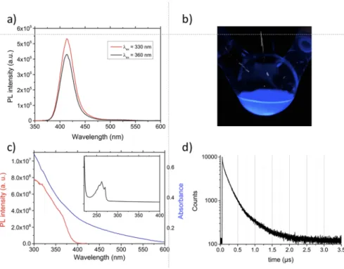
An interesting feature of the obtained NCs is their strong lumi-nescence in the deep blue range. In the PL spectrum of CsEuBr3NCs
[Fig. 3(a)], an emission peak centered at 413 nm with a linewidth of 30 nm (FWHM) is observed whose position is independent of the excitation wavelength (330 or 360 nm). This peak is blue shifted compared to Eu2+-doped bulk CsBr or CsBr NCs (440 nm) and to CsEuBr3single crystals (450 nm).32,38,43 In the former case, no perovskite structure is formed, but Eu2+occupies cesium positions, which leads to the creation of Cs+vacancies in their immediate sur-rounding for charge compensation.37The observed hypsochromic shift with respect to the single crystal data indicates differences within the coordination sphere of the Eu2+ion in CsEuBr3NCs, as already suggested by the X-ray data. For samples kept under inert atmosphere, no PL emission signals in the 600–700 nm range char-acteristic of the 5D0–7FJ transitions of Eu3+ ions were detected,44 even when using longer excitation wavelengths. This is an indirect proof that no oxidation took place. Moreover, also after storing the samples for one month in toluene or hexanes, no change of the PL spectra occurred (Fig. S6), demonstrating the long-term stabil-ity of the obtained CsEuBr3NCs. In the UV-vis absorption spec-trum [Fig. 3(c)] for strongly diluted samples, an absorption band comprising several features and peaking at 263 nm becomes vis-ible, attributed to the 4f7–4f65d1 transition.45 In contrast to the absorption spectra of Eu2+containing phosphors prepared in alkali halide melts,46no separation into two bands is observed. In partic-ular, no second absorption band in the range of 300–350 nm due to the splitting of the Eu2+ 5d orbitals by the crystal field is visi-ble. In the 300–800 nm range, the absorption spectrum is featureless [blue curve in Fig. 3(c)], while the photoluminescence excitation (PLE) spectrum exhibits two small humps at around 320 and 355 nm (red curve).
The PL decay curve obtained in time-resolved measurements [Fig. 3(d)] was fitted with a triexponential function, resulting in life-times of τ19.7 ns (A11.9), τ2124.5 ns (A235.7), and τ3296.0 ns (A3 62.5), yielding an average lifetime (intensity weighted) of 262.6 ns. As the underlying transition is Laporte-allowed, the lifetime is sev-eral orders of magnitude lower than that of well-known emitters involving Eu3+ions (τ in the millisecond range). The PLQY of the as-synthesized CsEuBr3NCs, measured at room temperature using an integration sphere, accounts for 39%. Except for one example (NCs of the 2D perovskite Cs3Sb2Br9 emitting at 410 nm with a PLQY of 46%),30 this is the highest value reported for lead-free metal halide perovskite NCs luminescing in the deep blue range,47
The Journal
of Chemical Physics
COMMUNICATION scitation.org/journal/jcpFIG. 3. (a) PL spectra of CsEuBr3
NCs using two different excitation wave-lengths in hexanes. The PL peak is cen-tered at 413 nm (FWHM 30 nm). (b) Pho-tograph of the colloidal solution under UV light (360 nm). (c) UV-vis absorp-tion spectrum (blue) and PLE spectrum (red,λem= 425 nm). Inset: absorption
spectrum of a strongly diluted colloidal solution. (d) Time-resolved PL spectrum.
indicating that CsEuBr3NCs are promising toxic heavy metal-free emitters for optoelectronic applications.
While for lead-based perovskite NCs, the variation of the reac-tion temperature is used to tune the NC size, here, this parameter had a critical influence on the optical properties of the product. At high temperatures (>130○C), either no emission was detectable (180○
C) or partial oxidation of Eu2+took place (150○
C), visible by the appearance of PL bands characteristic of Eu3+in the 600–700 nm range (cf. Fig. S4). As all reactions were carried out under strict air- and moisture-free conditions, oxidation could arise from side reactions of the used surface ligands, such as, for example, amide formation48 or ketonization,49 leading to thein situ formation of minute amounts of water. Degradation of the optical properties continues to progress within a few days of storage. At lower reac-tion temperatures (e.g., 110○C), only partial conversion of the precursors and low reaction yields are observed, combined with limited stability (cf. Fig. S5). 130○
C turned out to be an opti-mum value, leading to the long-term stability (at least 1 month) of the optical properties, both in terms of spectral shape and PLQY (Fig. S6).
To check the influence of the halide ion, additional experiments were conducted using EuCl2and EuI2as precursors. In the former case, EuCl2could not be solubilized in ODE containing a mixture of OA and OLA, even when higher temperatures (up to 180○C) and longer reaction times (up to two days) were applied. EuCl2has a much higher bond dissociation energy (414 kJ/mol) as compared to EuBr2(347 kJ/mol) and PbCl2(304 kJ/mol) or PbBr2(260 kJ/mol).50 In consequence, no reaction with Cs-oleate took place under the used experimental conditions: the isolated precipitate was identified by means of powder X-ray diffraction as EuCl2nanoparticles hav-ing a crystallite size of around 80 nm (Fig. S7). In contrast, EuI2
(bond dissociation energy: 272 kJ/mol) could be easily solubilized at 130○
C in the presence of OLA and OA. However, upon Cs-oleate injection, no visible change occurred, and only for longer reaction times (>15 min), a white precipitate could be isolated. The prod-uct was identified using XRD as 18-nm CsI nanoparticles of low size dispersion (cf. Fig. S8).
IV. CONCLUSION
CsEuBr3NCs were synthesized using a hot-injection method at 130○C. They exhibit a narrow PL peak at 413 nm with a PLQY of 39%, which represents the highest reported value for lead-free ABX3 perovskite NCs. No change in their optical properties was observed over one month when exposure to air and moisture was avoided. These features make CsEuBr3NCs promising candidates for optoelectronic applications, such as, for example, UV-to-blue color converters in LEDs.47 Nonetheless, unlike lead-based per-ovskite NCs, their luminescence cannot be varied in a wide spec-tral range. In CsPbX3NCs, excitonic emission takes place between band edge states constituted of Pb2+and X−electronic orbitals and the position of these levels can be tuned by changing the size of the particles (via the quantum confinement effect) or the nature of the halide.25In contrast, the PL emission of CsEuBr
3NCs results from the intrinsic 4f7–4f65d1transition of Eu2+whose energy can-not be tuned using similar approaches but is influenced by the coordination sphere of the europium ion. Nevertheless, other emis-sion ranges could be accessed by using different types of REs, e.g., Sm2+. As such, this first example of RE halide perovskite NCs can serve as a basis for the development of related systems involving the use of different lanthanide ions and/or different halides. To overcome the solubility issues of RE2+ halides, synthetic schemes
J. Chem. Phys. 151, 231101 (2019); doi: 10.1063/1.5126473 151, 231101-5
allowing for the separate introduction of the three individual con-stituents of ABX3, by decoupling the B and X precursors, will be particularly useful in this context. This has recently been achieved by using, for example, benzoyl halide or molecular halogen as the X source.51,52
SUPPLEMENTARY MATERIAL
See thesupplementary materialfor additional XRD, SEM, and photoluminescence data and for the description of the ITFA method used for XANES data analysis.
ACKNOWLEDGMENTS
The authors acknowledge the French Research Agency ANR for financial support (Grant Nos. SuperSansPlomb ANR-15-CE05-0023-01 and PERSIL ANR-16-CE05-0019-02). K.D.W. acknowl-edges the LABEX Serenade (Grant No. ANR 11-LABX-0064) for his post-doctoral funding. F.A. acknowledges support from the PHC program MERLION financing an exchange with Nanyang Technical University Singapore. Hanako Okuna is thanked for TEM imaging. The authors gratefully acknowledge the help of Tim Bohdan at the ID26 beamline of ESRF during the HERFD XANES measurements. L.A. and K.K. acknowledge support from the European Research Council under ERC Grant No. N759696.
REFERENCES
1
J. Shamsi, A. S. Urban, M. Imran, L. De Trizio, and L. Manna, “Metal halide perovskite nanocrystals: Synthesis, post-synthesis modifications, and their optical properties,”Chem. Rev.119(5), 3296–3348 (2019).
2
M. V. Kovalenko, L. Protesescu, and M. I. Bodnarchuk, “Properties and poten-tial optoelectronic applications of lead halide perovskite nanocrystals,”Science 358(6364), 745–750 (2017).
3
Q. A. Akkerman, G. Rainò, M. V. Kovalenko, and L. Manna, “Genesis, challenges and opportunities for colloidal lead halide perovskite nanocrystals,”Nat. Mater. 17(5), 394–405 (2018).
4
L. C. Schmidt, A. Pertegás, S. González-Carrero, O. Malinkiewicz, S. Agouram, G. Mínguez Espallargas, H. J. Bolink, R. E. Galian, and J. Pérez-Prieto, “Nontem-plate synthesis of CH3NH3PbBr3perovskite nanoparticles,”J. Am. Chem. Soc.
136(3), 850–853 (2014).
5S. Gonzalez-Carrero, R. E. Galian, and J. Perez-Prieto, “Maximizing the emissive
properties of CH3NH3PbBr3perovskite nanoparticles,”J. Mater. Chem. A3(17),
9187–9193 (2015).
6S. Gonzalez-Carrero, L. Francés-Soriano, M. González-Béjar, S. Agouram, R.
E. Galian, and J. Pérez-Prieto, “The luminescence of CH3NH3PbBr3perovskite
nanoparticles crests the summit and their photostability under wet conditions is enhanced,”Small12(38), 5245–5250 (2016).
7
F. Zhang, H. Zhong, C. Chen, X.-g. Wu, X. Hu, H. Huang, J. Han, B. Zou, and Y. Dong, “Brightly luminescent and color-tunable colloidal CH3NH3PbX3(X =
Br, I, Cl) quantum dots: Potential alternatives for display technology,”ACS Nano 9(4), 4533–4542 (2015).
8H. Huang, J. Raith, S. V. Kershaw, S. Kalytchuk, O. Tomanec, L. Jing, A.
S. Susha, R. Zboril, and A. L. Rogach, “Growth mechanism of strongly emitting CH3NH3PbBr3perovskite nanocrystals with a tunable bandgap,”Nat. Commun.
8(1), 996 (2017).
9
W. Deng, X. Xu, X. Zhang, Y. Zhang, X. Jin, L. Wang, S. T. Lee, and J. Jie, “Organometal halide perovskite quantum dot light-emitting diodes,”Adv. Funct. Mater.26(26), 4797–4802 (2016).
10
Y. Ling, Z. Yuan, Y. Tian, X. Wang, J. C. Wang, Y. Xin, K. Hanson, B. Ma, and H. Gao, “Bright light-emitting diodes based on organometal halide perovskite nanoplatelets,”Adv. Mater.28(2), 305–311 (2016).
11
H. Huang, M. I. Bodnarchuk, S. V. Kershaw, M. V. Kovalenko, and A. L. Rogach, “Lead halide perovskite nanocrystals in the research spot-light: Stability and defect tolerance,” ACS Energy Lett. 2(9), 2071–2083 (2017).
12H. L. Wells, “Über die Cäsium- und Kalium-Bleihalogenide,”Z. Anorg. Chem.
3(1), 195–210 (1893).
13C. K. Möller, “Crystal structure and photoconductivity of cæsium
plumbo-halides,”Nature182, 1436 (1958).
14
L. Protesescu, S. Yakunin, M. I. Bodnarchuk, F. Krieg, R. Caputo, C. H. Hen-don, R. X. Yang, A. Walsh, and M. V. Kovalenko, “Nanocrystals of cesium lead halide perovskites (CsPbX3, X = Cl, Br, and I): Novel optoelectronic materials
showing bright emission with wide color gamut,”Nano Lett.15(6), 3692–3696 (2015).
15D. M. Jang, K. Park, D. H. Kim, J. Park, F. Shojaei, H. S. Kang, J.-P. Ahn, J.
W. Lee, and J. K. Song, “Reversible halide exchange reaction of organometal tri-halide perovskite colloidal nanocrystals for full-range band gap tuning,”Nano Lett.15(8), 5191–5199 (2015).
16
Q. A. Akkerman, V. D’Innocenzo, S. Accornero, A. Scarpellini, A. Petrozza, M. Prato, and L. Manna, “Tuning the optical properties of cesium lead halide perovskite nanocrystals by anion exchange reactions,”J. Am. Chem. Soc.137, 10276–10281 (2015).
17
G. Nedelcu, L. Protesescu, S. Yakunin, M. I. Bodnarchuk, M. J. Grotevent, and M. V. Kovalenko, “Fast anion-exchange in highly luminescent nanocrystals of cesium lead halide perovskites (CsPbX3, X = Cl, Br, I),”Nano Lett.15(8),
5635–5640 (2015).
18
S. G. R. Bade, J. Li, X. Shan, Y. Ling, Y. Tian, T. Dilbeck, T. Besara, T. Geske, H. Gao, B. Ma, K. Hanson, T. Siegrist, C. Xu, and Z. Yu, “Fully printed halide per-ovskite light-emitting diodes with silver nanowire electrodes,”ACS Nano10(2), 1795–1801 (2016).
19M. F. Aygüler, M. D. Weber, B. M. D. Puscher, D. D. Medina, P. Docampo,
and R. D. Costa, “Light-emitting electrochemical cells based on hybrid lead halide perovskite nanoparticles,” J. Phys. Chem. C 119(21), 12047–12054 (2015).
20S. Colella, M. Mazzeo, A. Rizzo, G. Gigli, and A. Listorti, “The bright side of
perovskites,”J. Phys. Chem. Lett.7(21), 4322–4334 (2016).
21
A. Swarnkar, A. R. Marshall, E. M. Sanehira, B. D. Chernomordik, D. T. Moore, J. A. Christians, T. Chakrabarti, and J. M. Luther, “Quantum dot-induced phase stabilization of α-CsPbI3perovskite for high-efficiency photovoltaics,”Science
354(6308), 92–95 (2016).
22
P. Ramasamy, D.-H. Lim, B. Kim, S.-H. Lee, M.-S. Lee, and J.-S. Lee, “All-inorganic cesium lead halide perovskite nanocrystals for photodetector applica-tions,”Chem. Commun.52(10), 2067–2070 (2016).
23L. Lv, Y. Xu, H. Fang, W. Luo, F. Xu, L. Liu, B. Wang, X. Zhang, D. Yang,
W. Hu, and A. Dong, “Generalized colloidal synthesis of high-quality, two-dimensional cesium lead halide perovskite nanosheets and their applications in photodetectors,”Nanoscale8(28), 13589–13596 (2016).
24
M. I. Saidaminov, M. A. Haque, M. Savoie, A. L. Abdelhady, N. Cho, I. Dur-sun, U. Buttner, E. Alarousu, T. Wu, and O. M. Bakr, “Perovskite photodetec-tors operating in both narrowband and broadband regimes,”Adv. Mater.28(37), 8144–8149 (2016).
25
D. Aldakov and P. Reiss, “Safer-by-design fluorescent nanocrystals: Metal halide perovskites vs semiconductor quantum dots,”J. Phys. Chem. C123(20), 12527– 12541 (2019).
26J. Sun, J. Yang, J. I. Lee, J. H. Cho, and M. S. Kang, “Lead-free perovskite
nanocrystals for light-emitting devices,”J. Phys. Chem. Lett.9(7), 1573–1583 (2018).
27
T. C. Jellicoe, J. M. Richter, H. F. J. Glass, M. Tabachnyk, R. Brady, S. E. Dutton, A. Rao, R. H. Friend, D. Credgington, N. C. Greenham, and M. L. Böhm, “Synthe-sis and optical properties of lead-free cesium tin halide perovskite nanocrystals,”
J. Am. Chem. Soc.138(9), 2941–2944 (2016).
28
M. Leng, Z. Chen, Y. Yang, Z. Li, K. Zeng, K. Li, G. Niu, Y. He, Q. Zhou, and J. Tang, “Lead-free, blue emitting bismuth halide perovskite quantum dots,”
Angew. Chem., Int. Ed.55(48), 15012–15016 (2016).
29
M. Leng, Y. Yang, K. Zeng, Z. Chen, Z. Tan, S. Li, J. Li, B. Xu, D. Li, M. P. Hautzinger, Y. Fu, T. Zhai, L. Xu, G. Niu, S. Jin, and J. Tang, “All-inorganic
The Journal
of Chemical Physics
COMMUNICATION scitation.org/journal/jcpbismuth-based perovskite quantum dots with bright blue photoluminescence and excellent stability,”Adv. Funct. Mater.28(1), 1704446 (2018).
30J. Zhang, Y. Yang, H. Deng, U. Farooq, X. Yang, J. Khan, J. Tang, and
H. Song, “High quantum yield blue emission from lead-free inorganic anti-mony halide perovskite colloidal quantum dots,”ACS Nano11(9), 9294–9302 (2017).
31
G. Pan, X. Bai, D. Yang, X. Chen, P. Jing, S. Qu, L. Zhang, D. Zhou, J. Zhu, W. Xu, B. Dong, and H. Song, “Doping lanthanide into perovskite nanocrystals: Highly improved and expanded optical properties,”Nano Lett.17(12), 8005–8011 (2017).
32Z. Yang, Z. Jiang, X. Liu, X. Zhou, J. Zhang, and W. Li, “Bright blue
light-emitting doped cesium bromide nanocrystals: Alternatives of lead-free perovskite nanocrystals for white LEDs,”Adv. Opt. Mater.7(10), 1900108 (2019).
33C. Gauthier, V. A. Solé, R. Signorato, J. Goulon, and E. Moguiline, “The ESRF
beamline ID26: X-ray absorption on ultra dilute sample,”J. Synchrotron Radiat. 6(3), 164–166 (1999).
34P. Glatzel, T.-C. Weng, K. Kvashnina, J. Swarbrick, M. Sikora, E. Gallo, N.
Smo-lentsev, and R. A. Mori, “Reflections on hard X-ray photon-in/photon-out spec-troscopy for electronic structure studies,”J. Electron Spectrosc. Relat. Phenom. 188, 17–25 (2013).
35
G. Meyer, “The synthesis and structures of complex rare-earth halides,”Prog. Solid State Chem.14(3), 141–219 (1982).
36H. Ehrenberg, H. Fuess, S. Hesse, J. Zimmermann, H. von Seggern, and
M. Knapp, “Structures of CsEuBr3and its degradation product Cs2EuBr5⋅10H2O,”
Acta Crystallogr., Sect. B: Struct. Sci.63(2), 201–204 (2007).
37Y. Wu, D. Han, B. C. Chakoumakos, H. Shi, S. Chen, M.-H. Du, I.
Gree-ley, M. Loyd, D. J. Rutstrom, L. Stand, M. Koschan, and C. L. Melcher, “Zero-dimensional Cs4EuX6 (X = Br, I) all-inorganic perovskite single crystals for
gamma-ray spectroscopy,”J. Mater. Chem. C6(25), 6647–6655 (2018).
38
S. Hesse, J. Zimmermann, H. V. Seggern, H. Ehrenberg, H. Fuess, C. Fasel, and R. Riedel, “CsEuBr3: Crystal structure and its role in the photostimulation
of CsBr:Eu2+,”J. Appl. Phys.100(8), 083506 (2006).
39
J. Zimmermann, S. Hesse, H. von Seggern, M. Fuchs, and W. Knüpfer, “Radia-tion hardness of CsBr:Eu2+,”J. Lumin.114(1), 24–30 (2005).
40P. Hackenschmied, G. Schierning, M. Batentschuk, and A. Winnacker,
“Precipitation-induced photostimulated luminescence in CsBr:Eu2+,”J. Appl. Phys.93(9), 5109–5112 (2003).
41
M. Nikl, K. Nitsch, K. Polak, G. P. Pazzi, P. Fabeni, D. S. Citrin, and M. Guri-oli, “Optical properties of the Pb2+-based aggregated phase in a CsCl host crystal:
Quantum-confinement effects,”Phys. Rev. B51(8), 5192–5199 (1995).
42
A. Rossberg, T. Reich, and G. Bernhard, “Complexation of uranium(VI) with protocatechuic acid-application of iterative transformation factor anal-ysis to EXAFS spectroscopy,” Anal. Bioanal. Chem. 376(5), 631–638 (2003).
43P. Hackenschmied, G. Zeitler, M. Batentschuk, A. Winnacker, B. Schmitt,
M. Fuchs, E. Hell, and W. Knüpfer, “Storage performance of X-ray irradi-ated doped CsBr,”Nucl. Instrum. Methods Phys. Res., Sect. B191(1), 163–167 (2002).
44
U. T. D. Thuy, A. Maurice, N. Q. Liem, and P. Reiss, “Europium doped In(Zn)P/ZnS colloidal quantum dots,”Dalton Trans.42, 12606–12610 (2013).
45
K. E. Johnson and J. N. Sandoe, “An interpretation of the spectra of bivalent rare-earth ions in crystals,”J. Chem. Soc. A1969, 1694–1697 (1969).
46R. Reisfeld and A. Glasner, “Absorption and fluorescence spectra of Eu2+in
alkali halide crystals,”J. Opt. Soc. Am.54(3), 331–333 (1964).
47
N. K. Kumawat, X.-K. Liu, D. Kabra, and F. Gao, “Blue perovskite light-emitting diodes: Progress, challenges and future directions,”Nanoscale11(5), 2109–2120 (2019).
48
M. Protiere and P. Reiss, “Amine-induced growth of an In2O3shell on colloidal
InP nanocrystals,”Chem. Commun.2007(23), 2417–2419 (2007).
49A. Cros-Gagneux, F. Delpech, C. Nayral, A. Cornejo, Y. Coppel, and B.
Chau-dret, “Surface chemistry of InP quantum dots: A comprehensive study,”J. Am. Chem. Soc.132(51), 18147–18157 (2010).
50J. E. Huheey, E. A. Keiter, and R. L. Keiter,Inorganic Chemistry: Principles of
Structure and Reactivity, 4th ed. (Harper Collins College Publishers, New York, 1993).
51M. Imran, P. Ijaz, D. Baranov, L. Goldoni, U. Petralanda, Q. Akkerman, A.
L. Abdelhady, M. Prato, P. Bianchini, I. Infante, and L. Manna, “Shape-pure, nearly monodispersed CsPbBr3 nanocubes prepared using secondary aliphatic
amines,”Nano Lett.18(12), 7822–7831 (2018).
52
S. Thapa, K. Bhardwaj, S. Basel, S. Pradhan, C. J. Eling, A. M. Adawi, J.-S. G. Bouillard, G. J. Stasiuk, P. Reiss, A. Pariyar, and S. Tamang, “Long-term ambi-ent air-stable cubic CsPbBr3perovskite quantum dots using molecular bromine,”
Nanoscale Adv.1, 3388–3391 (2019).
J. Chem. Phys. 151, 231101 (2019); doi: 10.1063/1.5126473 151, 231101-7
![Fig. S1). In the high-resolution TEM image [Fig. 1(b)], lattice planes can be identified throughout the entire particle, confirming the high crystallinity of the obtained NCs, which are sensitive to beam dam-age as visible by the darker areas in the imdam](https://thumb-eu.123doks.com/thumbv2/123doknet/13592444.423147/5.891.474.811.854.996/resolution-lattice-identified-particle-confirming-crystallinity-obtained-sensitive.webp)