HAL Id: hal-03091750
https://hal.archives-ouvertes.fr/hal-03091750
Submitted on 31 Dec 2020HAL is a multi-disciplinary open access archive for the deposit and dissemination of sci-entific research documents, whether they are pub-lished or not. The documents may come from teaching and research institutions in France or abroad, or from public or private research centers.
L’archive ouverte pluridisciplinaire HAL, est destinée au dépôt et à la diffusion de documents scientifiques de niveau recherche, publiés ou non, émanant des établissements d’enseignement et de recherche français ou étrangers, des laboratoires publics ou privés.
PLA2G1B is involved in CD4 anergy and CD4
lymphopenia in HIV-infected patients
Julien Pothlichet, Thierry Rose, Florence Bugault, Louise Jeammet, Annalisa
Meola, Ahmed Haouz, Frederick Saul, David Geny, José Alcami, Ezequiel
Ruiz-Mateos, et al.
To cite this version:
Julien Pothlichet, Thierry Rose, Florence Bugault, Louise Jeammet, Annalisa Meola, et al.. PLA2G1B is involved in CD4 anergy and CD4 lymphopenia in HIV-infected patients. Journal of Clinical Investi-gation, American Society for Clinical InvestiInvesti-gation, 2020, 130 (6), pp.2872-2887. �10.1172/JCI131842�. �hal-03091750�
TITLE: PLA2G1B IS INVOLVED IN CD4 ANERGY AND CD4
1LYMPHOPENIA IN HIV-INFECTED PATIENTS
23
Authors: Julien Pothlichet1,10, Thierry Rose2,10, Florence Bugault1,3, Louise Jeammet1,
4
Annalisa Meola1, Ahmed Haouz4, Frederick Saul4, David Geny5, José Alcami6, Ezequiel
Ruiz-5
Mateos Carmona7, Luc Teyton8, Gérard Lambeau9, and Jacques Thèze1,3
6 7
Affiliation : 1Diaccurate, Institut Pasteur, Paris, France.
8
2Center for Innovation and Technological Research, Institut Pasteur, Paris, France.
9
3Département Santé Globale, Institut Pasteur, Paris, France.
10
4Plate-forme Cristallographie, Institut Pasteur, Paris, France.
11
5NeuraImag Facility, Institute of Psychiatry and Neurosciences of Paris. INSERM U1266,
12
Paris, France. 13
6Unidad de Immunopatología del SIDA, Centro Nacional de Microbiologia, Instituto de Salud
14
Carlos III, ISCIII, Madrid and Hospital Clinic-Institut d’investigations Biomèdiques August 15
i Sunyer (IDIBASPS) Barcelona , Spain. 16
7Laboratorio de Infeccion por VIH y farmacocinetica de antivirales, UGC, Instituto de
17
Biomedicina de Sevilla, Hospitales Universitarios Virgen del Rocio, Sevilla, Spain. 18
8Department of Microbiology and Immunology, Scripps Research Institute, California, USA.
19
9Université Côte d’Azur, CNRS, IPMC, Valbonne Sophia Antipolis, France.
20
10These authors contributed equally: Julien Pothlichet, and Thierry Rose.
21 22
Address correspondence to : Jacques Thèze, DIACCURATE, Institut Pasteur, 25 rue du Dr 23
Roux, Bat . ROUX, 2nd Floor, 75015 Paris. Phone : 33.1.45.68.86.00.
24 Email: Jacques.theze@diaccurate.com 25 26 CONFLICT OF INTEREST 27
J.T. is cofounder and CEO of DIACCURATE, a spin-off of the Institut Pasteur. J.P., L.J. and 28
A.M. are employees of DIACCURATE. 29
ABSTRACT 30
The precise mechanism leading to profound immunodeficiency of HIV-infected patients is still 31
only partially understood. Here, we show that more than 80% of CD4 T cells from HIV-infected 32
patients have morphological abnormalities. Their membranes exhibited numerous large 33
abnormal membrane microdomains (aMMDs), which trap and inactivate physiological 34
receptors, such as that for IL-7. In patient plasma, we identified phospholipase A2 group IB 35
(PLA2G1B) as the key molecule responsible for the formation of aMMDs. At physiological 36
concentrations, PLA2G1B synergized with the HIV gp41 envelope protein, which appears to 37
be a driver that targets PLA2G1B to the CD4 T-cell surface. The PLA2G1B/gp41 pair induced 38
CD4 T cell unresponsiveness (anergy). At high concentrations in vitro, PLA2G1B acted alone, 39
independently of gp41, and inhibited the IL-2, IL-4, and IL-7 responses, as well as TCR-40
mediated activation and proliferation, of CD4 T cells. PLA2G1B also decreased CD4 T-cell 41
survival in vitro, likely playing a role in CD4 lymphopenia in conjunction with its induced IL-42
7 receptor defects. The effects on CD4 T-cell anergy could be blocked by a PLA2G1B-specific 43
neutralizing mAb in vitro and in vivo. The PLA2G1B/gp41 pair constitutes a new mechanism 44
of immune dysfunction and a compelling target for boosting immune responses in HIV-infected 45
patients. 46
INTRODUCTION 47
CD4 lymphocytes play a critical role in the severe immunodeficiency that characterizes HIV-48
infected patients. Although less than 0.5% of blood CD4 T cells are infected, almost all are 49
dysfunctional. Their progressive decline leads to CD4 lymphopenia. In addition, functional 50
defects of the remaining CD4 T cells lead to their unresponsiveness, or anergy, to certain 51
antigens and cytokines (1). Major progress has been made in treatment of the viral infection. 52
Antiretrovirals (ARVs) prevent acquired immunodeficiency syndrome (AIDS). However, 53
further improvement in HIV therapy will require a better understanding of the mechanisms 54
responsible for CD4 T-cell defects following HIV infection (2, 3). 55
The mechanism explaining CD4 T-cell loss during HIV infection is still debated (4, 5). 56
Persistent immune activation plays a critical role in the induction of this CD4 T-cell decline (6– 57
8). A major mechanism results from damage of the mucosal barrier and lymphoid tissues of the 58
gastrointestinal (GI) track that follows acute infection. HIV targets subpopulations of CCR5-59
expressing CD4 T cells, which are dense in the GI. Following this damage, microbial products 60
translocate across the GI barrier and cause general activation of the immune system (9–11). In 61
this context, it is noteworthy that HIV controller patients, who maintain high CD4 counts and 62
good control of viremia, show low inflammation (12). 63
Numerous investigations have described the impairment of CD4 T-cell function in HIV-64
infected individuals, in whom CD4 T cells fail to proliferate after stimulation by antigens or 65
mitogens (13, 14). A progressive loss of T helper function has also been reported (15–17). 66
These results may be partially explained by changes in the T-cell receptor repertoire (18), but 67
they may also result from a defect in the intrinsic capacity of the CD4 T cells to respond to 68
physiological signals. For example, a selective defect in IL-2 production, but not 69
IFNg synthesis, has been reported after anti-CD3 stimulation (19). In this context, we 70
previously analyzed CD4 T cell responses to IL-2 and IL-7, two cytokines that are crucial for 71
the control of the function, proliferation, and survival of CD4 T cells. We showed that the beta 72
and gamma c (gc) chains of IL-2 receptor (IL-2R) are under-expressed and non-functional, as 73
measured by reduced entry into the S+G2/M phases of the cell cycle (20). Similarly, decreased 74
expression of the IL-7R alpha chain (CD127) on the surface of CD4 T cells from HIV-infected 75
patients has been described and their function was shown to be defective (21). Altered induction 76
of the anti-apoptotic molecule Bcl-2 and decreased expression of CD25 after in vitro exposure 77
to IL-7 were measured (22). We subsequently showed that the Janus kinase (Jak)/Signal 78
Transducer and Activator of Transcription 5 (STAT5) signaling pathway is involved in these 79
defects (23, 24). Similar results involving gc have been published by other investigators (25– 80
30). 81
Most of the studies performed in lymphocytes from HIV-infected patients have used FACS 82
analysis and in vitro functional immunological assays. Here, we reinvestigated this question by 83
studying purified CD4 T lymphocytes from viremic HIV-infected patients using imaging 84
techniques (31) and molecular approaches rarely used in this field (32–34). We detected large 85
abnormal membrane microdomains (aMMDs) at the surface of CD4 T cells purified from HIV-86
infected patients in the absence of any stimulation. The aMMD-bearing cells were named 87
“Bumpy T cells”, due to their appearance after labeling. Their large aMMDs were shown to 88
trap all IL-7R chains (alpha and gc). IL-7R chains lose their function when embedded in these 89
aMMDs. Consequently, the Jak/STAT pathway was not functional and IL-7-induced phospho-90
STAT5 nuclear translocation (pSTAT5 NT) was inhibited. Bumpy T cells were recovered after 91
exposure of CD4 T cells from healthy donors (HD) to plasma from HIV-infected patients. After 92
characterization, we found that phospholipase A2 group IB (PLA2G1B) (35) was able to 93
recapitulate the effects of plasma from HIV-infected patients. It induced aMMDs and 94
consequently strongly affected numerous CD4 functions in vitro and in vivo (mouse model). 95
However, PLA2G1B was found to synergize with HIV gp41 envelope protein in the blood of 96
HIV-infected patients at physiological concentrations. Overall, our results provide new insights 97
into CD4 T-cell dysfunction and a mechanism for the CD4 anergy and lymphopenia observed 98
in chronically HIV-infected patients. 99
RESULTS 100
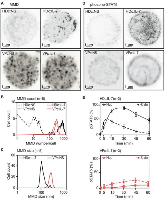
Membrane alterations and signaling defects in CD4 T cells from HIV-infected patients.
101
Stimulation emission depletion (STED) microscopy revealed numerous large aMMDs (up to 102
500/cell, with an average size > 200 nm) at the surface of purified CD4 T cells from VP, in the 103
absence of any activation. More than 80% of purified CD4 T cells, also called Bumpy T cells, 104
from all viremic patients (VPs) exhibited aMMDs (Figure 1, A-C). Under the same conditions, 105
resting CD4 T cells from healthy donors (HD) did not spontaneously exhibit any aMMDs, 106
whereas IL-7 stimulation promoted the formation of numerous physiological MMDs (pMMDs) 107
of smaller size (approximately 800/cell, with an average size of 100 nm) (Figure 1, A-C). In 108
contrast, IL-7 stimulation of CD4 T cells from VPs did not induce any observable changes in 109
their membranes (Figure 1, A and B). 110
We then examined the functional consequences of these morphological changes in the 111
membrane using the IL-7/IL-7R system as a readout. The function of IL-7Rs of VP CD4 T cells 112
was altered, as the IL-7-induced phosphorylation of STAT5 (pSTAT5) differed between CD4 113
T cells of HDs and VPs (Figure 1D); pSTAT5 nuclear translocation (pSTAT5 NT) was almost 114
completely abolished in the CD4 T cells from VPs after IL-7 stimulation. This resulted from 115
the difference in the kinetics of cytoplasmic phosphorylation of STAT5 and pSTAT5 NT 116
between CD4 T cells of VPs and HDs (Figure 1E). 117
We previously showed that IL-7-induced cytoskeletal organization is required for efficient 118
pSTAT5 NT in CD4 T cells of HDs and that colchicine and cytochalasin D treatment abolishes 119
pSTAT5 NT (34). These results are comparable to those obtained for non-treated VP CD4 T 120
cells (Supplemental Figure 1, A and B). Microfilaments and microtubules can be observed after 121
IL-7 stimulation. Due to the very small size of the cytoplasm of lymphocytes, these structures 122
were observed by pulsed STED microscopy. After staining by anti-tubulin antibodies, 123
microtubules could be observed in HD CD4 T cells but not VP CD4 T cells (Supplemental 124
Figure 1C). Similarly, microfilaments were visible in HD CD4 T cells after staining by anti-125
actin antibodies but not in VP CD4 T cells (Supplemental Figure 1D). This further supports 126
that the IL-7/IL-7R system is nonfunctional in VP CD4 T cells. 127
Overall, these results confirm and structurally characterize the activation status of CD4 T 128
lymphocytes from VPs and provide a new insight into the mechanism of unresponsiveness of 129
these CD4 T cells. 130
131
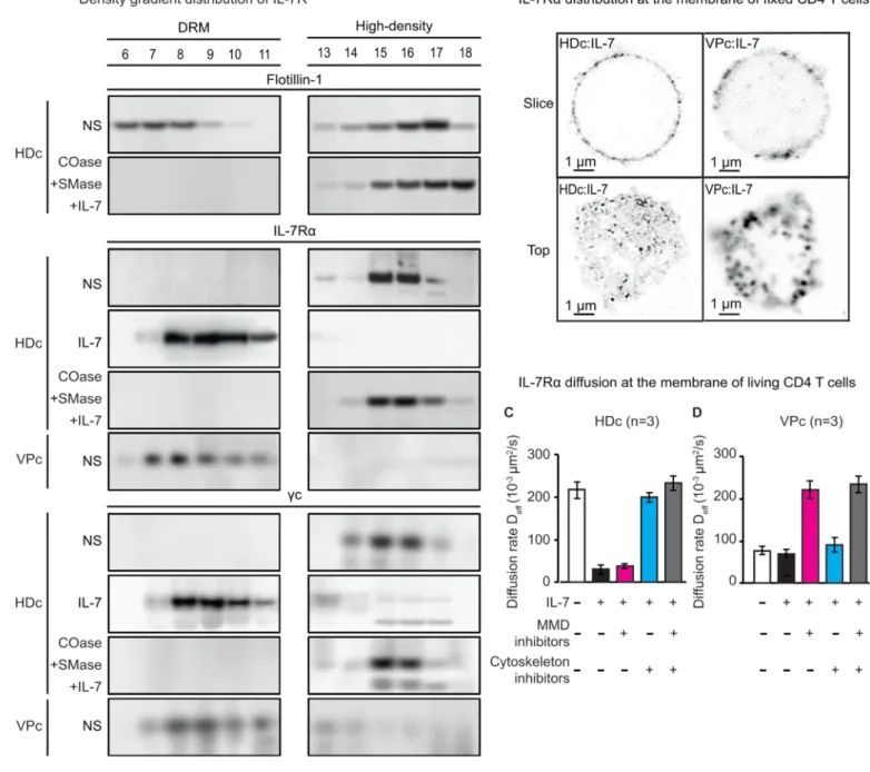
Biochemical analysis of aMMDs from the CD4 T lymphocytes of VPs.
132
We further examined the mechanism linking the presence of aMMDs at the surface of the CD4 133
T lymphocytes from VPs and the loss of function of the IL-7/IL-7R system by performing a 134
biochemical analysis of their membranes. Cell lysates obtained after moderate detergent 135
treatment were examined on sucrose gradients. This technique allowed us to separate free 136
molecules which migrate in high-density fractions and the detergent-resistant membranes 137
(DRMs), structurally related to MMDs, which are recovered in the low-density fractions. 138
Flotillin-1 was found in both fractions and was used as a marker in these experiments (Figure 139
2A and Supplemental Material). IL-7Ra chains and gc chains were found as free molecules in 140
the high-density fractions of HD CD4 T-cell lysates. They were found in low-density DRMs 141
only after IL-7 stimulation (Figure 2A). In contrast, IL-7Ra chains and gc chains were 142
spontaneously found associated with the low-density DRMs in lysates prepared from the 143
Bumpy T cells of VPs, in the absence of any stimulation (Figure 2A). IL-7Ra appeared as 144
clusters in STED images of the CD4 T-cell membranes from VPs (Figure 2B), further 145
supporting the presence of low-density DRMs containing IL-7R chains. We further verified 146
that 7Ra is included in aMMDs by studying its diffusion rate at the membrane surface. IL-147
7Ra was included in the aMMDs of VP CD4 T cells, as their diffusion was limited and could 148
be restored after disruption of the aMMDs by cholesterol oxidase and sphingomyelinase 149
treatment (Figure 2, C and D). 150
Overall these results demonstrate that IL-7Ra and gc are spontaneously embedded in specific 151
macrostructures of the membranes of CD4 T cells from VPs, measured as aMMDs or DRMs. 152
These data also show that the receptors lose their function when trapped in this abnormal 153
structure of Bumpy CD4 T cells. 154
155
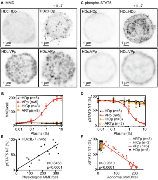
Plasma from VPs induces the Bumpy T-cell phenotype in HD CD4 T cells.
156
We investigated the molecular mechanism leading to the Bumpy T-cell phenotype. The addition 157
of plasma from VPs (Figure 3A) to HD CD4 T cells was sufficient to induce the Bumpy T-cell 158
phenotype. Titration showed the phenotype to be induced in 50% of the cells at approximately 159
1% VP plasma (Figure 3B). HD CD4 lymphocytes treated with VP plasma and Bumpy T cells 160
were microscopically undistinguishable, and the number and size of aMMDs at their surface 161
were not influenced by IL-7. In addition, plasma from elite controllers (HICp) (36, 37) and 162
patients with suppressed viremia after 10 years of ARV (ARTp) could not induce this 163
phenotype (Figure 3B). 164
We then studied the responsiveness of VP plasma-induced Bumpy T cells. IL-7-induced 165
pSTAT5 NT was inhibited by VP plasma, with a half maximum dose of 1% (Figure 3, C and 166
D). These results suggest a direct link between the induction of aMMDs and the mechanism 167
leading to the inhibition of pSTAT5 NT. We found a positive correlation between the number 168
of pMMDs and the frequency of cells with translocated pSTAT5 during IL-7 responses in HD 169
CD4 T cells (Figure 3E). Conversely, we found a negative correlation between the number of 170
aMMDs per CD4 T cell and the percent of cells with nuclear pSTAT5 in IL-7-stimulated 171
induced Bumpy T cells (Figure 3F). These correlations further support that plasma-172
induced aMMDs are responsible for the loss of IL-7 response. 173
174
Phospholipase A2 group IB (PLA2G1B) is a unique inducer of Bumpy T cells.
175
We biochemically characterized the plasma molecule involved in these morphological and 176
functional changes. Size-exclusion and ion exchange chromatography were used to determine 177
the apparent MW and pI of the bioactive molecule(s) from the plasma of three VPs, using 178
microscopy as a read-out (Supplemental Figure 2, A-C). A list of 103 10-15 kDa proteins with 179
a pI between 6.5 and 7.5 and a secretory signal peptide was determined. Differential mass 180
spectrometry analysis identified PLA2G1B, also known as pancreatic phospholipase (35), as 181
the top candidate (PA21B in Supplemental Figure 2D). Active PLA2G1B is produced after the 182
cleavage of seven N-terminal residues from non-active proPLA2G1B (38). Recombinant 183
PLA2G1B was produced, purified, crystallized and structurally characterized. The position of 184
residues H48 and D99 and the Ca2+-binding loop, critical for the activity of the enzyme, are
185
shown in Figure 4A. 186
Recombinant PLA2G1B alone was able to induce aMMDs (Figure 4B) and inhibit pSTAT5 187
NT in HD CD4 T cells (Figure 4C). This property was catalytic site-dependent, as the non-188
active H48Q mutant (39) was unable to induce aMMDs or inhibit pSTAT5 NT on human CD4 189
T cells (Figure 4, D and F). These effects were specific to PLA2G1B; indeed, other members 190
of the PLA2 family such as PLA2GIIA, PLA2GIID or PLA2GX showed no significant effect 191
on either aMMDs formation or pSTAT5 NT inhibition (Figure 4, E and G). Similarly, only 192
polyclonal antibodies specific for PLA2G1B decreased plasma-induced pSTAT5 NT 193
inhibition, whereas polyclonal antibodies specific for PLA2GIIA or PLA2GIID had no effect 194
(Figure 4H). 195
We developed mouse monoclonal antibodies (mAb) specific for PLA2G1B. Among them, mAb 196
14G9 efficiently inhibited the enzymatic activity of PLA2G1B and abrogated VP plasma-197
inhibition of pSTAT5 NT in a dose-dependent manner (Figure 4I). These experiments show 198
that, at physiological concentrations, PLA2G1B is involved in the phenotypic and functional 199
changes induced by VP plasma in HD CD4 T cells and the Bumpy T-cell phenotype observed 200
in VP. 201
202
PLA2G1B induces anergy of CD4 T cells: specificity and reversal of the effects.
203
The unresponsiveness of CD4 T cells to 7 induced by PLA2G1B was also observed for IL-204
2 and IL-4, two other gc cytokines. Similar to IL-7, pSTAT NT-induced by these two cytokines 205
was inhibited by PLA2G1B in a dose-dependent manner and with a comparable IC50 (Figure 206
5A). These observations were verified using VP plasma-induced Bumpy T cells (Figure 5B). 207
Unlike IL-2, IL-4, and IL-7, IFNa-induced pSTAT1 NT was not inhibited by PLA2G1B or the 208
plasma of VPs (Figure 5, C and D). IFN-a is known to signal by a mechanism independent of 209
MMDs (40, 41). Thus, these results suggest that PLA2G1B only affects signaling pathways that 210
involve compartmentalization into pMMDs. We then studied the effects of PLA2G1B first 211
observed on total unseparated CD4 T cells, on naïve (CD45RA+) and memory (CD45RA–) CD4
212
T cells. PLA2G1B was slightly more active on CD45RA+ CD4 T cells than CD45RA– CD4 T
213
cells (Figure 5E). Such differential sensitivity is not the consequence of selective modulation 214
of IL-7R (CD127) expression at the cell surface by PLA2G1B (Figure 5, F and G and 215
Supplemental Figure 3). As previously described, the percentage of CD127-positive cells was 216
slightly higher in CD45RA+ than CD45RA– CD4 T cells (Figure 5F) (42, 43). In addition MFI
217
analysis of CD127 expression (Figure 5G and Supplemental Figure 3) showed a slight reduction 218
in CD45RA- cells as previously reported (44). Overall, these analyses establish that PLA2G1B 219
does not influence CD127 expression and support our hypothesis that PLA2G1B acts on signal 220
transduction (as described above) and not by decreasing receptor expression. 221
The action of PLA2G1B appears to be specific to CD4 T cells. Indeed, PLA2G1B did not 222
induce aMMD formation in purified CD8 T cells from HDs (Figure 5H). Similarly, pSTAT5 223
NT was not affected in CD8 T cells by PLA2G1B, even at high concentrations (Figure 5I). 224
These results are consistent with ex vivo observations of VP CD8 T cells in which aMMDs 225
were not detectable and pSTAT5 NT continued to occur (Supplemental Figure 4, A-C). In 226
addition, physiological concentrations of PLA2G1B present in VP plasma inhibited pSTAT5 227
NT on CD4 T cells but had no functional effects in CD8 lymphocytes purified from HDs 228
(Supplemental Figure 4D). 229
PLA2G1B is known to digest lipids, we thus further explored the difference between the 230
response of CD4 and CD8 T cells to PLA2G1B by lipidomic analysis. There were significant 231
differences in the proportions of the ganglioside GM3, PC, PE, PI, PS, SM and TG between 232
HD CD4 and CD8 T cells (Supplemental Figure 4E). Similarly, differences in the lipid 233
proportions have been reported between murine CD4 and CD8 T cells (45). It is possible that 234
the differential effects of PLA2G1B on CD4 and CD8 T cells are associated with differences 235
in lipid composition, but direct evidence will require extensive studies. 236
We next investigated the reversal of the induced Bumpy T-cell phenotype in vitro and show the 237
results of one of three representative experiments (Figure 5J). Purified CD4 T cells were first 238
treated in vitro with PLA2G1B and then cultured for various periods of time up to 3 days. 239
Inhibition of pSTAT5 NT was examined every day. Under our experimental conditions, 30% 240
of the cells were anergized and did not respond to IL-7. After three days in culture, pSTAT5 241
NT returned to normal, clearly showing that the Bumpy T-cell phenotype is reversible. 242
Furthermore, neutralizing mAb 14G9 accelerated the reversion (Figure 5J). 243
244
PLA2G1B affects CD4 T-cell survival in vitro: inhibition by neutralizing mAb 14G9.
245
Aside from the unresponsiveness of CD4 T cells to physiological signals (anergy), HIV-246
infected patients suffer from CD4 lymphopenia. We thus tested the effects of PLA2G1B on the 247
half-life of CD4 T cells in vitro. Purified CD4 lymphocytes were cultured and the number of 248
live cells counted over time as described in Methods. The number of surviving CD4 T cells 249
varied between donors in control cultures. The effects of PLA2G1B on the CD4 lymphocytes 250
are thus expressed as the percentage of the surviving cells relative to that in control cultures in 251
the absence of PLA2G1B at each time point. The effect of PLA2G1B on CD4 T-cell survival 252
was first tested at various concentrations up to 100 µM (Figure 6A). We then verified that 50% 253
cells died after 18 days of culture in the presence of 1 µM PLA2G1B (Supplemental Figure 254
5A) and 40% of the cells died after 24 days of culture in the presence of 250 nM PLA2G1B 255
(Figure 6B). 256
During these experiments, we further analyzed the CD4 T cells. As expected, numerous dying 257
cells become Annexin positive Zombie-positive. However, we also detected Annexin V-258
negative Zombie-positive cells (Figure 6, C and D). Their percentage increased during culture, 259
reaching more than 70% of the recovered lymphocytes (Figure 6E). This was a specific 260
consequence of PLA2G1B treatment, as such cells were not detected when CD4 T cells were 261
cultured in the presence of the inactive PLA2G1B mutant H48Q (Figure 6C and Supplemental 262
Figure 5B). Similarly, their percentage was lower when cultures were performed in the presence 263
of mAb 14G9, which neutralizes the enzymatic activity of PLA2G1B (Figure 6, D and E). This 264
can be explained by the fact that PLA2G1B digested one of its substrates during culture, 265
phosphatidylserine, which is also the binding site of Annexin V. This confirms that the action 266
of PLA2G1B on CD4 T cells is mediated by its enzymatic activity. 267
We then tested the effect of mAb 14G9 on CD4 T-cell survival. The mAb significantly 268
increased the survival of CD4 T cells exposed to PLA2G1B (up to > 50%) relative to cultures 269
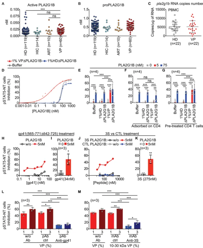
in the presence of the control isotype (Figure 6F and Supplemental Figure 5C). 270
271
In vitro and in vivo effects of human PLA2G1B in a mouse model.
272
We studied the effects of human PLA2G1B in mice after verifying its activity in vitro on mouse 273
CD4 T cells to extend our data in vivo. Upon exposure to PLA2G1B we also observed Annexin 274
V-negative Zombie-positive mouse CD4 T cells (Supplemental Figure 6). CD4 T cells from 275
C57BL/6 mice were purified from the spleen and stimulated by anti-CD3 plus anti-CD28 beads, 276
in the presence of IL-2. Human PLA2G1B was active on mouse CD4 T cells and induction of 277
CD25 (IL-2Ra) was inhibited by PLA2G1B by day 5 in a dose-dependent manner (Figure 7, 278
A and B). Similarly, survival and proliferation were profoundly altered (Figure 7, C-E). These 279
effects depended on the catalytic activity of PLA2G1B, as the H48Q mutant was ineffective 280
(Figure 7, A-E). Furthermore, neutralizing mAb 14G9 blocked CD25 induction and decreased 281
survival of the CD4 cells (Figure 7, F and G). These data demonstrate that PLA2G1B can also 282
inhibit TCR responses. In addition to the human experiments, these data establish that the 283
effects of PLA2G1B can be measured after several days in culture. 284
In vivo, PLA2G1B showed activity on mouse CD4 lymphocytes in a dose-dependent manner 285
(Figure 7H). Injection of 100 µg of PLA2G1B induced a long-lasting effect, which persisted 286
for up to 72 hours and began to diminish after 168 hours (Figure 7I). The effect of PLA2G1B 287
was maximal three hours after injection (Figure 7J). We tested the effects of pre-treatment with 288
the anti-PLA2G1B neutralizing mAb 14G9 under the same experimental conditions (Figure 289
7K). The blockade was close to 100%. We obtained a comparable result in a group of mice that 290
was pre-immunized against human PLA2G1B (Figure 7L). 291
The cell specificity of the effects of PLA2G1B was further verified in this experimental model. 292
Injection of PLA2G1B did not result in any loss of IL-7-induced pSTAT5 NT in mouse CD8 293
lymphocytes, as measured ex vivo three hours post-injection (Supplemental Figure 4F). 294
Overall, these results open the possibility of using mouse models to evaluate anti-PLA2G1B 295
neutralizing mAbs as an immunotherapeutic strategy. 296
297
Synergy between PLA2G1B and plasma HIV gp41 protein.
298
We then measured active and proPLA2G1B in the plasma of HD, HIC, ARV, and VP patients 299
using PLA2G1B ELISAs (Figure 8, A and B). VPs had similar levels of active PLA2G1B as 300
HIC and ART patients but slightly more active PLA2G1B than HDs (median increase of 1.4). 301
In addition, comparable copy numbers of pla2g1b RNA were found in the PBMCs of HDs and 302
VPs by qPCR (Figure 8C). We thought that these results cannot explain the difference in 303
PLA2G1B activity observed with the functional assays (Figure 3, B and D). This observation 304
led us to consider that one or more cofactors are present in the plasma of VPs and are required 305
for the induction of aMMDs and blockade of pSTAT5 NT. Indeed, the dose-response curve 306
(from 0.001 to 1,000 nM) of PLA2G1B diluted in HD or VP plasma previously depleted of 307
endogenous PLA2G1B showed striking differences in the IC50 values (Figure 8D), supporting 308
the presence of a cofactor. The IC50 of PLA2G1B was 75 nM when diluted in PBS buffer or 309
HD plasma, but decreased to 5 nM when diluted in PLA2G1B-depleted VP plasma. We 310
concluded that PLA2G1B was not acting alone but in synergy with another factor present in 311
VP plasma. A bioassay was developed, using a limiting amount of PLA2G1B inactive by itself 312
(5 nM, Figure 8D) and PLA2G1B-depleted VP plasma to detect the potential cofactor (Figure 313
8E), and used to show that the enhancement was lost after plasma was incubated with purified 314
CD4 T cells (Figure 8F), suggesting that the cofactor was adsorbed on CD4 T cells. After such 315
pretreatment, the addition of 5 nM PLA2G1B to the CD4 T cells, without further addition of 316
VP plasma, resulted in the inhibition of IL-7 driven pSTAT5 NT (Figure 8G). This cofactor 317
activity was sensitive to trypsin treatment and could be fractionated with an apparent MW 318
between 10 and 30 kDa. The search then focused on HIV peptides possibly released into the 319
plasma of infected patients. A gp41 fragment (MN 565-771 delta 642-725), and its 320
corresponding 3S peptide (46), of which the sequence is highly conserved among various HIV 321
isolates, exerted strong cofactor activity (Figure 8, H-K). In addition, the depletion of VP 322
plasma with anti-gp41-specific antibodies that do not bind to gp120 (Supplemental Figure 7) 323
resulted in the loss of cofactor activity (Figure 8L). This critical point was definitively 324
established after depletion by a mAb characterized in the laboratory. This mAb, 1C5, was raised 325
against the 3S peptide and shown to also recognize gp41 protein but not gp120 (Supplemental 326
Figure 7). It was also able to completely deplete various VP plasma samples of cofactor activity 327
(Figure 8M). These results support the hypothesis that fragments of gp41 containing the 3S 328
sequence may act as cofactors that target PLA2G1B to the surface of CD4 lymphocytes to exert 329
its enzymatic activity. 330
331
DISCUSSION 332
Here, we further characterize the activation status of the CD4 T-cell compartment in HIV-333
infected patients. Aside from currently used cell-surface activation markers, such as CD38 and 334
HLA-DR, we show structural alterations of CD4 T-cell membranes, consisting of large aMMDs 335
observed at the cell surface. They arise due to the activity of the PLA2G1B enzyme, acting in 336
synergy with gp41. These findings support a new mechanism of immunosuppression in HIV-337
infected patients. Here, we show that this mechanism is involved in CD4 anergy to gc cytokine 338
and TCR responses. Furthermore, its action on the decreased survival of CD4 T cells suggests 339
a role in CD4 lymphopenia. 340
Bumpy T cells represent a new phenotype characterizing activated CD4 T cells found in HIV-341
infected patients. It results from remodeling of the CD4 T-cell membrane under the influence 342
of the enzymatic activity of PLA2G1B. This exposes GM1 gangliosides, which are enriched in 343
aMMDs and recognized by labeled cholera toxin B using the STED imaging technique. Bumpy 344
T cells expressing numerous aMMDs up to 500/cell can be recovered from patient blood and 345
easily identified. In the blood of these individuals, aMMDs are spontaneously expressed by 346
CD4 T cells and their characteristic pattern is not modified by a strong stimulus, such as IL-7. 347
Given that this phenotype is observed in more than 80% of the CD4 T cells of HIV-infected 348
patients, a correlation with CD38 and HLA-DR activation markers should be investigated. In 349
any case, this observation offers new insights into our understanding of the dysfunction of the 350
CD4 compartment of HIV-infected patients. 351
At low concentrations, such as those found in the blood, PLA2G1B cannot act by itself. In HIV-352
infected patients, it requires a cofactor, which we identified as a soluble fragment of gp41. Gp41 353
appears to be a driver that targets PLA2G1B to the CD4 T-cell surface. Molecular dissection 354
of gp41 demonstrated that the 3S peptide of gp41 is the active moiety. Plasma gp41 is most 355
likely a degradation product of circulating HIV or HIV-infected dead cells. We analyzed the 356
spatiotemporal mode of action of these two molecules when in the plasma. Our results suggest 357
that they act in two separate steps. Gp41 acts first and possibly modifies the CD4 membrane. 358
Gp41 may bind to CD4 T-cell membranes by binding to the gC1qR through its 3S sequence 359
(47, 48). This may change the membrane composition, possibly through induction of the fusion 360
of exocytic vesicles with the plasma membrane, as previously shown for NKp44L induction 361
(47). In the second step, these changes in lipid composition of the outer leaflet of the CD4 T-362
cell may increase the binding and activity of PLA2G1B (49), making the modified membrane 363
a target of PLA2G1B. 364
PLA2G1B is the active moiety of the PLA2G1B/gp41 pair. At high concentrations, it appears 365
to act, in the absence of gp41, as a newly discovered immune modulator that acts on CD4 T 366
cells. Its unique properties, under these conditions, were defined in vitro or in vivo in mouse 367
experiments. PLA2G1B induces numerous aMMDs, trapping and inactivating the function of 368
receptors (gc family, TCR,…), thus decreasing physiological activation, proliferation, and 369
survival. The results obtained with IFNa show that PLA2G1B works selectively on systems 370
that use pMMDs for physiological signaling. It is possible that, by digesting phospholipids, 371
PLA2G1B modifies membrane composition and changes its fluidity, allowing pMMDs to fuse 372
and form receptor-inactivating aMMDs. 373
Despite this broad activity, the action of PLA2G1B remains specific and does not act directly 374
on CD8 T cells. However, this mechanism may indirectly affect CD8 responses in HIV-infected 375
patients, which are mostly highly CD4 T cell-dependent (50). It will be of interest to extend our 376
studies to other immune cells, such as NK cells, which are highly dependent on the gc cytokine 377
IL-15 for their activity (51). 378
In this context, we sought the origins of active PLA2G1B in the blood. We found the pancreas 379
to be a major source of PLA2G1B, followed by the duodenum, jejunum, and ileum 380
(Supplemental Figure 8, A-C). We used two different mAbs, 1C11 recognizing both 381
proPLA2G1B and active PLA2G1B and 14G9 specific for the active form, and observed that 382
proPLA2G1B is expressed in the endocrine pancreas while active PLA2G1B is mainly 383
expressed in the exocrine pancreas and intestinal tissues. We also observed low amounts of 384
pla2g1b transcripts in lymphoid cells (CD4 or CD8 T cells and Natural Killer cells) by qPCR,
385
whereas they were almost undetectable in myeloid cells (lung macrophages, mDC and pDC 386
dendritic cells) (Supplemental Figure 8D). The mechanism leading to the presence of active 387
and proPLA2G1B in the blood is still unknown. They may leak from the intestinal tract, where 388
proPLA2G1B is probably cleaved to form active PLA2G1B by proteolytic enzymes. 389
Conversion of proPLA2G1B into the active molecule may also take place at immune sites 390
where inflammatory cells produce proteolytic enzymes. Initially PLA2G1B was thought to play 391
solely a digestive role (35). Because of its function as an immunomodulator, demonstrated here, 392
it may also play a crucial role in the regulation of the lymphoid compartment of the gut immune 393
system. It may be involved in tolerance towards food and microbiota-derived components (52). 394
There is a high level of pla2g1b RNA in duodenum (Supplemental Figure 8A) and a high 395
concentration of active PLA2G1B protein in the intestinal lumen (Supplemental Figure 8B). 396
Thus, it may act directly on CD4 T cells or, alternatively, cofactors derived from bacteria or 397
viruses of the microbiota may be involved. These hypotheses may open new challenging areas 398
of investigation. 399
Our results also have other consequences for our understanding of the immunodeficiency of 400
HIV-infected patients. They show PLA2G1B to be the active component of the 401
PLA2G1B/gp41 pair and to contribute to the processes that renders most of the major 402
conventional CD4 T-cell subpopulations anergic. First, we have clearly demonstrated 403
dysfunction of the IL-7R/IL-7 induced microtubule and microfilament reorganization, therefore 404
blocking pSTAT5 NT. Furthermore, all gc cytokines lost their function because of sequestering 405
of the gc chains in macro-MMDs (aMMDs). This was verified by studying the blockade of IL-406
2- and IL-4-induced pSTAT NT after PLA2G1B treatment. Furthermore, the TCR responses 407
induced by anti-CD3/anti-CD28 were also inhibited after PLA2G1B treatment. Inhibition of 408
the function of gc cytokines and TCR responses leads to the blockade of antigen-specific 409
responses. Overall, our results show that Bumpy T cells obtained in vitro are anergic and 410
strongly suggest that the same is true for Bumpy T cells recovered from HIV-infected patients. 411
Furthermore, our data suggest that PLA2G1B is also involved in the CD4 lymphopenia 412
observed in HIV-infected patients. IL-7 is the main cytokine that controls homeostasis of the 413
CD4 compartment (53, 54). Thus, the PLA2G1B-induced defect of the IL-7R signaling 414
mechanism described here should significantly contribute to CD4 lymphopenia. Furthermore, 415
the number of surviving CD4 lymphocytes decreased after exposure to PLA2G1B in culture 416
(Figure 6, A and B). During these in vitro studies, we found numerous Annexin V-negative 417
Zombie-positive CD4 T-cells, which can be considered to be the direct consequence of 418
PLA2G1B activity, as they indicate the degradation of phosphatidylserine at the surface of the 419
dying cells (Figure 6, C-E). The activity of PLA2G1B on the membrane of dying cells led to 420
hypothesize an additional role of PLA2G1B in the removal of damaged cells, as previously 421
described for sPLA2 (35, 49). This may also contribute to the decreased in CD4 T-cell number. 422
In the future, Annexin V-negative dying cells could be used as a signature to study the effects 423
of PLA2G1B on the CD4 T cells of HIV-infected patients. This could be used to verify the 424
activity of PLA2G1B in vivo and to follow its neutralization after anti-PLA2G1B 425
immunotherapy. 426
PLA2G1B may represent a compelling therapeutic target for boosting immune responses in 427
people contaminated by HIV. In this context, the potential of the specific mAb 14G9 has to be 428
considered. It completely neutralized the effects of PLA2G1B in vitro, as measured by the 429
inhibition of pSTAT5 NT. This property was also verified in vivo, using a mouse model. 430
Furthermore, 14G9 accelerated the reversion of Bumpy cells in vitro, as measured by their 431
capacity to recover an IL-7 response. It also significantly reduced (up to >50%) the capacity of 432
PLA2G1B to decrease cell survival in vitro. Thus, neutralization of the deleterious effects of 433
the PLA2G1B/gp41 pair may be considered as a new therapeutic tool. It should be able to boost 434
the immune system early in infection, at a time when PLA2G1B is pathogenic as a consequence 435
of its synergy with gp41. Treatment during the beginning of ARV therapy, at a stage when the 436
viral load remains detectable, may increase the CD4 T cell-dependent immune defense and 437
improve the control of HIV infection. In addition, anti-PLA2G1B therapy could have positive 438
effects in patients infected by ARV-resistant HIV strains. After humanization, 14G9 could be 439
a drug candidate that could be used to boost the functions of the CD4 compartment and the 440
CD4-dependent immune responses of HIV-infected patients. By restoring IL-7 responses, 441
decreasing anergy, and contributing to an increase in CD4 counts, neutralization of PLA2G1B 442
may be one of the critical parameters towards remission of HIV infection (55–57). 443
The in vivo relevance of our data needs to be further underscored. The most significant data 444
comes from the analysis of the role of plasma from VPs. PLA2G1B in VP plasma is at 445
physiological concentrations and, in the presence of gp41, induces unresponsiveness of CD4 446
lymphocytes. The activity of plasma from VPs is well established. At the morphological level, 447
Bumpy T cells directly purified from the blood of VPs are indistinguishable from in vitro VP 448
plasma-induced Bumpy T cells. At the functional level, we found that aMMDs characterized 449
from purified CD4 T cells trap all gc chains and subsequently showed that HD CD4 T cells 450
treated with VP plasma become unresponsiveness to gc cytokines, such as IL-2, IL-4, and IL-7 451
(Figure 5, A and B). Inhibition or depletion of either PLA2G1B or gp41 abolishes the activity 452
of VP plasma (Figure 4, H and I and Figure 8, L and M). We thus propose a mechanism whereby 453
PLA2G1B is the active moiety and gp41 a cofactor or driver that targets PLA2G1B to the 454
surface of CD4 T cells. The activity of the VP plasma results from synergy between the two 455
molecules. 456
We then analyzed PLA2G1B and gp41 separately in vitro to understand the respective roles of 457
the two molecules. At high concentrations, PLA2G1B is active alone. High concentrations 458
appear to compensate for the absence of gp41. This allows characterization of the biochemical 459
and immunological properties of the active molecule. More significantly, at low concentrations, 460
cloned PLA2G1B was not active and did not induce unresponsiveness of the CD4 lymphocytes 461
in the absence of gp41. Under these experimental conditions, we clearly show that gp41 boosts 462
PLA2G1B activity (Figure 8, H-K). PLA2G1B becomes pathogenic only after CD4 463
lymphocytes have interacted with the gp41/3S peptide (Figure 8). Therefore, this in vitro 464
analysis further supports the model that synergy with viral gp41 is required to observe the 465
effects of low concentrations of PLA2G1B. 466
In addition, studies of three clinical settings confirm the relevance of our observations. 467
PLA2G1B activity strictly correlated with the presence of HIV particles. We only found 468
PLA2G1B activity in VPs who had HIV particles in the circulating blood and therefore gp41 469
in the plasma. In contrast, HIV controllers and patients treated for more than 10 years with 470
ARV, with an undetectable viral load in their plasma, had no detectable PLA2G1B activity. 471
Furthermore, we observed a negative correlation between the ability of plasma to induce 472
aMMDs in vitro and the CD4 counts of the patient source of plasma in a preliminary analysis 473
(Supplemental Figure 9A). Similarly, plasma from patients with CD4 counts below 300/mm3
474
more strongly inhibited pSTAT5 NT than plasma from patients with CD4 counts above 475
300/mm3 (Supplemental Figure 9B). Overall, these results corroborate the notion of a viral
476
cofactor/driver that synergizes with PLA2G1B to induce CD4 T lymphocyte unresponsiveness 477
in vivo. 478
It is noteworthy that the PLA2G1B/gp41 pair appears to be a new mechanism of 479
immunopathology, in which a physiological enzyme becomes pathogenic in the presence of 480
molecules derived from the pathogen. This system may play a role in diseases in which 481
immunodeficiency contributes to the emergence or progression of the disease. Preliminary data 482
obtained with hepatitis C, Staphylococcus aureus, and Porphyromonas gingivalis support this 483
concept. 484
METHODS 485
Characterization of membrane microdomains (MMDs) in primary human CD4 and CD8 T
486
cells.
487
Cell preparation and labelling of specific proteins were performed as previously described (34). 488
Briefly, purified cells were equilibrated in RPMI with 5% FBS for 2 h at 37°C and 5% CO2
489
before plating them onto poly-L-lysine-coated coverslips for 20 min at 37°C. Cells were treated 490
with IL-7 (2 nM, 15 min, 37°C) then fixed with 1.5% paraformaldehyde (PFA, Electron 491
Microscopy Sciences) and rehydrated for 15 min in PBS/5% FBS. GM1 gangliosides were 492
labelled with AlexaFluor-coupled cholera toxin subunit B (CtxB-AlexaFluor488, C22841; 493
CtxB-AlexaFluor633, C34778; or CTxB-Biotin, C34779 and Streptavidin-AlexaFluor647, 494
S32357, Life Technologies). 495
MMDs were analyzed at the surface of fixed CD4 T cells and CD8 T cells from viremic patients 496
(VPc) and healthy donors (HDc) in response to IL-7 stimulation or not (Figure 1, A-C and 497
Supplemental Figure 4) or purified HD CD4 or CD8 T cells upon treatment with plasma 498
samples (from HD, VP, ART, or HIC), WT or H48Q PLA2G1B, PLA2GIIA, PLA2GIID, or 499
PLA2GX recombinant proteins for 30 min and stimulation with the cytokine for 15 min 500
(Figures 3 to 5). 501
Images were acquired below the diffraction limit using a Leica TCS STED-CW (31) (Figures 502
1 and 3 and Supplemental Figure 1) or Leica TCS SP8 STED 3X (Figure 4B) or above the 503
diffraction limit using an inverted laser scanning confocal microscope (LSM700, Zeiss or 504
LSM780 ELYRA PS.1, Zeiss) as previously described (34). Deconvolution was performed 505
using Huygens Pro software (Scientific Volume Imaging, Hilversum, The Netherlands). For 506
each condition, the top half of a representative CD4 T cell is shown from Z-stack images. 507
MMDs were counted on the entire surface of the purified CD4 T cells; an average of 50 cells 508
were examined for HD and between 15 to 50 for VP. We determined the number of MMDs in 509
Figures 1 and 3 or the percentage of cells with aMMDs on their surface in Figures 4 and 5. 510
511
Phosphorylation and nuclear translocation of STAT (pSTAT NT).
512
STAT phosphorylation and nuclear translocation in VP and HD CD4 T cells were analyzed by 513
microscopy after IL-7 stimulation (2 nM), or in HD CD4 and CD8 T cells incubated with 514
plasma samples from HD, VP, ART, or HIC (30 min), WT or H48Q PLA2G1B, PLA2GIIA, 515
PLA2GIID, or PLA2GX recombinant proteins, with or without neutralizing antibodies (30 min) 516
before a 15 min stimulation with 2 nM IL-7, IL-2, or IL-4 or 1 nM IFN-a2. WT and H48Q 517
porcine PLA2G1B were used for the experiments shown in Figure 4D and F. The effect of the 518
anti-PLA2G1B 14G9 mAb on the recovery of a functional pSTAT5 NT response was studied 519
by pretreating CD4 T cells for 1 h with 250 nM PLA2G1B before the addition of 667 nM anti-520
PLA2G1B mAb (14G9). All pre-treatments and stimulations were performed at 37°C. 521
Stimulation was stopped by addition of 4% PFA and incubation for 15 min at 37°C. Cells were 522
then permeabilized overnight at -20°C in a 90% methanol/water solution. 523
CD4 and CD8 T cells were stained using respectively anti-human CD4 (mouse anti- CD4 clone 524
RPA-T4, 555344, BD Biosciences; or goat CD4, AF-379-NA, R&D/Novus) and anti-525
human CD8 (mouse anti-CD8 clone RPA-T8, 555364, BD Biosciences), labelled with donkey 526
anti-mouse-AlexaFluor488 (A21202, Thermofisher) or donkey anti-goat-AlexaFluor488 527
(A11055, Thermofisher). Phosphorylation of STAT5 in response to IL-2 or IL-7 stimulation 528
was then revealed by staining with rabbit anti-pSTAT5 (9356, Cell Signaling Technology) 529
labelled with goat anti-rabbit-Atto 647N (15068; Active Motif) or donkey anti-rabbit 530
AlexaFluor555 (A31572, Life Technologies), that of STAT6 in response to IL-4 stimulation by 531
rabbit anti-pSTAT6 (9361, Cell Signaling Technology) labelled with anti-rabbit-532
AlexaFluor488 (A11034 or A21206, Life Technologies), and that of STAT1 in response to 533
IFN-a2 stimulation by rabbit anti-pSTAT1 (9167, Cell Signaling Technology) labelled with 534
anti-rabbit-AlexaFluor 488 (A11034 or A21206, Life Technologies). 535
Images were acquired below the diffraction limit with a DM16000CS/SP5 inverted laser 536
scanning confocal microscope using pulsed excitation STED (TCS STED, Leica) (58) or above 537
the diffraction limit using an inverted laser scanning confocal microscope (LSM700 or LSM780 538
ELYRA PS.1, Zeiss) as previously described (34). Deconvolution was performed using 539
Huygens Pro software (Scientific Volume Imaging, Hilversum, The Netherlands). The 540
appearance of pSTAT was measured using ImageJ software. pSTAT5 was quantified in the 541
cytoplasm and nucleus of the cells (Figures 1 and 3 and Supplemental Figure 1) where 542
indicated. The number of cells positive for nuclear pSTAT among > 200 in response to 543
cytokines was analyzed by confocal microscopy in Figures 4, 5, 7, and 8 and Supplemental 544
Figure 4. 545
Study of the effect of PLA2G1B on human CD4 T-cell survival
546
Purified CD4 T cells were cultured (7x10⁶ cells/mL) in RPMI 1640 medium supplemented with
547
5% FBS (Life Sciences – Gibco). FBS was initially selected for its capacity to support efficient 548
CD4 T-cell activation in response to anti-CD3/CD28 stimulation, as measured by CD69 cell-549
surface expression. The same FBS was also later found to support long-term survival of these 550
lymphocytes. CD4 T cells were treated with PBS, PLA2G1B, or the inactive mutant H48Q 551
PLA2G1B alone or with the anti-PLA2G1B mAb 14G9 (Figure 6 and Supplemental Figure 5) 552
or control isotype (Mouse IgG1, 16-4714-85, Thermofisher, Figure 6 and Supplemental Figure 553
5). The effect of PLA2G1B on CD4 T-cell survival was evaluated by a Moxy Z Mini Automated 554
Cell Counter (Moxy Z, Orflo technologies). Moxy Z measures cell counts, cell size, and cell 555
health. Cell heath is evaluated via the Moxy Viability Index (MVI) value (59). The results based 556
on the MVI were analogous to those obtained by hemocytometer count of cells stained with 557
Trypan blue (0.1%). 558
For the Annexin V experiments, CD4 T cells were stained with AlexaFluor488-labelled 559
antibodies against CD4 (300519, Biolegend) for 30 min at 4°C, the Zombie Violet Fixable 560
Viability Kit (0.5 µL/test) (423114, Biolegend) and Annexin V-APC (5µL/test) (640941, 561
Biolegend) for 15 min at RT. Cells were analyzed with a cytoflex cytometer (Beckman Coulter) 562
and FlowJo software, version 10 (Tree Star). 563
564
Determination of gp41 and 3S plasma cofactor peptide activity on healthy donor CD4 T cells.
565
The effect of gp41 on PLA2G1B activity on CD4 T cells was assessed by incubating purified 566
CD4 T cells in PBS/1% BSA containing peptides, recombinant proteins, VP or HD plasma, or 567
the 10-30 kDa fraction previously depleted, or not, of PLA2G1B or gp41 (Supplemental 568
Material), together with recombinant PLA2G1B. 569
The binding of the viremic plasma cofactor to CD4 T cells was tested by first incubating the 570
PLA2G1B-depleted plasma with CD4 T cells for 15 min. Then, the adsorbed plasma was 571
collected and incubated with other CD4 T cells from the same donor for 30 min, alone or 572
together with PLA2G1B (Figure 8F). 573
The pretreatment effect of viremic plasma cofactor on CD4 T cells was tested by first incubating 574
the PLA2G1B-depleted plasma with CD4 T cells bound onto poly-L-lysine-coated coverslips 575
for 15 min. Then the supernatant was removed and the cells were washed and incubated for 30 576
min, with or without PLA2G1B (Figure 8G). pSTAT5 NT was analyzed in CD4 T cells 577
incubated with the adsorbed supernatants. 578
The effect of the recombinant gp41 and 3S gp41 peptide on PLA2G1B activity was tested by 579
pretreating the cell suspension for 15 min with 40 µl of the recombinant gp41 protein, peptides, 580
with subsequent addition of 10 µl PLA2G1B for 30 min (Figure 8, H-K). The regulation of 581
PLA2G1B by endogenous gp41 was tested by treating the cell suspension for 30 min with 50 582
µl of plasma dilutions or 10-30 kDa plasma fraction, previously depleted or not of gp41 583
(Figures 8, L and M). pSTAT5 NT inhibition was examined by microscopy as described above. 584
585
Additional reagents and procedures are detailed in the online Supplemental Material, which 586
includes information on study design and human sample collection, recombinant proteins and 587
peptides, cell purification and culture, detergent-resistant microdomain (DRMs) analysis, 588
western-blot analyses, analysis of the IL-7 receptor (IL-7R) diffusion rate at the surface of 589
living CD4 T lymphocytes, the study of the effect of cytoskeleton inhibitors on pSTAT5 NT, 590
analysis of cytoskeleton organization, identification of PLA2G1B as the active component in 591
VP plasma, active human PLA2G1B structure determination, lipidomic analysis, ELISA, 592
quantitative real-time PCR, immunodepletion experiments, flow cytometry analyses, in vitro 593
experiments on mouse T cells, and in vivo experiments in mice. 594
595
Statistics
596
Statistical parameters, including the exact value of n, precise measures (mean ± SD in all 597
Figures, with the exception of the mean ± SEM in Figure 7, B-G), statistical significance, and 598
tests used for each analysis are reported in the figures and figure legends. Analyses were 599
performed using GraphPad Prism (GraphPad Software Inc.). 600
For experiments on human cells, one donor represents one experiment. For experiments on 601
mice, the number of pooled mice from n independent experiments is shown. 602
Correlations between two variables were evaluated by Pearson’s correlation and linear 603
regression. 604
Data were analyzed using the two-tailed unpaired t-test for two groups or ANOVA with 605
correction for multiple comparisons (Tukey’s, Dunnett’s, or Sidak’s) when the distribution was 606
gaussian according to the D'Agostino & Pearson omnibus test. The effect of PLA2G1B on the 607
survival of mouse CD4 T cells in G0 to G5 was analyzed by two-way ANOVA with Dunnett’s 608
correction for multiple comparisons using the control condition as the control group. The anti-609
PLA2G1B effect on CD25 expression and the survival of PLA2G1B-treated mouse CD4 T 610
cells, as well as the kinetics of the effect of PLA2G1B injection on the percentage of cells 611
showing pSTAT5-NT, were analyzed by two-way ANOVA with Sidak’s correction for 612
multiple comparisons. When data were not Gaussian, Mann-Whitney’s non parametric test was 613
used to compare two groups and Kruskal-Wallis test was used when more than two groups were 614
compared. When Kruskal-Wallis test was significant, two-by-two comparisons were conducted 615
to identify groups which differed, but applying a Bonferroni correction. The level of 616
significance is indicated as *p < 0.05, **p < 0.01, and ***p < 0.001 in all figures.
617
Study approval
618
Human study: This study was supported by the ANRS and approved by the Comité des
619
Personnes Ile-de-France VII under number 05-15. All participants were adults and provided 620
written informed consent prior to inclusion in the study. 621
Animal studies: All animal experiments described in the present study were conducted at the
622
Institut Pasteur according to European Union guidelines for the handling of laboratory animals 623
(http://ec.europa.eu/environment/chemicals/lab_animals/home_en.htm) and were approved by 624
the Institut Pasteur Animal Care and Use Committee (CETEA 89, Institut Pasteur de Paris) and 625
the Direction Sanitaire et Vétérinaire de Paris under permit number 2016-0004 and 626
APAFIS#6453-2016071912038344 v2. All experiments were subject to the three R's of animal 627
welfare (refine, reduce, and replace). 628
AUTHOR CONTRIBUTIONS 629
J.P., T.R., and F.B. designed and conducted the experiments on human and mouse cells. The 630
study was initiated by T.R and continued by J.P, who participated in the writing of the 631
manuscript. D.G. performed the TCS SPS STED analysis. T.R. designed and conducted the 632
plasma chromatography, biochemical analysis, microscopy image analysis, MS, and 633
bioinformatics data analysis. L.J. participated in the mAb characterization, ELISA 634
development, and recombinant protein production. A.M. participated in the characterization of 635
anti-gp41 mAb. A.H. and F.S. designed and conducted the structural analysis. J.A. and E.R.-636
M. participated in the studies of plasma from HIV-infected patients. L.T. provided PLA2G1B 637
protein. G.L. contributed to the design of certain experiments, provided ideas and models, and 638
shared resources. J.T. was the lead senior author for this paper. 639
ACKNOWLEDGMENTS 640
This work was part of the ANRS programs EP20, EP33, and EP36 (J.-F. Delfraissy, O. 641
Lambotte). It was initially supported by the Institut Pasteur (PTR 424) and the Pasteur-642
Weizmann Foundation. We are grateful to P. Pouletty for continuous interest and support. We 643
wish to thank U. Schwarz (Leica Microsystems, Mannheim), E. Perret, P. Roux, A. Salles, and 644
S. Shorte (Imagopole, Institut Pasteur) for their microscopy expertise, as well as A.-H. Pillet 645
for her expertise in biochemistry and P. Bochet for data processing. We thank Yoann Madec 646
and Fredj Tekaia for their help and expertise in statistics. We acknowledge SOLEIL for the 647
provision of synchrotron radiation facilities and thank the staff of the PROXIMA-1 beamline 648
for their assistance. We benefited greatly from help and numerous discussions with C. Abrial, 649
L. Touqui, B. Colsch, D. Troisvallet, M.-L. Gougeon, P. Bruhns, and J. Tiollier. We also 650
gratefully acknowledge J.-P. Routy and B. Malissen for their critical review of the manuscript. 651
REFERENCES 652
1. Walker B, McMichael A. The T-cell response to HIV. Cold Spring Harb Perspect Med. 653
2012;2(11):a007054. 654
2. Klatt NR, Chomont N, Douek DC, Deeks SG. Immune activation and HIV persistence: 655
implications for curative approaches to HIV infection. Immunol Rev. 2013;254(1):326–342. 656
3. Pitman MC, Lau JSY, McMahon JH, Lewin SR. Barriers and strategies to achieve a cure for 657
HIV. Lancet HIV. 2018;5(6):e317–e328. 658
4. Doitsh G, et al. Cell death by pyroptosis drives CD4 T-cell depletion in HIV-1 infection. 659
Nature. 2014;505(7484):509–514.
660
5. Doitsh G, Greene WC. Dissecting How CD4 T Cells Are Lost during HIV Infection. Cell 661
Host Microbe. 2016;19(3):280–291.
662
6. Deeks SG, Tracy R, Douek DC. Systemic effects of inflammation on health during chronic 663
HIV infection. Immunity. 2013;39(4):633–645. 664
7. de Armas LR, et al. Reevaluation of immune activation in the era of cART and an aging 665
HIV-infected population. JCI insight. 2017;2(20):e95726. 666
8. Sousa AE, Carneiro J, Meier-Schellersheim M, Grossman Z, Victorino RMM. CD4 T cell 667
depletion is linked directly to immune activation in the pathogenesis of HIV-1 and HIV-2 but 668
only indirectly to the viral load. J Immunol. 2002;169(6):3400–3406. 669
9. George V, et al. Associations of Plasma Cytokine and Microbial Translocation Biomarkers 670
With Immune Reconstitution Inflammatory Syndrome. J Infect Dis. 2017;216(9):1159–1163. 671
10. Tincati C, Douek DC, Marchetti G. Gut barrier structure, mucosal immunity and intestinal 672
microbiota in the pathogenesis and treatment of HIV infection. AIDS Res Ther. 2016;13(1):19. 673
11. Brenchley JM, et al. Microbial translocation is a cause of systemic immune activation in 674
chronic HIV infection. Nat Med. 2006;12(12):1365–1371. 675
12. Hocini H, et al. HIV Controllers Have Low Inflammation Associated with a Strong HIV-676
Specific Immune Response in Blood. J Virol. 2019;93(10):e01690-18. 677
13. Palmer BE, Blyveis N, Fontenot AP, Wilson CC. Functional and phenotypic 678
characterization of CD57+CD4+ T cells and their association with HIV-1-induced T cell 679
dysfunction. J Immunol. 2005;175(12):8415–8423. 680
14. Harari A, Petitpierre S, Vallelian F, Pantaleo G. Skewed representation of functionally 681
distinct populations of virus-specific CD4 T cells in HIV-1–infected subjects with progressive 682
disease: changes after antiretroviral therapy. Blood. 2004;103(3):966–972. 683
15. Clerici M, et al. Detection of three distinct patterns of T helper cell dysfunction in 684
asymptomatic, human immunodeficiency virus-seropositive patients. Independence of CD4+ 685
cell numbers and clinical staging. J Clin Invest. 1989;84(6):1892–1899. 686
16. Boswell KL, et al. Loss of Circulating CD4 T Cells with B Cell Helper Function during 687
Chronic HIV Infection. PLoS Pathog. 2014;10(1):e1003853. 688
17. Pallikkuth S, de Armas L, Rinaldi S, Pahwa S. T Follicular Helper Cells and B Cell 689
Dysfunction in Aging and HIV-1 Infection. Front Immunol. 2017;8:1380. 690
18. Jiang W, et al. Cycling Memory CD4+ T Cells in HIV Disease Have a Diverse T Cell 691
Receptor Repertoire and a Phenotype Consistent with Bystander Activation. J Virol. 692
2014;88(10):5369–5380. 693
19. Sieg SF, Bazdar DA, Harding C V, Lederman MM. Differential expression of interleukin-694
2 and gamma interferon in human immunodeficiency virus disease. J Virol. 2001;75(20):9983– 695
9985. 696
20. David D, et al. Regulatory dysfunction of the interleukin-2 receptor during HIV infection 697
and the impact of triple combination therapy. Proc Natl Acad Sci U S A. 1998;95(19):11348– 698
11353. 699
21. Colle JH, Moreau JL, Fontanet A, Lambotte O, Delfraissy JF, Thèze J. The correlation 700
between levels of IL-7Ralpha expression and responsiveness to IL-7 is lost in CD4 lymphocytes 701
from HIV-infected patients. AIDS. 2007;21(1):101–103. 702
22. Colle JH, et al. Regulatory dysfunction of the interleukin-7 receptor in CD4 and CD8 703
lymphocytes from HIV-infected patients--effects of antiretroviral therapy. J Acquir Immune 704
Defic Syndr. 2006;42(3):277–285.
705
23. Landires I, et al. HIV infection perturbs interleukin-7 signaling at the step of STAT5 nuclear 706
relocalization. AIDS. 2011;25(15):1843–1853. 707
24. Juffroy O, et al. Dual Mechanism of Impairment of Interleukin-7 (IL-7) Responses in 708
Human Immunodeficiency Virus Infection: Decreased IL-7 Binding and Abnormal Activation 709
of the JAK/STAT5 Pathway. J Virol. 2010;84(1):96–108. 710
25. Villarino AV, Kanno Y, O’Shea JJ. Mechanisms and consequences of Jak–STAT signaling 711
in the immune system. Nat Immunol. 2017;18(4):374–384. 712
26. Lin JX, Leonard WJ. The Common Cytokine Receptor γ Chain Family of Cytokines. Cold 713
Spring Harb Perspect Biol. 2018;10(9):a028449.
714
27. Freeman ML, Shive CL, Nguyen TP, Younes SA, Panigrahi S, Lederman MM. Cytokines 715
and T-Cell Homeostasis in HIV Infection. J Infect Dis. 2016;214(suppl 2):S51–S57. 716
28. McLaughlin D, Faller E, Sugden S, MacPherson P. Expression of the IL-7 Receptor Alpha-717
Chain Is Down Regulated on the Surface of CD4 T-Cells by the HIV-1 Tat Protein. PLoS One. 718
2014;9(10):e111193. 719
29. Micci L, et al. Paucity of IL-21-producing CD4(+) T cells is associated with Th17 cell 720
depletion in SIV infection of rhesus macaques. Blood. 2012;120(19):3925–3935. 721
30. Shive CL, et al. Inflammatory Cytokines Drive CD4+ T-Cell Cycling and Impaired 722
Responsiveness to Interleukin 7: Implications for Immune Failure in HIV Disease. J Infect Dis. 723
2014;210(4):619–629. 724
31. Willig KI, Rizzoli SO, Westphal V, Jahn R, Hell SW. STED microscopy reveals that 725
synaptotagmin remains clustered after synaptic vesicle exocytosis. Nature.