508 © The Author 2014. Published by Oxford University Press on behalf of the European Orthodontic Society. All rights reserved.
For permissions, please email: [email protected]
Original article
Gingival labial recessions and the
post-treatment proclination of mandibular incisors
Anne-Marie Renkema
*
, Zuzanna Navratilova
**
, Katerina Mazurova
**
,
Christos Katsaros
***
and Piotr S. Fudalej
**
,***
*Department of Orthodontics and Craniofacial Biology, Radboud University Medical Centre, Nijmegen, The Netherlands, **Department of Orthodontics, Palacky University, Olomouc, Czech Republic and ***Department of Orthodontics and Dentofacial Orthopedics, University of Bern, Bern, Switzerland
Correspondence to: Piotr S. Fudalej, Department of Orthodontics and Dentofacial Ortopedics, University of Bern, Freiburg-strasse 7, 3010 Bern, Switzerland. E-mail: [email protected]
Summary
Introduction: A prerequisite for development of gingival recession is the presence of alveolar
bone dehiscence. Proclination of mandibular incisors can result in thinning of the alveolus and dehiscence formation.
Objective: To assess an association between proclination of mandibular incisor and development
of gingival recession.
Methods: One hundred and seventeen subjects who met the following inclusion criteria were selected:
1. age 11–14 years at start of orthodontic treatment (TS), 2. bonded retainer placed immediately after treatment (T0), 3. dental casts and lateral cephalograms available pre-treatment (TS), post-treatment (T0), and 5 years post-treatment (T5), and 4. post-treatment (T0) lower incisor inclination (Inc_Incl) <95° or >100.5°. Two groups were formed: non-proclined (N = 57; mean Inc_Incl = 90.8°) and proclined (N = 60; mean Inc_Incl = 105.2°). Clinical crown heights of mandibular incisors and the presence of gingival recession sites in this region were assessed on plaster models. Fisher’s exact tests, t-tests, and regression models were computed for analysis of inter-group differences.
Results: The mean increase of clinical crown heights (from T0 to T5) of mandibular incisors ranged from 0.75 to 0.83 mm in the non-proclined and proclined groups, respectively (P = 0.273). At T5, gingival recession sites were present in 12.3% and 11.7% patients from the non-proclined and proclined groups, respectively. The difference was also not significant (P = 0.851).
Conclusions: The proclination of mandibular incisors did not increase a risk of development of
gingival recession during five-year observation in comparison non-proclined teeth.
Introduction
A gingival recession is the displacement of the marginal tissue apical to the cemento-enamel junction (1). The resulting exposure of root surface can cause aesthetic concerns (2), tooth hypersen-sitivity (3), and can lead to caries of the root (4). Recession is relatively common in humans and their development depends on age—it is more prevalent in older than in younger persons. It was shown that almost all persons above 50 years of age have at least one recession site (5). Gingival recession has been found to be
more frequent in mandibular than maxillary teeth and on facial than lingual surfaces (6).
Biological mechanism of the development of gingival recession is not fully understood. It has been assumed, however, that the recession cannot develop without pre-existing dehiscence in the alveolar bone (7). As shown in animal experiments (8,9) pronounced labial move-ment of teeth led to the developmove-ment of bone dehiscences and loss of periodontal attachment. Importantly, the breakdown of periodontal apparatus occurred at sites with gingival inflammation. Therefore it seems that the co-occurrence of dehiscences in the alveolar bone and
doi:10.1093/ejo/cju073 Advance Access publication 6 December 2014
gingivitis is critical for the development of gingival recession in many clinical situations. This hypothesis is supported by the evidence from several large-scale investigations of the effects of orthodontic treat-ment in periodontally compromised patients (10,11). In general, the studies demonstrated that tooth movement in adults with reduced, but healthy, periodontium did not result in clinically relevant fur-ther loss of periodontal attachment provided that oral hygiene was strictly controlled.
Orthodontic therapy can contribute to the development of reces-sion (12,13). Slutzkey and Levin (12) observed that young adults (18–22 years old) who had been treated orthodontically many years before showed twice as high risk of developing gingival recession than their untreated peers (22.9% versus 11.4%, respectively). Renkema et al. (13) reported that the prevalence of gingival recession was considerably higher in patients 2 and 5 years after orthodontic therapy than in untreated controls. Comparable conclusions were made by Bollen et al. (14) who systematically reviewed available evi-dence regarding periodontal status after orthodontic treatment of various types of malocclusion. The authors found that periodontium demonstrated worsening after orthodontic therapy.
The change of the shape of dental arch may result in incisor pro-clination. Consequently incisor roots can approximate the buccal surface of the alveolus. It is reasonable to assume that if the alveolar bone has already been thin, a bone dehiscence can develop. Several studies addressed the problem of incisor proclination and devel-opment of recession (15,16). Their findings were contradictory— a negative effect of tooth proclination on periodontal tissues was shown by Årtun and Krogstad (15), whereas Ruf et al. (16) did not demonstrate such an association. These contrasting findings could be caused by methodological issues such as the moment of assessment (immediately after treatment versus long-term) or sample composi-tion (subjects with a given type of malocclusion versus subjects with various types of malocllusion). Moreover, orthodontic treatment is followed by a period of retention. In the mandible, fixed retainers are commonly used (17,18). However, the prolonged wear of bonded retainers can be associated with increased accumulation of inflam-mation-inducing dental plaque (19). A combination of significant proclination of anterior teeth with the presence of a fixed retainer can be a serious risk factor of gingival recession. To our knowledge, there is no publication in which the development of recession was assessed in a group of orthodontic patients with proclined incisors at the end of orthodontic treatment and retained with fixed retainer in comparison to patients without proclination of anterior teeth. In our previous study (20) we found that the change of inclination of mandibular incisors during orthodontic treatment had no effect on the development of gingival recession. However, the increase of the angle between mandibular incisor axis relative to the mandibular plane is not always tantamount with excessive proclination. In sub-jects with retruded teeth prior to treatment, proclining incisors can lead to their normal inclination relative to mandibular plane at the end of treatment. Therefore, the objective of this study was to test the research hypothesis (Hr) that proclination of mandibular incisors at the end of orthodontic treatment followed by a permanent reten-tion with fixed retainers results in an increase of the clinical crown heights and development of gingival recession.
Material and methods
In this retrospective study, a sample of orthodontically treated patients described in our previous study (20) was followed longi-tudinally from the start of treatment until 5 years post-treatment.
Subjects
The archive housed in the Department of Orthodontics and Craniofacial Biology, Radboud University Nijmegen Medical Center Nijmegen, the Netherlands, was searched to identify all subjects who met the following inclusion criteria: 1. from 11–14 years of age at start of orthodontic treatment (TS), 2. all mandibular incisors were fully erupted before treatment, 3. none of incisors was extracted dur-ing treatment, 4. a fixed lower retainer was bonded directly after active orthodontic treatment with full fixed appliances, 5. there was no visible wear of incisal edges, 6. no orthodontic retreatment, and 7. dental casts and lateral cephalometric radiographs available before treatment (TS), after treatment (T0), and 5 years after treat-ment (T5). Exclusion criteria were: 1. combined orthodontic/surgi-cal treatment, 2. restorative treatment of mandibular incisors after orthodontic therapy, and 3. dental casts of poor quality, particularly in the area of gingival margin.
Demographic data, i.e. gender, age at TS, T0, and T5, were obtained from the patient files. All subjects were born between 1967 and 1986 and all were treated with fixed appliances in both dental arches. The type of fixed appliance (i.e. slot size, manufacturer, etc.) could not be determined.
One hundred seventy nine subjects (77 males and 102 females) met the inclusion criteria. Based on the post-treatment inclination of the mandibular incisors relative to the mandibular plane (Inc_Incl at T0) the sample was divided into three groups of comparable size: 1. Inc_Incl < 95°, 2. Inc_Incl ≥ 95° and ≤100.5°, and 3. Inc_Incl > 100.5°. Only subjects with Inc_Incl < 95° (non-proclined group, N = 57) and with Inc_Incl > 100.5° (proclined group, N = 60) were used in further analy-sis. Study size analysis was not performed before an initiation of the investigation. Instead, all eligible subjects were included in the study. Methods
Three types of assessments of post-treatment changes were made: 1. measurements of clinical crown heights, 2. scoring the presence of gingival recession sites, and 3. cephalometric analysis.
The clinical crown heights were determined as the distances between the incisal edges and the deepest points of the curvature of the vestibulo-gingival margins. They were measured on the plaster models made at TS, T0, and T5 for all mandibular incisors. The measurements were made with an electronic calliper (Digital 6, Mauser, Winterthur, Switzerland) by one investigator with an accuracy of 0.01 mm.
The presence of pre-treatment (TS) recession in all teeth was scored as Yes/No on the plaster models independently by two cali-brated observers. The presence of gingival recessions 5 years after treatment (at T5) was scored only for the lower incisors. A reces-sion was noted (scored Yes) if the labial cementoenamel junction was exposed. The measurement methods were described by Renkema et al. (20) and the validity of scoring gingival recession on plaster models was confirmed (13).
The following landmarks were identified and traced on the lat-eral cephalometric radiographs taken at TS, T0, and T5: incisal edge (ie) and apex (ap) of the lower incisor, Menton (the lowest point of the mandibular symphysis) and Gonion (the most inferior posterior point of the mandibular angle). The inclination of the incisors was determined at all time points as the angle between the line connecting ie and ap and the line connecting Menton and Gonion landmarks. Method error
To determine the reliability of determination of the clinical crown heights, inclination of the lower incisors, and presence of gingi-val recessions, 80 dental casts and 20 lateral cephalograms of 20
randomly selected subjects were re-evaluated by two observers after more than 1 month. Spearman’s correlation coefficients, duplicate measurement errors (DME; calculated as the standard deviation of the difference between paired scores divided by √2), and paired
t-tests were calculated to assess error of measurement of clinical
crown heights and lower incisor inclination. The kappa statistics was done to assess the strength of agreement for scoring of the presence of recession sites.
Statistical analysis
Descriptive statistics (means and standard deviations) were calcu-lated. Fisher’s exact tests were computed to evaluate the inter-group difference in distribution of gender, extraction versus non-extraction treatment type, and presence of recession. T-tests for independent samples were used to assess the inter-group differences regarding age at TS, T0, and T5, incisal inclination at TS, T0, and T5, treatment time, and post-treatment time (from T0 to T5).
Regression analysis was performed to investigate an association between the change of clinical crown heights from T0 to T5 (depend-ent variable) and age at T0, group (non-proclined and proclined), and gender (independent variables).
Results
Method errorThe assessment of the error of measurements of clinical crown heights showed that the coefficients of reliability were greater than 0.970. One statistically significant difference of the clinical crown height measurements between the both observers was found at TS
(tooth 42). No differences were found at T0, whereas seven differ-ences were identified at T5. All these differences were small, with a
maximum of 0.04 mm. The DME for the clinical crown height was from 0.07 mm to 0.17 mm.
The reliability of measurements of incisal inclination at the four points in time (Inc_Incl) performed by the two observers was greater than 0.98. The difference between the two observers was statistically significant at all points in time, with the mean difference between the observers ranging between 0.23° and 0.46°. The DME for the inclination ranged between 0.81° and 0.91°.
The kappas for the presence of recession sites were calculated for each tooth and each point in time. The mean kappa (κ) for inter-observer agreement for all teeth suggests almost perfect agreement (κ > 0.850).
Sample
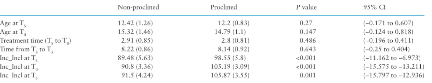
The gender proportion in the proclined group was different from that in the non-proclined group (P = 0.002, Table 1). The proportion of extrac-tion versus non-extracextrac-tion treatment was comparable in the groups (P = 0.114). Both groups were well-matched regarding age at TS, age at T0, and treatment time. Other data of the sample are in Table 2.
Pre-treatment Inc_Incl was larger in the proclined group (98.55°) than in the non-proclined group (89.48°); end-of-treatment (T0) Inc_Incl was also larger in the proclined group (105.19°) than in the non-proclined group (91.39°). From T0 to T5, Inc_Incl remained constant in both groups.
Gingival recession
No gingival recession sites were found before treatment (TS) in any of the subjects from the non-proclined and proclined group. Five years after treatment (T5), there was no difference in the number of subjects with gingival recession—seven subjects (12.3%) from the non-proclined and seven subjects (11.7%) from proclined group had labial recession (P = 0.851).
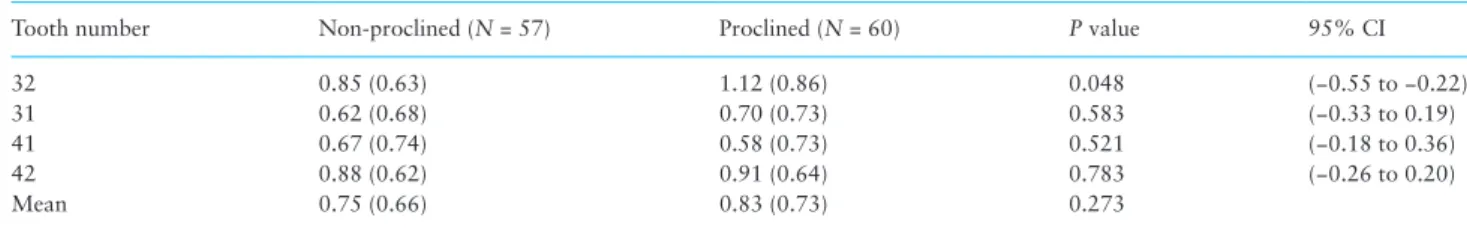
Clinical crown height
The mean increase of clinical crown heights of the lower incisors dur-ing treatment (from Ts to T0) was 0.1 mm (SD = 0.65) and −0.12 mm (SD = 0.64) in the non-proclined and proclined group, respectively. The mean increase of clinical crown heights of the lower incisors after treatment (from T0 to T5) ranged from 0.58 to 1.12 mm in the non-proclined and proclined group, respectively (Table 3). The only statistically significant inter-group difference was a larger increase of the clinical crown height of tooth number 32 in the proclined group in comparison with the non-proclined group—1.12 mm in the former and 0.85 mm in the latter group (P = 0.048; 95% CI: −0.55 to −0.22).
The regression analysis (Table 4) showed that none of the inde-pendent variables had an effect on the change of clinical crown heights of lower incisors.
Table 1. Gender proportion and extraction versus non-extraction treatment alternative in non-proclined (N = 57) and proclined (N = 60) groups assessed with the Fisher’s exact tests.
Non-proclined Proclined P value
Males/females 40.4%/59.6% 70%/30% 0.002
Extraction/non-extraction 40.4%/59.6% 25%/75% 0.114
Table 2. Characteristics of the non-proclined (N = 57) and proclined (N = 60) groups.
Non-proclined Proclined P value 95% CI
Age at TS 12.42 (1.26) 12.2 (0.83) 0.27 (−0.171 to 0.607) Age at T0 15.32 (1.46) 14.79 (1.1) 0.147 (−0.124 to 0.818) Treatment time (TS to T0) 2.91 (0.85) 2.8 (0.81) 0.486 (−0.196 to 0.411) Time from TS to T5 8.22 (0.86) 8.14 (0.92) 0.643 (−0.25 to 0.404) Inc_Incl at TS 89.48 (5.63) 98.55 (5.8) <0.001 (−11.162 to −6.973) Inc_Incl at T0 90.8 (3.36) 105.19 (3.09) <0.001 (−15.575 to −13.211) Inc_Incl at T5 91.5 (4.24) 105.87 (3.55) 0.001 (−15.797 to −12.936)
All values are in years or degrees. Standard deviations are in the brackets. Inter-group differences were analysed with analysis of variance (ANOVA) tests; paired comparisons were made with post hoc Tukey’s tests. CI, confidence interval.
Discussion
The current trend in orthodontics is that more and more patients are treated without extraction of teeth. For example, in 1986 almost 35% orthodontic patients in the USA had teeth extracted, whereas in 2008 only 18% patients were treated with extractions (21). On one hand reduction of extraction therapy has been possible due to the development of new methods of distalization of teeth. On the other hand, it likely results in more proclination of anterior teeth because the anterior teeth (i.e. anterior anchorage) may not remain completely stationary during distalization of posterior teeth (22). Animal experiments showed that movement of the tooth outside the bony envelope, as may occur in excessive proclination of incisors, might result in the development of gingival recession (8). As a result, it has been speculated that labial movement of incisors in humans is also a risk factor for the development of recession (15).
Our findings do not support the claim that a proclined position of the lower incisors at the end-of-treatment promotes the occurrence of gingival labial recession. The prevalence of recession sites in patients who had proclined mandibular incisors at the end of treatment and in those, in whom mandibular incisors remained at roughly the same inclination throughout treatment and retention phase, was compara-ble. Also the mean increase of clinical crown heights in proclined and non-proclined groups showed no difference. Although we found that the increase of the clinical crown height in tooth number 32 in the proclined group was larger than in the non-proclined group (Table 3), the difference was limited to only one tooth and the change of clinical crown heights of the remaining incisors demonstrated no difference.
The results of several other recent investigations in which the effect of post-treatment proclination of mandibular incisors on the development of gingival recession was evaluated (16,23–25) in gen-eral support our findings. Ruf et al (16) assessed how therapy of Class II adolescents with the Herbst appliance influenced the occur-rence of gingival recession. The authors found that proclining lower incisors by almost 9° did not increase the risk of recession. Also the comparison of maximal (16 subjects; mean = 16.4°) and minimal proclination (17 subjects; mean = 2.7°) did not show any signifi-cant differences for crown height or for the incidence of recession sites between the subgroups. However, the authors did not report the value of end-of-treatment inclination of incisors relative to
mandibular plane and one can only assume that incisors were at excessive inclination after treatment. Årtun and Grobéty (23) fol-lowed the group of young patients with Class II malocclusion (mean age = 10.2 years) treated with reverse headgear attached to the mandible (mean treatment time = 4.3 years) until 22 years of age. The post-treatment inclination of lower incisors was 99.1° in the ‘Pronounced advancement’ group and 96.2° in the control group. The authors found no difference in the increase in clinical crown height from post-treatment to follow-up. Djeu et al. (24) found that in patients treated with fixed appliances, in whom post-treatment inclination of mandibular incisors was 99.4°, there was no increased risk of development of recession. Allais and Melsen (25) compared periodontal status in adult patients (mean age = 34 years) immedi-ately after orthodontic treatment and in age- and sex-matched con-trols. The pre-treatment inclination of mandibular incisor was 94°. The estimated change of inclination of lower incisors during treat-ment was 7°. The authors found that mandibular incisors showed more gingival recession than untreated controls. However, the dif-ference in crown heights between treated and untreated individuals was minimal (<0.2 mm) and clinically irrelevant.
The current investigation has several advantages in comparison with the studies discussed above. First, the post-treatment inclina-tion of mandibular incisors in our proclined group was 105°, more than 1 SD from population mean (26) and considerably more than in the samples evaluated by Årtun and Grobéty (23), Djeu et al. (24) and Allais and Melsen (25). Secondly, a relatively long follow-up time (>5 years) allowed to evaluate the effects of mandibular incisor proclination on the development of gingival recession in a long-term perspective. In contrast, the studies by Ruf et al. (16), Djeu et al. (24), and Allais and Melsen (25) assessed the prevalence and severity of recession immediately and/or within a few months after treatment. Thirdly, the patients in our group had a fixed retainer in the mandibu-lar dental arch. The presence of fixed retention warranted that incisors proclined during treatment remained at their end-of treatment posi-tions. Moreover the application of fixed retention resembles a typical situation after orthodontic treatment when the relapse is prevented with the long-term use of retainers (17,18).
All subjects in our sample had fixed retention during the whole post-treatment period. We selected patients with bonded retainers
Table 3. The increase (in millimetres) of mean clinical crown height of lower incisors after treatment (from T0 to T5).
Tooth number Non-proclined (N = 57) Proclined (N = 60) P value 95% CI
32 0.85 (0.63) 1.12 (0.86) 0.048 (−0.55 to −0.22)
31 0.62 (0.68) 0.70 (0.73) 0.583 (−0.33 to 0.19)
41 0.67 (0.74) 0.58 (0.73) 0.521 (−0.18 to 0.36)
42 0.88 (0.62) 0.91 (0.64) 0.783 (−0.26 to 0.20)
Mean 0.75 (0.66) 0.83 (0.73) 0.273
Standard deviation in brackets. CI, confidence interval.
Table 4. Results of regression analysis.
Coefficients (B) P value Lower limit of 95% CI Upper limit of 95% CI
(Constant) 87.97 <0.001 60.27 115.67
Age at TS* −12.86 0.085 −27.51 1.79
Gender (female = 0; male = 1) −1.934 0.874 −26.1 22.23
Proclined group 7.191 0.547 −16.42 30.81
The mean increase of crown length of the mandibular incisors after the orthodontic treatment (from T0 to T5) was dependent variable. CI, confidence interval. *Age above 11 years.
because of a trend among orthodontists to use compliance-free retention (17,18,27). Several studies demonstrated that the long-term use of fixed retention had a limited effect on periodontal health (28–30). Heier et al. (29) evaluated periodontal condition at debond-ing and after 1, 3, and 6 months of fixed or removable retention with indices such as plaque index (PI), calculus index (CI), modified gin-gival index (GI), bleeding on probing, and gingin-gival crevicular fluid flow. The authors observed that slight gingival inflammation present during orthodontic treatment decreased from baseline to a 6-month follow-up irrespective of the retainer type used. However, more dental plaque and calculus accumulated in subjects having bonded than removable retention. The authors concluded that periodontal health should not be compromised provided patients receive regu-lar oral hygiene instructions. Årtun et al. (28) found no difference in PI, CI, and GI in patients wearing three different types of fixed retainers and removable retainers for 3 years following completion of orthodontic treatment. Booth et al. assessed periodontal status in a group of patients having fixed retention for minimum 20 years and reported that bonded retainers left for prolonged time had no detrimental effects to the mandibular anterior gingiva. Nevertheless, Pandis et al. (19) pointed to the fact that although retainers bonded to mandibular anterior teeth had overall only small effect on perio-dontal tissues, their detrimental effect could be observed if they were attached to proclined incisors. Pandis et al. formulated this hypothe-sis because they found that patients being in retention for 9–11 years had more gingival recession than patients wearing fixed retainers for 3–6 months (25% versus 0%, respectively). They described a poten-tial mechanism according to which the proclination of incisors was associated with gingival recession. If proclined teeth were retained with bonded retainer, which promotes the inflammation-inducing adhesion of dental plaque and calculus (31), the environment con-ducive to the development of recession of marginal gingiva could have been created. Our findings do not support this hypothesis. The increase of clinical crown heights and prevalence of recession sites were similar irrespective of the amount of post-treatment proclina-tion. However, it cannot be ruled out that it would be possible to identify an association between incisor proclination and develop-ment of gingival recession if the observation period were longer.
The occurrence of gingival recession may be associated with past orthodontic treatment (12,13,32). Both Slutzkey and Levin (12) and Renkema et al. (13) found that the proportion of subjects with gingi-val recession was consistently higher among orthodontically treated than untreated individuals. The difference was observed already at the end of treatment and continued until 5 years post-treatment (13). Zachrisson and Alnaes (32), in turn, found a significant loss of gingival attachment in orthodontic patients in comparison to untreated individuals. Unfortunately, no single cause of recession in humans has been identified. Instead, several putative contributing factors such as individual biological predisposition toward reces-sion, a gingival biotype, and a narrow width of keratinized gin-giva were discussed in depth in several recent systematic reviews (15,33,34).
The demand for orthodontic treatment steadily increases. Unfortunately studies to date suggest that a cause-and-effect rela-tionship between some element(s) of orthodontic therapy and/or retention phase may exist. The difficulty to identify predictor(s) of gingival recession, however, urgently warrants a large prospective study with clinical examination before, during and after treatment, stratification for gingival biotype and various types of malocclusion, and a long follow-up. Because smoking and inadequate hygiene resulting in gingival inflammation are associated with gingival
recession (7), these parameters should also be monitored during the advocated study.
Conclusions
Based on the findings of this study we conclude that 5 years after orthodontic treatment gingival recession sites were present, on aver-age, in 12% subjects; however, overall the amount of proclination of lower incisors at the end of treatment seemed not to affect the development of labial gingival recession nor the change of clinical crown heights in this patient group.
Acknowledgement
We would like to thank Dr. E. Bronkhorst for statistical analysis.
References
1. Armitage, G.C. (1999) Development of a classification system for peri-odontal diseases and conditions. Annals of Periodontology, 4, 1–6. 2. Smith, R.G. (1997) Gingival recession. Reappraisal of an enigmatic
condi-tion and a new index for monitoring. Journal of Clinical Periodontology, 24, 201–205.
3. Al-Wahadni, A. and Linden, G.J. (2002) Dentine hypersensitivity in Jorda-nian dental attenders. A case-control study. Journal of Clinical
Periodon-tology, 29, 688–693.
4. Lawrence, H.P., Hunt, R.J. and Beck, J.D. (1995) Three-year root caries incidence and risk modeling in older adults in North Carolina. Journal of
Public Health in Dentistry, 55, 69–78.
5. Löe, H., Anerud, A. and Boysen, H. (1992) The natural history of peri-odontal disease in man: prevalence, severity, and extent of gingival reces-sion. Journal of Periodontology, 63, 489–495.
6. Khocht, A., Simon, G., Person, P. and Denepitiya, J.L. (1993) Gingival recession in relation to history of hard toothbrush use. Journal of
Peri-odontology, 64, 900–905.
7. Wennström, J.L. (1996) Mucogingival considerations in orthodontic treat-ment. Seminars in Orthodontics, 2, 46–54.
8. Batenhorst, K.F., Bowers, G.M. and Williams, J.E. Jr. (1974) Tissue changes resulting from facial tipping and extrusion of incisors in monkeys. Journal
of Periodontology, 45, 660–668.
9. Steiner, G.G., Pearson, J.K. and Ainamo, J. (1981) Changes of the marginal periodontium as a result of labial tooth movement in monkeys. Journal of
Periodontology, 52, 314–320.
10. Nelson, P.A. and Årtun, J. (1997) Alveolar bone loss of maxillary anterior teeth in adult orthodontic patients. American Journal of Orthodontics and
Dentofacial Orthopedics, 111, 328–334.
11. Re, S., Corrente, G., Abundo, R. and Cardaropoli, D. (2000) Orthodontic treatment in periodontally compromised patients: 12 year report.
Interna-tional Journal of Periodontics and Restorative Dentistry, 20, 31–39.
12. Slutzkey, S. and Levin, L. (2008) Gingival recession in young adults: occur-rence, severity, and relationship to past orthodontic treatment and oral piercing. American Journal of Orthodontics and Dentofacial Orthopedics, 134, 652–656.
13. Renkema, A.M., Fudalej, P.S., Renkema, A.A., Abbas, F., Bronkhorst, E. and Katsaros, C. (2013a) Gingival labial recessions in orthodontically treated and untreated individuals: a case - control study. Journal of
Clini-cal Periodontology, 40, 631–637.
14. Bollen, A.M., Conha-Cruz, J., Bakko, D.W., Huang, G.J. and Hujoel, P.P. (2008) The effects of orthodontic therapy on periodontal health. A sys-tematic review of controlled evidence. Journal of the American Dental
Association, 139, 413–422.
15. Årtun, J. and Krogstad, O. (1987) Periodontal status of mandibular inci-sors following excessive proclination. A study in adults with surgically treated mandibular prognathism. American Journal of Orthodontics and
16. Ruf, S., Hansen, K. and Pancherz, H. (1998) Does orthodontic proclination of lower incisors in children and adolescents cause gingival recession?
Amer-ican Journal of Orthodontics and Dentofacial Orthopedics, 114, 100–106.
17. Renkema, A.M., Sips, E.T., Bronkhorst, E. and Kuijpers-Jagtman, A.M. (2009) A survey on orthodontic retention procedures in The Netherlands.
European Journal of Orthodontics, 31, 432–437.
18. Lai, C., Grossen, J.M., Renkema, A.M., Bronkhorst, E., Fudalej, P.S. and Katsaros, C. (2014) Orthodontic retention procedures in Switzerland – a survey. Swiss Dental Journal, 124, 655–661.
19. Pandis, N., Vlahopoulos, K., Madianos, P. and Eliades, T. (2007) Long-term periodontal status of patients with mandibular lingual fixed reten-tion. European Journal of Orthodontics, 29, 471–476.
20. Renkema, A.M., Fudalej, P.S., Renkema, A.A., Bronkhorst, E. and Kat-saros, C. (2013b) Gingival recessions and the change of inclination of mandibular incisors during orthodontic treatment. European Journal of
Orthodontics, 35, 249–255.
21. Keim, R.G., Gottlieb, E.L., Nelson, A.H. and Vogels, D.S. 3rd. (2008) JCO study of orthodontic diagnosis and treatment procedures, part 1: results and trends. Journal of Clinical Orthodontics, 42, 625–640.
22. Antonarakis, G.S. and Kiliaridis, S. (2008) Maxillary molar distalization with noncompliance intramaxillary appliances in Class II malocclusion. A systematic review. Angle Orthodontist, 78, 1133–1140.
23. Årtun, J. and Grobéty, D. (2001) Periodontal status of mandibular incisors after pronounced orthodontic advancement during adolescence: a follow-up evaluation. American Journal of Orthodontics and Dentofacial
Ortho-pedics, 119, 2–10.
24. Djeu, G., Hayes, C. and Zawaideh, S. (2002) Correlation between man-dibular central incisor proclination and gingival recession during fixed appliance therapy. The Angle Orthodontist, 72, 238–245.
25. Allais, D. and Melsen, B. (2003) Does labial movement of lower incisors influence the level of the gingival margin? A case-control study of adult orthodontic patients. European Journal of Orthodontics, 25, 343–352.
26. Riolo, M.L., Moyers, R.E., McNamara, J.A. Jr and Hunter, W.S. (1974) An
Atlas of craniofacial growth: cephalometric standards from the University School Growth Study. Monograph 2, Craniofacial Growth Series. Center
for Human Growth and Development, University of Michigan, Ann Arbor, MI, pp. 1–2.
27. Valiathan, M. and Hughes, E. (2010) Results of a survey-based study to identify common retention practices in the United States. American
Jour-nal of Orthodontics and Dentofacial Orthopedics, 137, 170–177.
28. Årtun, J., Spadafora, A.T. and Shapiro, P.A. (1997) A 3-year follow-up study of various types of orthodontic canine-to-canine retainers. European
Journal of Orthodontics, 19, 501–509.
29. Heier, E.E., De Smit, A.A., Wijgaerts, I.A. and Adriaens, P.A. (1997) Peri-odontal implications of bonded versus removable retainers. American
Journal of Orthodontics and Dentofacial Orthopedics, 112, 607–616,
30. Booth, F.A., Edelman, J.M. and Proffit, W.R. (2008) Twenty-year follow-up of patients with permanently bonded mandibular canine-to-canine retainers. American Journal of Orthodontics and Dentofacial
Orthope-dics, 133, 70–76.
31. Tacken, M.P., Cosyn, J., De Wilde, P., Aerts, J., Govaerts, E. and Vannet, B.V. (2010) Glass fibre reinforced versus multistranded bonded orthodon-tic retainers: a 2 year prospective multi-centre study. European Journal of
Orthodontics, 32, 117–123.
32. Zachrisson, B.U. and Alnaes, L. (1973) Periodontal condition in ortho-dontically treated and untreated individuals. I. Loss of attachment, gin-gival pocket depth and clinical crown height. Angle Orthodontist, 43, 402–411.
33. Joss-Vassalli, I., Grebenstein, C., Topouzelis, N., Sculean, A. and Katsaros, C. (2010) Orthodontic therapy and gingival recession: a systematic review.
Orthodontics and Craniofacial Research, 13, 127–141.
34. Aziz, T. and Flores-Mir, C. (2011) A systematic review of the association between appliance-induced labial movement of mandibular incisors and gingival recession. Australian Orthodontic Journal, 27, 33–39.