Early cleavage of in-vitro fertilized human embryos to
the 2-cell stage: a novel indicator of embryo quality and
viability
Youssef Shoukir
1, Aldo Campana
1, Tim Farley
2and
Many methods have been suggested to evaluate embryoviability in IVF programmes. A limiting factor is that these
Denny Sakkas
1,3measurements need to be non-invasive and not time consuming. 1Clinic of Infertility and Gynaecological Endocrinology–WHO
Routinely, the embryos selected for transfer are chosen on the Collaborating Centre, Department of Obstetrics and Gynaecology,
basis of their morphology and rate of development in culture. University Hospital of Geneva, 1211 Geneva 14, Switzerland and
2UNDP/UNFPA/WHO/World Bank Special Programme of In one such study, Cummins et al. (1986) established an Development and Research Training in Human Reproduction embryo quality and embryo development rating and found that Program, WHO, Geneva, Switzerland good ratings for both result in clinical pregnancies. Other 3To whom correspondence should be addressed at: Laboratoire des studies have also found advantage in transferring embryos on Game`tes, Policlinique de Ste´rilite´, Hoˆpital Cantonal Universitaire the basis of a morphological and developmental assessment de Gene`ve, 30 Bd. de la Cluse, 1211 Gene`ve 14, Switzerland
(Edwards et al., 1984; Hill et al., 1989; Steer et al., 1992). In addition to the above methods, the measurement of several A number of non-invasive methods have been proposed to
metabolic parameters of the embryos by using non-invasive evaluate embryo viability in human in-vitro fertilization
procedures has also been proposed (Leese, 1987). In one programmes. In addition to biochemical analyses, a
com-such study Conaghan et al. (1993) showed that an inverse mon method for the selection of embryos prior to transfer
relationship existed between pyruvate uptake and human involves assessment of embryo quality and morphology.
embryo viability. Although these methods are potentially of We propose a new method to evaluate embryo viability
great benefit they unfortunately require additional expertise based on the timing of the first cell division. Fertilized
and cost, which may be prohibitive in smaller IVF centres. embryos that had cleaved to the 2-cell stage 25 h
post-We therefore propose a new simple method to evaluate insemination were designated as ‘early cleavage’ embryos
embryo viability based on the timing of the first cell division. while the others that had not yet reached the 2-cell stage
The earliest time the human zygote can reach the 2-cell stage were designated as ‘no early cleavage’. In all cases the
ranges between 20 and 27 h post-insemination (Trounson et al., early cleavage embryos were transferred when available.
1982; Balakier et al., 1993; Capmany et al., 1996). We have Early cleavage was observed in 27 (18.9%) of the 143
defined a sub-group of embryos that have undergone ‘early cycles assessed. There were significantly (χ2 J 4.0; P J
cleavage’; in that they divide and reach the 2-cell stage 25– 0.04) more clinical pregnancies in the early cleavage group,
26 h post-insemination. We therefore present results examining 9/27 (33.3%), compared with the no early cleavage group,
the relationship between early cleavage of embryos and the 17/116 (14.7%). No difference was found when comparing
incidence of pregnancy. Furthermore, we propose that this key parameters (age, stimulation protocol and semen
char-may be used as a simple and effective selection criterion for acteristics) of couples belonging to both groups, pointing
selecting embryos for transfer. to an intrinsic property or factor(s) within the early cleaving
embryos. We propose ‘early cleavage’ as a simple and effective non-invasive method for selection and evaluation
of embryos prior to transfer. Materials and methods
Key words: early cleavage/embryo quality/in-vitro fertiliza- The study was performed on patients entering the IVF programme
tion/viability of the Clinic of Infertility and Gynaecological Endocrinology,
Depart-ment of Obstetrics and Gynaecology, University Hospital of Geneva, Geneva, Switzerland between January 1992 and December 1995. In all, 143 cycles were assessed where the patient underwent a routine
Introduction IVF treatment with transfer. All patients who failed to achieve
fertilization or who underwent subzonal microinjection or intracyto-The success rate of human in-vitro fertilization (IVF) remains
plasmic sperm injection (ICSI) were not included. The stimulation relatively low when the number of pregnancies is considered
protocol adopted by our group has been previously described (Sakkas in proportion to the number of embryos transferred. The low
et al., 1994).
pregnancy rates per embryo transferred leads to a need to
Collected oocytes (day 0) were fertilized using our standard culture transfer more than one embryo, which in turn increases the medium, Whittingham’s T6 (Quinn et al., 1982) supplemented with chance of multiple pregnancies. To increase pregnancy rates 10% maternal human serum. Oocyte retrieval took place between and limit the occurrence of multiple births, a more efficient 0830 and 1030 hrs. Insemination was performed between 1530 and and rigorous procedure for embryo selection prior to transfer 1630 (~1600) hrs in tubes. The following morning (day 1) the oocytes were removed from the tubes, washed and placed in 20µl culture is therefore needed.
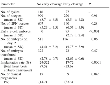
Table I. The total and mean number (6SD) of oocytes, 2-pronucleate
(2PN) oocytes, embryos observed on day 2, embryos transferred,
implantation rate and pregnancies according to whether embryos had or had not undergone early cleavage to the 2-cell stage by 25 h post-insemination Parameter No early cleavage Early cleavage P
No. of cycles 116 27 –
No. of oocytes 999 229 0.91
(mean6 SD) (8.7 6 6.5) (8.5 6 4.8)
No. of 2PN oocytes 607 160 0.28
(mean6 SD) (5.236 3.5) (6.076 3.9)
Early 2-cell embryos 0 75 ,0.001
(mean6 SD) (2.786 2.4)
Figure 1. The clinical pregnancy rate in relation to the number of
No. of embryos on 511 156 0.06
day 2 early cleaving 2-cell embryos observed at 25 h post-insemination.
(mean6 SD) (4.416 3.2) (5.786 3.9) The number of cycles for each group is shown in parentheses.
No. of embryos 322 72 0.47 (χ25 10.6, DF 5 2, P 5 0.005.)
transferred
(mean6 SD) (2.786 0.7) (2.676 0.6)
Implantation rate (%) 24/322 17/72 0.0001
Table II. Female parameters for cycles with and without early cleavage to (fetal heart beat/ (7.5) (23.6)
the 2-cell stage by 25 h post-insemination embryo transferred)
No. of clinical 17 9 0.045
Parameter No early cleavage Early cleavage P
pregnancies
(%) (14.7) (33.3)
No. of cycles 116 27 –
Female age (years) 35.16 4.5 34.76 3.9 0.72
Oestradiol concentrations drops under oil (Light white mineral oil; Sigma Pharmaceuticals, (pg/ml) on:
Day 0 (day of HCG) 1834.06 1043.4 1952.36 1063.2 0.60 Buchs, Switzerland) in Petri dishes and the presence of two pronuclei
Day 1 2280.16 1348.7 2181.06 1242.5 0.76
assessed. On the same day (day 1), at 1700 hrs (25 h
post-Day 2 1139.26 690.2 990.26 621.8 0.43
insemination), the embryos were examined to see if cleavage to the
Ampoules of Pergonal 27.06 12.0 30.06 10.3 0.23 2-cell stage had occurred. These embryos were designated as ‘early
cleavage’ embryos while the embryos that had not yet cleaved were Values are means6 SD. designated as ‘no early cleavage’. Both groups were maintained in
separate culture drops. The following day (day 2) the embryos were
and pregnancy outcome, in patients who had at least one early transferred between 1000 and 1800 hrs. Routinely, a maximum of
cleavage embryo compared with those that had none. There three embryos were transferred to the patient. When there were only
was no difference in the number of oocytes, number of 2-one or two early cleavage embryos the embryo transfer group was
pronucleate oocytes or the number of embryos transferred completed by adding the best one or two embryos from the no early
between both groups. Seventy-five early cleaving embryos cleavage group. In 11 cycles, four embryos were transferred due to
advanced maternal age, repeated failure in previous cycles or poor were observed in 27 cycles. The mean number of embryos quality embryos. Coincidentally, these patients were all in the no per patient on day 2 displayed a tendency to be higher in the
early cleavage group. early cleavage group compared with the no early cleavage
On the day of transfer all embryos were assessed for the number group. Early cleaving embryos implanted at a rate 3-fold of cells per embryo, to ascertain their cleavage rate, and given a higher than no early cleaving embryos. The pregnancy rate in quality score based on the presence of fragments and clarity of the
the early cleavage group was double the rate of the no cytoplasm of the blastomeres, similar to that previously described by
early cleavage group; nine pregnancies in 27 cycles (33.3%) Cummins et al. (1986). The ratings given for cell number were: 1
compared with 17 pregnancies in 116 cycles (14.7%) (χ2 5 for 1-cell, 2 for 2-cell, 3 for 3-cell, 4 for 4-cell, 6 for between 4–
4.0; P5 0.04). No cases of ectopic pregnancies were observed 8-cell and 8 forù8-cell embryos. The ratings given to embryos for
in either group. The chances of achieving a pregnancy increased quality were the same as those used by Cummins et al. (1986) except
along with the number of early cleavage embryos observed that the values of 0 for poorest and 3 for best embryo were given.
A pregnancy test was conducted 14 days after transfer. Patients per patient (Figure 1). In effect, patients in whom two or more that exhibited three consecutive human chorionic gonadotrophin early cleaving embryos were observed had 3-fold the pregnancy (HCG) values.5 mIU/ml and in which the fetus or fetuses displayed rate compared with those with no early cleavage.
a heart beat by ultrasound examination 4–5 weeks after transfer were When a number of key parameters were examined for both considered to have achieved a clinical pregnancy. groups, we failed to find any differences that influenced the The statistical evaluations used were analysis of variance followed occurrence of early cleavage. The mean age and progress of by Scheffe´’s F-test for comparison of mean values andχ2analysis with
stimulation, as indicated by oestradiol levels on the day of continuity correction for comparison of pregnancy rates. Statistical
HCG and oocyte retrieval and the number of ampoules significance was set at P, 0.05.
of Pergonal (Serono, Aubonne, Switzerland) used during stimulation, showed no differences between the two groups
Results (Table II). There were also no significant differences concerning
the mean age and the semen characteristics of the male partner Early cleavage was observed in 27/143 (18.9%) of the cycles.
implanted after transfer (Conaghan et al., 1993). More recently,
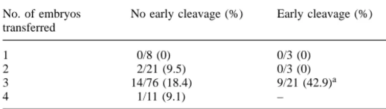
Table III. Clinical pregnancy rates according to the number of embryos Lane and Gardner (1996) examined glucose uptake and
transferred in patients who had early and no early cleaving embryos
glycolytic activity, using non-invasive quantitative microfluo-No. of embryos No early cleavage (%) Early cleavage (%) rescence, to prospectively select mouse blastocysts for transfer.
transferred They showed that a low glycolytic activity resulted in a
4-fold increase in the pregnancy rate. Although effective, many
1 0/8 (0) 0/3 (0)
2 2/21 (9.5) 0/3 (0) of these methods need special training and can prove to be
3 14/76 (18.4) 9/21 (42.9)a
time consuming, subsequently limiting their use in routine
4 1/11 (9.1) –
evaluation of embryo viability.
Currently, many IVF programmes depend on a simple aSignificantly different (P5 0.04) when compared to patients with no early
cleavage embryos. These data include mixed transfers as detailed in Table morphological assessment of embryos at the time of transfer. IV.
Several systems of embryo grading have been proposed accord-ing to the morphology (Hill et al., 1989; Scott et al., 1991), cleavage stage (Edwards, 1984; Cummins et al., 1986; Claman The clinical pregnancy rate according to the number of
et al., 1987; Bolton et al., 1989) or both (Puissant et al., 1987; embryos transferred is represented in Table III. When three
Steer et al., 1992). Cummins et al. (1986) confirmed the embryos were transferred significantly more pregnancies were
feasibility of such an approach in patients receiving single obtained in the early cleavage group compared with the no
embryo transfers by showing that combining a subjective score early cleavage group. No comparison was made when one or
of embryo quality and a rating of cleavage stage was a good two embryos were transferred due to the low number of cycles.
predictor of expected pregnancy. Table IV shows the implantation rate and the incidence of
In this study we introduce a novel assessment criterion and single, twin and triplet pregnancies when the cohort of the
show that when a patient has early cleaving 2-cell embryos three embryos transferred contained one, two or three early
the chances of achieving a pregnancy are significantly cleavage embryos or all no early cleavage embryos.
Interes-improved. More importantly, we also show that even in patients tingly, when all three embryos transferred had undergone early
who had transfers of high quality and better cleaving embryos cleavage the pregnancies obtained were multiple.
in the no early cleavage group the pregnancy rates achieved To examine whether the cohorts of embryos transferred to
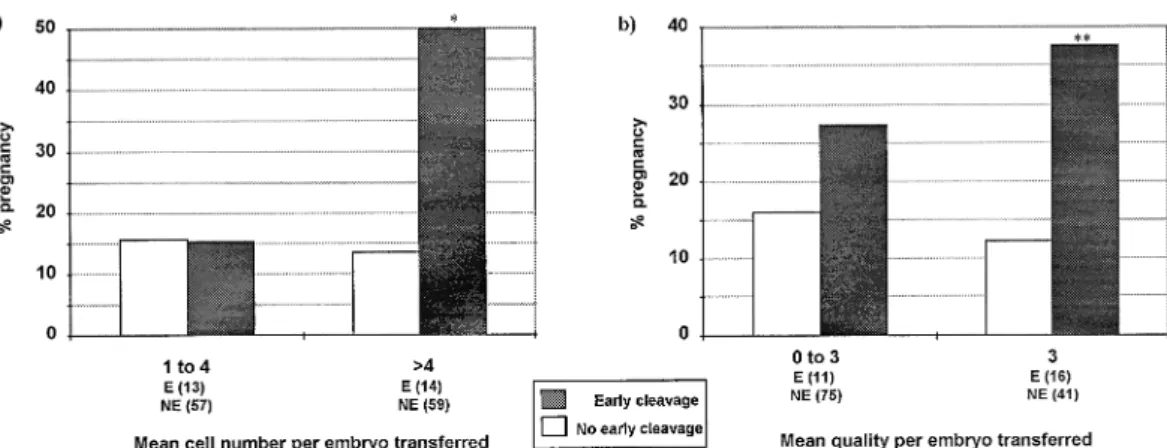
were still higher for the early cleavage group. This raises the the patients with and without early cleaving embryos differed,
question as to why selecting early cleaving embryos appears we calculated the mean score of the number of cells and mean
to be more advantageous than selecting the best embryos at quality per embryo. The mean number of cells per embryo
the time of transfer. The timing of the first cell division in and the mean quality of cells per embryo on day 2 for those
humans has been reported by several investigators. Trounson transferred are presented in Table V. The results show that,
et al. (1982) reported that the earliest time the 2-cell stage can while no significant difference was evident in the mean cell
be observed was 27 h post-insemination. However, more numbers of the embryos transferred, the quality rating was
recently, Balakier et al. (1993) reported this time to be at 20– significantly higher for the embryos transferred in the patients
22 h post-insemination while Capmany et al. (1996) reported that had early cleavage embryos. Finally, when a comparison
it to be 25 h post-insemination. In fact, Balakier et al. (1993) between both groups was made of the pregnancy rates in
found that 1% of the zygotes reached the 2-cell stage at 20 h relation to the number of cells and the quality of embryo
post-insemination, 5% at 24 h and 38% by 27 h. These times transferred, the transfer of embryos of higher cell number and
are in accordance with our observations. quality in the early cleavage group provided a significantly
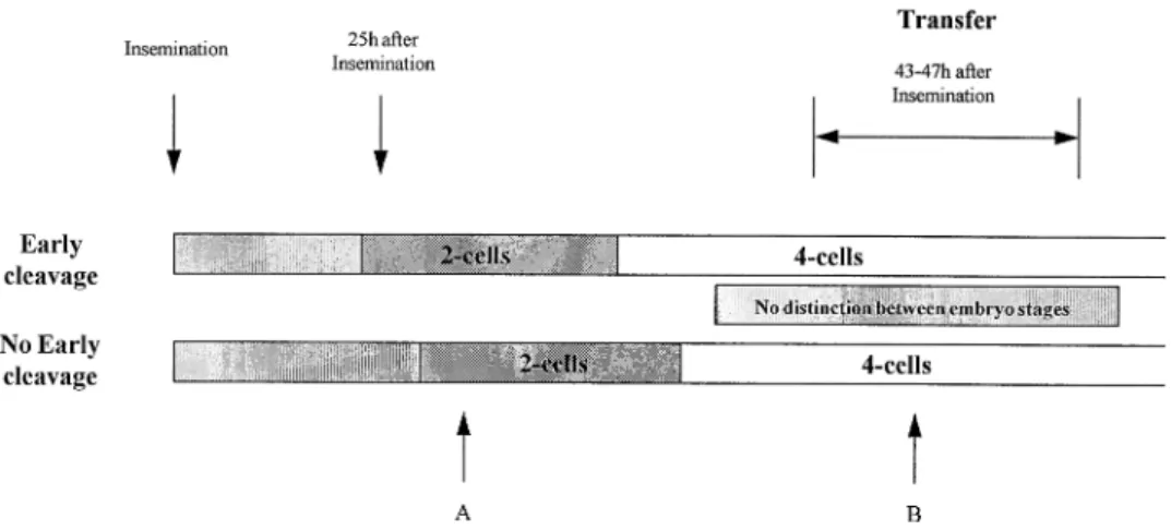
The selection of a critical time-point is essential so as to higher pregnancy rate (Figure 2).
maximize the differences between embryos. Observations of embryo development in culture are sometimes made
infre-Discussion quently (commonly at 16–18 h post-insemination to check for
pronuclei and again at ~40 h just before transfer) so that The need for a non-invasive method to assess embryo viability
has been highlighted previously by Leese (1987). Methods precise data on cleavage timing is usually not available. Bavister (1995) highlighted this problem by stating that the currently available are those that examine biochemical markers
such as the measurement of several metabolic parameters, for examination of embryos at arbitrary time points during develop-ment can be quite misleading with respect to categorizing the example: O2consumption (Magnusson et al., 1986 ), pyruvate
uptake (Leese et al., 1986; Hardy et al., 1989; Conaghan et stage of development reached and ‘timeliness’ of development. This problem is illustrated in Figure 3. In two groups of al., 1993), glucose uptake and lactate production (Wales et al.,
1987; Hardy et al., 1989), the secretion of factors such as embryos (early and no early cleavage) observation of embryos at point A shows no difference between embryos of the same platelet activating factor (O’Neill and Saunders, 1984) and the
activity of the enzymes involved in fatty acid catabolism, the cell stage (2-cell stage), although in the early cleavage group the embryo has divided before the no early cleavage embryo. citrate cycle and the pentose pathway (Chi et al., 1988; Martin
et al., 1993). Of these methods the assessment of metabolic The same applies to point B for the selection of embryos for transfer. In both groups the embryos are at the 4-cell stage criteria appear to be the most effective. For example, analysis
of 2–8-cell stage human embryos prior to transfer revealed and are of the same stage despite one having cleaved hours before (in the early cleavage group) while the other has just that pyruvate uptake was significantly lower by embryos that
Figure 2. The clinical pregnancy rate in relation to (a) the mean cell number per embryo transferred and (b) the mean quality per embryo transferred for the early cleavage (E) and no early cleavage (NE) groups. The number of cycles for each group is shown in parentheses. *Significant difference (χ25 7.1, P 5 0.008) in pregnancies when compared to the no early cleavage group. **Increased but no significant difference (χ25 3.3, P 5 0.07) in pregnancies when compared to the no early cleavage group.
Table IV. Pregnancy in relation to the number of early and no early cleavage embryos transferred in patients
receiving three embryos at transfer
No. of embryos transferred Pregnancy
No early cleavage Early cleavage Single Twins Triplets Implantation ratea(%) Totalb(%) Patients with no early cleaving embryos
3 0 10 2 2 20/228 (8.8) 14/76 (18.4)
Patients with early cleaving embryos
2 1 1 1 0 3/9 (33.3) 2/3 (66.6)
1 2 1 1 0 3/18 (16.7) 2/6 (33.3)
0 3 0 4 1 11/36 (30.6) 5/12 (41.7)
aNumber of fetal heart beats per embryo transferred. bNumber of clinical pregnancies per cycle.
Table V. Mean number of cells per embryo and mean quality per embryo on day 2 of embryos transferred in
patients with and without early cleavage to the 2-cell stage by 25 h post-insemination Embryos of no early Embryos of early cleavage patients cleavage patients:
No early cleavage All embryos No early cleavage Early cleavage
No. of embryos 322 72 21 51
transferred
No. of cellsa 4.36 1.7 4.56 1.2 4.26 1.7 4.86 1.9
Quality of cellsa 2.36 0.8b,c 2.66 0.6b 2.66 0.5 2.76 0.5c
aMeans6 SD.
b,cSame letters differ significantly:bP5 0.04 andcP5 0.02.
cleaved (the no early cleaved group). Although in a group of the female partners as regards to the age, type of infertility (data not shown), oestradiol concentrations and stimulation embryos there will be those present that have a higher cell
number, the embryos at the same stage cannot be distinguished. protocol excludes possible female factors such as endometrial receptivity as the cause of the differences in pregnancy rates For example, if a patient had one 8-cell and six 4-cell embryos
of similar quality, the 8-cell embryo is selected but a problem between both groups. As no ascertainable difference was evident in the male and female characteristics, this points to arises as to which 4-cell embryo is more advanced. Examination
of early cleavage therefore gives more discrimination the possibility of an enhanced intrinsic oocyte or embryo quality in the cases where early cleavage is observed. The between groups.
An analysis of the patient groups giving rise to early finding of a higher number of embryos on day 2 in the early cleavage group, although not significant, also indicates that a cleavage and no early cleavage showed no differences in the
number of oocytes, the rate of fertilization, nor when examining better embryo development in the early cleavage group may indeed be present. One possibility is that the early cleavage for parameters associated with males and females. The fact
sub-Figure 3. Importance of specific time points for distinguishing embryo cleavage during screening (see Discussion for details).
sequent events simply due to the difference in the time required same authors demonstrated a correlation of HLA-G mRNA expression with improved cleavage rate in human preimplanta-for sperm penetration through the cumulus cells and zona
pellucida. The latter depends on the maturity of the oocytes, tion embryos.
In this study we have shown that the evaluation of early as less mature oocytes are usually fertilized later than the
mature ones, leading to a delayed cleavage (Balakier et al., cleaving 2-cell embryos in an IVF programme could be an effective and valuable method of assessment of embryo viabil-1993). Contrary to this hypothesis, however, are similar results
we have obtained when separating early cleavage and no early ity, in that it provides a strong prognostic indication of the likelihood of pregnancy. The verification of this observation cleavage 2-cell embryos after ICSI. In this case the time of
sperm entry and the maturity of the oocytes is regulated and in a larger study and by independent groups is eagerly awaited. The cause of early cleavage in embryos is not obvious but an even more marked significant difference is evident in
pregnancy rates between the two groups (unpublished data). indications exist that it could be an intrinsic factor within the oocyte or embryo. An inherent genetic control of such early These data would exclude a possible influence of the time of
fertilization and oocyte maturity on early cleavage. cleavage could be postulated. The search for such a factor Whereas early cleavage could be due to a selection of the may be important in understanding the timing of events during oocytes that were primed to fertilize, a further explanation of the first cell cycle in the human and could possibly provide the higher viability of the early cleavage embryos could also an indicator for pregnancy in a given cycle.
arise due to the presence of an unknown factor(s) within the oocyte which favours or promotes the early division of embryos
after fertilization. The intrinsic nature of a certain population Acknowledgements
of embryos to cleave faster has previously been observed in We thank all members of our IVF team for their assistance during mouse embryos. Mouse embryos have been shown to possess this study.
a gene, associated with the major histocompatibility complex, which manifests itself as two functional alleles. This gene
has been called the preimplantation embryo development (Ped) References
gene. Mouse embryos homozygous for the dominant fast Ped Balakier, H., MacLusky, N.J. and Casper, R.F. (1993) Characterization of the allele cleave at a faster rate than those which are heterozygous, first cell cycle in human zygotes: implications for cryopreservation. Fertil.
Steril., 59, 359–365. while those homozygous for the slow Ped allele cleave at the
Bavister, B.D. (1995) Culture of preimplantation embryos: facts and artefacts. slowest rate (Goldbard and Warner, 1982). It has been shown
Hum. Reprod. Update, 1, 91–148.
that the Ped phenotype is an intrinsic property of the embryo Bolton, V.N., Hawes, S.M., Taylor, C.T. and Parsons, J.H. (1989) Development independent of the uterine environment (Brownell and Warner, of spare human preimplantation embryos in vitro: an analysis of the correlation among gross morphology, cleavage rates and development to 1988). In addition, there is no effect of the Ped gene on time
blastocyst. J. In Vitro Fertil. Embryo Transfer, 6, 30–35. of ovulation or fertilization as it initially acts at the time of
Brownell, M.S. and Warner, C.M. (1988) Ped gene expression by embryos the first cleavage division. The Ped phenotype has been linked cultured in vitro. Biol. Reprod., 39, 806–811.
with production of the Qa-2 antigen. The way in which the Capmany, G., Taylor, A., Braude, P.R. and Bolton, V.N. (1996) The timing of pronuclear formation, DNA synthesis and cleavage in the human 1-cell presence of the Qa-2 antigen on the cell surface induces the
embryo. Mol. Hum. Reprod., 2, 299–306. embryo to divide at an earlier and faster rate is unknown. It
Chi, M.M., Manchester, J.K., Yang, V.C. et al. (1988) Contrast in levels of is likely that the mouse Ped gene has a human homology metabolic enzymes in human and mouse ova. Biol. Reprod., 39, 295–307. within human leukocyte antigen (HLA)-F (Stroynowski, 1990). Claman, P., Armant, D.R., Seibel, M.M. et al. (1987) The impact of embryo quality and quantity on implantation and the establishment of viable More recently, Jurisicova et al. (1996) have demonstrated the
pregnancies. J. In Vitro Fertil. Embryo Transfer, 4, 218–222. expression of HLA-G throughout preimplantation development
Conaghan, J., Hardy, K., Handyside, A.H. et al. (1993) Selection criteria for in the human, and proposed that it could be a functional human embryo transfer: a comparison of pyruvate uptake and morphology.
J. Assist. Reprod. Genet., 10, 21–30. homologue to the mouse Qa-2 antigen. Most importantly, the
Cummins, J.M., Breen, T.M., Harrison, K.L. et al. (1986) A formula for scoring human embryo growth rates in in vitro fertilization: its value in predicting pregnancy and in comparison with visual estimates of embryo quality. J. In Vitro Fertil. Embryo Transfer, 3, 284–295.
Edwards, R.G. (1984) In vitro fertilization and embryo replacement: opening lecture. Ann. NY Acad. Sci., 442, 1–22.
Edwards, R.G., Fishel, S.B., Cohen, J. et al. (1984) Factors influencing the success of in vitro fertilization for alleviating human infertility. J. In Vitro Fertil. Embryo Transfer, 1, 3–23.
Goldbard, S.B. and Warner, C.M. (1982) Genes affect the timing of early mouse embryo development. Biol. Reprod., 27, 419–424.
Hardy, K., Hooper, M.A., Handyside, A.H. et al. (1989) Non-invasive measurement of glucose and pyruvate uptake by individual human oocytes and preimplantation embryos. Hum. Reprod., 4, 188–191.
Hill, G.A., Freeman, M., Bastias, M.C. et al. (1989) The influence of oocyte maturity and embryo quality on pregnancy rate in a program for in vitro fertilization–embryo transfer. Fertil. Steril., 52, 801–806.
Jurisicova, A., Casper, R.F., MacLusky, N.J. et al. (1996) HLA-G expression during preimplantation human embryo development. Proc. Natl. Acad. Sci. USA, 93, 161–165.
Lane, M. and Gardner, D.K. (1996) Selection of viable mouse blastocysts prior to transfer using a metabolic criterion. Hum. Reprod., 11, 1975–1978. Leese, H.J. (1987) Analysis of embryos by non-invasive methods. Hum.
Reprod., 2, 37–40.
Leese, H.J, Hooper, M.A., Edwards, R.G. and Ashwood-Smith, M.J. (1986) Uptake of pyruvate by early human embryos determined by a non-invasive technique. Hum. Reprod., 1, 181–182.
Magnusson, C., Hillensjo, T., Hamberger, L. and Nilsson, L. (1986) Oxygen consumption by human oocytes and blastocysts grown in vitro. Hum. Reprod., 1, 183–184.
Martin, K.L., Hardy, K., Winston, R.M. and Leese, H.J. (1993) Activity of enzymes of energy metabolism in single human preimplantation embryos. J. Reprod. Fertil., 99, 259–266.
O’Neill, C. and Saunders, D.M. (1984) Assessment of embryo quality. Lancet,
ii, 1035.
Puissant, F., Van Rysselberge, M., Barlow, P. et al. (1987) Embryo scoring as a prognostic tool in IVF treatment. Hum. Reprod., 2, 705–708.
Quinn, P., Barros, C. and Whittingham, D.G. (1982) Preservation of hamster oocytes to assay the fertilizing capacity of human spermatozoa. J. Reprod. Fertil., 66, 161–168.
Sakkas, D., Jaquenoud, N., Leppens, G. and Campana, A. (1994) Comparison of results after in vitro fertilized human embryos are cultured in routine medium and in coculture on Vero cells: a randomised study. Fertil. Steril.,
61, 521–525.
Scott, R.T., Hofmann, G.E., Veek, L.L. et al. (1991) Embryo quality and pregnancy rates in patients attempting pregnancy through in vitro fertilization. Fertil. Steril., 55, 426–428.
Steer, C.V., Mills, C.L., Tan, S.L. et al. (1992) The cumulative embryo score: a predictive embryo scoring technique to select the optimal number of embryos to transfer in an in-vitro fertilization and embryo transfer program. Hum. Reprod., 7, 117–119.
Stroynowski, I. (1990) Molecules related to class-I major histocompatibility complex antigens. Annu. Rev. Immunol., 8, 501–530.
Trounson, A.O., Mohr, L.R., Wood, C. and Leeton, J.F. (1982) Effect of delayed insemination on in-vitro fertilization, culture and transfer of human embryos. J. Reprod. Fertil., 64, 285–294.
Wales, R.G., Whittingham, D.G., Hardy, K. and Kraft, I.L. (1987) Metabolism of glucose by human embryos. J. Reprod. Fertil., 79, 289–297.