Early cleavage of human embryos to the two-cell stage
after intracytoplasmic sperm injection as an indicator of
embryo viability
Denny Sakkas
1, Youssef Shoukir,
Didier Chardonnens, Patrizia Grace Bianchi
and Aldo Campana
Clinic of Infertility and Gynaecological Endocrinology–WHO Collaborating Centre in Human Reproduction, Department of Obstetrics and Gynaecology, University Hospital of Geneva, 1211 Geneva 14, Switzerland
1To whom correspondence should be addressed at: Laboratoire des Game`tes, Clinique de Ste´rilite´ et d’Endocrinologie Gyne´cologique, Hoˆpital Cantonal Universitaire de Gene`ve, 30 Bd. de la Cluse, 1211 Gene`ve 14, Switzerland
In-vitro fertilization (IVF) embryos are selected for transfer on the basis of morphology and rate of development. However, when a number of embryos have similar charac-teristics, the selection of the best embryos is left to chance. Recently, we proposed a simple, novel method to overcome this problem, based on pre-selection of embryos cleaving early to the two-cell stage. In this study we have adopted the same method to choose embryos fertilized after intra-cytoplasmic sperm injection (ICSI). Fertilized embryos that had cleaved to the two-cell stage by 27 h post-injection were designated as ‘early cleavage’ embryos, while those that had not yet reached the two-cell stage were designated as ‘no early cleavage’. In all cases, the early cleavage embryos were transferred when available. Early cleavage was observed in 54 (61.4%) of the 88 cycles assessed. There were significantly (PJ 0.04) more clinical pregnancies in the early cleavage group, 14/54 (25.9%), compared with the no early cleavage group 2/34 (3.2%). No differences between the groups were found when comparing key parameters (age, stimulation protocol and semen character-istics) of the couples. Using the ICSI technique, we have shown that early cleavage to the two-cell stage is not influenced by the timing of fertilization, and is more likely due to intrinsic factors within the oocyte or embryo that promote embryo cleavage after fertilization.
Key words: early cleavage/embryo quality/intracytoplasmic
sperm injection/viability
Introduction
One of the major factors that can influence the success of an in-vitro fertilization (IVF) cycle is the selection of the best embryos for transfer. Routinely, the embryos selected for transfer are chosen on the basis of their morphology and rate of development in culture. This line of thinking stems from the idea that faster developing embryos in culture have a greater capacity to establish pregnancies. In one study, Cummins et al.
(1986) established an embryo quality and embryo development rating and found that good ratings for both resulted in a greater chance of establishing clinical pregnancies. Other studies have also demonstrated an advantage in transferring embryos on the basis of morphological and developmental assessment (Edwards et al., 1984; Hill et al., 1989; Steer et al., 1992). In the report by Edwards et al. (1984), it was clearly shown that embryos cleaving more rapidly had a greater chance of implanting. For example, embryos that had cleaved to the eight-cell stage prior to 55 h after insemination gave rise to a pregnancy rate of .31% when transferred while embryos reaching the eight-cell stage 56 h after insemination or later gave rise to a pregnancy rate of,17%. A similar pattern in pregnancy rates was also evident when the earliest cleaving two-cell embryos were transferred compared to zygotes. In contrast to the above methods, the measurement of several metabolic parameters of embryos by using non-invasive pro-cedures has also been proposed (Leese, 1987); however these methods unfortunately require additional expertise and cost to IVF centres preventing their widespread use.
Whereas the selection of embryos at the time of transfer on the basis of cell number and quality is of significant benefit (Cummins et al., 1986), it does not take into account variability in the time of cleavage. In fact, as discussed by Bavister (1995), difficulties arise in assigning precise cleavage times to embryos due to differences in the timing of sperm penetration and in the duration of the cell cycle in different embryos. Subsequently, if for example a group of four-cell embryos is observed at the time of transfer, it is not possible to distinguish which has just cleaved to the four-cell stage or which has been at the four-cell stage for several hours. Hence, selection of the more advanced embryo is left to chance. The importance of establishing a precise timing of development was demonstrated in the hamster by McKiernan and Bavister (1994) and Gonzales
et al. (1995). In their studies hamster embryos that had reached
the eight-cell stage in a normal time frame had a greater ability to develop to the blastocyst stage and a higher viability when transferred, compared to embryos that had not reached the eight-cell stage at the same timepoint.
In a recent article (Shoukir et al., 1997), we proposed a novel method to evaluate human embryo viability based on the timing of the first cell division. The earliest time the human zygote can reach the two-cell stage after routine IVF ranges between 20 and 27 h post-insemination (Trounson et al., 1982; Balakier et al., 1993; Capmany et al., 1996). By pre-selecting embryos that had undergone early cleavage to the two-cell stage by 25 h post-insemination and then transferring these embryos the following day, we found that patients with early cleaving embryos had more than twice the pregnancy rate and
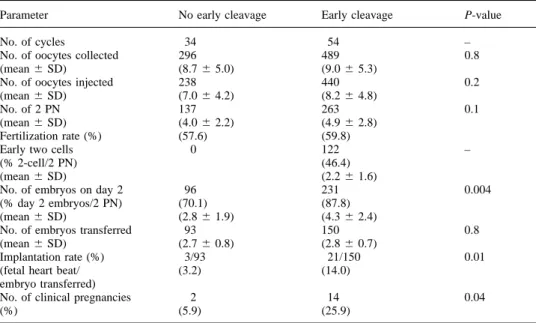
Table I. The total and mean number (6SD) of oocytes, two-pronucleate (2PN) oocytes, embryos observed
on day 2, embryos transferred, implantation rate and pregnancies in patients who had at least one embryo that had undergone early cleavage to the two-cell stage by 27 h after ICSI, compared to those who had none
Parameter No early cleavage Early cleavage P-value
No. of cycles 34 54 –
No. of oocytes collected 296 489 0.8
(mean6 SD) (8.76 5.0) (9.06 5.3)
No. of oocytes injected 238 440 0.2
(mean6 SD) (7.06 4.2) (8.26 4.8)
No. of 2 PN 137 263 0.1
(mean6 SD) (4.06 2.2) (4.96 2.8)
Fertilization rate (%) (57.6) (59.8)
Early two cells 0 122 –
(% 2-cell/2 PN) (46.4)
(mean6 SD) (2.26 1.6)
No. of embryos on day 2 96 231 0.004
(% day 2 embryos/2 PN) (70.1) (87.8)
(mean6 SD) (2.86 1.9) (4.36 2.4)
No. of embryos transferred 93 150 0.8
(mean6 SD) (2.76 0.8) (2.86 0.7)
Implantation rate (%) 3/93 21/150 0.01
(fetal heart beat/ (3.2) (14.0)
embryo transferred)
No. of clinical pregnancies 2 14 0.04
(%) (5.9) (25.9)
three times the implantation rate (Shoukir et al., 1997). In this paper, we have therefore investigated whether the same improvement in pregnancy rate is observed when early cleavage to the two-cell stage is used as an indicator to select embryos for transfer after intracytoplasmic sperm injection (ICSI). The results obtained after ICSI are of particular interest, as the timing of fertilization is more defined than after routine IVF, hence the cleavage rates of embryos are less likely to be determined by the time of fertilization.
Materials and methods
The study was performed on patients entering the IVF programme of the Clinic of Infertility and Gynaecological Endocrinology, Depart-ment of Obstetrics and Gynaecology, University Hospital of Geneva, Geneva, Switzerland between October 1994 and March 1997. In all, 88 cycles were assessed where the patient underwent ICSI treatment with embryo transfer. Patients that failed to achieve fertilization were not included. The stimulation protocol adopted by our group has been previously described (Sakkas et al., 1994).
Collected oocytes (day 0) were maintained in our standard culture medium Whittingham’s T6 (Quinn et al., 1982) supplemented with 10% maternal human serum. Oocyte retrieval took place between 0830 and 1030 h. ICSI was performed between 1300 and 1500 h (~1400 h) as previously described (Sakkas et al., 1996). The following morning (day 1), the oocytes were washed in 20 µl culture drops under oil (light white mineral oil; Sigma Pharmaceuticals, Buchs, Switzerland) in Petri dishes and the presence of two pronuclei assessed. On the same day (day 1), at 1700 h (27 h post-injection), the embryos were examined to see if cleavage to the two-cell stage had occurred. These embryos were designated as ‘early cleavage’ embryos, while those that had not yet cleaved were designated as ‘no early cleavage’. Both groups were maintained in separate culture drops. The following day (day 2) the embryos were transferred between 1000 and 1500 h. Routinely, a maximum of three embryos was transferred to the patient. When there were only one or two early cleavage embryos, the embryo transfer group was completed by
adding the best one or two embryos from the no early cleavage group. In nine cycles, four embryos were transferred due to advanced maternal age, repeated failure in previous cycles or poor quality embryos.
On the day of transfer all embryos were assessed for the number of cells per embryo, to ascertain their cleavage rate, and given a quality score based on the presence of fragments and clarity of the cytoplasm of the blastomeres, similar to that previously described by Cummins et al. (1986). The ratings given to embryos for cell number were 1 for one cell, 2 for two cells, 3 for three cells, 4 for four cells, 6 for between four and eight cells and 8 for more than eight cells. The ratings given to embryos for quality were the same as those used by Cummins et al. (1986), except that the values of 0 for poorest and 3 for best embryo were given.
A pregnancy test was conducted 14 days after transfer. Patients that exhibited three consecutive human chorionic gonadotrophin (HCG) values.5 mIU/ml and where the fetus or fetuses displayed a heart beat by ultrasound examination 4–5 weeks after transfer were considered to have achieved a clinical pregnancy.
The statistical evaluations used were analysis of variance followed by Scheffe´’s F-test for comparisons of mean values (Tables I, IV and V) and χ2 analysis with continuity correction for comparison of pregnancy rates (Table I and II) (Statview 4.5, Abacus Concepts Inc., Ca, USA). Statistical significance was set at P, 0.05.
Results
Early cleavage to the two-cell stage was observed in 54/88 (61.4%) of the cycles. This is a greater percentage than when IVF embryos were checked in our laboratory (Shoukir et al., 1997) at the same time point, and is explained by the fact that ICSI was performed earlier than when oocytes were inseminated in routine IVF. Table I shows the overall results, including the implantation rate and clinical pregnancy outcome, in patients who had at least one early cleavage embryo compared to those that had none. There was no significant difference in the number of oocytes injected or fertilization
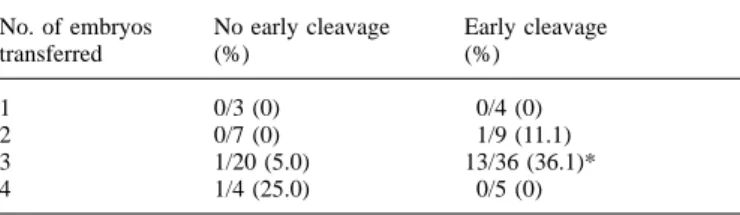
Table II. Clinical pregnancy rates according to the number of embryos
transferred in patients who had early and no early cleaving embryos after ICSI
No. of embryos No early cleavage Early cleavage
transferred (%) (%)
1 0/3 (0) 0/4 (0)
2 0/7 (0) 1/9 (11.1)
3 1/20 (5.0) 13/36 (36.1)*
4 1/4 (25.0) 0/5 (0)
*Denotes significantly differentχ25 5.1, P 5 0.02 when compared to patients with no early cleavage embryos.
rate between both groups. The early cleavage patient group however had significantly more embryos per patient on day 2 compared with the no early cleavage group. Patients with early cleaving embryos displayed an implantation rate four times higher than patients without early cleaving embryos. The pregnancy rate in the early cleavage group was also more than four times higher than the no early cleavage group. No cases of ectopic pregnancies were observed in either group, while there were an additional four biochemical pregnancies in the early cleavage group, where three positive HCG values were measured but no positive confirmation of the pregnancy was obtained by ultrasound.
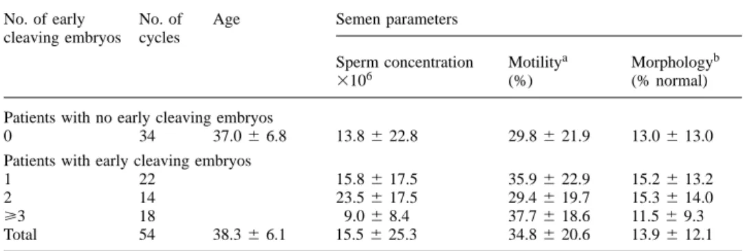
The clinical pregnancy rate according to the number of embryos transferred is represented in Table II. When three embryos were transferred, significantly more pregnancies were obtained in the early cleavage group compared to the no early cleavage group. No comparison was made when one, two or four embryos were transferred due to the low number of cycles. Table III shows the implantation rate and the incidence of single, twin and triplet pregnancies when the cohort of the three embryos transferred contained either: one, two or three early cleavage embryos or all no early cleavage embryos. When all three embryos transferred had undergone early cleavage, the majority of pregnancies obtained were multiple. When a number of key parameters in the couples were examined, we failed to find any differences between the groups linked to the male or female partner that influenced the occurrence of early cleavage. The mean age (6SD) of the females was 33.8 6 3.9 and 34.5 6 4.9 in the no early and early cleavage group respectively. When the progress of stimulation, as indicated by oestradiol concentration on the day of HCG and oocyte retrieval and the number of ampoules of Pergonal (Serono, Aubonne, Switzerland) used during stimulation was examined no statistically significant differences were evident between the two groups (data not shown). More importantly, as the majority of patients being treated were male factor patients, we examined whether semen parameters influenced the differences between the groups. There were no significant differences concerning the mean age and the semen characteristics of the male partner between the groups, and semen characteristics did not influence the number of early cleaving embryos observed (Table IV). In addition, no signific-ant trends in pregnancy rates in relation to the different sperm parameters (sperm concentration, motility and morphology) in patients who had early cleaving embryos could be observed.
To examine whether the cohort of embryos transferred to the patients with and without early cleaving embryos differed, we calculated the mean score of the number of cells and mean quality per embryo. The mean number of cells per embryo and the mean quality of cells per embryo on day 2 for those transferred are presented in Table V. The results show that the mean cell numbers of the embryos transferred and the quality rating was significantly higher for early cleavage embryos. When taking this observation into consideration, it could be argued that the early cleaving embryos may have been selected for transfer anyway. However, in 25 of the cycles with early cleaving embryos the patient had more than three embryos available for transfer hence the embryos of the highest cell number and best quality had to be distinguished. In eight of the 25 (32%) cycles, three embryos could be selected at the time of transfer according to cell number and quality. In the remaining 17 cycles (68%), a selection had to be made between two or more embryos of the same cell number and quality to complete the cohort of embryos selected for transfer. It is in these cases that a prior selection of the early cleaving embryos helps to distinguish which of the embryos is more advanced.
Discussion
The kinetics of fertilization and cleavage to the two-cell stage after ICSI have been previously documented in a study by Nagy et al. (1994). In their study, cleavage to the two-cell stage was initially observed 20 h after ICSI, whereby 10 out of the 93 normally fertilized oocytes (11%) had cleaved to the two-cell stage. In this study, fertilized oocytes were examined at 27 h after ICSI and at this stage a total of 122/400 (30.5%) had cleaved to the two-cell stage. A comparative study examining the number of fertilized oocytes cleaving to the two-cell stage after sub-zonal insemination reported that between 25.5 and 27 h post-treatment just over 30% of the embryos had cleaved (van Wissen et al., 1995).
Early cleavage was observed in 54 of the 88 cycles (61.4%) examined. Subsequently, in the 54 cycles where early cleavage had taken place a significantly higher number of embryos implanted and pregnancies were established. The question as to why the selection of early cleavage embryos leads to a higher pregnancy rate may be related to a number of factors. Firstly, the early cleaving embryos may simply be fertilized earlier. Although this argument can be used in the case of routine IVF, the direct injection of spermatozoa into the cytoplasm using ICSI ensures a shorter time frame of fertiliza-tion. Hence by showing that early cleavage embryos have a greater viability after both routine IVF (Shoukir et al., 1997) and ICSI, it appears that the ability of an embryo to undergo early cleavage is not related to the timing of fertilization. A second parameter that can also influence fertilization is related to the maturity of the oocytes; less mature oocytes are usually fertilized later than the mature ones, leading to a delayed cleavage (Balakier et al., 1993). The removal of the cumulus complex prior to performing ICSI, however, overcomes the presence of immature oocytes. The elimination of these factors influencing the early cleavage of embryos points to the presence of an unknown factor(s) within the oocyte which favours or
Table III. Pregnancy in relation to the number of early cleavage and no early cleavage embryos transferred
in patients receiving three embryos at transfer
No. early and no early cleavage embryos Pregnancy Implantation Totalb
transferred ratea(%) (%)
No early cleavage Early cleavage Single Twins Triplets Patients with no early cleaving embryos
3 0 1 0 0 1/60 1/20
(1.7) (5.0)
Patients with early cleaving embryos
2 1 1 0 0 1/27 1/9 (3.7) (11.1) 1 2 4 1 0 6/36 5/12 (16.7) (41.7) 0 3 3 2 2 13/45 7/15 (28.9) (46.7) aNumber of fetal heart beats per embryo transferred.
bNumber of clinical pregnancies per transfer.
Table IV. Semen parameters (mean6 SD) for cycles with and without early cleavage to the two-cell stage
No. of early No. of Age Semen parameters
cleaving embryos cycles
Sperm concentration Motilitya Morphologyb
3106 (%) (% normal)
Patients with no early cleaving embryos
0 34 37.06 6.8 13.86 22.8 29.86 21.9 13.06 13.0
Patients with early cleaving embryos
1 22 15.86 17.5 35.96 22.9 15.26 13.2
2 14 23.56 17.5 29.46 19.7 15.36 14.0
ù3 18 9.06 8.4 37.76 18.6 11.56 9.3
Total 54 38.36 6.1 15.56 25.3 34.86 20.6 13.96 12.1
aMotility includes all spermatozoa with forward progression.
bSeven cycles with no early cleavage and six with early cleavage could not be assessed for morphology due to low sperm numbers.
Table V. Mean number of cells per embryo and mean quality per embryo of embryos transferred in patients
with and without early cleavage to the two-cell stage (see Materials and methods for a description of the scoring system)
Embryos of no early
cleavage patients Embryos of early cleavage patients
No early cleavage All embryos No early cleavage Early cleavage
Number of embryos transferred 93 150 47 103
Mean number of cells6 SD 2.76 1.4a 3.66 1.1a 3.16 1.3b 3.86 1.0b Mean quality of cells6 SD 1.86 1.1c 2.56 0.7c 2.26 0.9d 2.76 0.5d Like letters a–a; b–b; c–c; d–d are significantly (P, 0.01) different.
promotes the early division of embryos after fertilization. This unknown factor subsequently improves the viability of embryos. A number of candidates could be proposed to explain early cleavage. One of these is the expression of human leukocyte antigen (HLA)-G, as in human pre-implantation embryos it has been demonstrated that a correlation exists between HLA-G mRNA expression and improved cleavage rate (Jurisicova et al., 1996). It would therefore be of interest to ascertain whether early cleavage is observed repetitively in certain patients, indicating a genetic influence over this phenomenon. A further candidate can be linked to the metabolic fitness of oocytes from different patients. For example, Van
Blerkom et al. (1995) proposed that grossly normal human embryos may not progress to implantation due to a chronically low ATP content that is first established in the oocyte. A further advantage to embryos that have undergone early cleavage is that they will also be less likely to arrive at a critical minimal level of maternal mRNA levels (Bacharova and de Leon, 1980; Clegg and Piko, 1983) prior to activation of the embryonic genome. Human embryos that have experienced retarded fertilization or have a slower cleavage rate may deplete their vital maternal mRNA stores and their viability would be compromised prior to reaching the four- to eight-cell stage when the embryonic genome is activated (Braude
et al., 1988). As the manifestation of the above mentioned
challenges to embryo development would likely lead to a poor morphology of the developing embryo it was of interest to note that embryos that had undergone early cleavage had a significantly higher quality compared to those that had cleaved later.
The early cleavage of embryos was also investigated in relation to the sperm parameters of the male. It has previously been shown that a strong paternal effect is associated with the development of human embryos to the blastocyst stage (Janny and Me´ne´zo, 1994). These authors found evidence that frozen and abnormal spermatozoa gave rise to embryos with a significantly lower cleavage rate and potential to form blasto-cysts. Prior to this study, a number of other authors also suggested that semen quality and embryo development may be related (Ron El et al., 1991; Chan et al., 1993; Parinaud et al., 1993). When examining the influence of sperm parameters on the number of early cleavage embryos and the outcome of pregnancy, we found no difference between males with varying sperm parameters. The previous studies that have indicated a paternal effect have however been based on using routine IVF insemination procedures where cleavage rates and embryo quality may be influenced by the timing of fertilization. In this study however, the timing of fertilization is more definite, because of the use of ICSI, hence the influence of a poor sperm parameter on fertilization is largely removed. Indeed a number of studies have already shown that sperm parameters have little influence on fertilization rates and embryo quality after ICSI (Van Steirteghem et al., 1993; Payne et al., 1994; Nagy et al., 1995; Tournaye et al., 1995).
A final point of consideration arises from certain studies that provide evidence that male embryos cleave faster than female embryos (Tsunoda et al., 1985; Burgoyne, 1993; Peippo and Bredbacka, 1995). In the human, it has also been proposed that sex selection is inadvertently performed in IVF pro-grammes because the faster cleaving and higher quality embryos are selected for transfer (Tarin et al., 1995). The technique of embryo selection used in this study would therefore predictably give rise to more males. In this study, and our previous study (Shoukir et al., 1997), there do not appear to be any tendencies to a selection of males; however, the number of pregnancies is too limited to come to any conclusion.
In this and our previous study (Shoukir et al., 1997), we have shown that the assessment of early cleavage to the two-cell stage can be used as an indicator of embryo viability and is subsequently a strong prognostic factor of the likelihood of pregnancy. The simple action of selecting early cleaving two-cell embryos alleviates the problem of guessing which are the more advanced embryos at the time of transfer. Using the ICSI technique, we have also shown that the early cleavage of embryos to the two-cell stage is not entirely influenced by the timing of fertilization, and is therefore more likely due to intrinsic factors within the oocyte or embryo that promote embryo cleavage after fertilization.
Acknowledgements
We thank all members of our IVF team for their assistance during this study.
References
Bacharova, R. and de Leon, V. (1980) Polyadenylated RNA of mouse ova and loss of maternal mRNA in early development. Dev. Biol., 74, 1–8. Balakier, H., MacLusky, N.J. and Casper, R.F. (1993) Characterization of the
first cell cycle in human zygotes: implications for cryopreservation. Fertil.
Steril., 59, 359–365.
Bavister, B.D. (1995) Culture of preimplantation embryos: facts and artefacts.
Hum. Reprod. Update, 1, 91–148.
Braude, P., Bolton, V. and Moore, S. (1988) Human gene expression first occurs between the four- and eight-cell stages of preimplantation development. Nature, 332, 459–461.
Burgoyne, P.S. (1993) A Y-chromosomal effect on blastocyst cell number in mice. Development, 117, 341–345.
Capmany, G., Taylor, A., Braude, P.R. and Bolton, V.N. (1996) The timing of pronuclear formation, DNA synthesis and cleavage in the human 1-cell embryo. Mol. Hum. Reprod., 2, 299–306.
Chan, S.Y.W., Tucker, M.J., Leung, C.K.M. and Leong, M.K.H. (1993) Association between human in vitro fertilization rate and pregnancy outcome: a possible involvement of spermatozoal quality in subsequent embryonic viability. Asia-Oceania J. Obstet. Gynaecol., 19, 357–373. Clegg, K.B. and Piko, I. (1983) Quantitative aspects of RNA synthesis and
polyadenylation in 1-cell and 2-cell mouse embryos. J. Embryol. Exp.
Morphol., 74, 169–182.
Cummins, J.M., Breen, T.M., Harrison, K.L. et al. (1986) A formula for scoring human embryo growth rates in in vitro fertilization: its value in predicting pregnancy and in comparison with visual estimates of embryo quality. J. In Vitro Fert. Embryo Transf., 3, 284–295.
Edwards, R.G. Fishel, S.B., Cohen, J. et al. (1984) Factors influencing the success of in vitro fertilization for alleviating human infertility. J. In Vitro
Fert. Embryo Transf., 1, 3–23.
Gonzales, D.S., Pinheiro, J.C. and Bavister, B.D. (1995) Prediction of the developmental potential of hamster embryos in vitro by precise timing of the third cell cycle. J. Reprod. Fertil., 105, 1–8.
Hill, G.A., Freeman, M., Bastias, M.C. et al. (1989) The influence of oocyte maturity and embryo quality on pregnancy rate in a program for in vitro fertilization–embryo transfer. Fertil. Steril., 52, 801–806.
Janny, L. and Me´ne´zo, Y.J.R. (1994) Evidence for a strong paternal effect on human preimplantation embryo development and blastocyst formation. Mol.
Reprod. Dev., 38, 36–42.
Jurisicova, A., Casper, R.F., MacLusky, N.J. et al. (1996) HLA-G expression during preimplantation human embryo development. Proc. Natl Acad. Sci.,
93, 161–165.
Leese, H.J. (1987) Analysis of embryos by non-invasive methods. Hum.
Reprod., 2, 37–40.
McKiernan, S.H. and Bavister, B.D. (1994) Timing of development is a critical parameter for predicting successful embryogenesis. Hum. Reprod.,
9, 2123–2129.
Nagy, Z., Liu, J., Joris, H. et al. (1994) Time-course of oocyte activation, pronucleus formation and cleavage in human oocytes fertilized by intracytoplasmic sperm injection. Hum. Reprod., 9, 1743–1748.
Nagy, Z., Liu, J., Joris, H. et al. (1995) The result of intracytoplasmic sperm injection is not related to any of the three basic sperm parameters. Hum.
Reprod., 10, 1123–1129.
Parinaud, J., Mieusset, R., Vieitez, G. et al. (1993) Influence of sperm parameters on embryo quality. Fertil. Steril., 60, 888–892.
Payne, D., Flaherty, S.P., Jeffrey, R. et al. (1994) Successful treatment of severe male factor infertility in 100 consecutive cycles using intracytoplasmic sperm injection. Hum. Reprod., 9, 2051–2057.
Peippo, J. and Bredbacka, P. (1995) Sex-related growth rate differences in mouse preimplantation embryos in vivo and in vitro. Mol. Reprod. Dev.,
40, 56–61.
Quinn, P., Barros, C. and Whittingham, D.G. (1982) Preservation of hamster oocytes to assay the fertilizing capacity of human spermatozoa. J. Reprod.
Fertil., 66, 161–168.
Ron-El, R., Nachum, H., Herman, A. et al. (1991) Delayed fertilization and poor embryonic development associated with impaired semen quality. Fertil.
Steril., 55, 338–344.
Sakkas, D., Jaquenoud, N., Leppens, G. and Campana, A. (1994) Comparison of results after in vitro fertilized human embryos are cultured in routine medium and in coculture on Vero cells: a randomised study. Fertil. Steril.,
61, 521–525.
Sakkas, D., Urner, F., Bianchi, P.G. et al. (1996) Sperm chromatin anomalies can influence decondensation after intracytoplasmic sperm injection. Hum.
Shoukir, Y., Campana, A., Farley, T. and Sakkas, D. (1997) Early cleavage of in-vitro fertilized human embryos to the 2-cell stage: a novel indicator of embryo quality and viability. Hum. Reprod., 7, 1531–1536.
Steer, C.V., Mills, C.L., Tan, S.L. et al. (1992) The cumulative embryo score: a predictive embryo scoring technique to select the optimal number of embryos to transfer in an in-vitro fertilization and embryo transfer program.
Hum. Reprod., 7, 117–119.
Tarin, J.J., Bernabeu, R., Baviera, A. et al. (1995) Sex selection may be inadvertently performed in in-vitro fertilization–embryo transfer programmes. Hum. Reprod., 11, 2992–2998.
Tournaye, H., Liu, J., Nagy, Z. et al. (1995) Intracytoplasmic sperm injection: the Brussels experience. Reprod. Fertil. Dev., 7, 269–281.
Trounson, A.O., Mohr, L.R., Wood, C. and Leeton, J.F. (1982) Effect of delayed insemination on in-vitro fertilization, culture and transfer of human embryos. J. Reprod. Fertil., 64, 285–294.
Tsunoda, Y., Tokunaga, T. and Sugie, T. (1985) Altered sex ratio of live young after transfer of fast- and slow-developing mouse embryos. Gamete Res.,
12, 301–304.
Van Blerkom, J., Davis, P.W. and Lee, J. (1995) ATP content of human oocytes and developmental potential and outcome after in-vitro fertilization and embryo transfer. Hum. Reprod., 10, 415–424.
Van Steirteghem, A.C., Liu, J., Joris, H. et al. (1993) Higher success rates by intracytoplasmic sperm injection than by subzonal insemination. Report of a second series of 300 consecutive treatment cycles. Hum. Reprod., 8, 1055–1060.
van Wissen, B. Wolf, J.P., Bomsel-Helmreich, O. et al. (1995) Timing of pronuclear development and first cleavages in human embryos after subzonal insemination: influence of sperm phenotype. Hum. Reprod., 10, 642–648.