Attenuation of myocardial reperfusion injury in pigs by Mirococept, a
membrane-targeted complement inhibitor derived from human CR1
Yara Banz
a,c, Otto M. Hess
b, Simon C. Robson
c, Eva Csizmadia
c, Daniel Mettler
a,
Pascal Meier
b, André Haeberli
a, Sidney Shaw
a, Richard A. Smith
d, Robert Rieben
a,⁎
aUniversity of Bern, Department of Clinical Research, Switzerland bCardiology, Swiss Cardiovascular Center, University Hospital, Bern, Switzerland
cHarvard University, Beth Israel Deaconess Medical Center, Boston, MA, USA dInflazyme Pharmaceuticals Ltd., Richmond, BC, Canada
Received 18 January 2007; received in revised form 20 July 2007; accepted 25 July 2007 Time for primary review 21 days
Available online 2 August 2007
Abstract
Objectives: Membrane-targeted application of complement inhibitors may ameliorate ischemia/reperfusion (I/R) injury by directly targeting damaged cells. We investigated whether Mirococept, a membrane-targeted, myristoylated peptidyl construct derived from complement receptor 1 (CR1) could attenuate I/R injury following acute myocardial infarction in pigs.
Methods: In a closed-chest pig model of acute myocardial infarction, Mirococept, the non-tailed derivative APT154, or vehicle was administered intracoronarily into the area at risk 5 min pre-reperfusion. Infarct size, cardiac function and inflammatory status were evaluated. Results: Mirococept targeted damaged vasculature and myocardium, significantly decreasing infarct size compared to vehicle, whereas APT154 had no effect. Cardioprotection correlated with reduced serum troponin I and was paralleled by attenuated local myocardial complement deposition and tissue factor expression. Myocardial apoptosis (TUNEL-positivity) was also reduced with the use of Mirococept. Local modulation of the pro-inflammatory and pro-coagulant phenotype translated to improved left ventricular end-diastolic pressure, ejection fraction and regional wall motion post-reperfusion.
Conclusions: Local modification of a pro-inflammatory and pro-coagulant environment after regional I/R injury by site-specific application of a membrane-targeted complement regulatory protein may offer novel possibilities and insights into potential treatment strategies of reperfusion-induced injury.
© 2007 European Society of Cardiology. Published by Elsevier B.V. All rights reserved.
Keywords: Ischemia; Reperfusion; Complement; Complement receptor 1; Endothelium; Mirococept
1. Introduction
Reperfusion following ischemia is known to induce a pro-inflammatory reaction, involving activation of the classical, alternative and lectin complement pathways[1,2]. The im-portance of each of the three different complement pathways may, in part, depend on the organ affected. Furthermore, the injured organ itself up-regulates the production of comple-ment components, which contribute to reperfusion damage
[3].
In vivo, membrane-bound regulators of complement, in-cluding membrane cofactor protein (MCP, CD46),
decay-Abbreviations: AAR, area at risk; ANR, area not at risk; aPTT, activated partial thromboplastin time; CH50, (classical pathway) complement activity; CR1, complement receptor 1; EC, endothelial cell; EF, ejection fraction; ELISA, enzyme-linked immunosorbent assay; ET-1, endothelin-1; FS, fractional shortening; HR, heart rate; I/R, ischemia/reperfusion; LAD, left anterior descending artery; LVEDP, left ventricular end-diastolic pressure; LVM, left ventricular mass; MAP, mean arterial pressure; MI, myocardial infarction; MPO, myeloperoxidase; NIT, necrotic ischemic tissue; TF, tissue factor; TTC, triphenyl tetrazolium chloride; TUNEL, terminal deoxynu-cleotidyl transferase-mediated dUTP nick end-labeling; VIT, vital ischemic tissue.
⁎ Corresponding author. Department of Clinical Research University of Bern Murtenstrasse 31, P.O. Box 33 CH-3010 Bern, Switzerland. Tel.: +41 31 632 9669; fax: +41 31 632 8837.
E-mail address:[email protected](R. Rieben).
0008-6363/$ - see front matter © 2007 European Society of Cardiology. Published by Elsevier B.V. All rights reserved. doi:10.1016/j.cardiores.2007.07.016
accelerating factor (DAF, CD55), protectin (CD59) as well as complement receptor type 1 (CR1, CD35) protect against unwanted, harmful complement activation [4]. CR1 influ-ences the classical and alternative pathway by inhibiting the C3/C5 convertases, acting as a cofactor for factor-I mediated cleavage of C4b/C3b and for the subsequent cleavage of iC3b to C3c and C3dg complement fragments[4]. Further-more, CR1 is up-regulated on human vascular endothelial cells following hypoxic stress and mediates immune com-plex processing [5]. These properties render it ideal to ex-ploit therapeutically in complement-mediated disease states. In a soluble recombinant form, it has been used in several studies to successfully treat I/R- and cardiopulmonary by-pass-associated injury[6–8]. Local, directed cytoprotection and complement inhibition with CR1, modified to bind to (damaged) cell membranes, holds out the prospect of im-proved efficacy through increasing the effective concentra-tion of the complement regulatory pharmacophore at the site where it is needed.
In this report we describe the use of Mirococept[9], an engineered fragment of soluble (s)CR1, which, through coupling to a reagent comprising a peptide motif and a lipophilic tail[9,10], enables tethering of the inhibitor to cell surfaces. We hypothesized that since pro-inflammatory com-plement components are formed and assembled at the sur-face of damaged cells, intracoronary infusion of Mirococept to damaged endothelium and myocardium may reduce I/R injury in a pig model of acute myocardial infarction. 2. Materials and methods
Care and use of animals in the present study were in compliance with the Guide for the Care and Use of Labo-ratory Animals (NIH publication no. 85-23, revised 1996) as well as Swiss National guidelines.
2.1. Materials
Mirococept is a soluble, recombinant protein derived from soluble complement receptor 1 (sCR1) modified with membrane-binding properties through a myristoylated pep-tide sequence. The membrane-binding moiety was designed for posttranslational chemical attachment to the short con-sensus repeats 1–3 [9,10] and is based on the myristoyl electrostatic switch structural feature of intracellular proteins
[11]. Membrane localization of Mirococept is rapid (com-plete in vitro within 2 min at 25 °C) and the dissociation half-life averages several hours. Radiolabelling experiments sug-gest that the modified protein is retained 5–10 times longer than the unmodified form (see below). Further details on construction and structure of Mirococept can be found in the publication of Pratt et al.[12]and elsewhere[10,13,14]. The recombinant non-tailed CR1 fragment APT154 possesses identical chemical and complement inhibitory properties as Mirococept, but is untagged and therefore does not contain the membrane-binding moiety. A vehicle control was used
which contains all but the active Mirococept or APT154, respectively. All substances were produced and provided by Inflazyme (Inflazyme Pharmaceuticals Ltd., Richmond, Canada).
2.2. Experimental model
Eighteen large white pigs (30 ± 2 kg) were premedi-cated with intramuscular ketamine (20 mg/kg) and xylazine (2 mg/kg), followed by intravenous midazolam (0.5 mg/kg) and atropine (0.05 mg/kg), intubated and mechanically ventilated with a Draeger respirator (O2/N2O 1:3,
Isoflur-ane 1–1.5 vol.%). Central venous and arterial lines were introduced and a single bolus of unfractionated heparin (2500 IU) was administered intravenously. Baseline values were recorded during a 30-minutes stabilization period.
The left anterior descending artery (LAD) was occluded with a semi compliant over-the-wire percutaneous coronary intervention catheter (Concerto, Occam, the Netherlands, balloon length 10 mm, diameter 2.5 mm to 3 mm) just distally of the first diagonal branch. The balloon was inflated under angiographic control to completely occlude the vessel (Encore™ 26 inflation device, Boston Scientific, Ireland) for 60 min.
Ischemia was followed by two hours of reperfusion. Five minutes prior to reperfusion, 10 ml of Mirococept (1.5 mg/ ml; corresponding to 0.5 mg/kg [22 nmol/kg]; the dose and application scheme i.e. one-time application were chosen according to previous experience and studies[12,15], n = 6), APT154 (non-tailed control, 1.37 mg/ml [equimolar to Mi-rococept], n = 6) or vehicle control (n = 6) were slowly injected intracoronarily, through the catheter tip, into the area at risk. Thereafter the balloon was deflated to allow for two hours of reperfusion. Following reperfusion the balloon was re-inflated, 60 ml Evan's Blue injected intravenously and the animals sacrificed by intravenous potassium chloride. The heart was excised for further analysis.
ECG and invasive arterial pressure were recorded through-out with a Hewlett-Packard CMS patient monitor. Ejection fraction (EF) was determined angiographically and calculated using the area-length-method according to Dodge [16]. Fractional shortening (FS) was also determined angiographi-cally and was assessed in three regions (apical, medial and basal). The geometric mean of the three regional values was used for further analyses.
All experimenters were blinded with regard to treatment regimen. Randomization of the animals into the three groups was done using a randomization code with a random num-ber generator (SAS, version 9.1.2, SAS Institute Inc., Cary, NC, USA), prior to the begin of the series (Mirococept = 0, APT154 = 1, vehicle controls = 2). The 10 ml samples (Miro-cocept, APT154 or vehicle solution) were prepared accord-ing to the randomization output and stored at−80 °C until use. Randomization and sample preparation was performed by an independent laboratory technician. Prior to pre-medi-cation of the animals, the corresponding vial was allocated
to the pig (sequential number of vial = sequential number of the pig). All 18 consecutively enlisted pigs were treated according to the above-described protocol. No animal was excluded from or dropped out during the experiment. Sam-ple size (n = 6) was determined in advance, estimated from previous work [12,17] and experience and not by formal sample size calculations.
In case of ventricular fibrillation a biphasic defibrillator (150 J) was used for cardioconversion.
2.3. Infarct size
The area at risk (AAR) was determined by re-occlusion of the LAD and intravenous injection of 60 ml Evan's Blue (2% wt/vol solution) at the end of the experiment. Tissue stained blue was defined as the area not at risk. The left ventricle was cut perpendicular to the septum into 3 mm slices. Incubation with triphenyl tetrazolium chlo-ride (TTC, Sigma, pH 7.4, 1% in phosphate buffered saline) for 20 min at 37 °C was used to stain viable myocardium (vital ischemic tissue, VIT) from the area at risk bright red (formazan complex in presence of active dehydrogenases) whereas infarcted tissue (necrotic ische-mic tissue, NIT) remains unstained. All three tissue sec-tions were dissected from each other and weighed. The area at risk was defined as the sum of necrotic and vital ischemic tissue. Data were expressed as area at risk as percent of LVM (AAR/LVM) and necrotic ischemic tissue as percent of the area at risk (NIT/AAR).
2.4. Complement and coagulation
Venous blood samples were collected (EDTA plasma/ serum), kept on ice until centrifugation (1750 x g for 10 min at 4 °C) and stored at−80 °C until further analysis.
C3a and C5a were measured by sandwich ELISA as previously described [18]. In brief, microtiter plates were coated with a monoclonal antibody (IgG2b) against porcine C3a/C3a(desArg) or rabbit anti-mouse IgG (Dako, Glostrup, Denmark) followed by a monoclonal antibody (IgG1) against porcine C5a. After washing with PBS–Tween, plasma sam-ples were incubated at a 1:50 (C3a ELISA) or 1:2 (C5a ELISA) dilution in PBS/Tween/EDTA for 2 h. Biotinylated monoclonal anti-C3/C3a antibody or rabbit anti-C5a, fol-lowed by streptavidin-alkaline phosphatase conjugate (Amersham Pharmacia Biotech, Bucks, UK) and 4-nitrophe-nyl phosphate substrate (Sigma) were used to detect bound C3a and C5a (all non-commercial antibodies were obtained from the Georg-August University, Goettingen— Science-bridge GmbH, Germany).
Classical pathway complement activity was determined by standard a CH50 assay with antibody-coated sheep erythrocytes[19].
Activated partial thromboplastin time (aPTT) was mea-sured using Dade Actin FS reagent in a standard coagulation assay as a global parameter for the coagulation system.
2.5. Troponin I
Troponin I was measured by liquid suspension array technique using the Luminex fluorescent bead platform. Mouse anti-human troponin I (Abcam Ltd, clone 19C7), biotinylated using the EZ-Link NHS-PEO Solid Phase Biotinylation Kit mini spin columns from Pierce (Pierce, Rockford, IL, USA), was used as the detection antibody and mouse anti-human troponin I, cross-reactive with porcine troponin I (Abcam Ltd., Cambridge, UK, clone 16A11), was used as the capture antibody. Covalent coupling of the capture antibody to the region-specific microspheres was performed using the Bio-Plex Amine Coupling Kit (Rad). Analysis was done with the Plex system and Bio-Plex Manager version 4.0 software (Bio-Rad) with five-parametric curve fitting. Assay conditions were derived from those of commercial tests.
2.6. Histology and immunostaining
Tissue samples from the area not at risk as well as necrotic and vital ischemic tissue were fixed in 4% buffered for-maldehyde, paraffin-embedded, cut into 3μm sections and stained with hematoxylin-eosin for histological evaluation. Neutrophil tissue numbers were determined by myeloper-oxidase (MPO) staining on additional tissue sections. Anti-gen-retrieval was performed with target retrieval solution (Dako, Glostrup. Denmark) and demasking at 96 °C for 30 min. Following peroxidase block (Dako) MPO was de-tected using a polyclonal antibody to MPO (Dako) and visualized with a horseradish peroxidase-labeled secondary antibody and EnVision+ System (Dako). Neutrophil numb-ers were quantified in 10 randomly selected high-power viewing fields from the tissue samples of each experiment. The intravascular/interstitial ratio was calculated for each section and experiment and averaged for all experiments in each of the three groups.
For complement and tissue factor detection, 5 μm sec-tions were cut from all snap frozen tissue samples, air-dried, acetone fixed, hydrated and labeled using a two-step indirect immunofluorescence technique. The following antibodies were used: rabbit anti-human C1q, C3b/c and C4b/c (Dako); goat anti-human C6 (Quidel, for detection of the membrane attack complex). Cross-reactivity with the respective por-cine antigens was verified. Secondary antibodies were goat anti-rabbit IgG(H+L)-FITC (Southern Biotechnology Asso-ciated) and rabbit anti-goat Ig-FITC (Dako). Immunohisto-chemical staining for tissue factor (TF) with polyclonal rabbit anti-human TF antibody was carried out as reported previously
[20].
For every individual experiment four samples per area (area not at risk, ischemic viable and necrotic areas) were graded according to the following scoring system: 0: no staining, 1: minimal focal or diffuse staining, 2: moderately strong focal or diffuse staining, 3: extensive focal or diffuse staining.
Tissue binding of Mirococept was visualized on ace-tone-fixed cryosections using Cy3-labeled 3E10 antibody (mouse anti-human sCR1-3 monoclonal antibody, provided by Inflazyme).
2.7. Quantification of myocyte apoptosis by TUNEL Myocyte apoptosis was detected on paraffin-embedded sections in high-power fields by terminal deoxynucleotidyl transferase-mediated dUTP nick end-labeling (TUNEL, Chemicon Inc., Millipore, Billerica, MA, USA). TUNEL-positive nuclei that were not definitively of myocyte origin were excluded from counting. The extent of apoptosis was expressed by normalizing the results to the total number of myocyte nuclei per high-power field.
2.8. Statistics
Data from triphenyl tetrazolium chloride staining (AAR/ LVM in % and NIT/AAR in %, primary endpoint) were compared between the three groups by use of one-way-ana-lysis of variance (ANOVA). Results for all other parameters (secondary endpoints) were compared by use of two-way repeated-measures ANOVA with time and treatment as fixed factors. Subject number was included as random. Effects of interest were overall between-group differences and interac-tion between group and time. The assumpinterac-tion of the sphericity of our data was analyzed by Mauchly's test and corrections were made where appropriate (Greenhouse-Geisser). In case of a significant p-value in the overall ANOVA with respect to
group ⁎ time interaction, post-hoc analyses were performed. Holm's procedure was used in post-hoc pair-wise compar-isons to control for type I error. For all analyses p-values were two-sided and differences were considered to be statistically significant with a p-value ofb0.05. SAS Version 9.1.2 (SAS Institute Inc., Cary, NC, USA) and SPSS Version 12.0.1 (SPSS Inc., Chicago, IL, USA) software were used for all analyses. Data, unless otherwise specified, are presented as average ± standard deviation in text and figures.
3. Results
3.1. Tissue and plasma levels of Mirococept
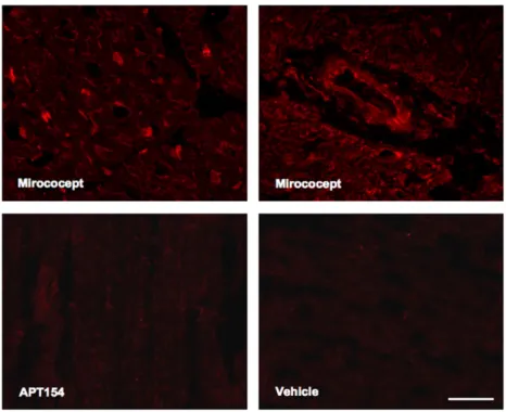
Bound Mirococept was found mainly in the area at risk, both in viable and necrotic ischemic tissue, on the inner lining of small and large blood vessels and in the myo-cardium, and to a lesser extent in non-ischemic myocardial tissue (Fig. 1). Staining for APT154 essentially revealed no tissue positivity. No staining was observed at distant sites (lung, kidney, liver). In an ELISA for Mirococept, small amounts were detected in circulation 5 min after reperfusion (3.4 ± 0.8μg/ml). Plasma levels decreased after 1 h and 2 h reperfusion (1.8 ± 0.5 μg/ml, 0.8±0.2 μg/ml respectively, results not shown).
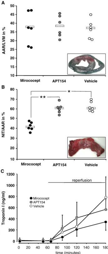
3.2. Infarct size
The myocardial area at risk (AAR) as percentage of left ventricular mass (LVM) was not different in the three groups:
Fig. 1. Staining for Mirococept using Cy3-labeled 3E10 antibody (mouse anti-human sCR1-3 monoclonal antibody). Specific deposition of Mirococept is detected in damaged vessels and myocardium in Mirococept-treated animals. No staining is detected in representative samples from APT154- or vehicle-treated animals. Scale bar: 50μm.
The measured mean values ± standard deviations were 37.1 ± 9.4%, in the Mirococept animals, 38.3 ± 6.5% in the APT154 animals, and 36.5 ± 4.8% in the vehicle controls ( p = 0.968 Mirococept vs. APT154; p = 0.886 Mirococept vs. vehicle
controls, p = 0.900 APT154 vs. vehicle controls). The corres-ponding wet weights in grams were 33.7 ± 6.7 g, 34.7 ± 8.5 g and 29.5 ± 3.3 g, respectively. However, infarcted tissue (necrotic ischemic tissue, NIT) as percentage of AAR was
Fig. 2. Ratio of area at risk (AAR) to left ventricular mass (LVM), as detected by Evan's Blue staining (A). Ratio of necrotic ischemic tissue (NIT) to AAR as detected by triphenyl tetrazolium chloride (TTC) staining (B). Individual values and means (bars) are shown. Comparable AAR in all groups (p = ns between groups). NIT as percent of AAR: p = 0.0003 (⁎) Mirococept vs. controls, p = 0.0001 (⁎⁎) Mirococept vs. APT154, p = 0.880 APT154 vs. controls. Representative pictures of the respective staining are shown as inserts (in A: whole section of left and right ventricle, in B: magnified image of Evan's Blue negative section after TTC staining). Levels of troponin I as measured in EDTA plasma samples by sandwich immunoassay (C). Results are presented as mean ± standard deviation.
significantly reduced from 61.9 ± 6.3% in vehicle controls to 42.5 ± 4.0% in the Mirococept treated animals ( pb0.001). Administration of APT154 did not reduce infarct size, which, at 60.8 ± 5.1% of the AAR, was not significantly different to vehicle controls (p = 0.880). Wet weights of NIT were 14.4 ± 3.3 g in the Mirococept animals, 21.0 ± 4.8 g in the APT154 group, and 18.1 ± 1.6 g in the vehicle controls (Fig. 2A and B).
3.3. Plasma troponin I
Plasma troponin I increased in all groups following reperfusion from similar baseline- (average for all groups 6 ± 8 ng/ml) and ischemia-levels to 776 ± 372 ng/ml in the
vehicle controls, 566 ± 246 ng/ml in APT154 animals, and 344 ± 153 ng/ml in Mirococept treated animals after 2 h reperfusion ( p = 0.888 for overall group-time interaction,
Fig. 2C).
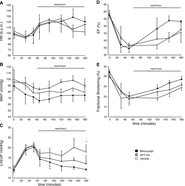
3.4. Hemodynamics
Heart rate did not differ significantly amongst the three groups throughout the experimental procedure (p = 0.888 for overall group-time interaction,Fig. 3A).
Baseline mean arterial pressures (MAP) as well as pres-sures during the experiments were also not significantly different in the three groups. (p = 0.888 for overall group-time interaction,Fig. 3B).
Fig. 3. Heart rate (HR, A), mean arterial pressure (MAP, B), left ventricular end-diastolic pressure (LVEDP, C), ejection fraction (EF, D) and regional wall motility (fractional shortening, FS, E; geometric means of basal, medial and apical values) recorded throughout the experiments. HR and MAP: p = ns between all groups throughout. LVEDP: p = 0.033 (⁎) for Mirococept vs. vehicle controls during reperfusion. EF: p = 0.048 for overall group-time interaction during ischemia, p = 0.081 for Mirococept vs. vehicle controls, p = 0.096 for Mirococept vs. APT154 during reperfusion. Fractional shortening: p = 0.032 for overall group-time interaction during ischemia, p = 0.078 for Mirococept vs. vehicle controls, p = 0.098 for Mirococept vs. APT154 during reperfusion. Data are mean ± standard deviations.
Left ventricular end-diastolic pressure (LVEDP) rapidly and significantly increased in all three groups during ische-mia (p = 0.009 for overall group-time interaction). Following reperfusion, LVEDP decreased in all groups, most markedly in the Mirococept treated group. LVEDP was significantly lower in the Mirococept group as compared to vehicle con-trols (p = 0.033). Other between group differences (Miroco-cept vs. APT154 and APT154 vs. vehicle controls) were not significant (Fig. 3C).
Ejection fraction significantly decreased in all three groups during LAD occlusion from similar baseline values (overall group-time interaction p = 0.048). Recovery during reperfusion was more rapid in the Mirococept group. Post-hoc analyses for between group differences showed p = 0.081 for Mirococept vs. vehicle controls, p = 0.096 for Mirococept vs. APT154 and p = 0.678 for APT154 vs. vehicle controls (Fig. 3D).
Basal and medial, but particularly apical (not shown separately) regional wall motility (fractional shortening, FS) was impaired in all three groups during ischemia as com-pared to baseline, and only showed some recovery towards the end of the reperfusion period, with a tendency towards better recovery in the Mirococept group as compared to vehicle controls and the APT154 animals (overall group-time interaction p = 0.032, post-hoc analysis: p = 0.078 for Mirococept vs. vehicle controls, p = 0.098 for Mirococept vs. APT154 and p = 0.323 for APT154 vs. vehicle controls,
Fig. 3E).
The need for defibrillation for ventricular fibrillation was not significantly different in the three groups (2 animals per group).
3.5. Histology and neutrophil numbers
Reperfusion damage was evident in the necrotic myocar-dium of all three groups, comprising contraction bands and coagulation necrosis. Gross hemorrhage was observed in two animals of each group. Microscopic hemorrhagic infarction was detected in all groups (n = 2 for Mirococept and APT154, n = 3 for vehicle controls). Focal minimal signs of ischemia (wavy fibers) were noted in all three groups in the viable ischemic tissue. Samples from non-ischemic myocar-dium showed normal histological findings.
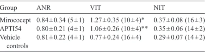
The total numbers and intravascular/interstitial ratios of neutrophils in tissue samples are shown in Table 1. Miro-cocept significantly prevented neutrophil extravasation, particularly in vital ischemic tissue (ratio 1.27 ± 0.35, p = 0.021 vs. vehicle controls). Ratios in the area not at risk and in necrotic ischemic tissue were not significantly different between the three groups. Neutrophil numbers in non-ischemic and necrotic tissue from all three groups did not differ significantly. However, treatment with Mirococept significantly reduced MPO positive cells within the vital ischemic tissue as compared to sections from APT154 and vehicle control animals (p = 0.031).
3.6. Complement deposition and soluble complement parameters
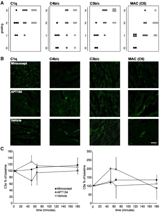
Fig. 4 summarizes the data of complement deposition and soluble complement parameters. Samples from all ex-periments were stained and graded: 0: no staining, 1: mi-nimal focal or diffuse staining, 2: moderately strong focal or diffuse staining, 3: extensive focal or diffuse staining. Normal tissue samples from healthy pigs (not shown) and from myocardium not at risk served as controls. Mirococept markedly reduced complement deposition (C1q, C4b/c, C3b/c, and membrane attack complex) in necrotic ischemic tissue as compared with vehicle controls and APT154 treated animals. Essentially no, or only minimal, comple-ment staining was observed in myocardium of healthy pigs or in myocardium not at risk of all three treatment groups (Fig. 4A and B).
Levels of the anaphylatoxins C3a and C5a, expressed as percentage of baseline, did not vary greatly throughout the experiment and were not significantly different be-tween the three groups after 2 h reperfusion (p = 0.422 Mirococept vs. vehicle controls, p = 0.277 Mirococept vs. APT154, p = 0.395 APT154 vs. vehicle controls for C3a; p = 0.317 Mirococept vs. vehicle controls, p = 0.162 Miro-cocept vs. APT154, p = 0.243 APT154 vs. vehicle controls for C5a, Fig. 4C).
CH50 tests were performed to evaluate systemic com-plement activity of the classical pathway. CH50 values were calculated as % lysis, where lysis of red blood cells in water was set to 100%. Baseline CH50 values were not signi-ficantly different in the groups and values remained largely unaffected in both the APT154 and vehicle groups. Miro-cocept significantly inhibited the classical pathway of com-plement, as apparent CH50 values dropped and remained significantly lower after 2 h reperfusion (14 ± 2% at 5 min reperfusion, 25 ± 5% at 2 h reperfusion; p = 4.3 × 10− 7 for overall group-time interaction. Post-hoc analysis at 120 min reperfusion: p = 0.0018 Mirococept vs. vehicle controls, p = 0.0012 Mirococept vs. APT154, p = 0.332 APT154 vs. ve-hicle controls, results not shown).
Table 1
Representation of intravascular/interstitial ratio and total number of tissue neutrophils in myocardial samples
Group ANR VIT NIT
Mirococept 0.84 ± 0.34 (5 ± 1) 1.27 ± 0.35 (10 ± 4)⁎ 0.37 ± 0.08 (16 ± 3) APTI54 0.80 ± 0.21 (4 ± 1) 1.06 ± 0.26 (10 ± 4)⁎⁎ 0.35 ± 0.06 (14 ± 2) Vehicle
controls
0.81 ± 0.22 (4 ± 1) 0.77 ± 0.24 (16 ± 4) 0.29 ± 0.07 (14 ± 2) Four samples per area and experiment were examined and an average calculated from these. ⁎ denotes p = 0.021 Mirococept vs. vehicle controls (neutrophil ratio) and p = 0.040 Mirococept vs. vehicle controls (neutrophil numbers); ⁎⁎ denotes p = 0.043 APT154 vs. vehicle controls (neutrophil numbers). ANR: area not at risk, VIT: vital ischemic tissue, NIT: necrotic ischemic tissue.
3.7. Tissue factor expression and aPTT
Compared to vehicle controls, Mirococept treatment re-duced vascular TF expression in necrotic ischemic tissue. Upregulation of TF in the vehicle controls as well as APT154 treated animals within the injured vasculature was associated with the infarcted areas. Some weak TF staining was noted in vessels obtained from the area not at risk and vital ischemic tissue, with no differences between the groups. Minimal expression of TF was observed within native cardiac vascu-lature of healthy swine (Fig. 5).
Values for activated partial thromboplastin time (aPTT) did not differ significantly at baseline or any other time between the groups (p = 0.875 for overall group-time interaction, p = 0.698 for overall between group differ-ences, not shown).
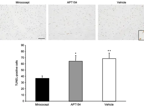
3.8. TUNEL staining
Fig. 6illustrates TUNEL-positive myocytes at high-power magnification from representative experimental samples. Although a large fraction of myocytes were TUNEL-positive
Fig. 4. Grading for complement tissue deposition of C1q, C3b/c, C4b/c and membrane attack complex (detected as C6), as well as ELISA for C3a and C5a in plasma. Four samples per area and experiment were graded with respect to intensity for complement staining. Each point represents the average of all evaluations for one experiment. 0 = essentially no staining, 1 = minimal focal or diffuse staining, 2 = moderately strong focal or diffuse staining 3 = extensive diffuse staining. Results for staining in the necrotic ischemic tissue (NIT) are shown for Mirococept- (black circles), APT164-treated animals (grey circles) as well as vehicle controls (white circles, A). Representative samples from the NIT of all three experiments for complement staining are visualized (B). Scale bar: 50μm. Plasma C3a and C5a levels are presented as a percentage of baseline (set to 100%). Systemic C3a and C5a generation was not influenced by treatment with Mirococept (C).
within the ischemic necrotic area, the proportion of positive cells was significantly greater in samples from vehicle con-trols (69 ± 8) and from the APT154 (63 ± 7) group as com-pared to the Mirococept (37 ± 4) treated animals (p = 1.0 × 10− 6and p = 1.6 × 10− 6, respectively). Within the non-ische-mic and vital ischenon-ische-mic area, the numbers of TUNEL-positive myocytes were few and not significantly different between the three groups (not shown).
4. Discussion
Our data show that site-specific application of the mem-brane-targeted recombinant complement inhibitor Miroco-cept to ischemic myocardium significantly prevented I/R injury and in part ameliorated cardiac function in the early reperfusion period. Cardioprotection was associated with binding of Mirococept to ischemic tissue and was not
Fig. 5. Staining for pro-coagulant tissue factor in vital, ischemic tissue (VIT) and necrotic, ischemic tissue (NIT) sections. Positivity is revealed as brown staining (black arrow). Representative sections from each of the three groups are shown. Scale bar: 50μm.
Fig. 6. Terminal deoxynucleotidyl transferase-mediated dUTP nick end-labeling (TUNEL) of myocardial tissue (necrotic ischemic tissue, NIT). TUNEL-positive myocytes stain brown. Representative sections from each of the three groups are shown. Inlay with magnification of two TUNEL-positive myocytes. Scale bar: 100μm. Mirococept vs. APT154: p=1.6×10− 6(⁎), Mirococept vs. vehicle: p = 1.0 × 10− 6(⁎⁎).
observed with the use of the untagged, non-membrane binding form (APT154).
As organs injured following ischemia and reperfusion locally up-regulate complement[3], it may be advantageous to target protective agents to the actual site of injury. Indeed, previous work by Horstick et al. has documented the efficacy of local, intracoronary C1-inhibitor in reducing myocardial damage in a porcine infarction model [21]. Furthermore, although the therapeutic success of systemic complement inhibition, for example using soluble CR1, has been well documented[6,22–24], strategies chiefly, but not necessarily exclusively, targeting the organ locally may provide several advantages over systemic application. Undesired binding to non-target organs, particularly excretory organs such as the kidney, may largely be avoided and lower doses may be required to achieve the same effect, avoiding complications potentially associated with systemic complement inhibition. Indeed, in our study, Mirococept was effective at a dose of 0.5 mg/kg, as compared to studies using soluble CR1 in pigs, where up to 10 mg/kg were used [25]. However, as the results reveal, Mirococept's beneficial effect was not entirely complete for all investigated aspects. Possibly an interme-diate dose (between 0.5 and 10 mg/kg) may be more effective for local application strategies. Although such dosage considerations may be of less importance where short-term complement inhibition is needed, they may well prove significant for long-term therapies. Clearly however, pronounced systemic inflammation in addition to local I/R injury, such as occurs during cardiopulmonary bypass sur-gery, may require systemic complement inhibition in addi-tion to site-targeted cytoprotecaddi-tion. Also, it remains to be investigated whether concurrent low-dose systemic inhibi-tion may provide addiinhibi-tional benefits.
Strategies to target complement inhibitors to cell mem-branes have included linking these to sialyl Lewis-x moieties
[26]. Other strategies include taking advantage of the fact that endothelial damage causes shedding of the glycocalyx[27]to reveal a pro-inflammatory and pro-coagulant surface. We previously demonstrated that the complement inhibitor[28–
30] and glycosaminoglycan analog low molecular weight dextran sulfate selectively binds to endothelial cells denu-dated of their protective glycocalyx layer in vitro[30]and protects from I/R injury in acute myocardial infarction in pigs following intracoronary administration [17]. The current data with Mirococept confirm the possibility of achieving a similar degree of protection, albeit using a substance with different cell membrane-tethering properties.
Unlike the myristoyl-tailed Mirococept, non-tagged APT154 was not detected in myocardial tissue samples and did not provide cardioprotection; this, despite infusion of an equivalent molar dose and local, intracoronary application. We attribute the failure of APT154 to protect from I/R injury to the fact that the infused agent was not targeted to bind to the site of damage and, once systemically in circulation, did not suffice to offer protection. Results from the CH50 test, which was used to evaluate systemic complement inhibition, would
indeed suggest that the APT154 in circulation did not sys-temically affect complement activity. Results with Miroco-cept imply that the applied dose not only sufficed to locally bind to and inhibit tissue complement activation, but also affected complement activity systemically. This may, in part, represent an in vitro phenomenon [10], whereby small amounts of unbound, quasi-surplus Mirococept, added in vitro to antibody-sensitized sheep erythrocytes, were enough to bind to the comparatively few erythrocytes and substan-tially inhibit complement. However, these results may also reflect a certain amount of true systemic complement inhi-bition in vivo. Whether this is due to shedding of initially bound Mirococept into the blood stream or represents excess, unbound Mirococept, washed out of the coronary circulation without initial local binding to the vasculature, remains to be investigated. Nonetheless, the peak amounts detected in vivo suggest that most of the injected Mirococept was bound locally. Indeed, no staining for Mirococept was detectable in distant organs (including liver and kidneys).
The mechanism by which Mirococept exerts its cardio-protective effects may, in part, be deduced from the known mode of action of native CR1, as Mirococept retains enough activity of CR1 necessary for breakdown of complexed complement fragment C3b within C3 convertases[14]and hence for an inhibitory effect on generation of the anaphy-latoxins C3a and C5a and the membrane attack complex. Indeed, all of the complement components studied, from C1q and C4b/c of the classical complement pathway to C3b/c, and C6 of the membrane attack complex, were reduced in myocardial tissue samples from Mirococept-treated animals. Reduction of anaphylatoxins has been described in vena cordis magna blood with the use of C1-inhibitor. Systemic C3a and C5a levels however did not vary greatly and were not influenced by Mirococept administra-tion. This may be attributed to the minimal amounts of systemically-available unbound Mirococept and its largely restricted local action. C3b and membrane attack complex are associated with promotion of leukocyte activation via upregulation of adhesion molecules [31]. Compared to vehicle controls and APT154, Mirococept significantly reduced total neutrophil numbers in the viable ischemic tissue and decreased the extravasation of neutrophils into this area. sCR1 and sialyl Lewis-x glycosylated sCR1 have been shown to reduce neutrophil accumulation in a model of stroke[32]. However, maximal tissue neutrophil accumula-tion is not observed following only two hours of reperfusion and non-complement-dependent mechanisms of leukocyte recruitment may also be of importance in this setting. As a certain reduction in neutrophil extravasation was also observed in the APT154 group, it remains to be verified if Mirococept's protective effects are, at least in part, attributable to modification of neutrophil extravasation, particularly in later stages of more pronounced tissue extravasation.
This endothelial protective effect translated into reduced tissue factor (TF) expression in Mirococept treated animals,
the induction of which critically contributes to I/R damage. While Mirococept does not influence coagulation systemi-cally (no effect on aPTT), attenuating intravascular throm-bosis and inflammation by limiting endothelial TF expression may prove fundamental in reducing I/R damage[33].
Myocardial ischemia has also been shown to induce apoptosis in animal models and humans[34,35]. Treatment with Mirococept not only significantly reduced myocardial necrosis but also I/R-induced apoptosis. This reduction may be related to the observed reduced terminal complement complex deposition in the ischemic myocardium, which has been shown to play an important role not only in I/R-induced necrosis but also apoptosis[36].
4.1. Limitations
TTC staining, though under debate for determining in-farct size following short reperfusion times, was used here, as its staining validity for 120 min reperfusion in pigs has previously been confirmed, where viable TTC positive islets within TTC negative necrotic tissue correlated with electron microscopy findings[37]. Isoflurane, known to have a pre-conditioning effect[38], may have contributed to a certain degree of cardioprotection. However, this effect would be equal in all groups and would not explain the benefits observed with the use of Mirococept.
Although Mirococept provides protection in the critical initial phase of reperfusion, further studies are required to validate whether a one-time application of Mirococept offers extended protection from complement-mediated damage. In particular, functional analysis, including regional wall moti-lity, would also need to be evaluated at a later stage, as myocardial stunning, a likely significant finding in all groups in the early post-reperfusion phase, may hamper initial in-vestigations and skew early results.
5. Conclusions
In summary, we have demonstrated the feasibility of modifying the local pro-inflammatory and pro-coagulant en-vironment encountered following regional myocardial ische-mia and reperfusion by targeting a complement regulatory protein to the site of injury. This protection was associated with a single dose application of a molecule previously proven safe for use in humans. The concept of targeted cytoprotection of ischemic tissue pre-reperfusion using substances such as Mirococept, may offer an attractive therapeutic option in treating reperfusion injury and warrants further investigation. Acknowledgements
This study is supported by the Swiss Heart Foundation, the Swiss National Science Foundation (3200B0-104228, 7SUPJ062207) and the Bern University Hospital (study no. 898). Yara Banz is a recipient of an MD-PhD grant of the Swiss Academy of Medical Sciences.
The authors wish to thank Sarah Fritchley for help with aspects of the work with Mirococept and the Protein Technologies group at Inflazyme for the provision of materials as well as Katja Matozan, Olgica Beslac, Daniel Zalokar, Jane Boden and Trinh Cung for excellent technical assistance.
References
[1] Rossen RD, Michael LH, Kagiyama A, Savage HE, Hanson G, Reis-berg MA, et al. Mechanism of complement activation after coronary artery occlusion: evidence that myocardial ischemia in dogs causes release of constituents of myocardial subcellular origin that complex with human C1q in vivo. Circ Res 1988;62:572–84.
[2] Collard CD, Vakeva A, Morrissey MA, Agah A, Rollins SA, Reenstra WR, et al. Complement activation after oxidative stress: role of the lectin complement pathway. Am J Pathol 2000;156:1549–56. [3] Yasojima K, Schwab C, McGeer EG, McGeer PL. Human heart
generates complement proteins that are upregulated and activated after myocardial infarction. Circ Res 1998;83:860–9.
[4] Ross GD, Lambris JD, Cain JA, Newman SL. Generation of three different fragments of bound C3 with purified factor I or serum. I. Requirements for factor H vs CR1 cofactor activity. J Immunol 1982;129: 2051–60.
[5] Collard CD, Bukusoglu C, Agah A, Colgan SP, Reenstra WR, Morgan BP, et al. Hypoxia-induced expression of complement receptor type 1 (CR1, CD35) in human vascular endothelial cells. Am J Physiol 1999;276:C450–8.
[6] Weisman HF, Bartow T, Leppo MK, Marsh Jr HC, Carson GR, Concino MF, et al. Soluble human complement receptor type 1: in vivo inhibitor of complement suppressing post-ischemic myocardial inflam-mation and necrosis. Science 1990;249:146–51.
[7] Homeister JW, Satoh PS, Kilgore KS, Lucchesi BR. Soluble com-plement receptor type 1 prevents human comcom-plement-mediated dam-age of the rabbit isolated heart. J Immunol 1993;150:1055–64. [8] Lazar HL, Bao Y, Gaudiani J, Rivers S, Marsh H. Total complement
inhibition: an effective strategy to limit ischemic injury during coronary revascularization on cardiopulmonary bypass. Circulation 1999;100: 1438–42.
[9] Smith GP, Smith RA. Membrane-targeted complement inhibitors. Mol Immunol 2001;38:249–55.
[10] Smith RA, Dodd I, Mossakowska DEI; Conjugates of soluble peptidic compounds with membrane-binding agents. US patent WO 98/02454. 1998.
[11] Arbuzova A, Murray D, McLaughlin S. MARCKS, membranes, and calmodulin: kinetics of their interaction. Biochim Biophys Acta 1998;1376:369–79.
[12] Pratt JR, Jones ME, Dong J, Zhou W, Chowdhury P, Smith RA, et al. Nontransgenic hyperexpression of a complement regulator in donor kidney modulates transplant ischemia/reperfusion damage, acute rejection, and chronic nephropathy. Am J Pathol 2003;163: 1457–65.
[13] Linton SM, Williams AS, Dodd I, Smith R, Williams BD, Morgan BP. Therapeutic efficacy of a novel membrane-targeted complement regu-lator in antigen-induced arthritis in the rat. Arthritis Rheum 2000;43: 2590–7.
[14] Mossakowska D, Dodd I, Pindar W, Smith RA. Structure-activity relationships within the N-terminal short consensus repeats (SCR) of human CR1 (C3b/C4b receptor, CD35): SCR 3 plays a critical role in inhibition of the classical and alternative pathways of complement activation. Eur J Immunol 1999;29:1955–65.
[15] Souza DG, Esser D, Bradford R, Vieira AT, Teixeira MM. APT070 (Mirococept), a membrane-localised complement inhibitor, inhibits inflammatory responses that follow intestinal ischaemia and reperfu-sion injury. Br J Pharmacol 2005;145:1027–34.
[16] Dodge HT, Sandler H, Ballew DW, Lord Jr JD. The use of biplane angiocardigraphy for the measurement of left ventricular volume in man. Am Heart J 1960;60:762–76.
[17] Banz Y, Hess OM, Robson SC, Mettler D, Meier P, Haeberli A, et al. Locally targeted cytoprotection with dextran sulfate attenuates expe-rimental porcine myocardial ischaemia/reperfusion injury. Eur Heart J 2005;26:2334–43.
[18] Hopken U, Struber A, Oppermann M, Mohr M, Mucke KH, Burchardi H, et al. Production and characterization of pepetide-specific mono-clonal antibodies that recognise a neoepitope on hog C5a. In: Faist E, Meakins JL, Schildberg FW, editors. Host Defense Dysfunction in trauma, schock and sepsis. Berlin: Springer Verlag; 1993.
[19] Mayer MM. On the destruction of erythrocytes and other cells by antibody and complement. Cancer Res 1961;21:1262–9.
[20] Thiruvikraman SV, Guha A, Roboz J, Taubman MB, Nemerson Y, Fallon JT. In situ localization of tissue factor in human atherosclerotic plaques by binding of digoxigenin-labeled factors VIIa and X. Lab Invest 1996;75:451–61.
[21] Horstick G, Heimann A, Gotze O, Hafner G, Berg O, Boehmer P, et al. Intracoronary application of C1 esterase inhibitor improves cardiac function and reduces myocardial necrosis in an experimental model of ischemia and reperfusion. Circulation 1997;95:701–8.
[22] Shandelya SM, Kuppusamy P, Herskowitz A, Weisfeldt ML, Zweier JL. Soluble complement receptor type 1 inhibits the complement pathway and prevents contractile failure in the postischemic heart. Evidence that complement activation is required for neutrophil-mediated reperfusion injury. Circulation 1993;88:2812–26.
[23] Lazar HL, Bokesch PM, van Lenta F, Fitzgerald C, Emmett C, Marsh Jr HC, et al. Soluble human complement receptor 1 limits ischemic damage in cardiac surgery patients at high risk requiring cardiopul-monary bypass. Circulation 2004;110:II274–9.
[24] Keshavjee S, Davis RD, Zamora MR, de Perrot M, Patterson GA. A randomized, placebo-controlled trial of complement inhibition in is-chemia-reperfusion injury after lung transplantation in human beings. J Thorac Cardiovasc Surg 2005;129:423–8.
[25] Lazar HL, Hamasaki T, Bao Y, Rivers S, Bernard SA, Shemin RJ. Soluble complement receptor type I limits damage during revascular-ization of ischemic myocardium. Ann Thorac Surg 1998;65:973–7. [26] Mulligan MS, Warner RL, Rittershaus CW, Thomas LJ, Ryan US,
Foreman KE, et al. Endothelial targeting and enhanced antiinflammatory effects of complement inhibitors possessing sialyl Lewisx moieties. J Immunol 1999;162:4952–9.
[27] Mulivor AW, Lipowsky HH. Inflammation and ischemia induced shedding of the venular glycocalyx. Am J Physiol Heart Circ Physiol 2004;286:H1672–80.
[28] Meri S, Pangburn MK. Regulation of alternative pathway complement activation by glycosaminoglycans: specificity of the polyanion binding site on factor H. Biochem Biophys Res Commun 1994;198:52–9. [29] Wuillemin WA, te Velthuis H, Lubbers YT, de Ruig CP, Eldering E,
Hack CE. Potentiation of C1 inhibitor by glycosaminoglycans: dextran sulfate species are effective inhibitors of in vitro complement acti-vation in plasma. J Immunol 1997;159:1953–60.
[30] Laumonier T, Walpen A, Maurus C, Mohacsi PJ, Matozan K, Kor-chagina E, et al. Dextran sulfate acts as an endothelial cell protectant and inhibits human complement- and NK cell-mediated cytotoxicity against porcine cells. Transplantation 2003;76(5):838–43.
[31] Vaporciyan AA, Mulligan MS, Warren JS, Barton PA, Miyasaka M, Ward PA. Up-regulation of lung vascular ICAM-1 in rats is comple-ment dependent. J Immunol 1995;155:1442–9.
[32] Huang J, Kim LJ, Mealey R, Marsh Jr HC, Zhang Y, Tenner AJ, et al. Neuronal protection in stroke by an sLex-glycosylated complement inhibitory protein. Science 1999;285:595–9.
[33] Golino P, Ragni M, Cirillo P, Scognamiglio A, Ravera A, Buono C, et al. Recombinant human, active site-blocked factor VIIa reduces infarct size and no-reflow phenomenon in rabbits. Am J Physiol Heart Circ Physiol 2000;278:H1507–16.
[34] Gottlieb RA, Burleson KO, Kloner RA, Babior BM, Engler RL. Reperfusion injury induces apoptosis in rabbit cardiomyocytes. J Clin Invest 1994;94:1621–8.
[35] James TN. Normal and abnormal consequences of apoptosis in the human heart. From postnatal morphogenesis to paroxysmal arrhyth-mias. Circulation 1994;90:556–73.
[36] Vakeva AP, Agah A, Rollins SA, Matis LA, Li L, Stahl GL. Myo-cardial infarction and apoptosis after myoMyo-cardial ischemia and reper-fusion: role of the terminal complement components and inhibition by anti-C5 therapy. Circulation 1998;97:2259–67.
[37] Horstick G, Berg O, Heimann A, Gotze O, Loos M, Hafner G, et al. Application of C1-esterase inhibitor during reperfusion of ischemic myocardium: dose-related beneficial versus detrimental effects. Circu-lation 2001;104:3125–31.
[38] Piriou V, Chiari P, Lhuillier F, Bastien O, Loufoua J, Raisky O, et al. Pharmacological preconditioning: comparison of desflurane, sevoflur-ane, isoflurane and halothane in rabbit myocardium. Br J Anaesth 2002;89:486–91.