Improving mitral valve coaptation with adjustable rings: outcomes
from a European multicentre feasibility study with a new-generation
adjustable annuloplasty ring system
†
Francesco Maisano
a,*, Volkmar Falk
b, Michael A. Borger
c, Hugo Vanermen
d, Ottavio Alfieri
a,
Joerg Seeburger
c, Stefan Jacobs
b, Michael Mack
eand Friederich W. Mohr
ca San Raffaele University Hospital, Milan, Italy
b Cardiovascular Center, University Hospital Zurich, Zurich, Switzerland
c
Heart Center, University of Leipzig, Leipzig, Germany
d Department of Cardiovascular and Thoracic Surgery, Onze-Lieve-Vrouw Clinic, Aalst, Belgium
e Heart Hospital Baylor Plano, Dallas, TX, USA
* Corresponding author. Cardiochirurgia Ospedale San Raffaele, Via Olgettina 60, 20132 Milano, Italy. Tel: +39-02-26437111; fax: +39-02-26437125, e-mail: [email protected] (F. Maisano).
Received 8 October 2012; received in revised form 22 January 2013; accepted 27 January 2013
Abstract
OBJECTIVES: To evaluate the performance and safety of an adjustable semi-rigid annuloplasty ring for mitral regurgitation (MR) in a multicentre study.
METHODS: Between March 2010 and December 2011, 30 subjects underwent mitral valve (MV) repair using the Cardinal adjustable annuloplasty ring. This device is a semi-rigid ring allowing postimplantation size adjustment, under beating-heart conditions, to opti-mize leaflet coaptation under echocardiographic guidance. Coaptation length was determined before and after adjustment by transoe-sophageal echocardiography.
RESULTS: The study enrolled 21 (70%) male and 9 (30%) female subjects with a mean age of 64 years. The approach was conventional midline sternotomy or mini-invasive right thoracotomy. Leaflet resection was done in 17 subjects, and chordal repair was used in 13. Concomitant procedures included coronary artery bypass grafting in 2 (7%) subjects, atrial ablation in 4 (13%) and tricuspid repair in 4 (13%). There was 1 (3%) early death unrelated to the study device. Intraoperative ring adjustment was performed in 24 of the 30 sub-jects. Residual MR was detected prior to adjustment in 6 subjects (4 mild and 2 moderate MR). Following adjustment, 5 subjects had no MR and 1 had trace MR. After adjustment, mean coaptation length improved from 7 ± 3 to 10 ± 3 mm (P < 0.0001). All patients who completed 1-year follow-up had less-than-mild MR, with the exception of 1 patient with ring dehiscence (and resultant 2+ MR) and 1 functional MR patient who developed recurrent 2+ MR due to persistent leaflet tethering.
CONCLUSIONS: MV repair with the Cardinal adjustable annuloplasty ring is a reliable technique that enables the adjustment of the ring diameter on a beating heart under echocardiographic control. Such technology allows the optimization of leaflet coaptation, providing minimal residual MR and durable repair.
Keywords:Mitral valve• Annuloplasty • Repair • Echocardiography • Coaptation length • Image-guided surgery • Reconstruction
INTRODUCTION
The ultimate objective of mitral valve (MV) repair is to restore the largest possible coaptation surface by acting at multiple levels: annular, leaflet and subvalvular levels. MV annuloplasty is routinely performed to increase coaptation and to reinforce the annulus following leaflet and subvalvular repair [1]. Correct sizing of the annuloplasty ring is a crucial step to restoring the appro-priate annular-to-leaflet ratio [2]. Conventional annuloplasty ring sizing is based on anatomical assessment of the leaflets under
cardioplegic arrest. This static approach could lead to inappro-priate device selection since the mitral complex is a dynamic entity. Inappropriate sizing has been associated with insufficient leaflet coaptation and recurrent mitral regurgitation (MR) [3–5] or systolic anterior motion (SAM) of the anterior mitral leaflet [6,7]. To overcome this limitation, adjustable ring devices are under investigation [8,9]. The new-generation adjustable rings enable echo-guided, off-pump, beating-heart modulation of the valve annulus under physiological conditions to optimize mitral-repair anatomy and haemodynamics at various stages following the implant.
Here, we report the 1-year outcome from a single-arm, non-blinded, non-randomized, multicentre, prospective clinical study
†Presented at the 26th Annual Meeting of the European Association for
Cardio-Thoracic Surgery, Barcelona, Spain, 27–31 October 2012.
© The Author 2013. Published by Oxford University Press on behalf of the European Association for Cardio-Thoracic Surgery. All rights reserved.
ADUL T C ARD IA C
European Journal of Cardio-Thoracic Surgery 44 (2013) 913–918
ORIGINAL ARTICLE
with intrapatient comparisons designed to assess feasibility, safety and efficacy of the Cardinal adjustable ring (Valtech Cardio, Or Yehuda, Israel) implantation during MV repair.
MATERIALS AND METHODS
Patients with indication for MR correction [10] were enrolled between March 2010 and December 2011 in four centres in Europe. To date, 24 patients have completed 1-year follow-up. Patients with acute endocarditis, heavily calcified leaflets or annulus, previously implanted prosthesis, concomitant aortic or pulmonary valve implant, or life expectancy <12 months were excluded by protocol. The feasibility endpoint included the tech-nical implantation success rate of the Cardinal ring defined as the ability to size the annulus, to place the sutures in the annulus, to place the sutures in the ring, to lower the ring onto valve and to tie the sutures, with no damage to adjacent tissue from the ring or handle. The operator assigned a score from 1 to 3 for each step of the procedures (3 being operator-friendly and 1 unacceptable performance). The technical feasibility of postim-plantation adjustment was defined as the ability to modify the size of the annuloplasty ring, when required, after weaning from
cardiopulmonary bypass (CPB). The efficacy endpoint was
defined as the ability of the Cardinal ring to reduce MR
post-CPB, at hospital discharge, and at 30-day postoperatively. Additional data collected included the effect of ring annuloplasty on left ventricular reverse remodelling and on mitral leaflet length of coaptation. Serious adverse events were adjudicated by an independent Clinical Event Committee.
The Cardinal adjustable annuloplasty system
The Cardinal adjustable annuloplasty system received the CE mark in 2012 (Fig.1): it is designed for beating-heart adjustment of ring size after implantation and is applicable to both degen-erative and functional MR patients. The implant is a semi-rigid ring having a metal core enclosed in a polyester fabric covering, and attached to an adjustment mechanism comprised of a spool with an inner wire.
The ring is D-shaped with oneflexible segment corresponding to the posterior leaflet and a shorter, more rigid, segment corre-sponding to the anterior leaflet. The adjustment mechanism, integrated within the mid-portion of the anterior segment ring, enables size adjustment before and after implantation. Three ring sizes are currently available—small, medium and large (cov-ering sizes from 24 to 40 mm)—while at the time of the clinical trial only 2 sizes (small and large) were available.
A remote-adjustment tool temporarily attached to the adjust-ment mechanism enables ring adjustadjust-ment to the desired size from outside the heart. Adjustment of ring size is obtained by rotating a dial on the handle. Adjustment is bidirectional (both contraction and expansion of the ring is possible) and is com-pletely reversible until the adjustment handle is removed. All instruments are long-shafted so that they can be used in min-imally invasive surgery.
A set of sizers (covering sizes from 24 to 40 mm) is used to measure the MV. The sizers are also used as ring holders.
Implantation procedure
The Cardinal ring system is implanted on CPB, under cardiople-gic arrest and direct view, with a conventional suturing tech-nique. Following leaflet repair, ring sizers are used to assess the intercommissural distance, the surface area and the height of the anterior leaflet, as for conventional MV ring sizing. The corre-sponding Cardinal adjustable annuloplasty ring is selected and mounted on the holder. In patients with degenerative MR, a slightly oversized annuloplasty ring may be chosen in order to retain the option of ring expansion after implantation. In the case of functional MR, undersizing is not required since it is achievable by ring contraction following implantation.
Figure 1:The Cardinal adjustable annuloplasty ring system. (A) The Cardinal
ring has two portions, an anterior and a posterior one, identifiable by a green marker. The anterior portion is rigid and non-compressible. The posterior
one isflexible and adjustable. The adjustment tool is positioned in the mid of
the anterior portion. (B) An intraoperative view of a cardinal ring with the remote adjusting tool connected to the ring prior to adjustment. (C) The ad-justment tool allows remote ring size modifications by rotating the posterior knob on the handle. A numeric indicator is used to monitor the amount of size adjustment.
Following completion of the implant, ring size can be adjusted according to standard water testing of the MV. The left atrium is then closed in the usual fashion, with the exception that the ad-justment handle traverses the atriotomy suture line in order that final ring size adjustment can be performed after weaning from CPB. Haemodynamic status should be stable at the time of ad-justment, with near-physiological arterial pressure and optimal loading conditions.
Adjustment is guided by transesophageal echocardiography (TEE), focusing on residual MR, degree of leaflet coaptation and the presence of transmitral gradients. If residual MR is present, size reduction can be attempted. In the case of no residual MR, the operator can still consider adjusting the ring size in order to obtain a larger surface of coaptation. The length of coaptation is assessed in left ventricular outflow tract (LVOT) view, scanning the coaptation line in different segments. The length of coapta-tion should ideally be 8 mm or more throughout the coaptacoapta-tion line. In the case of SAM, the ring may be enlarged until normal leaflet coaptation is obtained. In the case of functional mitral stenosis, the ring may be re-expanded.
Followingfinal ring size adjustment, the adjustment handle is removed. A final TEE evaluation of MV function is performed. The operation is then completed in standard fashion.
Echocardiography
Echocardiogram was conducted in a standardized fashion using a prespecified protocol. Clinical follow-up and transthoracic echocardiography (TTE) assessment of left ventricular volumes and function and MR degree/grade were made at hospital dis-charge, 30 days, 6 months and annually postindex procedure.
Independent monitoring visits were routinely performed.
Analysis of echocardiographic data was reviewed by an inde-pendent core laboratory (Medstar Health, WA, USA).
Statistical analysis
Data were analysed with a per-protocol approach. Statistical ana-lysis was performed using the JMP 5.1.1 software (SAS Institute, Inc., Cary, NC, USA). Continuous variables are presented as mean ± SD throughout the manuscript. Univariable comparisons were performed using the pairedt-test. The level of significance was set atP = 0.05.
RESULTS
A total of 30 (21 males and 9 females) patients were enrolled (mean age of 64 ± 14 years, range 30–5 years). Comorbidities included hypertension in 16 (53%) patients, arrhythmias in 10 (30%), dyslipidaemia in 8 (27%), diabetes in 3 (10%), coronary artery disease in 3 (10%), chronic lung disease in 1 (3%) and history of cerebrovascular accident in 1 (3%). Most of the patients (22, 73%) were in New York Heart Association (NYHA) functional Class I–II, and 8 were in NYHA functional Class III–IV (27%).
According to core-lab assessment, all patients but 2 (with moderate MR) had severe MR at baseline. There were 3 (10%) patients with Carpentier Type I MR, 26 (87%) with Type II and 1 (3%) with Type III. There were 4 patients with mild associated aortic regurgitation, and 12 with mild or moderate tricuspid re-gurgitation. Baseline echocardiographic parameters are reported in Table1. A full sternotomy was performed in 12 patients, while minimally invasive right thoracotomy was done in the remaining 18.
MV leaflet resection was performed in 17 (57%) patients and chordal replacement in 13 (43%), while in 3 (10%), isolated ring implantation was performed. Associated cardiovascular proce-dures included tricuspid repair in 4 patients, atrialfibrillation ab-lation in 4 and coronary bypass surgery in 2.
Ring implantation was attempted in all patients and was suc-cessful in all but one. The unsucsuc-cessful implant was in a patient with fragile annular structure and the surgeon decided to proceed with MV replacement.
The scores for the technical feasibility endpoint were: 2.9 ± 0.2 for sizing, 2.9 ± 0.2 for placing sutures in the annulus, 2.9 ± 0.3 for placing sutures in the ring, 2.9 ± 0.2 for lowering the ring onto annulus and 2.9 ± 0.4 for tying the sutures; a score of 1 was given only once to tying the sutures due to difficulties related to the fragility of annulus, in the patient who underwent MV re-placement. Average annular sizing prior to implant was 35 ± 4 mm. A small ring was implanted in 16 (53%) patients and a large ring in 14 (47%).
Ring adjustment was done in 24 (80%) patients. It was per-formed under cardioplegic arrest, according to water testing, in 3 patients and post-CPB, under TEE guidance in 16, and both on- and post-CPB in 5. Adjustment was technically successful in all patients (Fig.2).
Residual MR was detected prior to adjustment in 6 subjects (4 mild and 2 moderate). Following adjustment, 5 subjects had no MR and 1 had trace MR. Ring enlargement was used to correct SAM in 1 patient and to reduce transmitral gradients in another patient.
Table 1: Left ventricular volumes, ejection fraction and septolateral annular dimension at baseline and follow-up
Baseline 1 month 6 months 12 months
LVEF (%) 63 ± 8 57 ± 9 (0.06) 59 ± 6 (0.13) 59 ± 3 (0.02)
LVEDV (ml) 136 ± 41 104 ± 29 (0.0002) 100 ± 25 (0.0006) 94 ± 19 (<0.0001)
LVESV (ml) 56 ± 21 50 ± 17 (0.18) 45 ± 13 (0.03) 42 ± 10 (0.001)
Annular SL (mm) 36 ± 4 21 ± 7 (<0.0001) 18 ± 4 (<0.0001) 17 ± 2 (<0.0001)
All variables are reported as mean ± 1 SD: theP-value is reported in brackets according to the paired t-test comparison with baseline.
LVEF: left ventricular ejection fraction; LVEDV: left ventricular end-diastolic volume; LVESV: left ventricular end-systolic volume; SL: septolateral.
ADUL T C ARD IA C
In the 16 patients who underwent post-CPB ring reduction, coaptation length after adjustment was significantly greater than before adjustment (10 ± 3 vs 7 ± 3 mm;P < 0.0001, Figs3and4). More importantly, after adjustment, no patient had a coaptation length <7 mm, when compared with 7 of the 16 patients with a coaptation length <7 mm before adjustment.
There was 1 hospital death (30-day mortality, 3%), due to is-chaemic lesion of the right coronary artery in a patient undergo-ing combined mitral and tricuspid repairs by a minimally invasive approach. This event was not related to the Cardinal ring implantation.
At discharge, average left ventricular ejection fraction was 56 ± 9% and slightly reduced when compared with baseline (Table 1). At 12-month follow-up, all patients but 3 were in NYHA Class I or II. Reverse remodelling of the left ventricle was observed with significant volume reductions over time (Table1).
The annular septolateral dimension was significantly reduced at 1 month from the procedure when compared with baseline (21 ± 7 vs 36 ± 4 mm,P < 0.0001), while the dimensions did not
change significantly at 6 months (18 ± 4 mm, P = 0.4, compared with 1 month) and 1 year.
One patient required reoperation at 6 months from the index procedure due to ring dehiscence and 2+ MR (Type II MR treated with the minimally invasive approach).
One patient who had functional MR developed recurrent 2+ MR at 1-year follow-up due to ongoing remodelling.
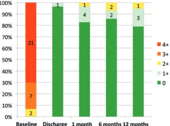
The core-lab assessment of MR degrees at various follow-up times compared with baseline are reported in Fig.5.
DISCUSSION
The study proved both the safety and the efficacy of the
Cardinal adjustable annuloplasty ring. The ring allows postim-plantationfine-tuning of annular size, to optimize leaflet coapta-tion under TEE guidance during normal physiological condicoapta-tions. Mitral annuloplasty is considered a cornerstone of MV repair, minimizing future recurrent MR after repair [1,11].
The creation of a large area of leaflet coaptation is the primary goal of durable mitral repair surgery [4, 12, 13]. Kunzelman et al. [14] showed that ring annuloplasty reduces leaflet stress and improves coaptation. Several factors influence
Figure 3: Coaptation length changes following Cardinal ring size tuning.
Individual coaptation lengths before and after ring adjustments. The two dots depict mean ± SD.
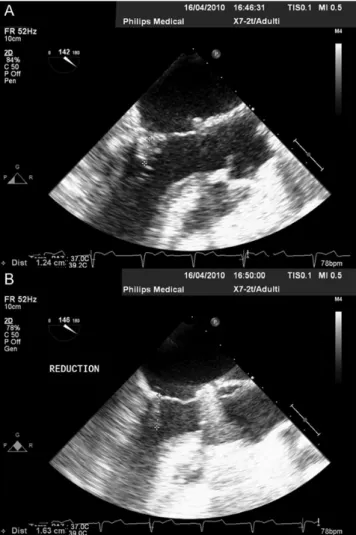
Figure 4:LVOT view of coaptation length. Coaptation length (mm) before (A)
and after adjustment (B) in a patient undergoing off-pump ring adjustment under TEE echo-guidance (LVOT view).
Figure 2:Ring size adjustment. Of 30 patients undergoing Cardinal ring
im-plantation, 6 required no adjustment and 24 had the ring size modified after the implant: in 3 cases under direct view, in 16 cases off-pump, and in 5 cases both on- and off-pump. (A) The Cardinal ring on three-dimensional live transesophageal echocardiography showing the saddle shape of the ring on the beating heart.
leaflet coaptation, including annular size and shape, and the amount of mobile leaflet tissue.
Surgeons can directly manipulate thefirst two factors by ring annuloplasty. The size of the ring annuloplasty is usually chosen according to the intraoperative sizing of the anterior leaflet. However, we previously reported that suboptimal mitral repair may require an immediate surgical revision in up to 11% of patients undergoing intraoperative TEE evaluation [15]. Early repair failure is often related to annuloplasty ring sizing issues. Incorrect ring undersizing may induce residual MR due to insuf-ficient leaflet coaptation. Early identification of limited coapta-tion is associated with poor long-term results and high MR recurrence rates [4,11,16]. On the contrary, SAM occurs in up to 15% of patients following complex MV repair [6,17] due to ex-cessive ring undersizing [18]. Rings with increased septolateral distance have been designed to reduce the occurrence of SAM [19], although their effect on MR reduction may be questioned.
The Cardinal annuloplasty ring is a semi-rigid device preserv-ing the natural movements of the native annulus. The rpreserv-ing is specifically designed to provide anisotropic compliance: the anterior segment is less compliant (similar to the heart’s skeletal structure near the trigones) and the posterior segment more compliant. This design feature promotes posterior annular plication and preferential reduction of the septolateral dimen-sion and may make this ring ideal to treat both degenerative and functional MR. The ring size obtained intraoperatively is maintained over time, as demonstrated in our study showing that the annular septolateral dimension at 1 month remains stable at 1 year. At follow-up, reverse remodelling of the left ventricle and stable MR reduction are observed in most of the patients.
The main advantage of the Cardinal annuloplasty system is that size adjustment can be achieved after weaning from CPB, on the beating heart. If residual MR is observed or leaflet coap-tation is not adequate, the septolateral distance can be reduced. Conversely, if SAM or increased gradients are observed, septolat-eral distance can be increased. In our experience, adjustment of the ring size was performed in 80% of the cases: the ring size
was more commonly reduced to improve leaflet coaptation
(from 7 ± 3 to 10 ± 3 mm), while in 2 cases it was increased to reverse SAM or transmitral gradients. In all patients undergoing
size reduction under TEE guidance, final coaptation length was >7 mm, when compared with 44% of patients with <7 mm coap-tation length before size adjustment. The adjustment man-oeuvres were safe and totally reversible with no device-related complications during adjustments.
In the present feasibility study, the number of patients treated with functional MR was too small to generate conclusions. Although the ring is designed to target functional MR, a more dedicated study is warranted to determine the feasibility and the efficacy in this setting.
The use of an adjustable ring may be particularly helpful in functional MR, because the operator could aggressively under-size to improve leaflet coaptation and to adjust the septal lateral diameter, while avoiding the risk of generating gradients [20–22]. Moreover, in the case of a suboptimal result, the surgeon may avoid a second CPB run.
Late adjustment option could be intriguing since recurrent MR is known to occur months to years after MV repair in a signi fi-cant proportion of patients with ongoing LV remodelling. Langer et al. [9] recently reported the 6-month results of the MiCardia DYANA annuloplasty system (MiCardia Corp., Irvine, CA, USA), a nitinol-based dynamic complete ring, which allows late modi fi-cation of the septolateral diameter by radiofrequency. As an al-ternative, transcatheter valve-in-ring procedures may successfully treat recurrent MR [23] after MV annuloplasty.
The Cardinal ring can be adjusted only intraoperatively, but allowsfine-tuning of annuloplasty ring size with greater degrees of adjustments when compared with the nitinol-based technol-ogy. In addition, the shape and size modification is bidirectional and totally reversible until the adjustment handle is removed.
In 1 patient with preoperative Type I MR, ring dehiscence oc-curred. This risk may be mitigated by the ring implantation tech-nique: more sutures may be required since ring reshaping determines higher loads on the annular structures. Further studies are required in order to confirm this hypothesis.
In conclusion, the current study shows that implantation of the adjustable Cardinal ring is safe and effective. This study has shown that size adjustment was used in 80% of the patients, with a positive effect on the surface of leaflet coaptation. Adjustment may be used to correct either the lack of coaptation with re-sidual MR, or to prevent SAM in the case of ring undersizing. The clinical use of the adjustable annuloplasty rings can improve acute and long-term results of mitral-repair surgery.
Funding
The study has been sponsored by ValtechCardio.
Conflict of interest: Francesco Maisano, Volkmar Falk, Hugo Vanermen, Ottavio Alfieri, Joerg Seeburger are consultants for ValtechCardio.
REFERENCES
[1] Gillinov AM, Cosgrove DM, Blackstone EH, Diaz R, Arnold JH, Lytle BW et al. Durability of mitral valve repair for degenerative disease. J Thorac Cardiovasc Surg 1998;116:734–43.
[2] Maisano F, La Canna G, Grimaldi A, Vigano G, Blasio A, Mignatti Aet al.
Annular-to-leaflet mismatch and the need for reductive annuloplasty in patients undergoing mitral repair for chronic mitral regurgitation due to mitral valve prolapse. Am J Cardiol 2007;99:1434–9.
Figure 5:MR severity at TTE examination (core-lab adjudicated). Coaptation:
degree of MR as adjudicated by the core lab in patients at different stages during the study period. Data are shown as percentages, with indication of the total number of patients in each MR degree group.
ADUL T C ARD IA C
[3] Armen TA, Vandse R, Crestanello JA, Raman SV, Bickle KM, Nathan NS. Mechanisms of valve competency after mitral valve annuloplasty for is-chaemic mitral regurgitation using the Geoform ring: insights from
three-dimensional echocardiography. Eur J Echocardiogr 2009;10:74–81.
[4] Bax JJ, Braun J, Somer ST, Klautz R, Holman ER, Versteegh MI et al.
Restrictive annuloplasty and coronary revascularization in ischemic mitral regurgitation results in reverse left ventricular remodeling.
Circulation 2004;110:II103–8.
[5] Yamauchi T, Taniguchi K, Kuki S, Masai T, Noro M, Nishino M et al.
Evaluation of the mitral valve leaflet morphology after mitral valve
re-construction with a concept‘coaptation length index’. J Card Surg 2005;
20:432–5.
[6] Crescenzi G, Landoni G, Zangrillo A, Guarracino F, Rosica C, La Canna G et al. Management and decision-making strategy for systolic anterior motion after mitral valve repair. J Thorac Cardiovasc Surg 2009;137:
320–5.
[7] Sorrell VL, Habibzadeh MR, Kalra N, Chitwood WR. Transient severe mitral regurgitation after mitral valve repair is associated with a poor clinical outcome: a small case series. Echocardiography 2008;25:835–9. [8] Czesla M, Gotte J, Voth V, Roser D, Weimar T, Doll N. Successful
post-operative activation of an adjustable annuloplasty ring (MiCardia) in re-current ischemic mitral valve regurgitation. Ann Thorac Surg 2012;94: 39–40.
[9] Langer F, Borger MA, Czesla M, Shannon FL, Sakwa M, Doll Net al.
Dynamic annuloplasty for mitral regurgitation. J Thorac Cardiovasc Surg
2012;145:425–9.
[10] Vahanian A, Alfieri O, Andreotti F, Antunes MJ, Baron-Esquivias G,
Baumgartner Het al. Guidelines on the management of valvular heart
disease (version 2012): The Joint Task Force on the Management of Valvular Heart Disease of the European Society of Cardiology (ESC) and the European Association for Cardio-Thoracic Surgery (EACTS). Eur J
Cardiothorac Surg 2012;33:2451–96.
[11] Maisano F, Caldarola A, Blasio A, De Bonis M, La Canna G, Alfieri O. Midterm results of edge-to-edge mitral valve repair without annulo-plasty. J Thorac Cardiovasc Surg 2003;126:1987–97.
[12] Greenhouse DG, Dellis SL, Schwartz CF, Loulmet DF, Yaffee DW,
Galloway ACet al. Regional changes in coaptation geometry after
reduc-tion annuloplasty for funcreduc-tional mitral regurgitareduc-tion. Ann Thorac Surg 2012;93:1876–80.
[13] Padala M, Powell SN, Croft LR, Thourani VH, Yoganathan AP, Adams DH. Mitral valve hemodynamics after repair of acute posterior leaflet pro-lapse: quadrangular resection versus triangular resection versus neochor-doplasty. J Thorac Cardiovasc Surg 2009;138:309–15.
[14] Kunzelman KS, Reimink MS, Cochran RP. Flexible versus rigid ring
annuloplasty for mitral valve annular dilatation: afinite element model.
J Heart Valve Dis 1998;7:108–16.
[15] Agricola E, Oppizzi M, Maisano F, Bove T, De Bonis M, Toracca Let al.
Detection of mechanisms of immediate failure by transesophageal echo-cardiography in quadrangular resection mitral valve repair technique for
severe mitral regurgitation. Am J Cardiol 2003;91:175–9.
[16] Rizza A, Sulcaj L, Glauber M, Trianni G, Palmieri C, Mariani Met al.
Predictive value of less than moderate residual mitral regurgitation as assessed by transesophageal echocardiography for the short-term out-comes of patients with mitral regurgitation treated with mitral valve repair. Cardiovasc Ultrasound 2007;5:25.
[17] Brown ML, Abel MD, Click RL, Morford RG, Dearani JA, Sundt TMet al.
Systolic anterior motion after mitral valve repair: is surgical intervention
necessary? J Thorac Cardiovasc Surg 2007;133:136–43.
[18] Jebara VA, Mihaileanu S, Acar C, Brizard C, Grare P, Latremouille Cet al.
Left ventricular outflow tract obstruction after mitral valve repair. Results
of the sliding leaflet technique. Circulation 1993;88:II30–34.
[19] McCarthy PM, McGee EC, Rigolin VH, Zhao Q, Subacius H, Huskin AL et al. Initial clinical experience with Myxo-ETlogix mitral valve repair
ring. J Thorac Cardiovasc Surg 2008;136:73–81.
[20] Matsunaga A, Tahta SA, Duran CM. Failure of reduction annuloplasty for
functional ischemic mitral regurgitation. J Heart Valve Dis 2004;13:390–7;
discussion 397–98.
[21] Lee AP, Acker M, Kubo SH, Bolling SF, Park SW, Bruce CJ et al.
Mechanisms of recurrent functional mitral regurgitation after mitral valve repair in nonischemic dilated cardiomyopathy: importance of
distal anterior leaflet tethering. Circulation 2009;119:2606–14.
[22] Kubota K, Otsuji Y, Ueno T, Koriyama C, Levine RA, Sakata R et al.
Functional mitral stenosis after surgical annuloplasty for ischemic mitral regurgitation: importance of subvalvular tethering in the mechanism and dynamic deterioration during exertion. J Thorac Cardiovasc Surg 2010;140:617–23.
[23] de Weger A, Ewe SH, Delgado V, Bax JJ. First-in-man implantation of a trans-catheter aortic valve in a mitral annuloplasty ring: novel treatment modality for failed mitral valve repair. Eur J Cardiothorac Surg 2011;39: 1054–6.
APPENDIX. CONFERENCE DISCUSSION
Dr G. Dellgren (Gothenburg, Sweden): Dr Maisano and colleagues from four European centres have described in this manuscript the clinical use of an ad-justable mitral valve ring called the Cardinal. As described by the authors, it is a device that allows adjustment of the ring after testing with saline, prior to and after coming off bypass but not after sternal wound closure. Innovative it is, and inevitably a device that may be of use since all of us have experienced patients with residual MR after mitral valve repair, although not very commonly.
In my opinion, Dr Maisano and colleagues have shown in a limited popula-tion that the device can be used with a good short-term outcome, although the conclusions drawn have to be cautious because of the small population.
I have one comment and one question. When I read this study, I became curious about what the inclusion criteria really were because it includes 30 patients, not many, although performed in four centres, with different surgical procedures or approaches in patients with different forms of MR, and with a need for readjustment of the ring in about 80% of the cases. In addition, as you referenced yourself, perhaps 5 or 10% of patients required re-exploration intraoperatively due to residual MR.
So please comment on why the device was not used only in functional MR, for which I think it was originally designed, or how you were planning on performing the inclusion of patients into this study?
Dr Maisano: Your comments will allow me to elaborate a little bit more on the baseline concept of these devices. First of all, this device has not been designed specifically for degenerative or for functional MR. I think it could be
applied in both conditions, because in different ways, both would benefit
from an adjustable, dynamic device. In the case of functional MR, obviously the challenge is to identify the ring reduction which is associated with a rea-sonable improvement of coaptation while preventing mitral stenosis.
In the case of degenerative MR, the concept of using an adjustable device is again to improve coaptation to attempt to improve durability, as well as to have the opportunity of enlarging the ring in case a SAM happens, and, therefore, to optimize coaptation under beating conditions.
The study has not been designed to properly demonstrate the need for such an adjustment. This was just a study to demonstrate the feasibility and the safety of using such devices. In the study, which included only 30 patients, there was one ring dehiscence which happened at the beginning of the study, and I think this is already one lesson we have learned: these rings should be implanted with a very careful technique to provide better control
and stablefixation in case of ischaemic mitral regurgitation.
Dr P. Tozzi (Lausanne, Switzerland): Two quick questions. The first one is, is the correction reversible? And the second one, did you measure the gradient across the valve just to avoid any stenosis?
Dr Maisano: The adjustment is fully reversible until the adjustment cable is left in place. Before that time you can both reduce and enlarge the ring as many times as you want until you get the intended result. In terms of docu-menting the gradients, unfortunately we did not include this in our database, although obviously this was one of the targets of the study, to obtain maximum coaptation while avoiding SAM or increase in gradients.