TRANSACTIONS OF THE ROYAL SOCIETY OF TROPICAL MEDICINE AXE HY~ENE (1989) 83, 317-321
Recombinant
polypeptides
for serology
of malaria
317
Indresh K. Srivastava’, Bela Takacs’, Patrick Caspers’, U. Certa’, I. A. McGregoI$, J. Scaife4 and Luc H. Perrin’ ‘Blood Transfusion Centre, H@ital Cantonal, 1211 Geneva 4, Switzerland; 2Central Research Unit, Hoffman-La-Roche and Co. Ltd, 4002 Basel, Switzerland; 3Department of Tropical Medicine, Liverpool School of Tropical Medicine, Liverpool, UK; 4Department of Molecular Biology, King’s Building, University of Edinburgh, EH9 3FR, UK
Abstract
We have evaluated 3 molecularly defined nolvneutides encoded bv encloned Plasmodium falci- iark ‘genes for their ability to serve as antigens for detecting antimalaria antibodies. The recombinant proteins-correspond to (i) a conserved part of 190- 200 kDa schizont merozoite surface comnonent. (ii) the carboxy terminal part of the P. ‘falcip&k aldolase, and (iii) the 5.1 antigen. Antibodies were detected using enzyme-linked immunosorbent assays (ELBA) in a high percentage of sera from individuals from a malaria endemic area in The Gambia (up to 99% for some adult groups). These results were further improved, especially for detection of anti- malaria antibodies in children, when a pool of all 3 polypeptides (ELISA MIXT) was used as antigen. This ELISA MIXT improves presently available assays for the detection of antimalaria antibodies directed against asexual blood stages in respect of standardization, sensitivity and specificity.
Introduction
Qualitative and quantitative determination of spe- cific antibodies is important for measuring the impact of malaria eradication or control programmes at the population level, in malaria endemic areas. In the future antimalarial antibodies in defined areas should be monitored before, during and after vaccination trials. In developed countries, measurement of malar- ia antibodies can be employed to screen sera of blood donors returning from areas where malaria is en- demic.
Several methods are available for the detection of antimalarial antibodies directed against asexual blood stages. Earlier tests relied on gel diffusion or on passive agglutination of sensitized erythrocytes. Other, more sensitive, tests include the indirect fluorescent antibody assay (IFA), radioimmunoassay (RIA) and enzyme-linked immunosorbent assay (EL- ISA) (PERRIN et al., 1984). RIA and ELISA are now used increasingly for epidemiological studies because they combine sensitivity, low cost and the possibility of automation.
The major limitation of these assays is the supply of standardized reagents, particularly defined antigens. The common sources of antigens are infected red blood cells (NBC) collected from either infected human individuals or animals (VOLLER et al., 1974) and more recently asexual blood stages of Plasmodium falciparum produced by culture in vitro (SPENCER et Corresponding author: Dr Luc H. Perk, Centre de Transfusion Sanguine, Hbpital Cantonal, UniversitC de Genke, 1211 Genkve 4, Switzerland.
al., 1979). The IRRC extracts contain a wide array of non-parasitic antigens including erythrocyte compo- nents and break-down products of the parasite. Conseauentlv, the use of total IRRC extract as antigen in EL&A could result in errors resulting from lthe high incidence of heterophile antibodies, antiglobu- lins or autoantibodies (ROSENBERG et al.. 1973) in the sera of individuals living in malaria endemic regions. The genes encoding a number of parasite polypeptides have recently been cloned and expressed in appropriate vectors (KEMP et al., 1986). Some recombinant P. falciparum components are now produced in high yield and purity and thus provide a source of molecularly defined polypeptides which can be used for malaria serology.
We used the following 3 P. falciparum recombinant polypeptides at an identical molar concentration to measure antimalarial antibodies in a human popula- tion living in an endemic area, by means of micro- ELISAs.
(i) A conserved region of the main schizont- merozoite surface protein termed 190L (GENTZ et al. T
1988).
(ii) Two-thirds of the 41 kDa protein (parasite aldolase) with a molecular mass of 27 kDa expressed in Escherichia coli (aa 112 to aa 360; U. Certa, unpublished results).
(iii) An exported antigen of the erythrocytic stage having one or more cross-reactive epitope(s) in common with the circumsporozoite protein of P. falciparum (HOPE et al., 1985; COPPEL er al., 1985),
designated the 5:l antigen.
In this investigation, 3 main points are addressed: (i) the suitability of these recombinant polypeptides for the development of serological tests, (ii) the age-dependent development of antibody to these defined polypeptides and the correlation between antibody levels against the 3 polypeptides, and (iii) the effect of the pooled polypeptides on the sensitivity of the micro-ELISA assay.
Materials and Methods Sera
Sera samples were collected from 361 inhabitants (children and adults) of Keneba village in The Gambia during a malarial survey and stored frozen at -20°C until used. At the same time of sample collection, thick smears were also prepared for every individual and later examined microscopically for the presence of malaria parasites. All relevant informa- tion, including age., -sex and previous history, was collected for everv individual included in the studv.
Serum samples from 5 adults in Cameroon, who had frequent bouts of malaria and an IFA antimalarial
antibody titre of 16400 constituted a pool of positive sera. Similarly, a pool of 5 sera collected from individuals never exposed to malaria and negative for antimalarial antibodies by IFA served as a negative control.
Samples collected from 46 normal healthy blood donors without any past history of malaria were also included in the study as controls.
Preparation of antigens for micro-ELISA assay Antigen 5.1. Bacterial pellets, containing the recom- binant‘;nalaria antigen 5; 1, were suspended in 25 mu imidazole-HCl buffer. DH 7.4, to about 4x10” cells/ml. Deoxyribonuclease (DNase) I was added to 5 ug/ml. Protease inhibitors, trasylol and phenyl- methylsulphonyl fluoride (PMSF), were added to 100 units/ml and 1 mu respectively. Cells were broken in a French pressure cell at 138 000 kPa [20 000 lbfl in*]. Cell iysis, monitored by phase contrast micros- CODV. was ereater than 98%. The crude cell lvsate was ce&ifuged’ at 42 000 rpm for 60 min. More than 90% of the antigen was recovered in the supernatant as assessed by sodium dodecyl sulphate-polyacryla- mide gel electrophoresis (SDS-PAGE; TAKACS, 1979).
Solid urea was added to the supernatant to a final concentration of 6 M, and the 5-l antigen was purified by chromatofocusing on a polybuffer exchanger column (PBE 94, Pharmacia), equilibrated with 25 mM imidazole-HCl buffer, pH 7.4, containing 6 M urea. Approximately 100 mg of protein were applied with polybuffer 74-HCl, pH 4? diluted 1:8 with water and containing 6 M urea. Antigen purifica- tion was monitored by SDS-PAGE. Fractions con- taining the 5-l antigen were pooled and precipitated by dialysis against saturated ammonium sulphate at 4°C. This concentration step removed not only polybuffer (composed of amphoteric species) but also eliminated bacterial pigments which co-migrated with antigen 5.1. The precipitated antigen was pelleted by centrifugation, dissolved in phosphate-buffered saline (PBS) c&tain&g 4 M urea; and dialysed against the PBS-urea buffer to remove ammonium sulnhate. The purity of the antigen preparation was assessed as 95% by SDS-PAGE.
Mice immunized with the recombinant protein produced antibodies that reacted with a 22 kDa polypeptide on immunoblots prepared from crude extracts of P. falciparum merozoites (data not shown).
Antigen 190 L. E. coli W 3110 R- paste, containing the recombinant malaria orotein His6190 L, was suspended in 25 mM imidazole-HCl, pH 7.4; con- taining 10% glycerol and 1 mu MgSO+ Trasylol(lO0 units/ml) and DNase I (5 ug/ml) were added and the cells were broken by French pressure cell treatment at 138 000 kPa. The homogenate was centrifuged at 8000 rpm for 15 min to sediment debris and un- broken cells. Solid urea and dithiothreitol (DTT) were added to the supernatant fraction to a final concentration of 6 M and 5 mM respectively. The suspension was stirred magnetically for 30 mm at 4°C and centrifueed at 42 000 mm for 60 min to obtain the urea ext&t. The urea-soluble proteins were then fractionated by chromatofocusing as described above for the 5.1 antigen purification. Fractions containing
the recombinant protein were pooled and dialysed against saturated ammonium sulphate at 4” for 16 h. For further mu&cation the nroteins DreciDitated bv ammonium iulphate were dissolved in, and dialyseh against, 0.1 M sodium phosphate buffer, pH 8.0, containing 6 M guanidine HCl. Dialysed samples were filtered through a 0.45 um pore size membrane and applied over a Ni++ chelate affinity column (GENTZ et al., 1988), equilibrated with 0.1 M sodium phosphate buffer, pH 8.0, containing 6 M guanidine HCl. The column was washed with the same buffer with a progressive decrease of pH from 7.0 to 6.0, until absorbance at 280 nm was reduced to back- ground level. The absorbed recombinant peptide was eluted with 0.2 M sodium acetate buffer, pH 5, containing 6 M eq. guanidine HCl. Starting with 6 g (wet weight) of E. coli paste, 16 mg of recombinant protein of more than 95% purity was obtained.
Antigen C 41. E. coli W 3110 R- paste containing the recombinant malarial protein His6 C 41 was treated in the same way as the paste containing 190 L, and C 41 was ourified to >98% nuritv accordina to the procedure ‘described above. a - - Assay procedure
The micro-ELISA tests were performed as de- scribed by GABRA et al. (1986), with slight modilica- tions, in 96-well flat bottomed polystyrene plates (Nunc, Denmark: PS-SH-2-69620). Briefly, the plates were coated with 100 ul of antigen diluted in bicarbonate buffer (pH 9.6, O-2 M) supplemented with 1 mg/litre of RIA-grade bovine serum albumin (BSA, Sigma Chemical Co., St Louis, Missouri, USA). The amount of each antigen was adjusted on the basis of the molecular weight of each polypeptide to a concentration of 20 no. After adsorption of the antigens overnight at 4X, the plates were washed twice with borate-buffered saline (BBS: 100 mu HsBOs, 30 mM NarB407.10 HrO, and 75 mu NaCl; pH 8.4) plus 0.4% Tween-20 (BBST). Non-specific binding &es were then saturated by incubating each well with 150 ul of 0.5% bovine serum albumin (BSA) in BBS for one hour at room temperature. The plates were again washed and 100 ul of dilutions of the test sera were added to each well, followed by overnight incubation at 4°C. Each serum was tested in duplicate at dilutions of 1: 100 and 1:2000 (in 2% BSA in BBST). Afterincubation, the plates were washed 6 times with BBST. 100 ul of a I:300 dilution of goat anti-human immunoglobulins (h and x chain specific) coupled with alkalinephosphatase (Cappel Laborator- ies, Westchester, Pennsvlvania, USA) were added to each well and the plates were- left for 2 h at room temperature. After washing the plates, 100 ul of substrate solution (1 mglml of p-nitrophenylphos- phate in 10% diethanolamine and 0.5 mM MgClr, pH 9.8) were added to each well and the plates were incubated in the dark for 2 h. In order to minimize day-to-day variation in ELISA results., the incubation time was slightly adjusted for each mdividual plate with respect to positive and negative controls. Positive woled sera (at dilutions of l/100. l/400, l/1600 and i/6400) and-negative pooled sera (l/id and 11400) were included on each plate. The ELISA values were measured at h=405 nm using a multiscan photometer (Titerskan, Flow Laboratories, Baar, Switzerland).
319 varied considerably among the Gambian patients as reflected by the high standard deviation observed for the 3 antigens. At the I:100 dilution, a high propor- tion of patients (24.7% for 190 L, 454% for 5.1, and 0.6% for C 41) had ELISA values >1*9. At a serum dilution of 1:2000, only 3.1 and 8.9 percent of patients had values > 1.9 for 190 L and 5.1 respective- ly, while none of the patients had values >1*9 for C 41.
Statistical analysis
The results were analysed by computer using the Statistical Package for the Social Sciences (McGraw Hill, New York, NY, USA). Correlation coefficients (r) were calculated using Pearson’s test for pairs of variables.
Results
The age distribution of the Gambian patients in relation to detectable parasitaemia and haemoglobin levels is presented in Table 1. Most patients in the O-10 years age group had a patent parasitaemia and relatively low haemoglobin values. With increasing age, the percentage of the population with patent parasitaemia decreased significantly and comparative- ly higher haemoglobin values were observed.
Micro-ELISA results for Gambian patients and blood donors using the three polypeptides as antigens are summarized in Table 2. Related to control blood donors, the Gambian patients had very high ELISA values for all 3 antigens. Slightly higher background values were observed using C 41 polypeptide com- pared to the other antigens. The antibody levels
Table 1. Proportion of patients from The Gambii in different age groups with patent parasitaemia and hemoglobin levels
Age group No. of Percentage with Haemoglobin level (Yed patients patent parasitaemia mean &SD’ (g/d0
O-10 128 1 l-20 70 21-30 49 31-+0 39 >41 75 ‘SD=Standard deviation. 87.4 10.76Cl.91 60.0 12.2Ok1.37 32.0 12.92k1.86 30.0 12.95f2.07 17.3 12.25k1.93
In order to interpret results of the ELISA, baseline levels are rwuired. Therefore we selected as cut-off points the mean values measured in the control group of blood donors plus 2 standard deviations (SD) or plus 3 SD and determined for each age group of Gambian patients the percentage of sera with values higher than these cut-off points (Table 3). As expected, the percentage of patients with positive ELISAs for the 3 polypeptides increased with age. Fig. 1 presents the average ELISA values for the various age groups; the age-dependent increase in antibody levels for 190 L and 5.1 antigens is quite obvious, while there is only a slight increase in antibody levels to C 41.
Correlation between antibodies directed against the 3 antigens is shown in Table 4. A good correlation was observed between levels of antibodies against 190L and 5.1. In contrast, there was only a marginal correlation between antibody levels against C 41 and :9ia;d no significant correlation between C 41 and We next tried to optimize the assay in order to develop a sensitive assay for antimalarial antibody detection useful in epidemiological studies. The antigen source for this improved assay was a mixture of the 3 recombinant polypeptides. For the ELISAs, the antigen solutions were used at 20 nM which Table 2. ELISA values (absorbance at h=405 nm) in patients from The Gambia and in controls (blood donors)
Antigens 19OL
Patients Blood donors
Mean+ SD” Range Mean+SDa Range
0*97+0*74 0.05-2.0 0.051+0.025 0~006-0~148 (0.31kO.46) (0~02-2~0) (0.029_+0.023) (o-0.066) c41 0.56f0.36 0.07-2.0 0*145+0.043 O-072-0.264 (0*14+0.10) (0-1~15) (0.075f0.021) (0~031-0~117) 5.1 1.38kO.73 0.04-2.0 0~037+0~015 0~014-0~097 (0.66+0.689) (O-2.0) (0.028+0*011) (0~009-0~068)
“SD=Standard deviation. First line indicates results with a serum dilution of 1: 100; second line (in parentheses) indicates results with a serum dilution of 1:2000.
Table 3. Proportion of seropositive cases among the Gambian patients of various age groups reacting with three recombinant antigens
_II-
Age group 19OL
Seropositiv$yl(percent)
5.1
b-4 No. Mean+ZSD Mean+3SD Mean+ZSD Mean+3SD Mean+ZSD Mean+3SD
O-10 128 93 81.3 78.9 70.3 87.5 85.2 1 l-20 :t 87.1 85.7 85.7 82.9 21-30 95.9 93.9 91.8 91.8 ;: : 31-40 :; 97.4 92.3 92.3 89.7 100 100 >41 97.3 94.7 90.7 89.3 98.7 96
Fii. 1. Mean ELISA values for patients of various age groups using different recombinant polypeptides as am@.
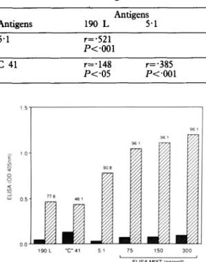
Table 4. Correlation between ELISA values obtained with diierent antigens
Antigens 5.1 Antigens 190 L 5-l r=-521 P<*OOl c 41 r=*148 r=.385 lv.05 P<*OOl 190 L “C” 41 5.1 75 150 300 I I
ELISA MIXT (ne/well)
Fig. 2. Comparison of single and pooled antigens in micro-ELISA assay. q Mean ELISA values for patients; q control blood donors. Numbers over each column indicate percentage seropositivity at reference value of mean +3SD (serum dilution l/100).
corresponded to 50,55 and 45 &well for 190 L, C 41 and 5.1 respectively. For the new assay (ELISA MIXT) a mixture containing 40 no of each antigen was prepared and the wells were coated with dilutions corresponding to 75 ng/well, 150 ng/well and 300 @well. To evaluate the sensitivity of the ELISA MIXT, 76 Garnbian sera with low ELISA values in one or more of the standard assays (most of them were
blood donors. The results of the various assays are shown in Fig. 2. A higher percentage of positive sera was detected by ELISA MIXT than by the standard assays. This was observed when the wells were coated with 75 ng of pooled antigens (in this mixture each single antigen was present at a lower concentration than in the assays based on a single recombinant polypeptide) and with wells coated with higher concentration of antigens. Only 3 sera were negative with the ELISA MIXT using 3 different concentra- tions of the pooled antigens. These 3 sera, collected from children, were also negative by IFA. Discussion
There is a need for improved and cheap immuno- diagnostic tests for the qualitative and quantitative measurement of antimalarial antibodies. The routine use of such tests requires a constant and standardized supply of malaria antigens. Conventional sources such as infected red blood cells do not meet these requirements (GABRA et al., 1986; DEMEDTS et al., 1987). Several genes coding for P. falciparum and P. vivux have been cloned and expressed in appropriate vectors. The sequences of the corresponding polypeptides have been determined and it has been shown that antibodies from individual patients with past malarial infection bind to some of these recom- binant polypeptides (BIANCO et al.,. 1988). No sys- tematic serodiagnostic study has been performed with cloned antigens with the exception of that by GABRA et al. (1986).
We have investigated the suitability of 3 recom- binant polypeptides as molecularly-defined malaria antigens for immunodiagnosis. These recombinant antigens were selected on the basis of(i) their known reactivity with antibodies from humans with past malarial infection, (ii) the fact that their amino acid sequences showed little or no variation between different isolates of P. fukiparum (GENTZ et al., 1988; CERTA et al., 1988), and (iii) a high level of expression in their vectors.
All the polypeptides were suitable for the detection of antimalarial antibodies. A very low background was observed using sera of individuals never exposed to malaria and positive reactivity was detected in almost all the sera of adults of a hyperendemic area (up to 100% positive sera in some groups of adults for the 5.1 antigen). Antibodies were also reliably de- tected in most of the sera of children. However, there was a clear difference in the amount of antibody reaction against the three recombinant polypeptides. Thus, levels of antibodies directed against 5.1 were the highest, followed by antibodies against 190 L and C 41. This was probably not a consequence of passive absorption of antibodies in zho during an acute attack of malaria, since differences in titre were observed for both the groups with a high incidence of patent parasitaemia (children) and a low incidence (adults). This may however be the reflection of better refolding of 5.1 and 190 L and C 41, or of the presence of more epitopes able to raise an antibody response on 5.1 and 190 L.
A clear correlation was observed between antibody levels directed against 5.1 and 190 L but not between antibodies directed against these 2 polypeptides and C 41. This may have been due to very high levels of
321 ant@en and red blood cell control antigen in the enzyme-linked immunosorbent assay for malaria. Amer- ican Journal of Tropical Medicine and Hygiene, 36,
257-263. -_. -__.
Gabra, M. S., Grossiord, D., Perrin, L. H., Shaw, A. R., Cheuna. A. & McGregor. I. A. (1986). Delined Plasmo- dium fikiparum antigens ‘m malaria serology. Bulletin of the World Health Organization, 64, 889-896.
antibody directed against 5.1 and 190 L. When a mixture of the 3 antigens was used, the percentage of positive sera increased significantly with a selection of sera poorly reactive with-the individual assays. The 3 sera negative with the ELISA MIXT were also negative-in the 3 assays using individual components and in IFA tests. In addition, the level of antibodies (measured by ELISA) was higher and the difference between negative and positive sera was greater.
These results suggest that an ELISA using the 3 recombinant polypeptides, available in relatively high amounts, would be useful for epidemiological studies and would also have the advantage of genuine standardization (use of molecularly defined antigens). Additional studies are required to determine the ability of this assay to detect cross-reactive antibodies to I’: vivan epitopes, and to compare the assay directly with IFA; however, published data concern- ing anti-asexual blood-stage antibody detection in children (MCGREGOR 1974; VOLLER et al., 1980; GABRA et al., 1986) seem to indicate that the ELBA MIXT has a higher sensitivity.
Acknowledgements
We thank Mrs Kim Zollinger for her expert technical assitance and Mrs C. Brown for preparing the manuscript. This work was supported by the UNDPWorld Bank/World Health Organization Special Pro8ramme for Research and Training in Tropical Diseases ID no. 870104 and by the Swiss National Research Foundation Grant 3.923.0-87.
References
Bianco, A. E., Crewther, P. E., Coppel, R. L., Stahl, H. D., Kemp, D. J., Anders, R. F. & Brown, G. V. (1988). Patterns of ant&en expression in asexual blood stages and gametocytes of Plasmodium falciparum. AmericanJournal of Tropical Medicine and Hygiene, 38, 258-267.
Certa, U., Ghersa, P., Diibeli, H., Matile, H., Kocher, H., Srivastava, I. K., Shaw, A. R. & Perrin, L. H. (1988). Aldolase activity of Plasmodium falcipanrm protein with protective properties. Science, 240, 1036-1038. Coppel, R. L., Favaloro, J. M., Crewther, P. E., Burkot, T.
R., Bianco, A. E., Stahl, H. D., Kemp, D. J., Anders, R. F. & Brown, G. V. (1985). A blood stage antigen of
Plasmodium falciparum shares determinants with the sporozoite coat protein. Proceedings of the National Academy of Science, USA, 82, 5121-5125.
Demedts, P,, Vermeulen-Van Overmeir, C. & W&y, M. (1987). Stmultaneous use of Plasmodium falciparum crude
Gem, R., Certa, U., T&KS, B;, Matile, H., Diibeli, H., Pink, R., Mackay? M., Bone, N. & Scaife, J. (1988). Major surface anugen p190 of Plasmodium falciparum:
detection of common epitopes present in a variety of plasmodia isolates. EMBO Journal, 7, 225-230.
Hope, I. A., Mackay, M., Hyde., J. E. & Scaife, J. (1985). The gene for an exported anugen of the malaria parasite
Plasmodium fakiparum cloned and expressed in E. coli. Nucleic Acid Research, 13, 369-379.
Kemp, D. J., Coppel, R. L., Stahl, H. D., Bianco, A. E., Corcoran, L. M., McIntyre, P., Langford, C. J., Favaloro, J. M., Crewther, P. E., Brown, G. V., Mitchell, G. F., Culvenor, J. G. & Anders, R. F. (1986). Genes for antigens of Plasmodium falcipamm. Parasitolo- gy, 91, S83-S108.
McGregor. I. A. (1974). Mechanism of acauired immunitv with epidemiololtical patterns of antibody responses in malaria in man. Bulletin of the World Health Organization,
50. 259-2M.
--1 --- ---.
Perrin, L. H., Parez, A. & Chixxolini, C. (1984). Malaria immunity, vaccination and immunodiagnosis. Experien- ria. 40. 1343-1350.
Rosen’ber8, E. B., Strickland, G. T., Yang, S. L. & Wahlen, G. E. (1973). IgM antibodies to red cells and autoim- mune anemia inpatients with malaria. American Journal of Tropical Medicine and Hygiene, 22, 146-151. Soencer. H. C.. Cohins. W. E.. Chin. W. & Skinner.1. C.
(1979). The enzyme-linked’ immunosorbent assay for malaria I. The use of in witro cultured Plasmodium
falciparum as antigen. American Journal of Tropical Medicine and Hygiene, 28, 927-932.
Takacs, B. (1979). Electrophoresis of proteins in polyacryla- mide slab gels. Immunological Me&&, 2, 81-105.
Voller, A., Bidwell, D. E., Huldt, G. & En&l, E. (1974). A microplate method of enzyme-linked immunosorbent assay and its application to malaria. Bulletin of the World Health Organization, 51, 209-211.
Voller, A., Comille Brogger, R., Storey, J. & Molineaux, L. (1980). A 1ongitudinal study of P&nod&m falcipatum
malaria in West African savanna using the ELISA technique. Bulletin of the World Health Organization, 58,
429-438.
Received 18 October 1988; revised 24 November 1988; accepted