Metagenesis by Unydmogeim peroxide foreataneimtt off mammaMaini eels;
a molecuilar analysis
mi
E.C.Moraes1, S.M.Keyse2 and R.M.Tyrrell3
Swiss Institute for Experimental Cancer Research, CH-1066 Epalinges sur Lausanne, Switzerland
'Permanent address: Biophysics Institute, Federal University of Rio de Janeiro, RJ 21941, Brazil
2Present address: ICRF Clare Hall Laboratories, Blanche Lane, South
Mimms, Potters Bar, Herts 3LD, UK
3To whom correspondence should be addressed
Hydrogen peroxide is an oxidizing agent which can be generated intracellularly either during normal metabolism or by treatment with external agents including solar UV radiation. Simian cells (CV-1) transfected with the SV40-based shuttle vector plasmid pZ189 have been treated with H2O2 and then incubated to allow repair and repli-cation of the plasmid. The frequency of mutations at the supF locus of the recovered plasmid increases by a factor of up to four over the spontaneous value. The nucleotide changes associated with 100 spontaneous and 100 H2O2-induced mutants have been determined directly by sequencing a 150 bp fragment that includes the entire supF tRNA coding region. Deletions were observed in - 4 5 % of both the spontaneous and induced mutants, whereas single or multiple base changes arose in 68 and 57% of the induced and spontaneous mutants respectively. The spectrum of induced mutations is characterized by (i) the occurrence of deletions associated with base changes (16% of all mutants analysed) and (ii) small deletions of 3 bp and less (51% of all deletion mutants sequenced). Sixty-five per cent (15 out of 23) of all small deletions (spontaneous and induced) are associated with runs of between two and five identical bases and eight of them arise at a mutational 'hotspot' region of five cytosines between bp 172 and 176. The majority (19 out of 30) of completely sequenced deletions observed in the spontaneous spectrum contain either (i) small (2-10 bp) direct repeat sequences that lie immediately outside one deletion terminus and immediately inside the second deletion terminus or (ii) small ( 2 - 3 bp) inverted repeat sequences lying immediately inside the two deletion termini. Most deletions that we have observed are therefore likely to arise as a consequence of specific aspects of DNA structure.
Introduction
Oxygen radicals appear to be involved in both the ageing process and in the initiation and progression of many human diseases, including cancer. A knowledge of the nature of mutations arising in DNA as a result of oxygen radical attack should provide important clues to understanding the molecular events that underlie these pathological changes. The characterization of the genotoxic action of the oxidizing agent, hydrogen peroxide, is particularly relevant in this respect for several reasons. In addition
•Abbreviations: IPTG, isopropyl-0-D-thiogalactoside; X-GAL, 5-brorno-4-chloro-3-indolyl-0-D-galactoside; LB, Luria broth.
to being produced during normal aerobic metabolism as a result of successive univalent reduction of molecular oxygen, H2O2 is also generated intracellularly by several drugs (1,2) and non-ionizing radiations (3,4). It appears likely that many of the biological effects of H2O2 are mediated by the highly reactive hydroxyl radical (• OH) generated in vivo by a Fenton reaction involving naturally occurring iron complexes (5,6).
Hydrogen peroxide is known to lead to mutations in the mitochondrial DNA of yeast and to a lesser extent in the nuclear genome (7,8). More recently this agent has been shown to be weakly mutagenic in bacteria (9). There is also evidence that peroxides arising during normal metabolism may increase spontaneous mutagenesis in Salmonella typhimurium strains containing deletions at the oxyR locus, a positive regulator of proteins involved in cellular defence against oxidative stress (10). Despite several reports showing that treatment of mammalian cells in culture with H2O2 results in DNA damage, sister chromatid exchange and chromosome aberrations (11-13), initial attempts to demonstrate the induction of gene mutations in mammalian cells were unsuccessful (12,14). However, in a recent study, Ziegler-Skylakakis and Andrae (15) have shown that if cells are treated at high cell density with concentrations of H2O2 in the low millimolar range then mutations can be detected at the hypoxanthine guanine phosphoribosyl transferase locus. The increase in mutation frequency is dependent on the concentration of H2O2 employed.
Several recent studies have focused on the characterization of the molecular nature of mutations induced by oxygen radicals at the DNA sequence level. Thus it has been shown that singlet oxygen (O2) induces a spectrum of mutations in bacteriophage quite distinct from the spectrum of mutations observed for spontaneous mutants (16) and that iron-generated oxygen species (presumably H2O2 and -OH) generate specific types of mutations at a single bacteriophage amber site (17).
In the present study we have treated monkey CV-1 cells with H2O2 at high cell densities and used an SV40-based shuttle vector (pZ189) as a target for mutagenesis within the cells (18). This approach has allowed us to determine the DNA sequence changes characteristic of mutants that arise as a result of the
processing of H2O2-damaged DNA in eukaryotic cells.
Materials and methods Cells and plasmid
African green monkey kidney cells (CV-1) were grown as monolayers in Earle's MEM supplemented with 15% fetal calf serum (both Seromed) in a 5% CO2
atmosphere at 37°C. The shuttle vector plasmid pZ189 described by Seidman et al. (18) was prepared using standard techniques. The indicator host bacterial strain used to distinguish between wild-type and mutant plasmids was Escherichia coli MBM7070, which has the genotype F'lacZ (Am) CA7O20 lacYl hsdR hsdM A(araABC-leu) 7679 galUgalK rpsL thi. When grown in the presence of isopropyl-0-D-thiogalactoside (IPTG), an inducer of the lac operon, and 5-bromo-4-chloro-3-indoyl-/3-D-galactoside (X-GAL), a synthetic substrate for /3-galactosidase, MBM7070 gives rise to blue colonies only if it carries a pZ189 plasmid with an active supF suppressor tRNA gene. If the supFgenc on the plasmid is carrying an inactivating mutation then MBM7070 will form white or occasionally light blue colonies.
Transfection and mutation induction
CV-1 cells growing in 9 cm Petri dishes (50% confluent, i.e. ~ 1.5 x 10⁶
cells/dish) were transfected with pZ189 DNA (50 ng/dish) using the DEAE-dextran method (19). The transfected cells were incubated for 6 h at 37°C and then treated with H2O2 (Fluka AG) at a concentration of either 7 or
10 mM in phosphate-buffered saline. Treatments were for 30 min at 37°C in the dark. Following treatment, the growth medium was replaced and cells were reincubated at 37 °C. Forty-eight hours later, vector DNA was extracted from the cells using the method of Hirt (20) as modified by McMaster el al. (21). Mutant selection and characterization
The procedures followed were exactly as described previously (22,23). Briefly, following transformation of the indicator strain MBM7070 with vector DNA, transformants were selected on Luria broth (LB) plates containing ampicillin, X-GAL and IPTG. White and light blue (mutant) colonies were picked, restreaked on the same selective medium for confirmation of phenotype and stored as stabs in soft agar containing ampicillin until analysis. Plasmid DNA was prepared for further analysis by a modified alkaline lysis 'mini prep' method (24). Plasmids were then analysed by agarose gel electrophoresis to distinguish between those carrying large deletions or insertions and those that co-migrated with wild-type plasmid and probably contained point mutations. The latter were then subjected to DNA sequence analysis. The tRNA region of the pZ189 vector was sequenced using a modification of the Sanger dideoxyribonucleotide procedure which allows sequence determination directly from the double-stranded plasmid DNA (25). The possibility that mutations arise in the bacterial rather than the mammalian host cell as a result of a low level of persistent lesions formed in pZ189 DNA during H2O2 treatment of the transfected cells is difficult to exclude
experi-mentally. However, since the induction of mutations in bacteria by H2O2 is an
extremely rare event (9) and we have been unable to detect mutant plasmid after direct transformation of bacteria with pZ189 DNA treated with up to 50 mM H2O2 for 30 min, it appears highly probable that the mutations we have observed
arose in the mammalian host.
Results
Induction of mutants by hydrogen peroxide
In preliminary experiments we determined that levels of plasmid DNA sufficient to allow subsequent transformation of E.coli MBM7070 and detection of supF mutants could be extracted from CV-1 cells that had been transfected at high cell density 6 h prior to treatment with 7 — 10 mM H2O2 and then incubated for a further 48 h. The frequency of mutation at the supF locus in plasmid recovered from H2O2 treated cells rose to almost four times the spontaneous level of 0.027 ± 0.013% (Figure 1). Spontaneous mutants were analysed and sequenced from eight independent transfections and induced mutants were analysed from six and five independent transfections for 7 and 10 mM H2O2 treatments respectively. Plasmid DNA from mutant colonies was first analysed on agarose gels to distinguish those with gross alterations. Only vector DNAs that migrated with normal mobility were subjected to sequence analysis. When identical mutations were detected in mutants isolated from the same transfection, they were excluded from further analysis to ensure that all mutations arose independently. All mutants contained at least one change within the tRNA coding region. A broad classification of the types of mutations is shown in Table I. Deletion mutations arose in - 4 5 % of both the spontaneous and the H2O2-induced mutants. There are no important differences in the broad classification of mutants induced by either 7 mM or 10 mM H2O2. When the pooled mutants that we have classified as induced are considered (i.e. 42 from 7 mM H2O2 treatments and 58 from 10 mM treatments), we estimate that approximately one-third of them will actually have arisen spontaneously and this should be considered in the description that follows.
Base-change mutagenesis by hydrogen peroxide
As for previous mutation spectra obtained with both spontaneous and UV-induced mutants (22,23,26), H2O2-induced mutants contained both single and multiple base changes (Table II). The
CO O >. ^ O
III
l\\
s
>
z
cTz F X 3 H HI S DC U . 1 U " 8 6 4 2J
0 5 10 [H2O2] mMFig. 1. Enhancement of mutation frequency at the supF locus of the pZ189 plasmid following exposure of plasmid containing cells to 30 min H2O2
treatment.
Table I. Classes of mutations arising spontaneously or after hydrogen peroxide treatment
H,O, Spontaneous
Altered gel mobility Only deletion Only base change Deletion + base change Insertion Insertion + deletion Total 11 (11%) 18 (18%) 53 (53%) 16 (16%) 1 (1%) 1 (1%) 100 12 (12%) 31 (31%) 54 (54%) 3 (3%) -100
Table II. Distribution of single and multiple base changes among spontaneous and hydrogen peroxide-induced mutants
No. of base changes per mutant
1 2:5 15 bp apart< 15 bp apart H2O2 31 (45)a 7 (10) 11 (16) Spontaneous 18 (32) 3 (5) 7 (12.5) 11 (16) 5 (7)4 (6) 11 (20) 10 (18)7 (12.5)
aValues in parentheses are the percentage of total base change mutants (H2O2
or spontaneous) that occur in each category.
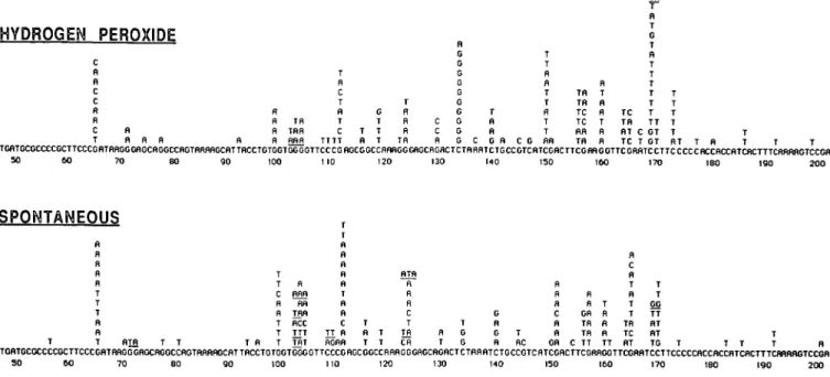
spectra of base changes that arise spontaneously in the region sequenced or that are induced by H2O2 treatment are compared in Figure 2. As observed previously for spontaneous and far UV (254 run) radiation-induced mutagenesis using this shuttle vector (22,23), the distribution of mutations within the supF gene is not random and several 'hotspots' are evident. Based on binomial probability analysis and the total number of base changes observed within the supF coding region (125 for both spontaneous and induced), six or more changes at a single site can be considered as a significant increase (P = 0.05) over that expected from a random distribution of base change mutations among the available sites. By this criterion, eight mutational 'hotspots' arise after H2O2 treatment at bp 111, 133, 149, 155, 156, 159, 168 and 172. Four of these sites (bp 133, 149, 168 and 172) contain significantly (P < 0.05) more alterations than arise in the spontaneous spectrum. Of the nine 'hotspot' sites for base change mutation apparent in the spontaneous spectrum, only two (bp 150 and 164) contain significantly more changes than the
corre-HYDROGEN PEROXIDE R R R R TR TRR RRR T R C T R T C TTTT G T T T fl T T R R R TR C C R R G G G G G G G G G G C T R R G R T T R R T T R T T C G flR Tfl Tfl TC TC RR Tfl R T B R T R R TC TR RT TC G T B T T T T T T TT C GT T GT T T T T T RT T R T T TGflTGCGCCCCGCTTCCCGBTBRGGGflGCflGGCCRGTflRflflGCRTTflCCTGTGGTGGGGTTCCCGflGCGGCCRRflGGGRGCflGflCTCTRRRTCTGCCGTCfiTCGflCTTCGRRGOTTCGRBTCCTTCCCCCRCCflCCfiTCRCTTTCftfiftflGTCCGfl 50 60 ?O 80 90 100 110 120 130 140 150 160 170 180 ISO 2 0 0
SPONTANEOUS
T T C R R T T T R flnfl RR TRfl RCC TTT THT T R fl fl R R T fl n c TT R RGRfl T R T T T RTfl H R fl C T Tfl Cfl R T T G G G R G R T RC R fl R C R n GR R R OR Tfl TR C TT T R R R TT fl C R T n T T Tfl TC flT T T GG TT flT RT TG TGflTGCGCCCCGCTTCCCGflTflflGGGflGCBGGCCRGTRflRflGCflTTRCCTGTGGTGGGGTTCCCGRGCGGCCRflflGGGflGCflGflCTCTRRflTCTGCCGTCflTCGRCTTCGRnOGTTCGRflTCCTTCCCCCflCCBCCRTCRCTTTCHflflBGTCCGfl SO 6 0 70 8 0 9 0 100 110 120 130 HO 150 160 170 180 190 200Fig. 2. The complete spectra of DNA base changes detected at the supF locus in spontaneous mutants and in those induced by H2O2. Tandem double and
triple base changes are underlined.
HYDROGEN PEROXIDE TCflTCCCCCCCGCTTCCCCflTflflGCCflCCflCCCCftCTnflflRGCflTTflCCTGTGCTCGGCTTCCCCflGCGGCCflflflGGGflOCflCflCTCTfiftRTCTGCCGTCflTCGfiCTTCCfiflGCTTCCfiftTCCTTCCCCCflCCflCCflTCRCTTTCflflnflCTCCGfi 50 60 70 BO 90 100 110 120 130 110 150 160 170 190 190 200 SPONTANEOUS fl C TT fl T T G C G G C fl TT T flT TGHTGCCCCCCGCTTCCCGflTHflGGGHGCHGGCCnGTOHBnGCnTTnCCTGTGGTGGGGTTCCCGnGCGGCCHHHGGGnGC(IGHCTCTni)flTCTGCCGTCnTCGflCTTCGnnGGTTCGnnTCCTTCCCCCnCCflCCnlCHCTTTCnflHnGTCCGB SO 60 70 SO 90 100 110 120 130 110 I50 I60 170 ISO 190 200
Fig. 3. The spectra of DNA base changes that are detected at the supF locus when only mutants containing a single base-change are considered. The asterisks denote the beginning and end of the tRNA coding region.
sponding site in the induced spectrum. Evidently, additional 'hotspot' locations and differences may become apparent on analysis of a larger population. The spectra arising from mutations involving only a single base-change are shown for reference in Figure 3. Despite the small sample population, the occurrence of four base changes at bp 123, 133, 159 and 168 in the H2O2 spectrum is higher (P < 0.1) than predicted by a random distribution of induced mutations. However, it appears that the majority of the base substitutions that give rise to the H2O2-induced 'hotspots' are caused by a multiple rather than single changes in the supF gene, particularly at position 149. The hotspot at bp 172 is of interest since it occurs at the start of the cytosine sequence run which is a 'hotspot' region for small deletions (see later).
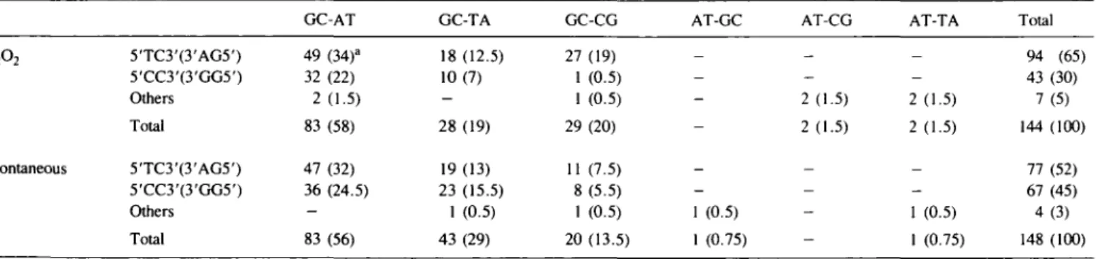
Almost all the base changes observed in both spectra occur at a G-C base pair: 140 out of 144 after H2O2 treatment and 146 out of 148 arising spontaneously. Previous studies of both spontaneous and UVC- or UVB-induced base change mutations using shuttle vectors (23,27) have shown a remarkable sequence specificity in that the majority of changes occur in the right-hand base pair of one of the two following sequences: 5'-TC-3':3'-AG-5' or 5'-CC-3':3'-GG-5'. In the present study, 95% of the H2O2-induced and 97% of the spontaneous base
changes arise in the right-hand base pairs of one of these sequences (Table 01). Furthermore, when a mutant has more than a single base change, the sites of these base changes are always in the right-hand base pair of one of the two possible base doublets that occur in the same orientation, i.e. 5'-TC-3' and 5'-CC-3' or 3'-AG-5' and 3'-GG-5'. This is a clear indication that all multiple base changes arising in a single mutant occur in the same strand (see also ref. 26). All four 'hotspots' which are more predominant after H2O2 treatment (positions 133, 149, 168 and 172) occur at the right-hand base of 5'-TC-3':3'-AG-5' sequences.
An analysis of the types of base changes arising spontaneously or after H2O2 treatment (Table III) shows that in both cases the GC to AT transition predominates.
Deletion mutagenesis by hydrogen peroxide
Approximately 10% of both spontaneous and H2O2-induced mutants show altered mobility when analysed on agarose gels (Table I), indicating gross changes-either large deletions or insertions-in the plasmid vector. These mutants were not subjected to DNA sequence analysis. Of the remaining mutants that were sequenced, 35 spontaneous and 37 H2O2-induced mutants contained deletions. These are listed in Table IV
Table III. Type
H2O2
Spontaneous
and site of base change
5TC3'(3'AG5') 5'CC3'(3'GG5') Others Total 5TC3'(3'AG5') 5'CC3'(3'GG5') Others Total mutations either GC-AT 49 (34)a 32 (22) 2 (1.5) 83 (58) 47 (32) 36 (24.5) -83 (56) induced by hydrogen GC-TA 18 (12.5) 10(7) -28 (19) 19(13) 23 (15.5) 1 (0.5) 43 (29) peroxide or arising GC-CG 27 (19) 1 (0.5) 1 (0.5) 29 (20) 11 (7.5) 8 (5.5) 1 (0.5) 20 (13.5) spontaneously AT-GC _ -_ -1 (0.5) 1 (0.75) AT-CG _ -2(1.5) 2 (1.5) — -AT-TA _ -2 (1.5) 2 (1.5) _ -1 (0.5) 1 (0.75) Total 94 (65) 43 (30) 7(5) 144 (100) 77 (52) 67 (45) 4 (3) 148 (100) aValues in parentheses are the percentage of total base changes (H2O2 or spontaneous) that occur at each site for each category of transition or transversion.
Table IV. Sequence of spontaneous deletion mutants
Mutant no.
Deletion position
Sequence deleted No. of
base pairs deleted Classification 5 ' — 117 5 ' - 135 5'-201 63-233 or 64-234 104-244 111 -208 or 116-213 CGC CCA TCT AAA CCG AAA TGG^GGT CTT^CTT CCC'GAGCG'GCC ..AJAGCG'TCA C*AAA 170 141 98 Out of sequence Out of sequence Out of sequence Unclassified (TPYR CTT) Direct repeat (5bp) 109-197 or 113-201 85-168 TTC'CCGA'GCG AGT'CCGA'AAG
GTAL AAA ATC'CTT
89 84 Direct repeat (4bp) (TP Y R CTT) 93-176
or 96-179 ATTVACCVTGT CCC
Irnl
vACCyACC84 Direct repeat (3bp) 10 11 12 43-125 or 44-126 71-179 68-119 or 78-129 GGA*G*CAG AAG'GGAG CACCVX 83 79 52 GATVAAGGGAGCAGVGCC CCAVAAGGGAGCAG ACT
Unclassified Inverted repeat (2bp) Direct repeat (10 bp) 13 130-176 or 132-178 C C c W \fcAC 47 Direct repeat (2bp)
Table IV. (contd) Mutant no. Deletion position Sequence deleted TTCCCG^ATA TTCC^GAG AAT AAG^GT^TCGAA AAA*TC*TGC ATCTTGCC GGTMCGA TCA T C G J V C T T A G G T T C G A ' A T C TTAXCT GGG?TTC GG'lWGGG CGA*G
v I
v
v
AGG TTC GAATCC TTCVCCC CCG^TC'ATCGACT'TC'GAA GGG'AGCAGACTVTA No. of base pairs deleted 45 40 37 24 Classification Direct repeat ( 5 - 6 bp) Direct repeat (4bp) Direct repeat (6 out of 7 bp) Direct repeat (5bp) 14 15 17 18 19 20 and 21 22 23 24 25 66-110 164-203 or 168-207 16 160-196 or 162-198 138-161 or 140-163 140-161 or 141-162 151-164 or 152-165 94-105 102-112 or 109-113 161-169 or 164-172 145-153 or 147-155 125-132 22 14 12 09 09 08 Unclassified Direct repeat (4bp) Inverted repeat (2bp) Unclassified Direct repeat (3 bp) Direct repeat (2bp) Inverted repeat (2bp) 26 27 114-121 or 116-213 174-180 or 175-181 ~or 176-182 AGC'GGTCAAA'GGVGAG TCC*C? 07 07 'ATC Direct repeat (2 bp) or inverted repeat (2 bp) Direct repeat (2bp) 28 142-145 or 143-146 CTGVCVCGTVCVATC 04 Unclassified 29 110-113 or 112-115 04 Direct repeat (2bp)30 127-129 GACrXAG*ACTACrXAG* 03 Direct repeat
Table IV. (contd) Mutant
no.
Deletion position
Sequence deleted No. of
base pairs deleted Classification 31 172,173 or 173,174 or 174,175 or 175,176 CWACC 02 Small deletion (identical base run)
32 102 or 103
or 104 or 105 GGT'GVGVGVGVTTC
01 Small deletion (identical base run)
33 142 or 143 'GTC 01 Small deletion
(identical base run)
+ 02 out of sequence (completely deleted) Out of sequence
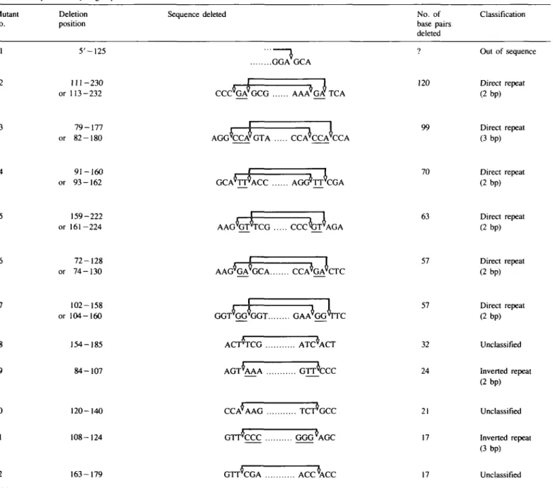
Table V. Sequence of hydrogen peroxide-induced deletion mutants Mutant
no.
Deletion position
Sequence deleted No. of
base pairs deleted 120 99 70 63 57 57 32 24 21 17 17 Classification Out of sequence Direct repeat (2bp) Direct repeat (3bp) Direct repeat (2bp) Direct repeat (2 bp) Direct repeat (2bp) Direct repeat (2bp) Unclassified Inverted repeat (2bp) Unclassified Inverted repeat (3bp) Unclassified 5'-125 2 3 4 5 6 7 8 9 10 11 12 288 111 - 2 3 0 or 113-232 79-177 or 82-180 91-160 or 93-162 159-222 or 161-224 72-128 or 74-130 102-158 or 104-160 154-185 84-107 120-140 108-124 163-179 GGA GCA
CCC^GA9GCG AAA^GA* TCA
AGG'CCVGTA CCA'CCA'CCA GCA'TT^ACC AGG*TT'CGA AAG^GT^TCG C C C ^ J T \ \ G A AAG^GA^GCA CCA^GA^CTC GGT'GG'GGT GAA'GGVTC ACT*TCG ATC*ACT A G T \ A A G r r c c c CCA*AAG GTTCCC GGG^AGC GTT*CGA ACC*ACC
Table V. (contd) Mutant
no.
Deletion position
Sequence deleted No. of
base pairs deleted Classification 13 14 159-170 137-145 GAAVGGT CCT*TCC TAAVATC CGT'CATC 12 09 Unclassified Direct repeat (imperfect) 15 145-152 or 146-153 CCGVTVCATCGACVTVTCG 08 Unclassified 16 17 18, 19 and 20 21 161-166 or 162-167 165-168 173-175 or 174-176 106,107 \GGVTVTCGAAVTVOCT TGAVAATCVCTT TCCVCVCCVCV GGG'TT'CCC 06 04 03 02 Unclassified Unclassified Small deletion (identical base run)
Unclassified 22 and 23 24 and 25 26 and 27 28 29 30 172.173 or 173.174 or 174.175 or 175,176 127 155 or or 102 104 119 121 or or or 103 105 120 ACC GAGVC\GA CTTCXJAA GGT WA'G 134 G C C W A G G G CTC'T'AAA 02 01 01 01 01 01 Small deletion (identical base run)
Small deletion unclassified
Small deletion unclassified
Small deletion (identical base run)
Small deletion (identical base run)
Small deletion unclassified 31 32 33 34 135 or 137 159 or or 136 160 165 or 166 167 TCTVAVAVAVTCT GAA'GVG'TTC TCG'WA'TCC GAA'T'CCT 01 01 01 01 Small deletion (identical base run)
Small deletion (identical base run)
Small deletion (identical base run)
Table V. (contd) Mutant
no.
Deletion position
Sequence deleted No. of
base pairs deleted Classification 35 172 or 173 or 174 or 175 or 176
crrcc'cc c ACC
Small deletion (identical base run)36 173 or 174
or 175 or 176
+ 01 out of sequence (completely deleted)
CTTC
Wc'clcc
01 Small deletion (identical base run) Out of sequence
(spontaneous) and Table V (induced) in descending order of size in base pairs. Several of the deletions run out of the region sequenced and cannot be further classified.
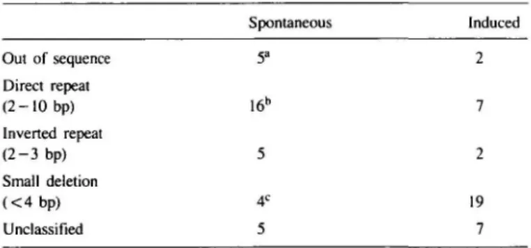
An important class of deletion mutants show regions of between 2 and 10 bp of homology between a region immediately flanking the outside of the deletion and the sequence immediately inside the deletion on the opposite side of the breakpoint. These are characteristic of the spontaneous mutants where 16 out of 33 (48%) of the deletion mutants sequenced contain such homologies as against only 7 out of 36 (19%) of the H2O2-induced mutants. Since the frequency of induced mutants is only ~ 3 times higher than the spontaneous level, the induced mutants that appear in this class may, at least partly, reflect spontaneous background. A third class of deletion mutations (Table VI) is comprised of very short 1 - 2 bp deletions which are much more predominant in the H2O2-induced set (19 out of 37 sequenced) than in the spontaneous set (4 out of 35 sequenced). This is shown diagrammatically in Figure 4. Deletions of a single base pair are clearly characteristic of the spectrum of mutations induced by H2O2. Deletions of between 4 and 100 bp are more predominant in the spontaneous spectrum. Many of the small deletions appear at runs of between three and five identical bases in the sequence. In particular the run of five consecutive cytosines from bp 127 to 176 in the tRNA gene appears to be a mutational hotspot for these deletions, accounting for 7 of the 19 induced mutations involving < 4 bp and a single, spontaneous 2 bp deletion.
Discussion
Studies on the induction of mutations in yeast by H2O2 have shown that the density at which cells are treated will influence the biological outcome and that higher cell concentrations will lead to a faster destruction of the added H2O2, presumably as a result of the action of cellular catalase and peroxidases. In the present study, we have not made a systematic investigation of the physiological conditions of treatment that influence the biological outcome but have chosen deliberately to treat cell populations at high density to minimize cell killing and allow detection of mutations. Under these specific conditions, we have shown that the frequency of mutants arising at the supF locus of the pZ 189 plasmid is enhanced by a factor of ~ 4 times over the spontaneous level when cells transfected with the plasmid are treated with H2O2 and incubated for a suitable period (Figure 1). This is not only consistent with a recent report that H2O2 is mutagenic to mammalian cells (15) but has also provided material for an analysis of such mutants at the DNA sequence level. The spectrum of deletion mutations induced by H2O2 differs markedly from that arising spontaneously. Both small deletions
1 bp 2-3 bp 4-20 bp 21-100 bp
DFl FTION <?IZF
Fig. 4. Spontaneous and H2C>2-induced deletions grouped as a function of
size and expressed as a percentage of the total number of deletions of < 100 bp in length.
Table VI. Classification of deletion mutants
Spontaneous Induced Out of sequence Direct repeat ( 2 - 1 0 bp) Inverted repeat ( 2 - 3 bp) Small deletion ( < 4 b p ) Unclassified 5a 16* 5 2 7 3 19 7 "The values given are the total numbers of mutants in each class as derived from Tables IV and V.
bMutant 26 (Table [V) has both a 2 bp direct repeat and a 2 bp inverted repeat. cMutant 30 (Table [V) is a direct repeat in a small deletion.
(Figure 4 and Table VI) and deletions with associated base changes (Table I) are clearly characteristic of the induced spectrum, whereas deletions involving direct sequence repeats appear to be primarily of spontaneous origin. A limitation of the particular shuttle vector system used here is that large deletions will go undetected because the small target gene is flanked on both sides by sequences necessary for the viability of the plasmid in the bacterial host. However, when compared with the 290
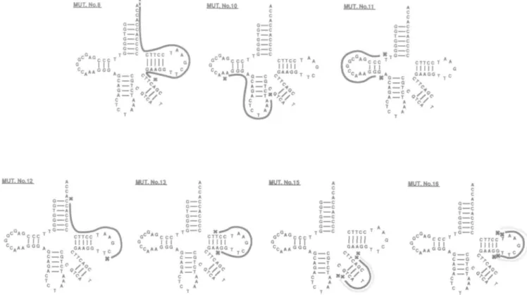
MUT. Nc.8 MUT. Wo 1? G c " C c A 0—C G—C T—A 0—C MUT. Mo 15 A ' c c c '1" " ' c^cfT^N I I I I I I I I G] C . A A0 0 0A GAAGG T , / CA«« *G—C O, K ^ J T C / 4G = C < A — T C A T A C A O C G C T A 0 C a—c a—c T ° c CTTCC ' Hill GAAGO » 0 — C C,, K C G A T 0 — C A — T MUT.No.1S "CCC i l l CTTCC T A G A A G OT A — T O. G — C f y1< A — T O / V C A V/ T A f C A T
Fig. 5. The structure of deletions observed in unclassified mutants induced by H2O2 shown superimposed on the potential cloverleaf structure of the coding
region of the supF tRNA gene.
techniques currently available for mutational analysis in chromosomal genes in mammalian cells (e.g. see ref. 28), the system is rapid and simple and both base changes and the two termini of deletions of 100 bp or less can be analysed from a much larger database than would otherwise be possible.
The majority of deletions that we have observed in both the spontaneous and induced spectra (Tables IV and V) are associated with distinct features of DNA structure which allow a broad classification, the details of which will be briefly discussed below.
Deletions associated with direct and inverted sequence repeats
Using the lad system in E. coli, Farabough et al. (29) observed that many small spontaneous deletions (9-123 bp) arose between regions of short homology (direct repeats). This observation was later found to hold for much larger spontaneous deletions (700- lOOO bp) in this same gene (30). Further analysis of such deletion data (31) showed that of those deletions which did not involve repeated DNA sequences, the majority contained palindromic or quasi-palindromic sequences capable of forming secondary structures that could juxtapose the end points of the deleted region and in this way direct the deletion specificity. Thus, in the latter case repeated DNA sequences are not required. Similar sequence-directed mechanisms of deletion formation were later seen in a small pool of both spontaneous (32) and ionizing radiation-induced mutants (28) in mammalian cells. In the present study we have confirmed the presence of these two classes of deletion mutant in mammalian cells. Fifteen out of 30 completely sequenced spontaneous deletion mutants show direct repeats of 2 —10 bp, one lying immediately outside the deletion and the second lying immediately inside the deletion on the opposite side. Several of the 'induced' mutants (Table V) also fall into this category at a frequency (7 out of 35) that indicates that they could
be of spontaneous origin. Of the 22 deletion mutants that were associated with direct repeats (Tables IV and V) none were repeated in independent transfections. The model that has been proposed to explain this association of direct sequence repeats with deletion termini has been termed 'slipped mispairing' (29) and involves DNA strand slippage and mispairing of the two repeated elements during replication.
We have also detected deletions with short (2—3 bp) inverted repeats that lie immediately inside the deletion termini. Five of the spontaneous deletion mutants (Table IV, nos I I , 20, 21, 25 and possibly 26) and two of the H2O2-induced mutants (Table V, nos 9 and 11) fall into this class. The presence of inverted repeat sequences could facilitate the looping out of a single-stranded region opposite a gapped structure. Although the thermodynamic stability of such stem loop structures is low, this is not incompatible with their possible role as intermediates in deletion formation since the latter is an extremely low probability event. Gapped structures could arise either during normal replication or as a result of structural damage to the DNA arising either spontaneously or during exposure to H2O2. In both cases deletion termini will be juxtaposed by the looping out of the intervening sequence thereby facilitating ligation. The final step in deletion formation would then either be replication of the shorter strand or incision of the entire loop from the heteroduplex DNA (33,34). Such a process is more likely for short deletions and the possibility of such a model is supported by the repetition of a small 12 bp deletion associated with an inverted repeat in two independent transfections (Table IV, nos 20 and 21). Glickman and Ripley (31) also found mutants that contained both direct and inverted repeats. In this case the formation of transient, single-stranded structural intermediates could draw the direct sequence repeats into close proximity and increase the probability of slipped mispairing. In our study a single spontaneous 7 bp
deletion mutant (Table IV, no. 26) falls into this category. However, most of the longer deletions contain inverted repeats within the region deleted which could participate in deletion formation.
Small deletions (1—3 bp)
Based on data obtained with bacteriophage T4, Streisinger et al. (35) proposed that frameshift deletions could result from the presence of a single-strand gap at or near a region containing a run of identical bases or base doublets in the sequence. The break would increase the probability of mispairing at the repeating bases, which in turn would lead to the addition or deletion of a base or bases during new DNA synthesis. This model would appear to be entirely consistent with our data for H2O2-induced mutations in the supF gene, where 19 out of 37 deletion mutants induced by H2O2 (51 %) involved small deletions of 3 bp or less (Table V) and 10 of these occur at runs of three or more identical bases. Four of the remaining single base pair deletions occur either between a repeated doublet (Table V, nos 24 and 25) or at the site of two identical bases (Table V, nos 21, 32 and 33). Of the three spontaneous mutations involving deletions of 1 or 2 bp, one occurs at a run of five cytosines (Table IV, no. 31), one at a run of four guanines (Table IV, no. 32) and one at the site of two identical bases (Table IV, no. 33). Overall, eight of the small deletions observed occur at the run of five cytosines between bp 172-176, a finding that clearly pinpoints this as a hotspot region for deletion mutagenesis. Since the small deletions are predominant in the spectrum of mutations induced by H2O2 (Figure 4), it appears likely that the strand breaks known to be induced by H2O2 in the DNA of mammalian cells (11) are involved in enhancing the probability of this type of mutational event. Strand breaks that occur in close proximity on the same DNA strand are probably directly responsible for the deletion of 1 or 2 bp from the five remaining small deletion mutants in the H2O2 series (Table V, nos 21, 25, 27, 30 and 34) that are not accounted for by the mispairing model.
Since the hydroxyl radical generated intracellularly via a Fenton-type reaction is probably the active intermediate leading to genetic damage after H2O2 treatment, it is instructive to compare our present data with that obtained in recent studies with ionizing radiation which generates hydroxy radical by the radiolysis of water. Of 51 mutants observed in the c 1 gene of lambda prophage, 11 were 1 and 2 bp frameshift deletions (36) and 4 out of 16 of the 'point mutations' arising in the endogenous adenine phosphoribosyl transferase gene of Chinese hamster ovary cells were small deletions (28).
Seven of the spontaneous deletion mutants fall into none of the classes described above. Five of these (Table IV, nos 4, 10, 13, 18 and 22) have single base repeats at similar positions in relation to the deletion termini as the direct repeats involving two or more bases. The remaining two (Table IV, nos 5 and 8) have the sequence T-pyr-CTT flanking the 3' deletion terminus. The significance of these weaker structural relationships is not clear. The six unclassified deletion mutants induced by H2O2 are shown superimposed on the potential cloverleaf structure of the coding region of the supF tRNA gene in Figure 5. Mutant no. 11 (Table V), which is classified as associated with an inverted repeat, is shown for comparison. Such secondary structure is likely to be formed in the presence of gapped structures such as would be induced by H2O2. Three of the potential inter-mediate structures (nos. 13, 15 and 16) bring deletion termini close together and could be involved in deletion formation.
Although the true nature of deletion intermediates is unknown, the above considerations indicate that DNA structure is involved
292
in directing deletion termini and that DNA damage induced by H2O2 (presumably DNA single-strand breaks) can strongly influence the spectrum of mutations observed.
Base change mutagenesis
Previous studies using shuttle vectors have demonstrated that the majority of base changes that occur in mutants that arise either spontaneously or as a result of UV treatment of the plasmid occur at G-C base pairs (23,27). We now show that 97% of the base changes that arise in the supF gene in the pZ189 plasmid following H2O2 treatment of transfected cells also occur at G-C base pairs (Table III) and that 60% of these base changes are G-C to A-T transitions.
It has been suggested that the changes at G-C base pairs that arise spontaneously may result from depurination (of guanine) or deamination (of cytosine) in the acid environment encountered by the plasmid DNA shortly after transfection (37), while the preference for changes at G-C base pairs seen in induced mutants after UV irradiation reflects the nature of the DNA breakage itself (38). However, since changes at G-C base pairs predominate both in spontaneous mutants and in mutants arising as a result of treatment with a range of different mutagenic agents including UVC, UVB and H2O2, it would seem more likely that such changes have a more general origin, possibly reflecting the influence of DNA structure, rather than specific types of DNA damage. Miller (39) has proposed that G-C to A-T transitions arise because adenine is preferentially inserted across from UV photoproducts and that mutational specificity is determined by the properties of the DNA polymerase rather than the specific damage to the DNA . Indeed it has been shown in studies of the replication fidelity of various DNA polymerases in vitro that for all templates and enzymes examined, purines rather than pyrimidines are preferentially misinserted, in most cases in the order A > G > C > T (40).
In summary, mutations arising in plasmids as a result of H2O2 treatment of the mammalian host cells have been characterized at the DNA sequence level. The most predominant feature of the mutational spectrum when compared with that which arises spontaneously is the high level of small deletion mutations, many of which arise in runs of identical base pairs.
Acknowledgements
We gratefully acknowledge the expert technical assistance of M.Pidoux and Y.Tromvoukis in certain of the experiments. This research was supported by grants from the Swiss National Science Foundation (3.186.088) and the Swiss League Against Cancer. E.C.M. was supported by a post-doctoral fellowship from the Brazilian National Research Council (CNPq).
References
1. Thor.H,. Smith,M.T., Hartzell.P., Bellomo.G., Jewell.S.A. and Orrenius.S. (1982) The metabolism of menadione (2-methyl-1,4-naphthoquinone) by isolated hepatocytes. J. Biol. Chem., 257, 12419-12425.
2. Rajagopolan,S. Politi.P.M., Sinha.B.K. and Myers,C.E. (1988) Adriamycin-induced free radical formation in the perfused rat heart implications for cardiotoxicity. Cancer Res., 48, 4766—4769.
3. McCormicU.P., Fischer.J.R., Pachlatko,J.P. and Eisenstark,A.A. (1976) Characterisation of a cell-lethal product from the photooxidation of tryptophan: hydrogen peroxide. Science, 191, 468—469.
4. Tyrrell,R.M. (1985) A common pathway for protection of bacteria against damage by solar UVA (334nm, 365nm) and an oxidising agent H2O2). Mutat. Res.. 145, 129-136.
5. Gutteridge,J.M.C. (1985) Superoxide dismutase inhibits the superoxide-driven Fenton reaction at two different levels. FEBS Lett., 185, 19-23. 6. Imlay,J.A., Chin.S.M. and Linn,S. (1988) Toxic DNA damage by hydrogen
peroxide through the Fenton reaction in vivo and in vitro. Science, 240, 640-642.
peroxide. Mutat. Res., 33, 147-156.
8. Thacker,J. and Parker.W.F. (1976) The induction of mutation in yeast by hydrogen peroxide. Mural. Res., 38, 4 3 - 5 2 .
9. Levin,D.E., Hollstein,M., Christman.M.F., Schwiers,E.A. and Ames,B.N. (1982) A new Salmonella tester strain with A T base pairs at the site of mutation detects oxidative mutagens. Proc. Natl. Acad. Sci. USA, 79, 7445-7449.
10. Stortz.G., Christman.M.F., Sies.H. and Ames.B.N. (1987) Spontaneous mutagenesis and oxidative damage to DNA in Salmonella typhimurium. Proc. Nail. Acad. Sci. USA, 84, 8917-8921.
11. Hoffmann,M.E. and Meneghini,R. (1979) Action of hydrogen peroxide on human fibroblasts in culture. Photochem. Photobiol., 30, 151-155. 12. Speit,G. (1986) The relationship between the induction of SCEs and mutations
in Chinese hamster cells. I. Experiments with hydrogen peroxide and caffeine. Mutat. Res., 174, 2 1 - 2 6 .
13. Schoneich,J. (1967) The induction of chromosomal aberrations by hydrogen peroxide in strains of ascites tumours of mice. Mutat. Res., 4, 384-388. 14. Bradley,M.O. and Erickson,L.C. (1981) Comparison of the effects of hydrogen peroxide and X-ray irradiation on toxicity, mutation, and DNA damage/repair in mammalian cells (V-79). Biochim. Biophys. Acta, 654, 135-141. 15. Ziegler-Skylakakis.K. and Andrae.U. (1987) Mutagenicity of hydrogen
peroxide in V79 Chinese hamster cells. Mutat. Res., 192, 6 5 - 6 7 . 16. Decuyper-Debergh.D., PietteJ. and Van de Vorst,A. (1987) Singlet
oxygen-induced mutations in M13 lacL phage DNA. EMBO J., 6, 3155-3161. 17. Loeb.LA., James.E.A., Waltersdorph.A.M. and Klebanoff,S.J. (1988)
Mutagenesis by the autoxidation of iron with isolated DNA. Proc. Natl. Acad. Sci. USA, 85, 3918-3922.
18. Seidman.M.M., Dixon,K., Razzaque,A. and Berman,M.L. (1985) A shuttle vector plasmid for studying carcinogen-induced point mutations in mammalian cells. Gene, 38, 233-237.
19. McCutchen,J.A. and Pagano.J.S. (1968) Enhancement of the infectivity of simian virus 40 deoxyribonucleic acid with diethyl-aminoethyl-dextran. J. Natl. Cancer Inst., 41, 351-357.
20. Hirt,B- (1967) Selective extraction of polyoma DNA from infected mouse cell cultures. J. Mol. Biol, 20, 365-369.
21. McMaster,G.K., Beard,P., Engers.H.D. and Hirt,B. (1981) Characterisation of an immunosuppressive parvovirus related to the minute virus of mice. J. Virol., 38, 317-326.
22. Hauser,J., Seidman.M.M., Sidur.K. and Dixon,K. (1986) Sequence specificity of point mutations induced during passage of a UV-irradiated shuttle vector plasmid in monkey cells. Mol. Cell. Biol., 6, 277-285.
23. Keyse.S.M., Amaudruz,F. and Tyrrell,R.M. (1988) Determination of the spectrum of mutations induced by defined-wavelength solar UVB (313nm) radiation in mammalian cells by use of a shuttle vector. Mol. Cell. Biol., 8, 5425-5431.
24. Sahli.R., McMaster.G.K. and Hirt,B. (1985) DNA sequence comparison between two-tissue specific variants of the autonomous parvovirus, minute virus of mice. Nucleic Acids Res., 13, 3617—3633.
25. Zagursky.R.J,. Baumeister.K., Lomax.N. and Berman,M.L. (1985) Rapid and easy sequencing of large linear double stranded DNA and supercoiled plasmid DNA. Gene Anal. Techn., 2, 89-94.
26. Seidman.M.M, Bredberg.A., Seetharam.S. and Kraemer.K.H. (1987) Multiple point mutations in a shuttle vector propagated in human cells: evidence for an error prone polymerase activity. Proc. Natl. Acad. Sci. USA, 84, 4944-4948.
27. Hauser,J., Levine.A.S. and Dixon.K. (1987) Unique pattern of point mutations arising after gene transfer into mammalian cells. EMBO J., 6, 63—67. 28.Grosovsky,A.J., de Boer,J.G., de Jong.P.J., Drobetsky.E.A. and
Glickman.B.W. (1988) Base substitutions, frameshifts, and small deletions constitute ionizing radiation-induced point mutations in mammalian cells. Proc. Natl. Acad. Sci. USA, 85, 185-188.
29. Farabough.P.J., Schmeissner,U,. Hofer,M. and Miller.J.H. (1978) Genetic studies of the lac represser VII. On the molecular nature of spontaneous hotspots in the lad gene of Escherichia coli. J. Mol. Biol., 126, 847—857. 30. Albertini,A.M, Hofer,M., Calos.M.P. and Miller.J.H. (1982) On the formation of spontaneous deletions: the importance of short sequence homologies in the generation of large deletions. Cell, 29, 319-328. 31. Glickman.B.W. and Ripley,L.S. (1984) Structural intermediates of deletion
mutagenesis: a role for palindromic DNA. Proc. Nail. Acad. Sci. USA, 81, 512-516.
32. Nalbangtoglu.J., Hartley.D., Phear,G., Tear,G. and Meuth.M. (1986) Spontaneous deletion formation at the aprt locus of hamster cells: the presence of short sequence homologies and dyad symmetries at deletion termini. EMBO J., 5, 1199-1204.
33. Ayares.D., Ganea,D., Chekuri.L., CampbeU.C.R. and Kucherlapati.R. (1987) Repair of single-stranded DNA nicks, gaps, and loops in mammalian cells.
Mol. Cell. Biol., 7, 1656-1662.
34. Weiss,U. and Wilson,J.H. (1987) Repair of single-stranded loops in heteroduplex DNA transfected into mammalian cells. Proc. Natl. Acad. Sci. USA, 84, 1619-1623.
35.Streisinger,G., Okada.Y., Emrich.J., Newton.J., Tsugita.A., Terzaghi.E. and Inouye,M. (1966) Frameshift mutations and the genetic code. Cold Spring Harbor Symp. Quant. Biol., 31, 7 7 - 8 4 .
36. Tindall,K.R., Stein.J. and Hutchinson,F. (1988) Changes in DNA sequence induced by gamma-ray mutagenesis of lambda phage and prophage. Genetics, 118, 551-560.
37. Miller,J.H., Lebkowski,J.S., Greisen,K.S. and Calos.M.P. (1984) Specificity of mutations induced in transfected DNA by mammalian cells. EMBO J., 3, 3117-3121.
38. Brash.D.E. and Haseltine.W.A. (1982) UV-induced mutation hotspots occur at DNA damaged hotspots. Nature, 298, 189-192.
39. Miller,J.H. (1985) Mutagenic specificity of ultraviolet light. J. Mol. Biol., 182, 4 5 - 6 8 .
40. Kunkel.T.A. and Alexander.P.S. (1986) The base substitution fidelity of eucaryotic DNA polymerases. J. Biol. Chem., 261, 160-166.
Received on August 7, 1989; revised on October 23, 1989; accepted on November 6, 1989