Regulation of Schwann cell proliferation and apoptosis in PMP22-deficient mice and mouse models of Charcot-Marie-Tooth disease type 1A
11
0
0
Texte intégral
(2) 2178. S. Sancho et al.. et al., 1996; Magyar et al., 1996) and rats (Sereda et al., 1996; Niemann et al., 2000). Interestingly, the neurological and pathological features of mutant mice correlated well with the findings in human patients and led to a pronounced reappraisal of the axonal defects in CMT1A (Sancho et al., 1999; Krajewski et al., 2000). Mice lacking PMP22 develop a demyelinating peripheral neuropathy reminiscent of severe CMT1 (Adlkofer et al., 1995) while, as expected from human genetics, heterozygous PMP22 knock-out mice revealed a pathology comparable with HNPP (Adlkofer et al., 1997a). Recent findings emphasize the crucial interplay between axons and Schwann cells in the cellular and molecular basis of CMT1A (for a review, see Naef and Suter, 1998). This conceptual framework originally has been derived from normal development of peripheral nerves where neurones regulate proliferation, survival and differentiation of Schwann cells (reviewed by Adlkofer and Lai, 2000). Schwann cells, in turn, support the suvival of neurones during development (reviewed by Jessen and Mirsky, 1999). In addition, Schwann cells are key determinants of the structure of myelinated fibres, in particular the establishment and maintenance of the nodes of Ranvier (reviewed by Arroyo and Scherer, 2000). CMT1 animal models have revealed that the alteration of Schwann cell physiology associated with demyelination and remyelination leads to both altered protein localization in Schwann cell membranes and loss of juxtaparanodal clustering of potassium channels in the axolemma (Neuberg et al., 1999). Furthermore, severe axonal atrophy was found in these mouse mutants (Sancho et al., 1999). Our current understanding of the regulation of Schwann cell proliferation in CMT1A is less clear and appears somewhat confusing at this time (reviewed by Naef and Suter, 1998; Muller, 2000). Data obtained from in vitro studies suggest a direct role for PMP22 in regulating cell growth. Retroviral PMP22 gene transfer in cultured Schwann cells caused growth suppression, while reduced PMP22 expression had the opposite effect (Zoidl et al., 1995). Consistent with these findings, cultivated Schwann cells isolated from nerve biopsies of CMT1A patients carrying the duplication showed decreased proliferation (Hanemann et al., 1998). Overexpression of PMP22 in Schwann cells and NIH-3T3 cells using microinjection of expression constructs, however, induced cell death in both cell types (Fabbretti et al., 1995; Brancolini et al., 1999). If cell death was blocked by co-expression of Bcl-2, these experiments also revealed a potential function for PMP22 in controlling cell morphology, possibly through the modulation of Rho small GTPase (Brancolini et al., 1999). Overexpression of PMP22 point mutations, including the Tr mutation, in the same in vitro paradigm led to a strongly reduced apoptotic response and the proteins behaved in a dominant-negative manner when co-expressed with wild-type PMP22 (Fabbretti et al., 1995). The molecular and cellular function of PMP22 is of cardinal importance for our understanding of CMT1A. In this report, we have examined the role of PMP22 in Schwann cell proliferation and cell death in early development and. disease. To this end, we have analysed and compared peripheral nerves of homozygous PMP22 knock-out mice (Adlkofer et al., 1995), the CMT1A mouse model Tr (PMP22 missense point mutation; for a review, see Suter et al., 1993) and PMP22 transgenic mice that carry extra copies of the PMP22 gene (Magyar et al., 1996).. Material and methods Animals and genotype analysis Homozygous PMP22 knock-out, heterozygous PMP22 transgenic and Tr mice were obtained from our own breeding colonies. Wild-type mice from the corresponding genetic backgrounds were used as controls (PMP22 knock-out, Agouti SV129EV/C57BL/6; PMP22 transgenic, B6C3; Tr, CBA). We have taken special care to analyse mice with comparable genetic backgrounds within each group with different PMP22 mutations at each time point. Comparisons between the three groups have to be viewed considering the fact that these strains do not have an identical genetic background. The genotypes were assessed by either Southern blot or polymerase chain reaction (PCR) analysis of mouse tail genomic DNA. Initially, PMP22 knock-out and PMP22 transgenic mice were genotyped by Southern blot as previously described (Adlkofer et al., 1995; Magyar et al., 1996). PCR protocols were later designed to genotype PMP22 knock-out and PMP22 transgenic mouse progeny. Two PCRs were carried out to distinguish the wild-type from the PMP22 knock-out allele. Oligonucleotide primer 5⬘-CACGTCTTGCCTGCTCTGAC-3⬘, located upstream of the homologous region of the targeting construct, was common to both PCRs. This common primer in combination with the neo gene internal oligonucleotide primer 5⬘-CGCAGCGCATCGCCTTCTATC-3⬘ was used to identify the PMP22 knock-out allele. Conditions for hot-start PCRs were 4 min initial denaturation, followed by 35 cycles of 95°C for 30 s, 66°C for 1 min and 72°C for 2 min using a thermal cycler (PerkinElmer, GeneAmp PCR System 9600). Oligonucleotide primer 5⬘-GAGACGAAGAGCAACAC-3⬘ within exon 1 of the PMP22 gene together with the common primer allowed the identification of the wild-type allele. Hot-start PCR conditions were as follows: 4 min initial denaturation, followed by 35 cycles of 95°C for 1 min, 52°C for 1 min and 72°C for 2 min. PMP22 transgenic progeny were identified using the pTCFupper oligonucleotide primer 5⬘-CCTCAACCTACTATGG-3⬘ and the PMP22 internal oligonucleotide primer 5⬘-AGGGCAATAAACACACTG-3⬘ (Magyar et al., 1996). Hot-start PCR conditions were as follows: 4 min initial denaturation, followed by 35 cycles of 94°C for 30 s, 60°C for 1 min and 72°C for 1.5 min. PCR analysis of Tr mice has been reported elsewhere (Adlkofer et al., 1997b). Experiments were performed in accordance with the legal requirements of the Eidgeno¨ ssische Technische Hochschule and Kanton Zu¨ rich (Switzerland)..
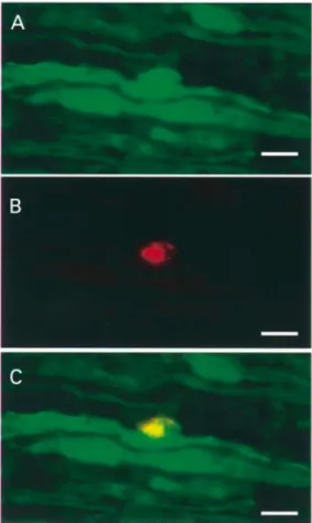
(3) Schwann cells in Charcot–Marie–Tooth mouse models. 2179. d-UTP-digoxigenin nick end-labelling (TUNEL) assay PMP22 mutant mice (PMP22 knock-out, PMP22 transgenic and Tr mice) and wild-types of the corresponding genetic background at postnatal days 1, 4, 10 and 21, and 10 weeks of age were sacrificed with a lethal dose of sodium pentobarbital (Nembutal®, Abbott, Ill., USA) and their sciatic nerves were removed. Nerves were fixed in 4% paraformaldehyde in PBS (phosphate-buffered saline) for 4 h at 4°C, dehydrated and embedded in paraffin. Longitudinal sections, 5 µm thick, were mounted on slides, dewaxed and rehydrated. Initially, slides were incubated in methanol containing 0.3% hydrogen peroxide to block endogenous peroxidase, followed by a 20 min digestion with proteinase K (20 µg/ml) at 37°C. Sections were pre-equilibrated in TdT buffer (30 mM Tris–HCl pH 7.2, 140 mM sodium cacodylate, 1 mM cobalt chloride) prior to the addition of the TUNEL reaction mix (0.2 U/µl TdT enzyme, 6 nM biotin-16-dUTP, 6 nM dATP in TdT buffer). The enzymatic reaction was carried out at 37°C for 1.5 h and was terminated by incubating in 2⫻ SSC (standard saline citrate) for 15 min at room temperature. After incubation of the sections in ABComplex (avidin–biotin complex) tagged to horseradish peroxidase (HRP) (Dako, Denmark) for 1 h at 37°C, incorporation of the labelled deoxynucleotide was visualized by applying the diaminobenzidine colour substrate solution for 12 min at room temperature in the dark. Nuclei were counterstained with methyl green (Elias, 1969). Positive and negative controls were run in parallel. Positive controls for the TUNEL reaction were paraffin sections of a E13.5 mouse embryo and sections of mouse small intestine previously digested with DNase I. As negative control, a section of an E13.5 mouse embryo was incubated in TUNEL reaction mix without adding the TdT enzyme. To quantify apoptosis, sciatic nerve longitudinal sections were visualized in a Zeiss Axiophot with the ⫻100 lens. A grid with 100 divisions mounted in one of the microscope objectives helped to define the field of analysis. The nuclei counting proceeded from the proximal to the distal end of the sciatic nerve sections until 1000 or 500 nuclei, depending on the age of the animals, were counted in consecutive microscopic fields. All TUNEL-labelled cells from the total number of counted nuclei were recorded at the same time. The cell death index was calculated as the percentage of nuclei that were TUNEL positive. At least four animals per genotype and age group were analysed. Statistical analysis was performed with the Mann–Whitney U-test (two-side test paradigm) using Statview, version 4.0 software. P values 艋 0.05 were considered to be statistically significant.. BrdU incorporation assay Mice of the same genotype and age as for the TUNEL assay were injected intraperitoneally with bromodeoxyuridine (BrdU; 100 µg/g of body weight) in 0.9% NaCl/7 mM NaOH.. Fig. 1 A proliferating Schwann cell on a longitudinal section of a sciatic nerve from a PMP22 transgenic mouse at P21. The immunoreactivity of S100 labels Schwann cells in green (A). BrdU labelling is shown in red (B), corresponding to a Schwann cell nucleus on the overlay (C, yellow). Scale bar ⫽ 10 µm.. One hour after BrdU injection, animals were sacrificed and tissues of interest were removed. Tissue-Tek-embedded sciatic nerves were frozen in liquid nitrogen-cooled isopentene and sectioned with a cryostat. Six consecutive longitudinal sections, 5 µm thick, were mounted alternatively onto two Superfrost slides, air dried and fixed for 5 min with 2% paraformaldehyde in PBS. Because our DNA denaturation protocol precluded the simultaneous identification of BrdUlabelled cells and staining of nuclei in the same section, one slide was stained with haematoxylin to count nuclei whereas the second slide was processed to reveal BrdU-positive cells. After blocking endogenous peroxidase activity, DNA denaturation was achieved by incubating the sections in 2 M HCl for 15 min at 37°C followed by neutralization in 0.1 M sodium borate pH 8.5 for 10 min. Nuclei that incorporated BrdU were labelled with a biotinylated anti-BrdU monoclonal antibody (Caltag Laboratories, Calif., USA) diluted 1 : 20 and detected by the ABComplex–horseradish peroxidase method as described above. Sections of small intestine from the same animals were used as positive controls. BrdU-labelled cells were counted in three alternated consecutive longitudinal sections. Starting from the proximal.
(4) 2180. S. Sancho et al..
(5) Schwann cells in Charcot–Marie–Tooth mouse models end of the sciatic nerve, 4–6 consecutive microscopic fields, as defined by the grid mounted in the microscope objective, were examined per section with a ⫻100 lens. All BrdUlabelled cells present within the defined fields were counted. An identical quantification method was used to assess the number of haematoxylin-stained nuclei in the other three alternated sections. The proliferation index was calculated as the percentage of BrdU-labelled cells in relation to the total number of haematoxylin-stained nuclei present in the same area. Cell density was defined as the total number of haematoxylin-stained nuclei per mm2. Usually five animals per genotype and age group were analysed. Statistical analysis was performed with the Mann–Whitney U-test (two-side test paradigm) using Statview, version 4.0 software. P values 艋 0.05 were considered to be statistically significant.. Immunohistochemistry To demonstrate that the BrdU- and TUNEL-positive cells were Schwann cells, selected nerves were immunostained with a polyclonal rabbit antiserum against S100 protein (Dako, Denmark) combined with either BrdU or TUNEL labelling. For these experiments, sciatic nerve sections were embedded in Tissue-Tek and 5 µm thick cryosections obtained. Thereafter, identical protocols to those described above were used to process sections for either BrdU or TUNEL labelling, except that Texas red-tagged streptavidin (Jackson ImmunoResearch Laboratories, Pa., USA) was used as detection system. S100-labelled cells were identified with a fluorescein-conjugated goat anti-rabbit secondary antibody (Jackson ImmunoResearch Laboratories).. Results Earlier analysis of PMP22 mouse mutants indicated that PMP22 plays a key role in the initial steps of myelination and in the maintenance of the myelin sheath (reviewed by Carenini et al., 1999; D’Urso et al., 1999; Naef and Suter, 1998). Since demyelination induces proliferation of Schwann cells in various mouse mutants, regardless of the underlying molecular defect (Giese et al., 1992; Anzini et al., 1997; Neuberg et al., 1999), we decided to start our analysis at postnatal day (P) 1, prior to the onset of appreciable myelination. Since PMP22 is expressed by peripheral glia, motor neurones and sensory neurones before birth (Baechner et al., 1995; Parmantier et al., 1995, 1997; Hagedorn et al., 1999), this strategy should allow the detection of alterations in. 2181. Schwann cell proliferation and cell death in early development without the interference of demyelination. We analysed three groups of PMP22 mutants to assess the influence of PMP22 gene dosage (PMP22 transgenic mice and PMP22 knock-out mice) and the effect of altered PMP22 protein (Tr mutant) on the proliferation and apoptosis of Schwann cells in comparison with wild-type controls. To follow the development and to evaluate the probable onset of alteration of proliferation and/or apoptosis, we examined sciatic nerves of P1, P4, P10 and P21 animals. In addition, 10-week-old young adult mice with completed peripheral myelination were also examined.. Schwann cell proliferation S100 was used as a Schwann cell marker in combination with BrdU incorporation (for example, see Fig. 1A–C). BrdU-positive cells without S100 immunoreactivity were not observed (data not shown). First, we compared the cell density in sciatic nerves of wild-type and mutants at P1. No significant differences between mutant mice and their corresponding control animals were observed within the three paired groups that were analysed (Fig. 2A, C and E). In all three wild-type control groups, cell density in the sciatic nerve decreased from P1 until the age of 10 weeks, to approximately one-third of the initial value (Fig. 2A, C and E). In the mutant animals, we observed a significant increase in cell density in Tr animals from P21 onwards (Fig. 2A) and in the transgenic PMP22 animals as early as P4 (Fig. 2E). Next, we determined the proliferation rate by calculating the number of cells with BrdU incorporation in relation to the total cell number (BrdU index). In wild-type mice, this index ranged from 15.8 ⫾ 2.5% to 18.9 ⫾ 1.5% at P1 and was reduced to undetectable levels in 10-week-old animals. In the Tr mutants, we found a strong increase of Schwann cell proliferation at P10 and in 10-week-old animals, while proliferation of Schwann cells at P21 did not reach significance in comparison with the age-matched wild-type controls with comparable genetic background (Fig. 2B). These findings are consistent with pathology data indicating that significant myelination occurs initially in these mice (Ayers and Anderson, 1976) followed by demyelination and, as shown here and suggested earlier (Perkins et al., 1981), accompanied by Schwann cell proliferation leading to cellular onion bulb structures. In PMP22 knock-out animals, a significant increase in the rate of proliferation was found. Fig. 2 Qualitative and quantitative analysis of cell densitiy and proliferation in normal and PMP22 mutant sciatic nerves. A, C and E refer to the cell density per mm2; B, D and F show the BrdU labelling index of proliferating cells in three groups of PMP22 mutant animals and their corresponding wild-type controls (wt). Asterisks indicate statistical significance (P ⬍ 0.025). Arrows show BrdUpositive nuclei. Scale bar ⫽ 20 µm. Genotypes are given in the upper right corner of each section: Tr ⫽ Trembler; ko ⫽ PMP22 knockout; Tg ⫽ PMP22 transgenic..
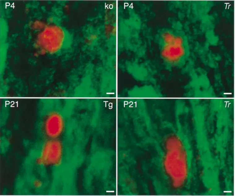
(6) 2182. S. Sancho et al.. Fig. 3 Confocal microscopy of TUNEL immunoreactivity on longitudinal sections of sciatic nerves from PMP22 mutant sciatic nerves. Green fluorescence (FITC) indicates S100-positive cells and red fluorescence marks TUNEL-positive nuclei. Note the pronounced nuclear fragmentation. Scale bar ⫽ 2 µm. Age is indicated in the upper left corner, while genotypes are given in the upper right corner of each section. Tr ⫽ Trembler; Tg ⫽ transgenic; KO ⫽ PMP22 knock-out.. only in 10-week-old animals (Fig. 2D). This result is consistent with the described pathology in these mice which are affected by focal hypermyelination (tomacula formation) early in life, followed by demyelination including onion bulb cell formation later (Adlkofer et al., 1995; Sancho et al., 1999). PMP22 transgenic mice showed a constant, significantly elevated proliferation index from P4 onwards in relation to the wild-type control group, consistent with the observed almost complete amyelination of the peripheral nerves of these animals throughout development (Fig. 2F; Magyar et al., 1996; Sancho et al., 1999). In contrast to wild-type animals, proliferating cells were detectable in the sciatic nerves of all three mutants at 10 weeks of age, but the rate was reduced to 1–2%.. Cell death of Schwann cells Cell death was analysed by TUNEL staining in combination with S100 immunohistochemistry. Examples of dying Schwann cells in the sciatic nerves of various PMP22 mutant mice, also characterized by fragmented nuclei, are shown in Fig. 3. Furthermore, a representative overview of apoptosis in a sciatic nerve of a Tr mutant at 10 weeks of age and the corresponding wild-type animal is shown (Fig. 4). In wildtype mice, considerable programmed cell death was observed from P1 onwards, peaking between P4 and P10, with very low levels at P21, and no detectable dying cells at the age of 10 weeks (Fig. 5). These findings are consistent with earlier suggestions that axon–Schwann cell interactions regulate the correct Schwann cell number during early postnatal.
(7) Schwann cells in Charcot–Marie–Tooth mouse models. 2183. Fig. 4 Longitudinal sections of sciatic nerves showing TUNEL-positive nuclei in a 10-week-old Trembler (Tr) mouse and an agematched control animal (wt). Arrows indicate TUNEL-positive, partially fragmented nuclei. Scale bar ⫽ 20 µm.. development through apoptosis (Grinspan et al., 1996; Syroid et al., 1999). The rate of apoptosis was similar in the three mutant groups and in the corresponding wild-type animals at P1 (Fig. 5A–C). In Tr animals, significantly increased cell death was observed starting at P21 and continuing at 10 weeks of age (Figs 4A and 5A). The results in PMP22 knock-out animals were qualitatively similar to Tr (Fig. 5B). In PMP22 transgenic mice, the rate of apoptosis was already doubled compared with wild-type at P4, and this strongly increased apoptosis continued up to adulthood (Fig. 5C). Earlier studies have indicated that the low affinity neurotrophin receptor p75 is involved in the induction of apoptosis in transected nerves (Syroid et al., 2000). Interestingly, p75 is also upregulated in peripheral nerves of PMP22 transgenic mice (Magyar et al., 1996). However, a potential causative function for p75 in the increased Schwann cell apoptosis of these animals remains to be established. In summary, all three groups of PMP22 mutant animals showed a significant increase of apoptotic Schwann cells at P21. At the age of 10 weeks, none of the corresponding wild-type groups showed any apoptotic Schwann cells, while apoptosis was still prominent in the mutant mice.. Discussion In this study, we have examined Schwann cell proliferation and apoptosis in three different mouse strains with altered expression of the PMP22 gene. The spontaneous mouse mutant Tr carries a point mutation in the PMP22 gene which leads to intracellular retention of the mutated protein in vitro and in vivo (Naef et al., 1997; Colby et al., 2000). Since PMP22 appears to form dimers, it is conceivable that wildtype PMP22 is also retained, at least partially, within the cell, suggesting a dominant–negative disease mechanism (Naef et al., 1997; Naef and Suter, 1999; Tobler et al., 1999;. Brancolini et al., 2000). However, a contribution by a gainof-function effect, possibly due to overloading of intracellular compartments, has also been demonstrated by genetic analysis (Adlkofer et al., 1997b). Similar to observations in animal models of Pelizaeus–Merzbacher disease where disrupted proteolipid protein trafficking appears to result in oligodendrocyte apoptosis (Gow et al., 1998), we show that a substantial increase in Schwann cell apoptosis is observed in Tr at P21 and at the age of 10 weeks. In support of a causative action of the Tr protein in the induction of apoptosis, we have also observed strongly increased programmed cell death in adult homozygous Tr mice compared with the heterozygous animals used in our study (preliminary observation). In addition, increased levels of apoptosis have also been described in the Tr-Ncnp mouse which carries an in-frame deletion in the PMP22 gene (Suh et al., 1997). The most straightforward interpretation of these data is that excessive intracellular accumulation of mutated PMP22 cannot be tolerated by Schwann cells in vivo and, consequently, the cells die. A similar mechanism may also explain the increased apoptosis in PMP22 transgenic mice, consistent with the abundant empty basal lamina present in the nerves of these animals (Magyar et al., 1996). The substantially earlier onset of apoptosis in PMP22 transgenic mice compared with Tr could be due to the high PMP22 gene dosage in these animals which may lead to fast intracellular accumulation of large amounts of misfolded PMP22 and immediate activation of the apoptosis pathway (Pareek et al., 1993). Alternative interpretations, however, are also possible. In the process of this study, we have confirmed that Schwann cell apoptosis occurs naturally in developing nerves of wild-type mice during the first two postnatal weeks (Grinspan et al., 1996; Syroid et al., 2000). It has been suggested that axon–Schwann cell interactions in this way regulate the correct Schwann cell numbers by competition for axonally derived neuregulins (Grinspan et al.,.
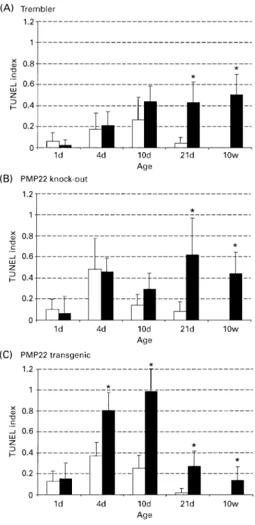
(8) 2184. S. Sancho et al.. Fig. 5 Quantification of apoptosis in the sciatic nerves of PMP22 mutant mice (black bars) and the corresponding wild-type control groups (grey bars). Asterisks indicate statistical significance (P ⬍ 0.025).. 1996; Syroid et al., 1996). Sciatic nerves of PMP22 transgenic animals contain virtually no myelin and generate an excess of Schwann cells starting already at P4, presumably since they are exposed continuously to mitogenic axolemma proteins. Thus, the early phase of increased Schwann cell apoptosis in PMP22 transgenic mice from P4 to P10, which is not present in Tr, may also involve processes regulating correct Schwann cell numbers in early development, including. the availability of and spatial exposure to limited amounts of survival factors (reviewed by Garratt et al., 2000). Importantly, there is a window of susceptibility of Schwann cells to apoptosis during development since programmed cell death could be enhanced by axotomy only during early development (Grinspan et al., 1996). Thus, the Schwann cell death observed from P21 onwards in PMP22 mutants is likely to be due to other mechanisms. Toxification through intracellular accumulation may contribute to the late phase of apoptosis in Tr and PMP22 transgenic mice, but the increased levels of Schwann cell apoptosis seen in PMP22 knock-out mice require other explanations. Based on the fact that this increase is correlated with morphological signs of abundant deymelination, remyelination and onion bulb formation (Adlkofer et al., 1995; Sancho et al., 1999), it is likely that dying supernumerary Schwann cells are responsible for our findings. The same mechanisms may also contribute to the apoptosis observed in Tr late in development since onion bulb formation associated with increased cell density is found within peripheral nerves (Ayers and Anderson, 1976; Perkins et al., 1981). Experiments in cell culture have suggested that PMP22 is directly involved in the regulation of the cell cycle (Zoidl et al., 1995, 1997) and the induction of cell death (Fabbretti et al., 1995; Brancolini et al., 1999). To address this issue, we have analysed the PMP22 mutants at P1. We reasoned that the function of PMP22 in myelination and myelin maintenance is not a substantially interfering factor at this time point since the sciatic nerve is still largely unmyelinated. Our analysis revealed no significant alterations in Schwann cell density, proliferation index or the rate of apoptosis in any PMP22 mutant compared with wild-type mice at this early time point. Thus, normal PMP22 expression is dispensable for correct Schwann cell proliferation and cell death up to P1. An influence of PMP22 in these processes later in development, however, cannot be excluded. This is difficult to assess due to the underlying myelin breakdown (demyelination) or amyelination pathologies which alter axon–Schwann cell interactions and, consequently, proliferation and apoptosis in ways that are not fully understood. How do our data compare with the human disease? In nerve biopsies of young CMT1A patients carrying the PMP22 duplication on chromosome 17p.12, Schwann cells show no signs of increased proliferation (Hanemann et al., 1997). Consistent with our observations in PMP22 transgenic mice, the authors concluded that there is no evidence for altered initial Schwann cell proliferation prior to the process of deand remyelination. Intriguingly, however, human Schwann cells obtained from CMT1A patients carrying the chromosomal duplication show a decrease in proliferation in vitro (Hanemann et al., 1998). This difference between in vitro and in vivo studies may reflect age-related changes and/or the course of disease. Alternatively, culturing CMT1A Schwann cells may unmask a difference in their potential for decreased proliferation that cannot be observed in vivo,.
(9) Schwann cells in Charcot–Marie–Tooth mouse models a common emerging theme in developmental biology (Anderson, 2001). Evidence for apoptosis of Schwann cells in CMT1A and HNPP nerve biopsies from patients with proven PMP22 duplications and deletions has been provided by Erdem and colleagues using the TUNEL method (Erdem et al., 1998). Quantitative studies showed a significantly reduced number of total Schwann cells in CMT1A compared with controls indicating a loss of Schwann cells. In HNPP, the number of total Schwann cells was increased and significant Schwann cell apoptosis was observed. The authors suggested on the basis of further morphological analysis that this Schwann cell apoptosis might be related to the regenerative state of the nerve resulting from the process of sprout pruning (Erdem et al., 1998). In summary, we show that altered expression of PMP22 does not affect the generation of the correct number of early Schwann cells. Furthermore, the proliferation index is not changed in PMP22 mutants in early postnatal development. However, increased proliferation and apoptosis are prominent features in all mutants at later developmental stages. Based on the differences in the kinetics of these alterations during the development of the different mutants, it is likely that overloading of intracellular compartments and altered access to trophic factors due to myelination deficiencies are responsible for the increased cell density, proliferation and apoptosis observed in PMP22 mutant peripheral nerve.. Acknowledgements We wish to thank Annette Kolar for excellent technical assistance. This work was supported by the Swiss National Science Foundation, the Swiss National Research Program for Neurodegenerative Disorders (NFP38), the Swiss Muscle Disease Foundation, the Wolfermann–Na¨ geli Stiftung and the Swiss Bundesamt for Science related to the Commission of the European Communities, specific RTD programme ‘Quality of life and Management of Living Resources’, QLK6-CT-2000-00179, ‘The role of neurosteroids in healthy aging: therapeutical perspectives’. It does not necessarily reflect its views and in no way anticipates the Commission’s future policy in this area. P.Y. is a recipient of a grant from the Deutsche Forschungsgemeinschaft (YO48/1-1) and is on sabbatical from the Department of Neurology, University of Mu¨ nster, Germany.. 2185. affected by a progressive demyelinating tomaculous neuropathy. J Neurosci 1997a; 17: 4662–71. Adlkofer K, Naef R, Suter U. Analysis of compound heterozygous mice reveals that the Trembler mutation can behave as a gain-offunction allele. J Neurosci Res 1997b; 49: 671–80. Anderson DJ. Stem cells and pattern formation in the nervous system: the possible versus the actual. Neuron 2001; 30: 19–35. Anzini P, Neuberg DH, Schachner M, Nelles E, Willecke K, Zielasek J, et al. Structural abnormalities and deficient maintenance of peripheral nerve myelin in mice lacking the gap junction protein connexin 32. J Neurosci 1997; 17: 4545–51. Arroyo EJ, Scherer SS. On the molecular architecture of myelinated fibers. [Review]. Histochem Cell Biol 2000; 113: 1–18. Ayers MM, Anderson RM. Development of onion bulb neuropathy in the Trembler mouse. Morphometric study. Acta Neuropathol (Berl) 1976; 36: 137–52. Baechner D, Liehr T, Hameister H, Altenberger H, Grehl H, Suter U, et al. Widespread expression of the peripheral myelin protein-22 gene (PMP22) in neural and non-neural tissues during murine development. J Neurosci Res 1995; 42: 733–41. Brancolini C, Marzinotto S, Edomi P, Agostoni E, Fiorentini C, Muller HW, et al. Rho-dependent regulation of cell spreading by the tetraspan membrane protein Gas3/PMP22. Mol Biol Cell 1999; 10: 2441–59. Brancolini C, Edomi P, Marzinotto S, Schneider C. Exposure at the cell surface is required for gas3/PMP22 to regulate both cell death and cell spreading: implication for the Charcot–Marie–Tooth type 1A and Dejerine–Sottas diseases. Mol Biol Cell 2000; 11: 2901–14. Carenini S, Neuberg D, Schachner M, Suter U, Martini R. Localization and functional roles of PMP22 in peripheral nerves of P0-deficient mice. Glia 1999; 28: 256–64. Chance PF, Alderson MK, Leppig KA, Lensch MW, Matsunami N, Smith B, et al. DNA deletion associated with hereditary neuropathy with liability to pressure palsies. Cell 1993; 72: 143–51. Colby J, Nicholson R, Dickson KM, Orfali W, Naef R, Suter U, et al. PMP22 carrying the trembler or trembler-j mutation is intracellularly retained in myelinating Schwann cells. Neurobiol Dis 2000; 7: 561–73. D’Urso D, Ehrhardt P, Muller HW. Peripheral myelin protein 22 and protein zero: a novel association in peripheral nervous system myelin. J Neurosci 1999; 19: 3396–403. Dyck PJ, Chance P, Lebo R, Carney JA. Hereditary motor and sensory neuropathies. In: Dyck PJ, Thomas PK, Griffin JW, Low PA, Poduslo JF, editors. Peripheral neuropathy. 3rd edn. Philadelphia: W. B. Saunders; 1993. p. 1094–136.. References Adlkofer K, Lai C. Role of neuregulins in glial cell development. [Review]. Glia 2000; 29: 104–11.. Elias JM. Effects of temperature, poststaining rinses and ethanol– butanol dehydrating mixtures on methyl green–pyronin staining. Stain Technol 1969; 44: 201–4.. Adlkofer K, Martini R, Aguzzi A, Zielasek J, Toyka KV, Suter U. Hypermyelination and demyelinating peripheral neuropathy in Pmp22-deficient mice. Nat Genet 1995; 11: 274–80.. Erdem S, Mendell JR, Sahenk Z. Fate of Schwann cells in CMT1A and HNPP: evidence for apoptosis. J Neuropathol Exp Neurol 1998; 57: 635–42.. Adlkofer K, Frei R, Neuberg DH, Zielasek J, Toyka KV, Suter U. Heterozygous peripheral myelin protein 22-deficient mice are. Fabbretti E, Edomi P, Brancolini C, Schneider C. Apoptotic phenotype induced by overexpression of wild-type gas3/PMP22: its.
(10) 2186. S. Sancho et al.. relation to the demyelinating peripheral neuropathy CMT1A. Genes Dev 1995; 9: 1846–56. Garratt AN, Britsch S, Birchmeier C. Neuregulin, a factor with many functions in the life of a schwann cell. [Review]. Bioessays 2000; 22: 987–96. Giese KP, Martini R, Lemke G, Soriano P, Schachner M. Mouse P0 gene disruption leads to hypomyelination, abnormal expression of recognition molecules, and degeneration of myelin and axons. Cell 1992; 71: 565–76. Gow A, Southwood CM, Lazzarini RA. Disrupted proteolipid protein trafficking results in oligodendrocyte apoptosis in an animal model of Pelizaeus–Merzbacher disease. J Cell Biol 1998; 140: 925–34. Grinspan JB, Marchionni MA, Reeves M, Coulaloglou M, Scherer SS. Axonal interactions regulate Schwann cell apoptosis in developing peripheral nerve: neuregulin receptors and the role of neuregulins. J Neurosci 1996; 16: 6107–18. Hagedorn L, Suter U, Sommer L. P0 and PMP22 mark a multipotent neural crest-derived cell type that displays community effects in response to TGF-beta family factors. Development 1999; 126: 3781–94. Hanemann, CO, Gabreels-Festen AA, Stoll G, Muller HW. Schwann cell differentiation in Charcot–Marie–Tooth disease type 1A (CMT1A): normal number of myelinating Schwann cells in young CMT1A patients and neural cell adhesion molecule expression in onion bulbs. Acta Neuropathol (Berl) 1997; 94: 310–15. Hanemann CO, Rosenbaum C, Kupfer S, Wosch S, Stoegbauer F, Muller HW. Improved culture methods to expand Schwann cells with altered growth behaviour from CMT1A patients. Glia 1998; 23: 89–98. Huxley C, Passage E, Manson A, Putzu G, Figarella-Branger D, Pellissier JF, et al. Construction of a mouse model of Charcot– Marie–Tooth disease type 1A by pronuclear injection of human YAC DNA. Hum Mol Genet 1996; 5: 563–9. Ionasescu VV, Searby CC, Ionasescu R, Reisin R, Ruggieri V, Arberas C. Severe Charcot–Marie–Tooth neuropathy type 1A with 1-base pair deletion and frameshift mutation in the peripheral myelin protein 22 gene. Muscle Nerve 1997; 20: 1308–10. Jessen KR, Mirsky R. Schwann cells and their precursors emerge as major regulators of nerve development. [Review]. Trends Neurosci 1999; 22: 402–10. Krajewski KM, Lewis RA, Fuerst DR, Turansky C, Hinderer SR, Garbern J, et al. Neurological dysfunction and axonal degeneration in Charcot–Marie–Tooth disease type 1A. Brain 2000; 123: 1516–27. Lupski JR, Garcia CA. Molecular genetics and neuropathology of Charcot–Marie–Tooth disease type 1A. [Review]. Brain Pathol 1992; 2: 337–49. Magyar JP, Martini R, Ruelicke T, Aguzzi A, Adlkofer K, Dembic Z, et al. Impaired differentiation of Schwann cells in transgenic mice with increased PMP22 gene dosage. J Neurosci 1996; 16: 5351–60. Muller HW. Tetraspan myelin protein PMP22 and demyelinating peripheral neuropathies: new facts and hypotheses. [Review]. Glia 2000; 29: 182–5.. Naef R, Suter U. Many facets of the peripheral myelin protein PMP22 in myelination and disease. [Review]. Microsc Res Tech 1998; 41: 359–71. Naef R, Suter U. Impaired intracellular trafficking is a common disease mechanism of PMP22 point mutations in peripheral neuropathies. Neurobiol Dis 1999; 6: 1–14. Naef R, Adlkofer K, Lescher B, Suter U. Aberrant protein trafficking in Trembler suggests a disease mechanism for hereditary human peripheral neuropathies. Mol Cell Neurosci 1997; 9: 13–25. Neuberg DH, Sancho S, Suter U. Altered molecular architecture of peripheral nerves in mice lacking the peripheral myelin protein 22 or connexin32. J Neurosci Res 1999; 58: 612–23. Nicholson GA, Valentijn LJ, Cherryson AK, Kennerson ML, Bragg TL, DeKroon RM, et al. A frame shift mutation in the PMP22 gene in hereditary neuropathy with liability to pressure palsies. Nat Genet 1994; 6: 263–6. Niemann S, Sereda MW, Suter U, Griffiths IR, Nave KA. Uncoupling of myelin assembly and schwann cell differentiation by transgenic overexpression of peripheral myelin protein 22. J Neurosci 2000; 20: 4120–8. Pareek S, Suter U, Snipes GJ, Welcher AA, Shooter EM, Murphy RA. Detection and processing of peripheral myelin protein PMP22 in cultured Schwann cells. J Biol Chem 1993; 268: 10372–9. Parmantier E, Cabon F, Braun C, D’Urso D, Muller HW, Zalc B. Peripheral myelin protein-22 is expressed in rat and mouse brain and spinal cord motoneurons. Eur J Neurosci 1995; 7: 1080–8. Parmantier E, Braun C, Thomas JL, Peyron F, Martinez S, Zalc B. PMP-22 expression in the central nervous system of the embryonic mouse defines potential transverse segments and longitudinal columns. J Comp Neurol 1997; 378: 159–72. Patel PI, Roa BB, Welcher AA, Schoener-Scott R, Trask BJ, Pentao L, et al. The gene for the peripheral myelin protein PMP22 is a candidate for Charcot–Marie–Tooth disease type 1A. Nat Genet 1992; 1: 159–65. Perkins CS, Aguayo AJ, Bray GM. Schwann cell multiplication in Trembler mice. Neuropathol Appl Neurobiol 1981; 7: 115–26. Raeymaekers P, Timmerman V, Nelis E, De Jonghe P, Hoogendijk JE, Baas F, et al. Duplication in chromosome 17p11.2 in Charcot– Marie–Tooth neuropathy type 1a (CMT 1a). The HMSN Collaborative Research Group. Neuromuscul Disord 1991; 1: 93–7. Roa BB, Garcia CA, Suter U, Kulpa DA, Wise CA, Mueller J, et al. Charcot–Marie–Tooth disease type 1A. Association with a spontaneous point mutation in the PMP22 gene. N Engl J Med 1993; 329: 96–101. Sancho S, Magyar JP, Aguzzi A, Suter U. Distal axonopathy in peripheral nerves of PMP22-mutant mice. Brain 1999; 122: 1563–77. Sereda M, Griffiths I, Puhlhofer A, Stewart H, Rossner MJ, Zimmerman F, et al. A transgenic rat model of Charcot–Marie– Tooth disease. Neuron 1996; 16: 1049–60. Skre H. Genetic and clinical aspects of Charcot–Marie–Tooth’s disease. Clin Genet 1974; 6: 98–118. Suh JG, Ichihara N, Saigoh K, Nakabayashi O, Yamanishi T, Tanaka K, et al. An in-frame deletion in peripheral myelin protein22 gene causes hypomyelination and cell death of the Schwann.
(11) Schwann cells in Charcot–Marie–Tooth mouse models cells in the new Trembler mutant mice. Neuroscience 1997; 79: 735–44. Suter U, Moskow JJ, Welcher AA, Snipes GJ, Kosaras B, Sidman RL, et al. A leucine-to-proline mutation in the putative first transmembrane domain of the 22-kDa peripheral myelin protein in the trembler-J mouse. Proc Natl Acad Sci USA 1992a; 89: 4382–6. Suter U, Welcher AA, Ozcelik T, Snipes GJ, Kosaras B, Francke U, et al. Trembler mouse carries a point mutation in a myelin gene. Nature 1992b; 356: 241–4. Suter U, Welcher AA, Snipes GJ. Progress in the molecular understanding of hereditary peripheral neuropathies reveals new insights into the biology of the peripheral nervous system. [Review]. Trends Neurosci 1993; 16: 50–6. Syroid DE, Maycox PR, Burrola PG, Liu N, Wen D, Lee KF, et al. Cell death in the Schwann cell lineage and its regulation by neuregulin. Proc Natl Acad Sci USA 1996; 93: 9229–34. Syroid DE, Zorick TS, Arbet-Engels C, Kilpatrick TJ, Eckhart W, Lemke G. A role for insulin-like growth factor-I in the regulation of Schwann cell survival. J Neurosci 1999; 19: 2059–68. Syroid DE, Maycox PJ, Soilu-Hanninen M, Petratos S, Bucci T, Burrola P, et al. Induction of postnatal schwann cell death by the low-affinity neurotrophin receptor in vitro and after axotomy. J Neurosci 2000; 20: 5741–7. Tobler AR, Notterpek L, Naef R, Taylor V, Suter U, Shooter EM. Transport of Trembler-J mutant peripheral myelin protein 22 is. 2187. blocked in the intermediate compartment and affects the transport of the wild-type protein by direct interaction. J Neurosci 1999; 19: 2027–36. Valentijn LJ, Baas F, Wolterman RA, Hoogendijk JE, van den Bosch NH, Zorn I, et al. Identical point mutations of PMP-22 in TremblerJ mouse and Charcot–Marie–Tooth disease type 1A. Nat Genet 1992; 2: 288–91. Windebank AJ. Inherited recurrent focal neuropathies. In: Dyck PJ, Thomas PK, Griffin JW, Low PA, Poduslo JF, editors. Peripheral neuropathy. 3rd edn. Philadelphia: W. B. Saunders, 1993. p. 1137–48. Young P, Wiebusch H, Stogbauer F, Ringelstein B, Assmann G, Funke H. A novel frameshift mutation in PMP22 accounts for hereditary neuropathy with liability to pressure palsies. Neurology 1997; 48: 450–2. Zoidl G, Blass-Kampmann S, D’Urso D, Schmalenbach C, Muller HW. Retroviral-mediated gene transfer of the peripheral myelin protein PMP22 in Schwann cells: modulation of cell growth. EMBO J 1995; 14: 1122–8. Zoidl G, D’Urso D, Blass-Kampmann S, Schmalenbach C, Kuhn R, Muller HW. Influence of elevated expression of rat wild-type PMP22 and its mutant PMP22 Trembler on cell growth of NIH3T3 fibroblasts. Cell Tissue Res 1997; 287: 459–70.. Received March 2, 2001. Revised April 10, 2001. Accepted July 2, 2001..
(12)
Figure

Documents relatifs
Elle permet d’abord d’interroger la pertinence et les limites de la notion de « patrimoine-ressource pour le développement », ainsi que les relations complexes entre
DocToBib: PubMed, the physician and the librarian...or the fantastic story of doctors and librarians producing videos together.. Journal of the European Association for
Le document 3 montre les résultats de dosages sanguins de deux hormones sexuelles Ⓐ et Ⓑ chez une femme au cours de deux cycles sexuels successifs. déduisez la nature des
Released zoospores were counted and the relative release rate was calculated by dividing values obtained for treatments by those obtained for the untreated controls (see
Le guide préparé pour l’entrevue de groupe comprend 10 questions ayant pour thèmes les stratégies d’empowerment développées par les infirmières, le sens du travail,
• aigu, ambigu, exigu, contigu font leur féminin en aiguë [aigüe], ambiguë [ambigüe], exiguë [exigüe], contiguë [contigüe] : une réponse ambiguë [ambigüe]. • andalou
We aim at (1) clar- ifying the internal structure of the tidal response by identifying dominating modes, their characteristics and their behaviour; (2) quantifying the order
hippocampus of rescue mice modified to express the gene exclusively in specific brain cell types : such as dorsal telencephalic glutamatergic neurons, or GABAergic neurons have
