Journal of Helminthology (1996) 70, 219-222 219
An improved test system for PCR-based
specific detection of Echinococcus
multilocularis eggs
A. Mathis, P. Deplazes and J. Eckert
Institute of Parasitology, University of Zurich, Winterthurerstr 266a,
CH-8057 Zurich, Switzerland
Abstract
For the sensitive detection of eggs of Echinococcus multilocularis in fox faeces by PCR we have evaluated a method based on the previous concentration of helminth eggs by a combination of sequential sieving of faecal samples and flotation of the eggs in zinc chloride solution. The eggs were microscopically detected in the fractions retained in 40 and 20 urn mesh sieves. DNA of the taeniid eggs retained in the 20 |im sieve was obtained after alkaline lysis and PCR was performed using E. multilocularis species-specific primers. Compared to the parasitological findings after examination of the small intestines of the foxes, the specificity of the PCR was 100% (no false-positive result with 20 foxes free of
E. multilocularis) and the sensitivity was 94% (33 positive results from total 35
foxes proven to be infected with E. multilocularis). Both false-negative results were obtained with faeces from foxes harbouring immature worms. Using faecal volumes between 2 and 20 ml, no inhibition of PCR was observed as was demonstrated by the amplification of size-modified target in parallel reactions. The tests were undertaken with fresh faeces stored in 70% ethanol, but egg detection by PCR was also possible after inactivation of eggs by freezing the faeces at -80°C for one week or by incubation at +70°C for 2 h.
Introduction
Human alveolar echinococcosis, caused by metacestode stages of Echinococcus multilocularis, is a life-threatening disease. For estimating the potential risk of human infection in endemic areas, accurate methods are needed to diagnose E. multilocularis in foxes and other definitive hosts. This is most reliably done at necropsy by careful examination of the small intestine by parasitological techniques. However, this is a time-consuming procedure linked with an infection risk for the investigators, which has to be avoided by extensive safety precautions (Eckert & Deplazes, 1995). Therefore, there is an urgent need for alternative and simplified examination techniques.
Recently, two novel approaches for the diagnosis of intestinal E. multilocularis infections were published: A coproantigen ELISA designed for the detection of E.
granulosus in dogs was shown to be specific for the genus Echinococcus and allowed detection of E. multilocularis in
heavily infected foxes (Deplazes et ah, 1992). Detection of
coproantigens has become a general focus in the diagnosis of infections with intestinal cestodes (Craig, 1993; Deplazes & Eckert, 1996).
The second approach is the PCR-based detection of E.
multilocularis eggs in fox faeces. Bretagne et ah (1993)
described such an assay using PCR primers derived from the noncoding flanking sequence of the Ul sRNA gene which is repeated at least 50 times in the genome. The primers were reported to be specific for E. multilocularis as no product was amplified with DNA from other tapeworm species, including E. granulosus.
In our experience, the test system when strictly performed as described by Bretagne et al. (1993) reached the reported sensitivity of detecting 1 egg in 4 g faeces, but many samples exhibited strong inhibitory effects on DNA amplification. Furthermore, several unspecific bands occurred as visualized on ethidium bromide-stained agarose gels (e.g. a product of almost the size of the diagnostic one repeatedly occurred when using faeces of dogs kept helminth-free) which both lower the sensitivity of the assay and make an additional hybridization step irrevocable.
https:/www.cambridge.org/core/terms. https://doi.org/10.1017/S0022149X00015443
220 A. Mathis et al. Therefore, we evaluated an alternative method for
sample preparation suitable for downstream PCR. The method is based on the combination of sieving and flotation to concentrate taeniid eggs and to remove both inhibitory substances and the majority of non-target organisms.
Material and methods
From the Zurich area, 55 foxes were collected after being shot by instructed hunters. Handling of the animals was performed following the safety precautions as described by Eckert et al. (1991). Faeces were taken from the rectum within 18 h after the foxes were shot and stored in 70% ethanol at 4°C. The small intestines, after being deep-frozen for at least 5 days at -80°C were incised longitudinally and examined macroscopically for helminths, then cut into 20 cm long pieces and placed in 1 litre bottles filled with physiological saline. The bottles were vigorously shaken for few seconds. After 15 min the supernatant as well as the intestines were removed and the sedimented fraction was transferred in small sub-samples to a square plastic petri dish (Falcon) and examined in transmission light under a stereomicroscope at 120 x magnification. Up to 100 Echinococcus specimens, the whole sediment was checked for worms; higher numbers were calculated from the count of one sub-sample.
Fox faeces (4-20 ml, suspended in 70% ethanol) stored up to 7 months as described above were passed through a 100 (im sieve to remove undigested particles, centrifuged (1000 xg, 5 min) and the pellets resuspended in zinc chloride solution (density 1.45 g/ml). After a centrifugation step (30 min, 1000 x g), the supernatant solution containing the taeniid eggs (diameter approximately 32 (im) was passed through sequential sieves (see below) with mesh sizes of 40 and 20 |xm, respectively. The sieves were washed thoroughly with water supplemented with 0.2% Tween 20 (Fluka, Buchs, Switzerland). The fractions retained in the last two sieves were each washed into a tube with a flattened side (Nunclon, Nunc, Denmark) allowing direct examination of helminth eggs with an inverted microscope. The pellet (about 100 (xl) obtained after centrifugation was used for DNA extraction. Appropriate safety precautions were taken according to Eckert et al. (1991). The sieves were in-house made with 50 ml falcon tubes (Becton-Dickinson, New Jersey) from which the bottom was cut off. The interior part from the lid was cut out leaving only the outer part with the winding. Nylon filters (Scrynel, Lanz-Anliker, Rohrbach, Switzerland) with the appropriate mesh size were pinched by screwing down the tube with the modified lid. The falcon tubes were reused after being submerged overnight in 2% sodium hypochlorite solution, washed thoroughly with distilled water and incubated at 80°C for 6 h.
DNA was obtained from the 20 |J.m sieve fraction by alkaline lysis and neutralization with Tris buffer and HC1 as described by Bretagne et al. (1993) but with only one tenth of the volume of all reagents. DNA was further purified with a silica matrix (Prep-a-Gene, BioRad) following the instructions of the manufacturer and thereby reducing the time of the procedure as compared to the protocol of Bretagne et al. (1993) using the same product. DNA was eluted in 100 ul 10 mM Tris-HCl, pH 8.3.
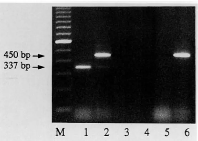
450
bp-337 bp.
M
1 2 3 4 5 6
Fig. 1. Detection of amplification products after gel electrophoresis and ethidium bromide staining. 337 bp fragment corresponds to Echinococcus multilocularis amplified DNA, 450 bp fragment corresponds to internal control amplified DNA. Inhibition of PCR is detected by abolished amplification of the control target in the parallel amplification reaction.M: 100 bp ladder; lane 1: sample no. 1 (positive); lane 2: sample no. 1 spiked with internal control; lane 3: sample no. 2 (inconclusive); lane 4: sample no. 2 spiked with internal control (inhibition); lane 5: no DNA template; lane
6: internal control alone.
PCR mixtures (100 ul) contained buffer (50 mM KC1, 20 mM Tris-HCl pH 8.4, 2.5 mM MgCl2, 0.5% Tween 20), 200 [Wi each deoxyribonucleotide, 1 |XM each primer, 2.5 U Taq polymerase (BRL, Basle, Switzerland) and 25 [i\ DNA solutions. PCR was performed in a Perkin Elmer thermal cycler. The 'hot start' technique was used and 35 cycles were performed of 30 sec at 94°C, 62°C, and 72CC. The primers used were those described by Bretagne et al. (1993) giving rise to a 337 bp fragment upon amplification. Amplicon production was determined by agarose gel electrophoresis (2%) with 20 p.1 of the reactions. In order to detect inhibition of the amplification reactions all specimens were tested in duplicate with the second reaction being spiked with 106 copies of the plasmid bearing the control target. This control target, constructed in analogy as described earlier (Mathis & Deplazes, 1995) yields an amplicon of about 450 bp upon amplification with the same primer pair and is easily discriminated from the E. multilocularis-specihc product of 337 bp (fig. 1). A negative control with no DNA added was included in all tests.
Cross-contamination problems were avoided by following good laboratory practice, including the use of aerosol-guarded tips and by performing the DNA extractions in a laminar flow hood which was subsequently irradiated by UV-light. The areas of DNA isolation, PCR and amplicon analysis were strictly separated.
Results and discussion
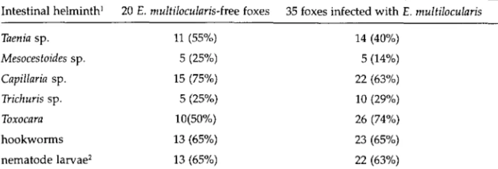
The spectrum of intestinal helminths of the foxes is summarized in table 1. Most foxes were infected with two to six helminth groups. Thirty five of 55 foxes (64%) were found to be infected with E. multilocularis with worm
https:/www.cambridge.org/core/terms. https://doi.org/10.1017/S0022149X00015443
PCR-based detection of E. multilocularis eggs 221
Table 1. Spectrum of intestinal helminths in the 55 foxes examined. Data from macroscopic/ microscopic determination of the parasites at necropsy and from the detection of eggs in faecal fractions retained in the 20 and 40 |im mesh sieves; prevalence data included in brackets. Intestinal helminth1 20 E. multilocularis-hee foxes 35 foxes infected with E. multilocularis
14 (40%) 5 (14%) 22 (63%) 10 (29%) 26 (74%) 23 (65%) 22 (63%) 'Parasite groups of single, double and multiple infections; 2not further differentiated.
Taenia sp. Mesocestoides sp. Capillaria sp. Trichuris sp. Toxocara hookworms nematode larvae2 11 (55%) 5 (25%) 15 (75%) 5 (25%) 10(50%) 13 (65%) 13 (65%)
Table 2. Detection of Echinococcus multilocularis infections in foxes by parasitological methods at necropsy and by PCR with faecal samples.
Necropsy
no E. multilocularis
specificity
with E. multilocularis infection (no. and non-gravid stages)
4-20 55-100 120-500 >500 sensitivity PCR positive / negative (no. of foxes) 0/20 100% of gravid 7/1 5/0 8/0 13/1 33/2 94%
burdens ranging from 4 to 60,000 per fox. The PCR results with faeces of foxes with and without natural infections of E. multilocularis are given in table 2. No PCR products were detected when using faeces from the 20 foxes that were found to be free of E. multilocularis at necropsy. The sensitivity with faeces from the 35 foxes infected with E.
multilocularis was 94%. Faeces from two foxes were scored
false-negative, and both foxes harboured immature worms of E. multilocularis (4 and 550 worms, respectively).
Faeces used in these tests was stored in 70% ethanol which is insufficient to kill the eggs and special safety precautions were needed during handling. Inactivation of the eggs can be achieved by freezing at -80°C for one week or by incubating at elevated temperatures (Veit et
al., 1995). We have tested eight aliquots of fox faeces from
E. multilocularis-iniected animals that were frozen for one week at -80°C. All proved positive in PCR. Furthermore, preliminary experiments showed that eggs isolated from adult E. multilocularis and kept at +70°C for 2 h did withstand the sieving procedure when added to two negative faeces and proved positive in the PCR. Hence, in
future investigations, the test can be performed with inactivated eggs which further simplifies the procedure.
PCR-based approaches for identification of £.
multilocularis eggs in faeces face a dilemma: E. multilocularis
eggs are excreted only intermittently or in low numbers (in cases of low worm burden) requiring as much faeces as possible for use in the detection of eggs. On the other hand, presence of PCR-inhibitory substances in faeces makes it desirable to use as little material as possible. The described assay, however, allows the concentration of taeniid eggs from large amounts of faecal samples (we used up to 20 ml) into a volume of less than 100 (il. None of the samples was inhibitory for the DNA amplification reaction and no unspecific bands were detected on the ethidium bromide-stained agarose gels thus making the reaction sensitive (as no competition for components of the amplification reaction occurs between such unspecific products and the diagnostic one) and rendering a time-consuming and laborious hybridization step with an internal probe superfluous.
Furthermore, a microscopical examination of the sieve fractions allows the detection of a wide spectrum of other intestinal parasites in the same sample. The sieving procedure replaces the washing steps needed in the 'total DNA extraction method' (Bretagne et al., 1993) and therefore no organosolvent extraction with hazardous chemicals is needed. However the whole PCR test system is still somewhat laborious. For mass screening of faeces of definitive hosts, coproantigen detection by ELISA (using anti-E. granulosiLS antibodies, Deplazesef al, 1992; or using anti-E. multilocularis antibodies, Deplazes et al., in preparation) is the method of choice. When screening animal populations with low prevalences by coproantigen detection (with a very high negative predictive value of the test), PCR is a valuable method for confirmation of a positive coproantigen result.
Our test should be applicable for processing different types and volumes of environmental samples allowing identification of recovered taeniid eggs (which are morphologically indistinguishable) by applying any specific PCR and possibly a hybridization assay (Bretagne
et al., 1993, for identification of E. multilocularis; Gottstein
& Mowatt, 1991, forEctonococcusspp.; Gottstein et al., 1991,
for Taenia saginata; Chapman et al, 1995, for T. saginata and
https:/www.cambridge.org/core/terms. https://doi.org/10.1017/S0022149X00015443
222 A. Mathis et al.
T. solhim). This will in t u r n c o n t r i b u t e to a better understanding of the transmission of taeniid eggs from definitive to intermediate hosts and humans.
Acknowledgement
The authors would like to thank Peter Alther for help in the collection of material.
References
Bretagne, S., Guillou, J.P., Morand, M. & Houin, R. (1993)
Detection of Echinococcus multilocularis DNA in fox faeces using DNA amplification. Parasitology 106, 193-199.
Chapman, A., Vallejo, V., Mossie, K.G., Ortiz, D., Agabian, N. & Flisser, A. (1995) Isolation and characterization of
species-specific DNA probes from Taenia solium and Taenia saginata and their use in an egg detection assay. Journal of Clinical Microbiology 33, 1283-1288.
Craig, P.S. (1993) Immunodiagnosis of Echinococcus granulosus.
pp. 85-118 in Anderson, F.L., Chai, J. & Liu, F. (Eds) Compendium on cystic echinococcosis. Provo (UT), Brigham Young University Print Service.
Deplazes, P. & Eckert, J. (1996) Diagnosis of the Echinococcus
multilocularis infection in final hosts. Applied Parasitology (in press).
Deplazes, P., Gottstein, B., Eckert, J., Jenkins, D.J., Ewald, D. & Jimenez-Palacios, S. (1992) Detection of Echinococcus
coproantigens by enzyme-linked immunosorbent assay in dogs, dingoes and foxes. Parasitology Research 78, SOS-SOS.
Eckert, J., Deplazes, P., Ewald, D. & Gottstein, B. (1991)
Parasitologische und immunologische Methoden zum Nachweis von Echinococcus multilocularis bei Fiichsen. Mitteilungen der Osterreichischen Gesellschaft fiir Tropenmedizin und Parasitologie 13, 25-30.
Gottstein, B. & Mowatt, M.R. (1991) Sequencing and
characterization of an Echinococcus multilocularis DNA probe and its use in the polymerase chain reaction. Molecular and Biochemical Parasitology 44, 83-194.
Gottstein, B., Deplazes, P., Tanner, I. & Skaggs, J.S. (1991)
Diagnostic identification of Taenia saginata with the polymerase chain reaction. Transactions of the Royal Society of Tropical Medicine and Hygiene 85, 248-249.
Mathis, A. & Deplazes, P. (1995) PCR and in vitro cultivation
for detection of Leishmania spp. in diagnostic samples from humans and dogs. Journal of Clinical Microbiology 33,1145-1149.
Veit, P., Bilger, B., Schad, V, Schafer, J., Frank, W. & Lucius, R. (1995) Influence of environmental factors on the
infectivity of Echinococcus multilocularis eggs. Parasitology
110, 79-86.
(Accepted 18 April 1996) © CAB INTERNATIONAL, 1996
https:/www.cambridge.org/core/terms. https://doi.org/10.1017/S0022149X00015443