HAL Id: inserm-02945829
https://www.hal.inserm.fr/inserm-02945829
Submitted on 22 Sep 2020
HAL is a multi-disciplinary open access
archive for the deposit and dissemination of
sci-entific research documents, whether they are
pub-lished or not. The documents may come from
teaching and research institutions in France or
abroad, or from public or private research centers.
L’archive ouverte pluridisciplinaire HAL, est
destinée au dépôt et à la diffusion de documents
scientifiques de niveau recherche, publiés ou non,
émanant des établissements d’enseignement et de
recherche français ou étrangers, des laboratoires
publics ou privés.
receptor in human pancreatic beta cell line and islets
Latif Rachdi, Alicia Maugein, Severine Pechberty, Mathieu Armanet, Juliette
Hamroune, Philippe Ravassard, Stefano Marullo, Olivier Albagli, Raphael
Scharfmann
To cite this version:
Latif Rachdi, Alicia Maugein, Severine Pechberty, Mathieu Armanet, Juliette Hamroune, et al..
Reg-ulated expression and function of the GABA B receptor in human pancreatic beta cell line and islets.
Scientific Reports, Nature Publishing Group, 2020, 10 (1), pp.13469. �10.1038/s41598-020-69758-6�.
�inserm-02945829�
1 Scientific RepoRtS | (2020) 10:13469 | https://doi.org/10.1038/s41598-020-69758-6
www.nature.com/scientificreports
Regulated expression and function
of the GABA
B
receptor in human
pancreatic beta cell line and islets
Latif Rachdi
1*, Alicia Maugein
1, Severine pechberty
1, Mathieu Armanet
2,
Juliette Hamroune
1, philippe Ravassard
3, Stefano Marullo
1, olivier Albagli
1&
Raphael Scharfmann
1G protein-coupled receptors are seven transmembrane signaling molecules that are involved in a wide variety of physiological processes. they constitute a large protein family of receptors with almost 300 members detected in human pancreatic islet preparations. However, the functional role of these receptors in pancreatic islets is unknown in most cases. We generated a new stable human beta cell line from neonatal pancreas. This cell line, named ECN90 expresses both subunits (GABBR1 and GABBR2) of the metabotropic GABAB receptor compared to human islet. In ECN90 cells, baclofen, a
specific GABAB receptor agonist, inhibits cAMp signaling causing decreased expression of beta
cell-specific genes such as MAFA and PCSK1, and reduced insulin secretion. We next demonstrated that in primary human islets, GABBR2 mRnA expression is strongly induced under cAMp signaling, while GABBR1 mRnA is constitutively expressed. We also found that induction and activation of the GABAB
receptor in human islets modulates insulin secretion.
Type 2 diabetes mellitus (T2DM) is the most common metabolic disease worldwide, affecting more than 350 million people. It is a multigenic disease showing increased insulin resistance progressively weakening pancre-atic beta cell response. In patients with T2DM, both beta cell function and beta cell mass are decreased. Thus, understanding the regulation of beta cell function is critical to identify mechanisms underlying the development of T2DM1.
G protein-coupled receptors (GPCRs), which modulate a variety of physiological responses, are potential targets for anti-diabetic compounds2. These seven transmembrane receptors are coupled to heterotrimeric G
proteins, such as Gαs, Gαi/o, Gαq/11 and Gα12/13. GPCR coupling to Gαs stimulates adenylyl cyclase, which increases
cyclic AMP levels while coupling through Gαi/o inhibits adenylyl cyclase activation3,4.
Gamma aminobutyric acid (GABA) is an inhibitory neurotransmitter that acts in an autocrine and/or par-acrine manner by activating GABAA and GABAB receptors at the plasma membrane. The GABAA receptor is a
five-subunits chloride ion channel whereas the GABAB receptor (also known as the metabotropic receptor) is
a GPCR heterodimer composed of two subunits, GABBR1 and GABBR2. The GABBR1 subunit binds GABA, whereas the GABBR2 subunit is responsible for Gαi/o-protein-coupled activation, leading to inhibition of
ade-nylyl cyclase and consequently, to decreased cAMP signaling. Previous studies demonstrated that the presence of both subunits is necessary for GABA-induced signaling in individual cells5,6. GABA plays major roles in the
brain. Interestingly, beta cells express glutamic acid decarboxylase (GAD1/GAD67 in mice and GAD2/GAD65 in human) the enzyme involved in the synthesis of GABA from glutamate7. However, how GABA signals in islets
and particularly in human beta cells has not been fully explored. Whereas the expression of GABAA receptors
in human islets and their altered expression and sensitivity in islets from T2DM patients has been reported6,8,9,
GABAB receptor mediated signaling in human beta cells remains controversial10–13.
Here, we investigated GABAB receptor signaling in human beta cells using in a newly developed human beta
cell line, ECN90 cells. Both GABBR1 and GABBR2 subunits are expressed in these cells. By using baclofen, a specific synthetic agonist of the GABAB receptor14, we demonstrated that the GABAB receptor is functional and
modulates beta cell differentiation and insulin secretion in ECN90 cells. We next demonstrated that human
open
1Institut Cochin, INSERM U1016, CNRS UMR 8104, Université de Paris, 123 bd du Port-Royal, 75014 Paris, France. 2Assistance Publique Hôpitaux de Paris, Cell Therapy Unit, Saint Louis Hospital, 75010 Paris, France. 3Paris Brain institute (ICM), INSERM U1127, CNRS UMR 7225, Sorbonne Université, 75013 Paris, France. *email: [email protected]
islets do not express functional GABAB receptors. We observed that while GABBR1 mRNA is expressed in a
constitutive fashion in human islets, induction of GABBR2 expression requires activation of the cAMP signaling to give rise to functional GABAB receptors. Our data also indicated that signaling through the GABAB receptor
is a previously unknown feedback mechanism regulating human beta cell differentiation and function.
Results
ECN90: a beta cell line derived from human neonatal pancreas.
The ECN90 cell line was derived from a fragment of pancreas from a 4-months old patient suffering of hyperinsulinemic hypoglycemia of infancy (PHHI). A protocol similar to the one previously developed to generate human beta cell lines from fetal pan-creas was used15. Briefly, the free margin of the neonatal pancreatic tissue was simultaneously transduced with2 lentiviral vectors expressing SV40T and hTERT both under the control of the rat insulin2 promoter and then transplanted under the kidney capsule of immune-incompetent SCID mice. Three months following transplan-tation, immunostainings indicated the presence of INSULIN+/SV40T+cell clusters with a fraction of INSULIN+
cells that proliferated, as shown by Ki67 staining (Fig. S1). Seven months post-transplantation, we observed large insulinomas positive for INSULIN, SV40T and Ki67 (Fig. S1). From serial transplantations 16, we derived a cell
line we named ECN90 (Fig. S2A) that stained positive for INSULIN, for PDX1, a transcription factor mainly expressed in beta cells, for SV40T and Ki67 (Fig. S2B,C).
ECN90 expresses both subunits of the metabotropic GABA
Breceptor.
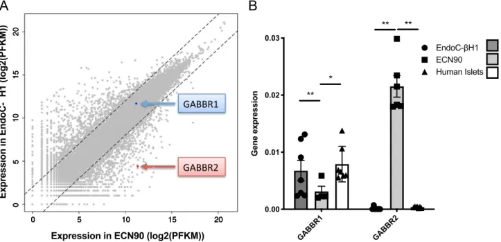
Comparative RNAseqanalyses were performed between ECN90 cells and the previously developed EndoC-βH1 cells. Expression profiles are depicted in Fig. 1A as scatter plots. Most of the transcripts are expressed at remarkably similar level in both cell lines, further indicating the beta cell identity of ECN90 cells. Both lines expressed GABBR1 at similar levels, whereas the expression of GABBR2 was more than 100 times higher in ECN90 cells compared to EndoC-βH1 cells. RT-qPCR analyses further indicated that GABBR1 was expressed in ECN90 cells, EndoC-βH1 cells and human islets. On the other hand, GABBR2 was only detected in ECN90 cells (Fig. 1B).
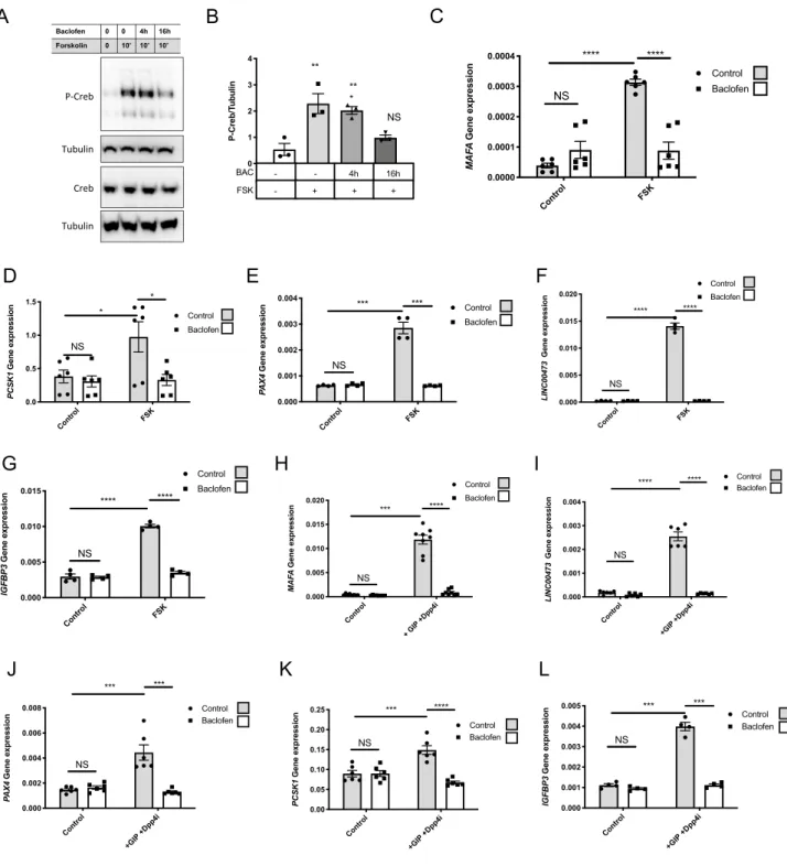
GABAB receptor function in ECN90 cells was tested using its specific agonist, baclofen (BAC). As the GABAB
receptor is a Gαi/o-coupled receptor17–19, we activated the ECN90 cell adenylyl cyclase with forskolin and tested
whether pretreatment with baclofen decreased such activation. Exposure of ECN90 to forskolin promoted the phosphorylation of CREB at Ser133 within 10 min while pretreatment (16 h, 100 µM) with baclofen inhibited CREB phosphorylation (Fig. 2A,B for quantification). To further investigate the function of the GABAB
recep-tor in ECN90, we searched for genes whose induction by forskolin would be blunted upon pretreatment with baclofen (16 h, 100 µM). Forskolin treatment induced a robust increase in MAFA, PCSK1, PAX4, IGFBP3 and the
non-coding long RNA Linc00473, as previously shown in our microarray analyses from forskolin treated human
EndoC-βH1 cells20. This induction was blunted upon pretreatment with baclofen (Fig. 2C–G). The repressive
effect of baclofen on MAFA, PCSK1, PAX4, Linc00473 induction by forskolin was reproduced when ECN90 were treated with the Gastric Inhibitory Polypeptide (GIP) (Fig. 2H–L), an incretin that is also an inducer of the cAMP pathway21,22.
GABBR1 GABBR2 0.00 0.01 0.02 0.03 Gene expression EndoC- H1 ECN90 Human Islets ** ** * **
Expression in ECN90 (log2(PFKM))
Expression in EndoC-H1 (log2(PFKM))
GABBR2
0 5 10 15 20 05 10 15 20 0 5 10 15 20 05 10 15 20 0 5 10 15 20 05 10 15 20GABBR1
A
B
Figure 1. Expression GABBR1 and GABBR2 in human beta cell lines and human islets. (A) Scatterplot
illustrating the comparative RNAseq analyses of 2 human beta cell lines EndoC-βH1 and ECN90. Blue and red arrows highlight GABBR1 and GABBR2 mRNA levels. (B) Expression of GABBR1 and GABBR2 mRNA by RT-qPCR in EndoC-βH1, ECN90 and human islets. Data are shown as the mean ± SEM; n = 6. *P < 0.05; **P < 0.01 relative to control by Student’s t test.
3 Scientific RepoRtS | (2020) 10:13469 | https://doi.org/10.1038/s41598-020-69758-6
www.nature.com/scientificreports/
As described above, ECN90 cells have been transformed using SV40T. To determine whether GABBR2 expres-sion is dependent of SV40T expresexpres-sion, we knocked-down SV40T using siRNA. SV40T depletion increased INSULIN staining and content (Fig. S3), IAPP and CDKN1A mRNA levels (Fig. S4A) as previously observed
Control FSK 10 ' BAC 4h + FSK 10 ' BAC 16h + FSK 10' 0 1 2 3 4 P-Creb/T ubuli n ** ** * NS BAC FSK -- 4h 16h + - + + P-Creb Tubulin Creb Tubulin Baclofen 0 0 4h 16h Forskolin 0 Contro l FSK 0.0000 0.0001 0.0002 0.0003 0.0004 MAF A Gene expression Control Baclofen NS **** **** Control FSK 0.0 0.5 1.0 1.5 PCSK 1 Gene expression Control Baclofen NS * * Control FS K 0.000 0.001 0.002 0.003 0.004 PA X4 Gene expression Control Baclofen NS *** ***
A
B
D
E
F
Control + GIP +Dpp4 i 0.000 0.005 0.010 0.015 0.020 MA FA Gene expression Control Baclofen NS **** *** Contro l FSK 0.000 0.005 0.010 0.015 IGFBP3 Gene expressio n Control Baclofen NS **** **** Contro l FSK 0.000 0.005 0.010 0.015 0.020 LINC00473 Gene expression Control Baclofen NS **** ****G
H
C
Contro l +GIP +Dpp4i 0.000 0.001 0.002 0.003 0.004 LINC0047 3 Gene expression Control NS **** **** Baclofen Contro l +GIP +Dpp4i 0.00 0.05 0.10 0.15 0.20 0.25 PCSK 1 Gene expression Control Baclofen NS **** *** Contro l +GIP +Dpp4i 0.000 0.002 0.004 0.006 0.008 PA X4 Gene expression Control Baclofen NS *** *** Contro l +GIP +Dpp4i 0.000 0.001 0.002 0.003 0.004 0.005 IGFBP3 Gene expression Control NS *** *** BaclofenI
J
K
L
Figure 2. Effects of baclofen treatment on ECN90. (A,B) Western blot and quantification of P-CREB (n = 3) in
ECN90 pretreated during 0-16 h with Baclofen (BAC) and next pulsed during 10′ with forskolin (FSK). (C–G) ECN90 were pretreated with or without Baclofen during 16 h and next pulsed for 1 h with FSK. RT-qPCR analyses indicate that Baclofen treatment blunts the induction by FSK of MAFA, PCSK1, PAX4, LINC00473 and
IGFBP3. (H–L) ECN90 were pretreated with or without Baclofen during 16 h and next pulsed for 1 h with GIP
and DPP4 inhibitor (used to inhibit GIP degradation). RT-qPCR analyses indicate that BAC treatment blunts the induction by GIP and DPP4 inhibitor for the same cAMP induced genes. Data are shown as the mean ± SEM (n = 3–6). *P < 0.05; **P < 0.01; ***P < 0.005; ****P < 0.001; NS = not significant relative to control by Student’s t test.
upon SV40T depletion in EndoC-βH1 cells23. Interestingly, SV40T knock-down did neither modify GABBR1
and GABBR2 mRNA levels, nor the ability of baclofen to inhibit the induction of MAFA by forskolin, indicating that GABBR2 expression and GABAB receptor function in ECN90 are independent of SV40T expression and
beta cell immortalization (Fig. S4B).
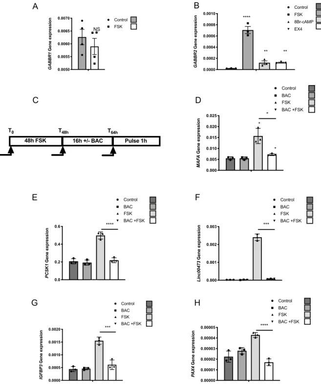
cAMp signaling regulates GABBR2 expression and function in EndoC‑βH1 cells and in human
islets.
Basal GABBR2 mRNA levels are extremely low both in EndoC-βH1 cells and in human islet prep-arations (Ct ~ 33–34 for cyclophilin at Ct ~ 20) (Fig. 1B). Moreover, GABAB receptor was not functional inEndoC-βH1 cells and in human islets as demonstrated by the lack of repressive effect of baclofen on forsko-lin-induced MAFA and PCSK1 induction (Fig. S5). Interestingly, mining our previous results from microarray analyses suggested that forskolin may increase GABBR2 mRNA levels in EndoC-βH1 cells20. We validated this
hypothesis by RT-qPCR that indicated that while a 48 h treatment with forskolin does not modify GABBR1 expression (Fig. 3A), it robustly increased GABBR2 mRNA levels in EndoC-βH1 cells (Fig. 3B). A similar induc-tion of GABBR2 was also observed upon treatments with either 8Br-cAMP or exendin4 (EX4) (Fig. 3B), 2 dif-ferent activators of the cAMP pathway. Moreover, following GABBR2 induction, baclofen treatment blunted forskolin-induced MAFA expression, indicating that the GABAB receptor is functional under such conditions
(Fig. 3C,D). The repressive effect of baclofen on PCSK1, Linc00473, IGFBP3 and PAX4 induction by forskolin was also observed in EndoC-βH1 (Fig. 3E–H).
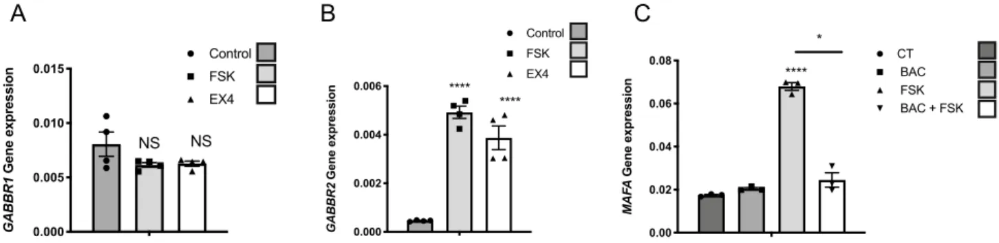
Based on this finding in EndoC-βH1 cells, we examined the cAMP dependent induction of GABBR2 on preparations of human islets. Forskolin and exendin4 (48 h treatments) did not modify GABBR1 expression (Fig. 4A), while, they both increased GABBR2 expression (Fig. 4B). As in EndoC-βH1 cells, under such condi-tions, GABAB receptor was functional, as demonstrated by the repressive effect of baclofen on forskolin-induced
MAFA expression in human islets (Fig. 4C).
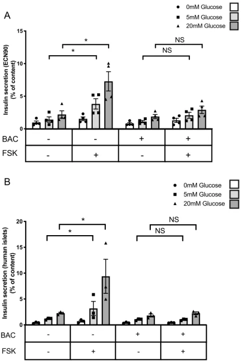
Signaling through the GABA
Breceptor limits the induction of insulin secretion by cAMp.
Wefinally evaluated whether signals through the GABAB receptor affect the beta cell function. We assessed the effect
of baclofen treatment on glucose and forskolin stimulated insulin secretion in ECN90 cells. Forskolin induced insulin secretion in a glucose-dependent manner and this effect was blunted when ECN90 cells were pre-treated with baclofen (Fig. 5A). Similar experiments were performed using human islets that had been treated for 48 h with forskolin to induce GABBR2 expression. As in ECN90 cells, baclofen treatment blunted forskolin-induced insulin secretion of human islets (Fig. 5B).
Discussion
Here we have developed a new human beta cell line from neonatal pancreas and demonstrated that both human beta cell lines and primary human islets express, in a tightly regulated fashion, functional GABAB receptors that
regulate insulin secretion.
We previously generated several functional human beta cell lines (EndoC-βH1 and EndoC-βH2) from human fetal pancreatic fragments15,16. In the present study, we generated an additional one, named ECN90. This line is
interesting for a number of reasons: (i). We derived ECN90 from a fragment of neonatal pancreas. Thus, with our protocol of targeted oncogenesis, while human adult beta cells are resistant to transformation16, fetal 15,16
but also neonatal (the present study) human pancreases are permissive for the generation of beta cell lines; (ii) ECN90 cells carry the HLA-I haplotype: HLA-A*02:01/03:01, -B*40:01/49:01, -C*03:04/07:01. This cell line provides a unique tool to model in vitro beta cell death in type 1 diabetes by assaying the cytotoxic effects of CD8T cell clones against human beta cells24 and thus to progress in the definition of ways to protect human beta
cells against destruction.
Here, we used ECN90 cells as a first model to study the expression and function of the GABAB receptor in
human beta cells. The GABAB receptor is a Gαi/o-protein-coupled receptor composed of 2 subunits, namely
GABBR1 and GABBR2 with both subunits necessary for signaling through this receptor25–28. The GABA
B receptor
is mainly present and functional in the central and peripheral nervous system where both GABBR1 and GABBR2 subunits are present29,30. Outside the nervous system, GABBR1 was detected in some visceral tissues such as the
stomach, intestine, heart, and spleen31, while gabbr2 expression has been observed in mouse liver and islets32. We
demonstrate here that ECN90 cells express both subunits of the GABAB receptor. We also show that this
recep-tor can be activated by baclofen, a specific synthetic agonist of the GABAB receptor that does not interact with
GABAA receptors5,14. The expression in beta cells of the GABAB receptor known to be highly enriched in neurons
is not fully unexpected, in the view of the large number of similarities between neurons and beta cells33. ECN90
cells can thus now be used as a model system to study signaling through the GABAB receptor in non-neural cells.
Our data indicate that under basal culture conditions, ECN90 cells express both GABBR1 and GABBR2, while EndoC-βH1, an independent human beta cell line and human islets express GABBR1 but not GABBR2. At that stage, we do not have clues to explain the differences from one cellular model to the other. However, it could be due to differences in the expression between ECN90 and EndoC-βH1 of transcription factors that regulate GABBR2 expression levels. But importantly, GABBR2 expression can be induced in EndoC-βH1 cells and in human islets upon treatment with compounds that increase the cAMP signaling pathway such as forskolin or exendin4, giving rise to functional GABAB receptors. It is well established that signaling by the GABAB receptor
is a tightly-regulated process. However, while information is available on signals implicated in post-translational regulation of GABBR2 signaling, such as its trafficking34, or its phosphorylation by the Protein Kinase A35,
infor-mation on its transcriptional regulation remains extremely scarce. For example, the mechanisms that explain the specificity of GABBR2 expression in the nervous system remain poorly defined. GABBR2 expression in
5 Scientific RepoRtS | (2020) 10:13469 | https://doi.org/10.1038/s41598-020-69758-6
www.nature.com/scientificreports/
human beta cells and its regulation by the cAMP pathway will represent an innovative model to progress on the mechanism that regulate its expression.
1 0.0050 0.0055 0.0060 0.0065 0.0070 GABBR 1 Gene expression Control FSK NS 1 0.0000 0.0002 0.0004 0.0006 0.0008 0.0010 GABBR2 Gene expression Control FSK 8Br-cAMP EX4 **** ** **
A
B
C
1 0.000 0.005 0.010 0.015 0.020 0.025 MAF A Gene expressio n Control BAC FSK BAC +FSK * * *D
48h FSK T0 T48h 16h +/- BAC T64h Pulse 1h 1 0.0 0.2 0.4 0.6 PCSK1 Gene expressio n Control BAC FSK BAC +FSK * **** 1 0.000 0.001 0.002 0.003 Linc00473 Gene expressio n Control BAC FSK BAC +FSK *** 0.0000 0.0005 0.0010 0.0015 0.0020 IGFBP 3 Gene expression Control BAC FSK BAC +FSK *** 0.00000 0.00001 0.00002 0.00003 0.00004 0.00005 PA X4 Gene expressio n Control BAC FSK BAC +FSK ****E
F
G
H
Figure 3. cAMP signaling induces GABBR2 expression in EndoC-βH1. (A) RT-qPCR analyses of GABBR1
mRNA in EndoC-βH1 treated with forskolin (FSK). (B) RT-qPCR analyses of GABBR2 mRNA in EndoC-βH1 following 48 h treatment with DMSO (0.1%; control condition), FSK, 8Br-cAMP or exendin4 (EX4). (C) Schematic representation of the timeline for the conditioning pulse experiment. (D–H) EndoC-βH1 were preconditioned during 48 h with FSK to induce GABBR2 expression. The medium was next changed and cells were further cultured during 16 h with or without Baclofen (BAC) and finally pulsed for 1 h with FSK. RT-qPCR analyses indicate that BAC treatment blunts MAFA induction by FSK. Result for PCSK1, LINC00473, PAX4 and IGFBP3 induction are also shown. Data are shown as the mean ± SEM (n = 3–4). *P < 0.05; ***P < 0.005; ****P < 0.001; NS = not significant relative to control by Student’s t test.
We demonstrate here that signaling through the GABAB receptor inhibits the cAMP pathway, known to
play major roles in beta cell. Through paracrine effects, cAMP signaling modulates glucose-stimulated insulin secretion36. Specifically, alpha cells release glucagon that signals on beta cells through the Gα
s-coupled Glucagon
receptor, increases cAMP levels and insulin secretion37,38. In parallel, delta cells secrete somatostatin that binds
the Gi-coupled somatostatin receptor 2 on beta cells, and decreases cAMP levels and insulin secretion39. Here,
we demonstrate that similarly, activation of the Gi-coupled GABAB receptor decreases cAMP levels and insulin
secretion. In this context, it is important to note that human beta cells express the enzyme GAD2 and thus pro-duce GABA that is co-secreted with insulin40. GABA may thus act through an autocrine loop to bring insulin
back to basal levels following stimulation, a major property of mature beta cells8,9,41.
The cAMP pathway is also important to maintain the beta cell differentiation status42 and signals through
the GABAB receptor counteract this process. Indeed, activating the cAMP pathways increased the expression
of a number of genes important for beta cell function. It is the case for MAFA, a factor implicated in insulin gene transcription43 and for the proconvertase PCSK1, an enzyme implicated in the processing of proinsulin44.
Forskolin treatment also increased PAX4 expression, a transcription factor essential for beta cell development during prenatal life45 that is also implicated in beta cell proliferation and protection against degeneration46.
Finally, forskolin treatment induced the expression of the long non-coding RNA LINC00473 with yet unknown function in beta cells. This cAMP induction of LINC00473 was previously described in non-small cell lung cancer47. Interestingly, such inductive effects are blunted upon activation of the GABA
B receptor, which suggests
a tight balance between positive and negative inducers of the cAMP pathway.
Antidiabetic medications such as incretins improve islet function through cAMP production. Here, we show that cAMP signaling induces functional GABAB receptors that counteract incretin effects. A number of type
2 diabetic patients are insensitive to incretins without any clue48. Whether it is due to GABA
B receptors
over-activation might be evaluated.
In conclusion, our data demonstrate that in human beta cells, signaling through the GABAB receptor
par-ticipates in an autocrine feedback inhibition loop that regulates beta cell specific gene expression and insulin secretion.
Methods
ethical statement.
This study was performed according to the Declaration of Helsinki and the Declaration of Istanbul. No tissues were procured from prisoners. As the French Biomedical Agency regulates the graft allo-cation system in France, every organ was allocated and approved by the ethics committee of the French Biomedi-cal Agency to be in accordance with French laws. Neonatal tissue was collected with written informed consents from the parents and in compliance with French bioethic legislation certified by the French Biomedical Agency. Human Islet collection was approved by the ethics committee of the French Biomedical Agency. Experiments using human graft in mice were approved by the animal experimentation ethics committee of Paris Descartes University and Sorbonne University (Paris, France). Experiments using mice were certified by the Direction Departementale de la Protection des Populations for the French Ministry of Research, Health and Agriculture (Paris) under agreement number A75-13–19 in accordance with approved guideline of French and European legislation. Human Islet collection was certified by the French Biomedical Agency Guidelines and registered in the French Ministry of Health under the number PFS12-006.Derivation from neonatal pancreas of ECN90, a human β cell line.
A fragment from neonatal pan-creas was collected, cut into pieces, digested with type IV collagenase (Sigma-Aldrich) and transduced with len-tiviral vectors. Two loxP sites flank the integrated sequences expressing SV40T and hTERT, allowing subsequent excision dependent on Cre recombinase expression15. The tissue was next transplanted under the kidney capsuleof immune-incompetent SCID (Charles River, L’Arbresle, France) as described49. Following 3 successive rounds
0.000 0.002 0.004 0.006 GABBR2 Gene expressio n Control FSK EX4 **** **** 0.000 0.005 0.010 0.015 GABBR1 Gene expression Control FSK NS EX4 NS
A
B
C
0.00 0.02 0.04 0.06 0.08 MA FA Gene expression CT BAC FSK BAC + FSK **** *Figure 4. cAMP signaling induces GABBR2 expression in human islets. (A) RT-qPCR analyses of GABBR1
mRNA in human islets treated with forskolin (FSK) or exendin4 (EX4). (B) RT-qPCR analyses of GABBR2 mRNA in human islets following 48 h treatment with DMSO (0.1%; control condition), FSK or EX4. (C) Human islets were preconditioned during 48 h with FSK to induce GABBR2 expression. The medium was next changed and cells were further cultured during 16 h with or without Baclofen (BAC) and finally pulsed for 1 h with FSK. RT-qPCR analyses indicate that under such conditions, BAC treatment blunts MAFA induction by FSK. Data are shown as the mean ± SEM (n = 3–4). *P < 0.05; ****P < 0.001; NS = not significant relative to control by Student’s t test.
7 Scientific RepoRtS | (2020) 10:13469 | https://doi.org/10.1038/s41598-020-69758-6
www.nature.com/scientificreports/
of transplantation, we derived the cell line ECN90 that is cultured at 37 °C in 5% CO2 in Advanced DMEM/F12
medium (Thermo Fisher Scientific) supplemented with 2% bovine serum albumin fraction V (Roche), 6.7 ng/ml sodium selenite, 10 mM nicotinamide (Calbiochem), 50 μM β-mercaptoethanol (Sigma-Aldrich) and penicillin/ streptomycin (Thermo Fisher Scientific).
Human islets.
Human islets were provided by the Human islet core facility of St-Louis Hospital (APHP, France). They were obtained from pancreata of seven brain-dead donors (mean age 55.67 ± 4.68 years; BMI 25.4 ± 4.36 kg/m2) with signed informed consents according to the procedures approved by the French Agencyof Biomedicine (Supplemental Table 1). Islets were isolated, handpicked and cultured 48 h in 12-well plates
Control FS K BAC FSK + BAC 0 5 10 15
Insulin secretion (ECN90)
(% of content) 0mM Glucose
*
NS NSBAC
FSK
-
+
+
+
-
+
-5mM Glucose 20mM Glucose*
A
B
Control FS K BAC FSK + BA C 0 5 10 15 20Insulin secretion (human islets)
(% of content) 0mM Glucose
*
*
NS NS -BAC FSK - + + + - - + 5mM Glucose 20mM GlucoseFigure 5. Baclofen treatment blunts forskolin-induced insulin secretion. (A) ECN90 were pretreated with
or without Baclofen (BAC) during 16 h and next pulsed for 1 h with or without forskolin (FSK) followed by a 40 min insulin secretion test at 0, 5 and 20 mM glucose. Secreted insulin is presented as % of content. (B) Human islets were preconditioned during 48 h with FSK to induce GABBR2 expression. They were next pretreated with or without BAC during 16 h and next pulsed for 1 h with FSK followed by a 40 min insulin secretion test at 0, 5 and 20 mM glucose. Secreted insulin is presented as % of content. Data are shown as the mean ± SEM (n = 3–4). *P < 0.05; NS = not significant relative to control by Student’s t test.
(50–100 islets per well) in CMRL medium supplemented with 10% fetal calf serum, Hepes and penicillin/strep-tomycin (all from Thermo Fisher Scientific).
cells and islets treatments.
The following compounds were used for treatments of EndoC-βH116, ECN90and human islets: forskolin (FSK) (Tocris;10 μM); R-Baclofen (Tocris;100 µM), linagliptin (Dpp4i; Selleckchem; 100 nM) GIP (Tocris, 100 nM), exendin4 (Tocris, 5 nM), 8Br-cAMP (Tocris; 1 µM).
Glucose‑stimulated insulin secretion (GSIS).
ECN90 cells were seeded onto Matrigel/fibronectin-coated 12-well plates at 2.5 × 10⁵ cells/well and human islets were cultured in 12-well plates (50 islets per well intriplicates). They were both starved in DMEM (Thermo Fisher Scientific) containing 0.5 mM glucose for 24 h, washed twice and then preincubated in Krebs–Ringer bicarbonate Hepes buffer (KRBH) containing 0.2% BSA in the absence of glucose for 1 h. Insulin secretion was measured following a 40 min incubation with KRBH containing 0.2% BSA that contained varying glucose concentrations. Glucose stimulation was performed in the presence or absence of 10 μM FSK. For insulin content measurements, cells and islets were lysed in the culture wells in 50 mM Tris, pH 8.0, 1% Nonidet P-40, 0.5% sodium deoxicolate, 0.05% SDS, 100 mM NaCl, 5 mM EDTA (Thermo Fisher Scientific), and anti-protease tablets (Roche) for 20 min on ice. Insulin secretion and content were measured by ELISA (Mercodia AB, Uppsala, Sweden) as described15.
RnA isolation, reverse transcription, and Rt-qpcR.
RNeasy Micro Kit (Qiagen) was used to extract total RNA from beta cell lines and human islets50. Genomic DNA was removed by DNAse treatment followingthe RNeasy Micro Kit protocol. Maxima First Strand cDNA Kit (Thermo Fisher Scientific) was used to synthe-size cDNA. RT-qPCR was performed using Power SYBR Green mix (Applied Biosystems) with a QuantStudio 3 analyzer. The comparative method of relative quantification (2ddCT) was used to calculate the expression levels of each target gene, normalized to Cyclophilin-A transcript. Reactions with and without reverse transcriptase (RT + and RT−) were used as control for the absence of genomic DNA contamination. Reactions with and with-out reverse transcriptase (RT + and RT−) were used as control of no genomic DNA contamination as RT- cDNA sample generate no signal with cyclophilin primers, indicating complete elimination of gDNA. Custom primers were designed with Primer-Blast online, and their efficiency was determined for each with a serial dilution of cDNA samples. The list of primers is presented in Supplemental Table 2.
RNA‑seq: library preparation and analysis.
RNA-seq was provided by our Genom’IC lab facility in Institut Cochin. RNA concentrations were measured using nanodrop (Thermo Fisher Scientific, USA). The quality of the RNA (RNA integrity number or RIN) was determined on the Agilent 2,100 Bioanalyzer (Agilent Technologies, Palo Alto, CA, USA). 800 ng of total RNA sample (RIN > 9) was processed to construct the librar-ies using TruSeq Stranded mRNA kit (Illumina). Librarlibrar-ies were quantified by RT-qPCR using the KAPA Library Quantification Kit for Illumina Libraries (KapaBiosystems, Wilmington, MA) and library profiles were assessed using the DNA High Sensitivity LabChip kit on an Agilent Bioanalyzer. Libraries were sequenced on an Illumina Nextseq 500 instrument using 75 base-lengths read V2 chemistry in a paired-end mode. After sequencing, a first analysis based on AOZAN software (ENS, Paris) was applied to demultiplex and control the quality of the raw data (based of FastQC modules / version 0.11.5). Obtained fastq files were then aligned using STAR algo-rithm (version 2.5.2b). Reads were then counted using Featurecount (version Rsubread 1.24.1) and the statistical analyses on the read counts were performed with the DESeq2 package version 1.14.1. RNA-seq data are available in the NCBI’s Gene Expression Omnibus (GEO) database (accession GSE155482).siRnA transfection.
ECN90 cells were transfected using Lipofectamine RNAiMAX (Thermo Fisher Scien-tific) as previously described50. Custom Silencer® Select siRNA for SV40T51 (Thermo Fisher Scientific), orON-TARGETplus nontargeting control pool (siCTRL) were used (Dharmacon, GE healthcare Life Sciences) at a final concentration of 80 nM. Briefly, siRNA and Lipofectamine RNAiMAX were combined in OptiMEM (Thermo Fisher Scientific) and applied to the cells. Medium was replaced 2.5 h later with fresh ECN90 culture medium50.
immunostainings.
Immunohistochemistry and immunocytochemistry were performed as previously described15 using the following antibodies guinea pig anti-insulin antibody (1/500; A0564, DakoCytomation);rabbit anti-human PDX1 antibody (1/2,000) 52 mouse anti-SV40T (1/50; DP-02, Calbiochem Merck Biosciences);
mouse anti-human Ki67 antigen (1/50; M7240, DakoCytomation). The Alexa fluor secondary antibodies were purchased from Thermo Fisher Scientific (1:200).
immunoblotting.
For Western blot, cells were lysed in RIPA buffer with anti-protease and PhosSTOP tab-lets (Roche) and sonicated as previously described50. Equal amounts of protein (20 μg) were resolved in a 4–12%Bis–Tris gel and transferred to a membrane using an iBLOT2 Dry Blotting System (Thermo Fisher Scientific). Membranes were immunoblotted with the following antibodies: mouse anti-SV40T (1/50; DP-02, Calbiochem Merck Biosciences), Phospho-Ser133 CREB (1:1,000; Cell Signaling Technology), CREB (1:1,000; Cell Signaling Technology), β-actin (1:2,000; Sigma), alpha-Tubulin (1:2,000; Sigma). Species-specific HRP-linked secondary antibodies (Cell Signaling Technology) were used for detection and visualization was performed on an Image-Quant LAS 4,000 following ECL exposure (GE Healthcare).
Statistics.
Data were analyzed using GraphPad Prism 6 software and are presented as the mean ± SEM. Quantitative data are presented as the mean ± SEM. The number of experiments is indicated in the figureleg-9 Scientific RepoRtS | (2020) 10:13469 | https://doi.org/10.1038/s41598-020-69758-6
www.nature.com/scientificreports/
ends. Statistical significance was estimated using a 2-tailed Student’s t test. A P value less than 0.05 was consid-ered significant.
Data and resource availability.
The datasets generated during and/or analyzed during the current study are available from the corresponding authors upon reasonable request. Most of the resources used during these studies are commercially available.Received: 6 November 2019; Accepted: 26 June 2020
References
1. Leahy, J. L. Pathogenesis of type 2 diabetes mellitus. Arch. Med. Res. 36, 197–209 (2005).
2. Ahrén, B. Islet G protein-coupled receptors as potential targets for treatment of type 2 diabetes. Nat. Rev. Drug Discov. 8, 369–385 (2009).
3. Wootten, D., Christopoulos, A., Marti-Solano, M., Babu, M. M. & Sexton, P. M. Mechanisms of signalling and biased agonism in G protein-coupled receptors. Nat. Rev. Mol. Cell Biol. 19, 638–653 (2018).
4. Pavlos, N. J. & Friedman, P. A. GPCR signaling and trafficking: the long and short of it. Trends Endocrinol. Metab. 28, 213–226 (2017).
5. Bowery, N. G. et al. International Union of Pharmacology. XXXIII. Mammalian gamma-aminobutyric acid(B) receptors: structure and function. Pharmacol. Rev. 54, 247–264 (2002).
6. Olsen, R. W. & Sieghart, W. International union of lassification on the basis of subunit composition, pharmacology, and function. Pharmacol. Rev. 60, 243–260 (2008).
7. Chessler, S. D. & Lernmark, A. Alternative splicing of GAD67 results in the synthesis of a third form of glutamic-acid decarboxylase in human islets and other non-neural tissues. J. Biol. Chem. 275, 5188–5192 (2000).
8. Braun, M. et al. Gamma-aminobutyric acid (GABA) is an autocrine excitatory transmitter in human pancreatic beta-cells. Diabetes
59, 1694–1701 (2010).
9. Korol, S. V. et al. Functional characterization of native, high-affinity GABAA receptors in human pancreatic β cells. EBioMedicine
30, 273–282 (2018).
10. Amisten, S., Salehi, A., Rorsman, P., Jones, P. M. & Persaud, S. J. An atlas and functional analysis of G-protein coupled receptors in human islets of Langerhans. Pharmacol. Ther. 139, 359–391 (2013).
11. Braun, M. et al. GABAB receptor activation inhibits exocytosis in rat pancreatic beta-cells by G-protein-dependent activation of calcineurin. J. Physiol. (Lond.) 559, 397–409 (2004).
12. Wang, Q., Ren, L., Wan, Y. & Prud’homme, G. J. GABAergic regulation of pancreatic islet cells: physiology and antidiabetic effects. J. Cell. Physiol. https ://doi.org/10.1002/jcp.28214 (2019).
13. Tian, J. et al. γ-Aminobutyric acid regulates both the survival and replication of human β-cells. Diabetes 62, 3760–3765 (2013). 14. Hill, D. R. & Bowery, N. G. 3H-baclofen and 3H-GABA bind to bicuculline-insensitive GABA B sites in rat brain. Nature 290,
149–152 (1981).
15. Scharfmann, R. et al. Development of a conditionally immortalized human pancreatic β cell line. J. Clin. Invest. 124, 2087–2098 (2014).
16. Ravassard, P. et al. A genetically engineered human pancreatic β cell line exhibiting glucose-inducible insulin secretion. J. Clin. Invest. 121, 3589–3597 (2011).
17. Hill, D. R. GABAB receptor modulation of adenylate cyclase activity in rat brain slices. Br. J. Pharmacol. 84, 249–257 (1985). 18. Kaupmann, K. et al. Human gamma-aminobutyric acid type B receptors are differentially expressed and regulate inwardly
rectify-ing K+ channels. Proc. Natl. Acad. Sci. USA. 95, 14991–14996 (1998).
19. Odagaki, Y. & Koyama, T. Identification of galpha subtype(s) involved in gamma-aminobutyric acid(B) receptor-mediated high-affinity guanosine triphosphatase activity in rat cerebral cortical membranes. Neurosci. Lett. 297, 137–141 (2001).
20. Richards, P. et al. MondoA is an essential glucose-responsive transcription factor in human pancreatic β-cells. Diabetes 67, 461–472 (2018).
21. Siegel, E. G. & Creutzfeldt, W. Stimulation of insulin release in isolated rat islets by GIP in physiological concentrations and its relation to islet cyclic AMP content. Diabetologia 28, 857–861 (1985).
22. Furman, B., Ong, W. K. & Pyne, N. J. Cyclic AMP signaling in pancreatic islets. Adv. Exp. Med. Biol. 654, 281–304 (2010). 23. Arda, H. E. et al. Age-dependent pancreatic gene regulation reveals mechanisms governing human β cell function. Cell Metab. 23,
909–920 (2016).
24. Culina, S. et al. Islet-reactive CD8+ T cell frequencies in the pancreas, but not in blood, distinguish type 1 diabetic patients from healthy donors. Sci Immunol 3, (2018).
25. Jones, K. A. et al. GABA(B) receptors function as a heteromeric assembly of the subunits GABA(B)R1 and GABA(B)R2. Nature
396, 674–679 (1998).
26. Kaupmann, K. et al. GABA(B)-receptor subtypes assemble into functional heteromeric complexes. Nature 396, 683–687 (1998). 27. Kuner, R. et al. Role of heteromer formation in GABAB receptor function. Science 283, 74–77 (1999).
28. White, J. H. et al. Heterodimerization is required for the formation of a functional GABA(B) receptor. Nature 396, 679–682 (1998). 29. Ong, J. & Kerr, D. I. GABA-receptors in peripheral tissues. Life Sci. 46, 1489–1501 (1990).
30. Bowery, N. G., Hudson, A. L. & Price, G. W. GABAA and GABAB receptor site distribution in the rat central nervous system. Neuroscience 20, 365–383 (1987).
31. Bowery, N. G. GABAB receptor pharmacology. Annu. Rev. Pharmacol. Toxicol. 33, 109–147 (1993).
32. Regard, J. B., Sato, I. T. & Coughlin, S. R. Anatomical profiling of G protein-coupled receptor expression. Cell 135, 561–571 (2008). 33. Arntfield, M. E. & van der Kooy, D. β-Cell evolution: How the pancreas borrowed from the brain: the shared toolbox of genes
expressed by neural and pancreatic endocrine cells may reflect their evolutionary relationship. BioEssays 33, 582–587 (2011). 34. Doly, S. et al. GABAB receptor cell-surface export is controlled by an endoplasmic reticulum gatekeeper. Mol. Psychiatry 21,
480–490 (2016).
35. Couve, A. et al. Cyclic AMP-dependent protein kinase phosphorylation facilitates GABA(B) receptor-effector coupling. Nat. Neurosci. 5, 415–424 (2002).
36. Seino, S., Takahashi, H., Fujimoto, W. & Shibasaki, T. Roles of cAMP signalling in insulin granule exocytosis. Diabetes Obes Metab
11(Suppl 4), 180–188 (2009).
37. Svendsen, B. et al. Insulin Secretion depends on intra-islet glucagon signaling. Cell Rep 25, 1127-1134.e2 (2018). 38. Zhu, L. et al. Intra-islet glucagon signaling is critical for maintaining glucose homeostasis. JCI Insight 5, (2019).
39. Tsonkova, V. G. et al. The EndoC-βH1 cell line is a valid model of human beta cells and applicable for screenings to identify novel drug target candidates. Mol. Metab. 8, 144–157 (2018).
40. Suckale, J. & Solimena, M. The insulin secretory granule as a signaling hub. Trends Endocrinol. Metab. 21, 599–609 (2010). 41. Rorsman, P. et al. Activation by adrenaline of a low-conductance G protein-dependent K+ channel in mouse pancreatic B cells.
Nature 349, 77–79 (1991).
42. Blanchet, E. et al. Feedback inhibition of CREB signaling promotes beta cell dysfunction in insulin resistance. Cell. Rep. 10, 1149–1157 (2015).
43. Hang, Y. & Stein, R. MafA and MafB activity in pancreatic β cells. Trends Endocrinol. Metab. 22, 364–373 (2011). 44. Steiner, D. F. & James, D. E. Cellular and molecular biology of the beta cell. Diabetologia 35(Suppl 2), S41-48 (1992).
45. Sosa-Pineda, B., Chowdhury, K., Torres, M., Oliver, G. & Gruss, P. The Pax4 gene is essential for differentiation of insulin-producing beta cells in the mammalian pancreas. Nature 386, 399–402 (1997).
46. Lorenzo, P. I., Cobo-Vuilleumier, N. & Gauthier, B. R. Therapeutic potential of pancreatic PAX4-regulated pathways in treating diabetes mellitus. Curr. Opin. Pharmacol. 43, 1–10 (2018).
47. Chen, Z. et al. cAMP/CREB-regulated LINC00473 marks LKB1-inactivated lung cancer and mediates tumor growth. J. Clin. Invest.
126, 2267–2279 (2016).
48. Herzberg-Schäfer, S., Heni, M., Stefan, N., Häring, H.-U. & Fritsche, A. Impairment of GLP1-induced insulin secretion: role of genetic background, insulin resistance and hyperglycaemia. Diabetes Obes. Metab. 14(Suppl 3), 85–90 (2012).
49. Castaing, M. et al. Blood glucose normalization upon transplantation of human embryonic pancreas into beta-cell-deficient SCID mice. Diabetologia 44, 2066–2076 (2001).
50. Oshima, M. et al. Virus-like infection induces human β cell dedifferentiation. JCI Insight 3, (2018).
51. Ihler, F. et al. Expression of a neuroendocrine gene signature in gastric tumor cells from CEA 424-SV40 large T antigen-transgenic mice depends on SV40 large T antigen. PLoS ONE 7, e29846 (2012).
52. Duvillié, B. et al. The mesenchyme controls the timing of pancreatic beta-cell differentiation. Diabetes 55, 582–589 (2006).
Acknowledgements
The authors thank Bruno Ragazzon and Marc Diedisheim (both from Institut Cochin), for their support in RNA seq analyses. We thank the Genom’IC facility of the Institut Cochin (INSERM U1016, UMR CNRS8104, Université Paris Descartes) for their technical and scientific expertises in genomic studies of this paper. We thank the Human islet core facility of St-Louis Hospital for providing human islets. The work leading to this publica-tion has received support from the Innovative Medicines Initiative 2 Joint Undertaking Rhapsody, under Grant Agreement Nos. 115881 (RHAPSODY) and 115797 (INNODIA) and from Lilly France. The R.S. laboratory is supported by the Dutch Diabetes Research Foundation, by the DON Foundation, by the Fondation pour la Recherche Médicale (EQU201903007793) and by the Fondation Francophone pour la Recherche sur le Diabete (FFRD). The RS laboratory belongs to the Laboratoire d’Excellence consortium Revive.
Authors contribution
LR, PR, SM, OA and RS designed the research. LR and RS wrote the manuscript. LR, AM, SP, JH, PR and OA performed research. LR, PR and OA acquired the data. MA provided human islets. All authors made substantial contributions to the analysis and interpretation of data. All authors approved the manuscript. LR is the guaran-tors of this work and, as such, had full access to all the data in the study and takes responsibility for the integrity of the data and the accuracy of the data analysis.
competing interests
The authors declare no competing interests.
Additional information
Supplementary information is available for this paper at https ://doi.org/10.1038/s4159 8-020-69758 -6.
Correspondence and requests for materials should be addressed to L.R. Reprints and permissions information is available at www.nature.com/reprints.
Publisher’s note Springer Nature remains neutral with regard to jurisdictional claims in published maps and
institutional affiliations.
Open Access This article is licensed under a Creative Commons Attribution 4.0 International
License, which permits use, sharing, adaptation, distribution and reproduction in any medium or format, as long as you give appropriate credit to the original author(s) and the source, provide a link to the Creative Commons license, and indicate if changes were made. The images or other third party material in this article are included in the article’s Creative Commons license, unless indicated otherwise in a credit line to the material. If material is not included in the article’s Creative Commons license and your intended use is not permitted by statutory regulation or exceeds the permitted use, you will need to obtain permission directly from the copyright holder. To view a copy of this license, visit http://creat iveco mmons .org/licen ses/by/4.0/.