CASE REPORT
Symptomatic syringomyelia occurring as a late complication
of posterior fossa medulloblastoma removal in infancy
in a boy also suffering from scaphocephaly
Yassine El Hassani&Karim Burkhardt&
Jacqueline Delavellle&Maria-Isabel Vargas&
Colette Boex&Benedict Rilliet
Received: 6 July 2009 / Published online: 7 August 2009 # Springer-Verlag 2009
Abstract
Introduction The association of a medulloblastoma and a syringomyelia has been already described in rare instances albeit without symptoms related to the syrinx.
Case report The case of a 23-year-old man operated in infancy for a medulloblastoma and then treated solely with adjuvant chemotherapy is reported. He was also operated in infancy for a scaphocephaly. With a very long time delay, he has developed a Chiari I and a symptomatic cervico-dorsal syringomyelia. The symptoms attributed to the syrinx consisted of a unilateral prurigo over the left arm which was so severe to lead to self-mutilation.
Discussion Clinical and magnetic resonance imaging follow-up after cervico-dorsal decompression shows a significant improvement of the symptoms together with a
reduction of the size of the syrinx. This case is discussed in the light of the presumed pathophysiology of the syrinx and its exceptional clinical presentation.
Keywords Medulloblastoma . Syringomyelia . Complications
Introduction
Syringomyelia is a cystic cavitation of the spinal cord. Seventy percent are associated with Chiari I malformation. It has also been described in association with a variety of posterior fossa tumours and usually resolving after the removal of the expanding mass [1, 5, 7, 9]. Classical symptoms of syringomyelia include mainly a suspended and dissociated sensory loss. The case of patient operated at the age of 8 months for a medulloblastoma who developed very lately a cervico-dorsal syrinx is reported. He was also operated 2 weeks later for a genuine scaphocephaly. As he was operated in infancy, he was never irradiated but was treated by chemotherapy for 2 years. The oncological outcome was very good and was declared cured from the medulloblastoma and practically lost of follow-up. One year before readmission (23 years later), he presented an important left upper limb prurigo. This was so severe that he began to have a compulsive behaviour with constant scratching of its left upper extremity. A few cases of syringomyelia associated with medulloblastoma have been reported at the time of diagnosis of the posterior fossa tumour but never with such a long delay. In rare instances intramedullary pathology such as syrinx, tumours arterio-venous malformations and split cord malformation can present with segmental prurigo.
Y. El Hassani
:
B. Rilliet (*)Service de Neurochirurgie, Hôpital Cantonal Universitaire, 24 rue Micheli-du-Crest,
1211 Geneva 14, Switzerland e-mail: [email protected] K. Burkhardt
Unité de Neuropathologie, Centre Médical Universitaire, 1211 Geneva 14, Switzerland
J. Delavellle
:
M.-I. VargasUnité de Neuroradiologie Diagnostique, Service de Radiologie, Hôpital Cantonal Universitaire,
24 rue Micheli-du-Crest, 1211 Geneva 14, Switzerland C. Boex
Neuromonitoring, Unité d’épileptologie, Service de Neurologie,
Hôpital Cantonal Universitaire, 24 rue Micheli-du-Crest, 1211 Geneva 14, Switzerland
Case report
B.T, a 23-year-old man was operated 23 years ago for a medulloblastoma. He started his medical history with an unexplained newborn hypotonia. The initial clinical exami-nation found a scaphocephaly, and some dysmorphic-like features were noted (retrognathism, cryptorchidism, acetabu-lar dysplasia, and a bilateral pes varus). An initial computed tomography (CT) cerebral done without iodine contrast did not give any explication for the hypotonia. The cerebral spinal fluid (CSF) was normal, and no tumour cells were found. A genetic analysis with numerous and structural chromosome chart analysis was normal.
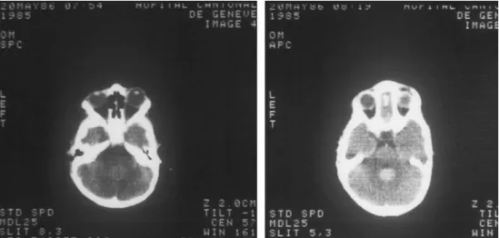
At the age of 6 months, clinical examination found sagittal synostosis and tense anterior fontanelle. A new CT cerebral with iodine contrast showed a hyperdense midline space-occupying lesion in the posterior cerebral fossa without hydrocephalus (Fig.1).
The boy was first operated at the age of 8 months, with a sub-occipital craniectomy to resect a fleshy tumour measuring 3 cm diameter that was easily separated for cerebellar tissue.
The microscopic analysis of the tumour tissue showed a population of densely packed small cells showing scanty cytoplasm, round or oval nuclei with dense chromatin, and occasional mitoses. The cells were organised in sheets interrupted by occasional rosettes. Pathologic diagnosis was World Health Organisation grade IV medulloblastoma (Fig.2).
After a period of 2 weeks, the correction the scaphocephaly was done with simple sagittal synostectomy.
Chemotherapy for 24 months with cisplatin and procarba-zine in alternation with vincristine and cyclophosphamide without any radiotherapy was achieved. Post-operative CT scan did not show any recurrence of the tumour nor tonsil herniation.
Although the patient remained with a short stature (all the percentile were below 3%), he enjoyed a normal life,
successfully pass his certificate to be a gardener, and was still working in that occupation when he came again with new symptoms at the age of 23. He progressively complained of tingling and itching in both upper limbs, especially on the left arm and hand. His clinical state worsened with self-mutilation of the left upper limb and was send to a dermatologist who then eventually referred him to a neurologist.
Clinical examination revealed a patient complaining of pruritus during all the period of consultation; neurological examination showed anaesthesia to light touch and pinprick, decreased pallaesthesia (7/8), and depressed osteotendinous reflexes in the upper left arm. There was no motor or sensitive deficit in the rest of the neurological examination.
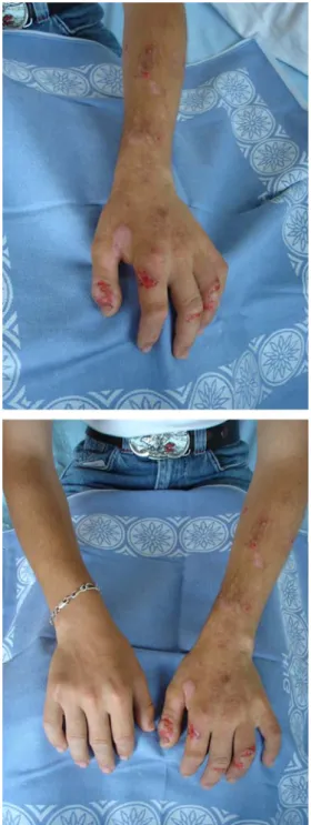
Dermatological examination found lesions in the whole upper left arm without a specific dermatome distribution, plaques with necrosis of different age, and excoriated and lichenified lesions. This was compatible with prurigo (Fig. 3).
Cerebral and spinal magnetic resonance imaging (MRI) with gadolinium showed an acquired Chiari type I, with a communicating syrinx cavity extending to the 11th dorsal vertebrae. On the axial plane, the syrinx was expanding more on the left side. There was no evidence for tethered cord, spina bifida, or any intra- or extra-medullary metastasis of the medulloblastoma (Fig.4).
Pre-operative sensitive and motor-evoked potential showed a left cervical radicular lesion with possible alteration in the posterior cord up to the 12th vertebrae.
Regarding to a good correlation between clinical and radiological findings, a C0 and C1 decompression with dura mater enlargement was performed. The occipital bone was particularly thick, and many adherences between the dura and the arachnoid over the tonsils were released; there was no evidence for tumour. Pre-operative, sensitive, and motor-evoked potentials in terms of latencies and amplitudes were reported as stable during the whole procedure. As observed
Fig. 1 Initial pre-operative axial computed tomography scan without (left) and with (right) iodine contrast showing a rounded, homogenous contrast enhancement situated just behind the fourth ventricle
previously to the surgery, the pre-operative somatosensory evoked potentials were slightly asymmetrical in disfavour of the left upper limb.
Surgery was uneventful and 3 and 6 months after surgery, the intensity pruritus has greatly abated, the dermatological lesions improved (Fig. 5), and The MRI shows a decrease although not total of the size of the syrinx (Fig.6).
Discussion
Medulloblastoma is a malignant primitive neuroectodermal tumour of the cerebellum, which mostly occurs in children. The outcome has significantly improved with new treatment modality, rising up to 90% of children with 5 years event-free survival [4].With improving the oncological outcome, some complications may appear in the long-term follow-up. The reported side effects of chemotherapy and radiotherapy may
occur during or after treatment and are well known to the neuro-oncology teams.
The association of posterior fossa tumour and associated syringomyelia is well described in the literature with several types of tumours encountered in the posterior fossa of adult patient such as meningiomas [1,9,13], epidermoid cyst [5], Lhermitte–Duclos disease [24], and midbrain gliomas [23]; and in children, giant craniopharyngioma [14], malignant posterior fossa growths such as medulloblastomas [7,8,12,
Fig. 2 Initial microscopic finding showing a lesion compatible with World Health Organisation grade IV medulloblastoma
Fig. 3 Pre-operative dermatological finding showing prurigo lesions strictly in the left arm without a specific dermatome distribution
17, 18, 20], or also benign tumours such as pilocytic astrocytoma as reported by Muzumdar and Ventureyra who made an extensive review on the subject [15]. Most of these syrinxes were asymptomatic and could be clearly explained by the added volume of the tumour in the posterior fossa that precipitated the downward movement of the tonsils, thus
creating an obstruction at the level of the foramen magnum. The syrinx eventually regressed in most of the cases after removal of the tumour.
We report a particular case, a Chiari malformation type I occurring with a long delay after the uncomplicated removal of a posterior fossa medulloblastoma causing syringomyelia and revealed by an important pruritus in the left arm and self-mutilations. Cervico-occipital decompression was indicated to improve CSF flow obstruction at the level of the foramen magnum, and 3 months after surgery, the pruritus has significantly decreased, and the size of syringomyelia too.
Focal dermatological lesions have been reported in rare instances associated with intramdedullary lesions.
Bond and Keough reported the first case of pruritus induced by transverse myelitis; they advocated that myelitis can interrupt pathways modulating the sensations of pain and pruritus [2]. A case of compulsive lip biting in a patient with Chiari type II malformation was also described [19].
The sensation of itch is thought to originate from the nerve endings of myelinated delta A fibres and unmyelinated C fibres near the dermo-epidermal junction. Focal pruritus may have a neurological cause. A neurological aetiology should always be considered in case of both localised pruritus and prurigo [2,10,21].
Vuadens et al. reported the case of woman with a 6-year history of pruritus of the inner side of the right arm accompanied by dysaesthesia that reveal a cavernous haemangioma at the level of T1 [22]. Some authors proposed that the lesion produced a hyperexcitable state by interfering with the descending pathways from an inhibitory centre responsible for pain and pruritus modulation [6].
Fig. 5 Six months follow-up showing dermatological findings with improvement of the lesions
Fig. 4 Pre-operative T2-weighted magnetic resonance imaging showing syringomyelia extending from C0 to T12, with a Chiari I malformation (white arrow)
Fig. 6 Post-operative T2-weighted magnetic resonance imaging showing decrease in the size of the syrinx cavity
Myles et al. reported a case of a 12-month-old female presenting with self-mutilation of the fingers due to sensory loss in the hands related to a split cord malformation at the cervico-dorsal junction [16]. Self-mutilation in the form of tongue biting, mutilation of the lips, fingertips reflect insensibility to pain, and the analgesia results from abnor-malities of the peripherals nerves, cutaneous receptor, or central sensory pathways, especially from chronic damage of the cord.
Syringomyelia is understood as a state of chronic interstitial oedema of the spinal cord due to accumulation of extracellular fluid; this accumulation is caused by a cascade of events starting with obstruction of cerebrospinal fluid flow and/or spinal cord tethering which ultimately alters CSF flow and increase extracellular fluid volume. One of the modality of treatment is to release CSF flow obstruction [11].
The exact mechanism by which our patient present syringomyelia is not clear, but it may be related to post-operative adherences causing scar and fibrosis around the brainstem and tonsils. Chronic tonsillar herniation has also been reported in severe cases of syndromic craniosynostosis with reduced volume of the posterior fossa due to premature fusion of the basal and lambdoid sutures and elevated pressure in the posterior fossa venous sinuses [3]. However, this has never been described in simple scaphocephaly, moreover, the sub-occipital bone was already removed at the time of the medulloblastoma surgery.
In conclusion, this patient cumulates a very rare late complication of the medulloblastoma surgery and a very infrequent clinical presentation of an intramedullary pathology.
References
1. Anegawa S, Hayashi T, Torigoe R, Iwaisako K, Higashioka H (1997) Cerebellopontine angle meningioma causing asymptomatic
syringo-myelia—case report. Neurol Med Chir (Tokyo) 37:624–626
2. Bond LD Jr, Keough GC (2003) Neurogenic pruritus: a case of
pruritus induced by transverse myelitis. Br J Dermatol 149:204–205
3. Cinalli G, Spennato P, Sainte-Rose C, Arnaud E, Aliberti F, Brunelle F, Cianciulli E, Renier D (2005) Chiari malformation in
craniosynostosis. Childs Nerv Syst 21:889–901
4. Crawford JR, MacDonald TJ, Packer RJ (2007) Medulloblastoma in childhood: new biological advances. Lancet Neurol 6:1073–1085 5. D’Osvaldo DH, Otero JM, Mosconi JB, Oviedo JD (2002)
Regression of symptomatic syringomyelia after resection of
posterior fossa tumour. Acta Neurochir (Wien) 144:385–388
6. Goodkin R, Wingard E, Bernhard JD (2003) Brachioradial pruritus: cervical spine disease and neurogenic/neuropathic pruri-tus. J Am Acad Dermatol 48:521–524
7. Hamlat A, Le Strat A, Boisselier P, Brassier G, Carsin-Nicol B (2005) Asymptomatic syringomyelia in the course of
medullo-blastoma. Pediatr Neurosurg 41:258–263
8. Hinokuma K, Ohama E, Oyanagi K, Kakita A, Kawai K, Ikuta F (1992) Syringomyelia. A neuropathological study of 18 autopsy
cases. Acta Pathol Jpn 42:25–34
9. Karttunen A, Heikkinen E, Tuominen J, Jartti P (2002) Secondary syringomyelia disappearing after removal of tentorial
meningio-ma. Acta Neurochir (Wien) 144:741–742
10. Kinsella LJ, Carney-Godley K, Feldmann E (1992) Lichen simplex chronicus as the initial manifestation of intramedullary
neoplasm and syringomyelia. Neurosurgery 30:418–421
11. Klekamp J (2002) The pathophysiology of syringomyelia—historical
overview and current concept. Acta Neurochir (Wien) 144:649–664 12. Klekamp J, Samii M, Tatagiba M, Sepehrnia A (1995) Syringo-myelia in association with tumours of the posterior fossa. Pathophysiological considerations, based on observations on three related cases. Acta Neurochir (Wien) 137:38–43
13. Kosary IZ, Braham J, Shaked I, Tadmor R (1969) Cervical syringomyelia associated with occipital meningioma. Neurology
19:1127–1130
14. Lee M, Rezai AR, Wisoff JH (1995) Acquired Chiari-I malfor-mation and hydromyelia secondary to a giant craniopharyngioma.
Pediatr Neurosurg 22:251–254
15. Muzumdar D, Ventureyra EC (2006) Tonsillar herniation and cervical syringomyelia in association with posterior fossa tumors
in children: a case-based update. Childs Nerv Syst 22:454–459
16. Myles LM, Steers AJ, Minns R (2002) Cervical cord tethering due to split cord malformation at the cervico-dorsal junction presenting with
self-mutilation of the fingers. Dev Med Child Neurol 44:844–848
17. Narayan P, Barrow DL (2003) Intramedullary spinal cavernous malformation following spinal irradiation. Case report and review of the literature. J Neurosurg 98:68–72
18. Nishio S, Matsuno H, Fukui M, Tateishi J, Kitamura K (1982) Cerebellar medulloblastoma associated with lumbosacral
syringo-myelia. J Neurol 227:67–73
19. Nurko C, Errington BD, Ben Taylor W, Henry R (1999) Lip biting in a patient with Chiari type II malformation: case report. Pediatr
Dent 21:209–212
20. Tachibana S, Harada K, Abe T, Yamada H, Yokota A (1995) Syringomyelia secondary to tonsillar herniation caused by posterior
fossa tumors. Surg Neurol 43:470–475 discussion 475–477
21. Thielen AM, Vokatch N, Borradori L (2008) Chronic hemi-corporal prurigo related to a posttraumatic Brown-Sequard
syndrome. Dermatology 217:45–47
22. Vuadens P, Regli F, Dolivo M, Uske A (1994) Segmental pruritus and intramedullary vascular malformation. Schweiz Arch Neurol
Psychiatr 145:13–16
23. Williams B, Timperley WR (1977) Three cases of communication syringomyelia secondary to midbrain gliomas. J Neurol Neuro-surg Psychiatry 40:80–88
24. Wolansky LJ, Malantic GP, Heary R, Maniker AH, Lee HJ, Sharer LR, Patel UJ (1996) Preoperative MRI diagnosis of Lhermitte-Duclos disease: case report with associated enlarged vessel and